Chapter 31
Anatomy of the Uveal Tract
DAVID BARSKY
Main Menu   Table Of Contents

Search

IRIS
CILIARY BODY
CHOROID
REFERENCES

The uveal tract is the densely pigmented, vascular tunic of the eye. It is sandwiched between the corneoscleral shell externally and the retina internally (Fig. 1). It is relatively firmly adherent to the sclera at the scleral spur anteriorly and at the optic nerve posteriorly and in between at the exit points of the four vortex veins. The component parts of the uveal tract are usually described in three anatomic sections, including the iris, the ciliary body, and the choroid.

Fig. 1. Normal eye: 1, conjunctiva; 2, sclera; 3, ciliary body; 4, angle of anterior chamber; 5, iris; 6, pupil; 7, cornea; 8, lens capsule (nucleus lost in cutting procedure); 9, anterior chamber; 10, limbus; 11, retina (detached due to fixation); 12, choroid; 13, optic nerve; 14, extraocular muscle (× 2, KEI 8982B).

The primary function of the uveal tract is to supply nutrition to the eye (Fig. 2), both in health and in disease. Clinically, the uveal tract mirrors vascular diseases. Inflammation in the uveal tract, or uveitis (e.g., iritis, cyclitis, choroiditis), reflects local and systemic noxious agents. Degenerations in the uveal tract are often due to vasculopathies such as arteriosclerosis, diabetes mellitus, and autoimmune diseases. Essentially vascular, the uveal tract provides sustenance for the retinal rods and cones through the choriocapillaris and for the lens through the aqueous. In addition, the muscles of the iris and the ciliary body (by, respectively, controlling pupillary size and altering the accommodation of the lens) assist in the refractive action of the eye. Contraction of the longitudinal portion of the ciliary muscle (Brucke's muscle), attached anteriorly to the trabecular fibers, may affect aqueous outflow and glaucoma.

Fig. 2. Vascular supply of the major arterial circle of the iris.

Back to Top
IRIS
The iris is the most anterior portion of the uveal tract. It composes the color of the eye. In essence, the iris is a musculovascular diaphragm with a central opening, the pupil. The function of the iris is related to its structure. The vascular stroma in the anterior three fourths of the iris provides for the nutrition of the anterior segment of the eye through diffusion of the aqueous. When inflammation occurs, exudate pours out into the anterior chamber. The sphincter and dilator muscles of the iris regulate the pupillary size, although the pigmented epithelium acts as a light barrier so that only the incident light passing through the pupil readily reaches the retina.

The embryologic development of the iris has two components. The origin of the anterior three fourths (i.e., the stroma) is from the neural crest cells, and that of the posterior one fourth (i.e., the muscles and the pigmented epithelium) is neuroectodermal. The sphincter and the dilator muscles are derived from the outer lamina of the primitive optic cup, whereas the pigmented epithelium layer is derived from the inner layer of the optic cup. Thus, the pupillary ruff (margin) in a fully developed eye represents the anterior tip of the primitive optic cup.

MACROSCOPIC FEATURES

Grossly, the iris appears as a ridged diaphragm, measuring about 12 mm in diameter and 0.5 mm in thickness, with a 3-mm, slightly nasal, off-center pupillary aperture in the resting state. The iris is thickest near the collarette and thinnest at the iris root. Therefore, after blunt trauma, the hyphema that occurs usually is due to a tear at the thin iris root: a traumatic iridodialysis. The degree of pigmentation of the iris determines the color of the eye (Fig. 3). The iris appears blue when the stroma is lightly pigmented and brown when the iris stroma is heavily pigmented. Thus, the assessment of “beauty” in eye color is merely an accident of chromatophore (pigment-bearing cell) collection. A portion of the iris in one eye may be heavily pigmented (brown) and the remainder of the iris lightly pigmented (blue). Thus, one eye alone may show color variations, or one eye may be totally blue and the other eye brown (heterochromia iridium).

Fig. 3. Brown iris. 1, Anterior dense surface pigmentation;2, thick-walled blood vessel; 3, pigmented epithelium (80% of× 195, KEI 8982B).

MICROSCOPIC FEATURES

The iris forms the posterior boundary of the anterior chamber angle (Fig. 4). The thin iris root is continuous with the pectinate ligaments of the trabecular meshwork. The surface of the anterior iris is irregular, with furrows and crypts. The pupillary zone is located between the collarette and the pupillary ruff. The remainder of the iris is called the ciliary zone. The fringed iris collarette is located approximately 2 mm from the pupillary border. The major arterial circle is located in the ciliary body, to which the thin iris base is attached (Fig. 5). Blunt trauma to the globe often tears the iris at its thin base (iridodialysis), and hemorrhage leaks into the anterior chamber (hyphema).

Fig. 4. Anterior chamber angle and iris base: 1, Descemet's membrane; 2, endothelium; 3, Schwalbe's line; 4, iris; 5, corneoscleral trabecula; 6, uveal trabecula; 7, ciliary muscle; 8, canal of Schlemm; 9, scleral spur (80% of × 175, KEI 8982B).

Fig. 5. Vascular supply of the iris.

The major arterial circle is formed by the junction of the long posterior ciliary arteries and the anterior ciliary arteries extending from the insertions of the extraocular rectus muscles. Two anterior ciliary artery branches extend from each rectus muscle scleral insertion at the level of the ora serrata, except for the lateral rectus muscle, which sends only one branch. Therefore, during extraocular muscle surgery, care should be taken not to sever more than three rectus muscles during one operation to avoid anterior segment ischemia. Tributaries from the major circle are sent radially to the lesser arterial circle at the iris collarette (Fig. 6).

Fig. 6. Vascular supply of the choroid.

The iris crypts are tiny caverns located between the vascular radiations, and they lead into the stroma. The iris contraction furrows are concentric with the periphery of the ciliary zone. They become prominent when the pupil dilates, thus permitting the telescoping effect during mydriasis. Although the iris has no definite anterior endothelial membrane, an irregular arrangement of stroma is condensed to form the anterior border layer. The iris stroma features pigment cells and clump cells interspersed between the blood vessels. The walls of the blood vessels are relatively thick, so that dilation or contraction of the pupil does not impair blood flow. The tight endothelial cell attachments may explain the relatively low permeability of normal iris vessels during angiography. The blood vessels are masked by the stromal pigment in brown irides but are often easily seen by biomicroscopy in blue irides. The sphincter muscle of the iris is a circular band of interwoven fibers surrounding the pupil (Fig. 7). It is innervated by the ciliary nerves (parasympathetic nerve fibers). The sphincter muscle is so constructed that even if part of the sphincter is cut or excised, the remaining fibers can still contract or relax. The dilator muscle of the iris is a long, thin band of radially arranged fibers lying just anterior to the pigmented epithelium (Fig. 8). The dilator fibers have cell bodies that are small and round, with long cytoplasmic extensions stretching out anteriorly. The fibers of the dilator muscles of the iris are innervated by the sympathetic nervous system, and stimulation causes mydriasis. The cell bodies of the dilator muscles are cuboidal, heavily pigmented, and closely adherent apex to apex to the cells of the posterior layer of iris pigmented epithelium. The pigmented epithelium of the iris is a lamina of heavily pigmented columnar cells; it lines the posterior surface from the pupillary ruff to the ciliary body. The pigmentation of this layer is so dense that the cell nucleus usually cannot be seen unless the microscopic sections of the iris are cut very thin or bleached (Fig. 9).

Fig. 7. Iris pupillary zone: 1, sphincter muscle; 2, pupillary ruff (margin); 3, pigmented epithelium (× 520, KEI 8982B).

Fig. 8. Iris midzone: 1, thick-walled stromal blood vessel; 2, dilator muscle; 3, pigmented epithelium (× 520, KEI 8982B).

Fig. 9. Iris midzone (bleached): 1, Anterior border layer; 2, thick-walled stromal blood vessel; 3, dilator muscle; 4, pigmented epithelium (× 420, KEI 8982B).

Back to Top
CILIARY BODY
The ciliary body is located between the iris and the ora serrata retinae. The innermost layer, the nonpigmented epithelium, is responsible for the secretion of the aqueous and possibly for the generation of vitreous. The ciliary muscles form the bulk of the ciliary body, and they moderate the refracting power of the lens by regulating zonular traction. Some clinicians believe that the ciliary muscles may alter the facility of aqueous outflow by tension on the scleral spur and compression of the trabecula.

The ciliary sulcus is the groove between the posterior surface of the iris and the anterior surface of the ciliary processes. This space has recently become very important for cataract surgeons, because if the lens capsule is broken during cataract extraction, or if the capsule bag is too weak to retain an implant, the haptics of the intraocular lens can be inserted into the ciliary sulcus or sutured transcleraly through the ciliary sulcus. With age the ciliary sulcus becomes smaller; surgeons should take note of this.

The epithelium of the ciliary body is derived from the midzone of the primitive optic cup. The outer layer of the primitive optic cup gives rise to the pigmented epithelium of the ciliary body, whereas the inner layer gives rise to the nonpigmented (secretory) epithelium. The ciliary body muscles are derived from neural crest cells, whereas the blood vessels are mesodermal in origin.

The ciliary body is triangular, 6 mm long, with its base abutting the root of the iris and the anterior chamber angle and its apex merging with the choroid and the ora serrata retinae. The ciliary body is commonly described in two sections, the anterior 2 mm (the pars plicata) and the posterior 4 mm (the pars plana). Therefore, surgery on the pars plicata to reduce aqueous secretion should be centered 2 to 3 mm from the limbus, whereas vitreous surgery through the pars plana should be initiated 3.5 to6 mm from the limbus to avoid injury to the lens anteriorly and to the retina posteriorly.

The pars plicata features approximately 80 radially placed ridged fronds representing the ciliary processes or crests (Fig. 10). The ciliary processes begin at the posterior border of the iris root and protrude into the posterior chamber. The processes have a vascularized connective tissue core covered by two layers of epithelium: a surface, nonpigmented, “secretory” cuboidal epithelium and an underlying pigmented epithelium layer (Figs. 11 and 12). The pigmented epithelial cells and the nonpigmented epithelial cells of the ciliary body are arranged apex to apex. The basement membrane of the pigmented epithelium faces the ciliary body stroma, whereas the basement membrane of the nonpigmented epithelium (internal limiting membrane) lines the posterior chamber and faces the vitreous and lens. During secretion of aqueous, nutrients from the capillaries in the ciliary processes pass through the endothelium and the basement membrane into the stroma, through the basement membrane of the pigmented epithelium to the pigmented epithelial cell, to the nonpigmented epithelial cell, and finally through its basement membrane (which forms the internal limiting membrane of the ciliary process). Tight junctions between the pigmented and nonpigmented epithelial cells inhibit leakage of nutrients between the cells. Aqueous humor formation is induced by active “secretion” by the inner cell layer (probably associated with the endoplasmic reticulum and the Golgi apparatus). Zonulae occludentes adjacent to the apical borders of the nonpigmented epithelial cells fill the lateral intercellular spaces to preserve the blood-aqueous barrier of the ciliary body.

Fig. 10. Ciliary body/pars plicata: 1, ciliary process; 2, ciliary muscle (radial and circular); 3, sclera; 4, anterior chamber angle trabecula; 5, iris; 6, cornea (× 105, KEI 8982B).

Fig. 11. Ciliary processes: 1, capillary; 2, nonpigmented (secretory) epithelium; 3, pigmented epithelium (× 195, KEI 8982B).

Fig. 12. Ciliary process epithelium:1, nonpigmented epithelium; 2, pigmented epithelium (× 580, KEI 8982B).

The long posterior ciliary arteries provide the vascular supply to the stroma of the ciliary body (see Fig. 6). The three bands of ciliary muscles (longitudinal, radial, and circular) are located between the ciliary processes and the sclera. The ciliary muscles are innervated by the parasympathetic nerve fibers, which synapse in the ciliary ganglion.

The pars plana is a 4-mm flat band area extending posterior to the ciliary processes. It joins with the peripheral cystoid retina at the ora serrata (Fig. 13). The nonpigmented epithelial layer flattens anteriorly but becomes columnar posteriorly as it approaches the retina. The pigmented epithelial layer of the pars plana continues as the pigmented epithelium of the retina. A ridged cuticular layer (the reticulum of Muller) lying adjacent to the pigmented epithelium layer of Bruch's membrane at the ora serrata is an avascular connective tissue that separates the inner cuticular layer from the outer elastic layer of Bruch's membrane in the pars plana region (Fig. 14).

Fig. 13. Pars plana adjoining the retina at the ora serrata: 1, peripheral cystic retina; 2, ora serrata; 3, nonpigmented pars plana epithelium; 4, choroid; 5, sclera (× 225, KEI 8982B).

Fig. 14. Pars plana: 1, nonpigmented epithelium; 2, pigmented epithelium; 3, cuticular layer of Bruch's membrane; 4, avascular connective tissue layer; 5, elastic layer of Bruch's membrane; 6, vascular layer of pars plana (× 225, KEI 8982B).

The nonpigmented epithelium of the pars plana secretes the acid mucopolysaccharide component of the vitreous body at its basal attachment. The lens zonules are firmly attached to the nonpigmented epithelial cells of the pars plicata, so that during cataract surgery traction may cause detachment of the nonpigmented epithelium if the zonules are broken.

Back to Top
CHOROID
The choroid is a thin (0.2-mm), spongy, pigmented, vascular lamina. It is located between the sclera and the retina, extending from the ora serrata 
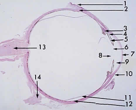
to the optic nerve (Fig. 15). Sensitive ultrasonographic techniques may detect increased thickness of the choroid as manifested by inflammation and neoplasms, focal or diffuse. Chromatophores are scattered within the choroid. The amount of pigmentation of the choroid determines the color of the fundus. Because the retina is transparent except for the blood vessels and the retinal pigmented epithelium (RPE), the variation in pigment accumulation within the choroid and retina determines the clinical picture of the ocular fundus. A heavily pigmented (negroid) fundus has the characteristic dark gray-green reflex, whereas a “blond” fundus has relatively little pigment and the pink choroidal vessel pattern is easily visible. Fluorescein and indocyanine green dye angiography readily provides clinicians with a means of evaluating the vascular integrity of the choroid and the changes that occur with disease. Also, the density of the choroidal pigmentation may affect the degree of retinal burn 
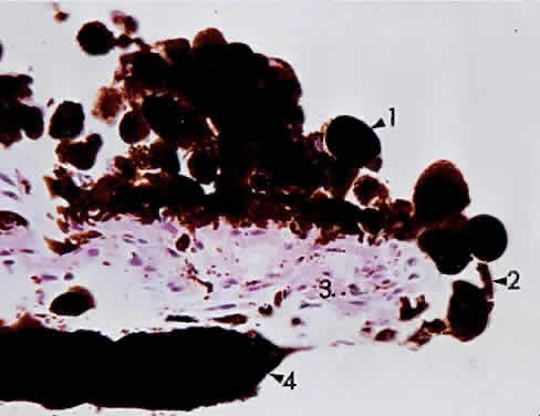
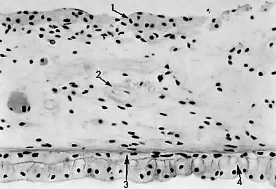
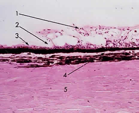
during photocoagulation of neovascular membranes.

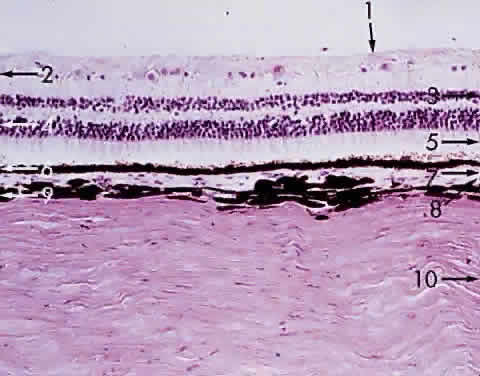
Fig. 15. Choroid wedged between the retina and sclera: 1, internal limiting membrane of retina; 2, ganglion cell layer of retina; 3, bipolar cell layer of retina; 4, nuclei of rods and cones; 5, rod and cone layer of retina; 6, pigment epithelium of retina; 7, Bruch's membrane; 8, choriocapillaris; 9, large blood vessel of choroid; 10, sclera (× 225, KEI 8982B).

During angiography, fluorescein dye is carried to the choriocapillaris from the larger vessels. Because the choriocapillaris leaks fluorescein easily, the dye diffuses rapidly throughout the choroid but cannot pass anteriorly into the retina because of the tight junctions between the RPE cells. Thus, clinically during an early phase of angiography, a shaded diffuse glow of fluorescein appears in the choroid as a result of the opaque RPE. If defects occur in the RPE (e.g., drusen, serous detachment), fluorescein can leak into the retina from the choroid. With the increasing incidence of age-related macular retinal degeneration, more attention is being directed to the subfoveal choroidal anatomy and changes in disease. Photodynamic therapy with verteporfin IV dye and laser treatment may help to prevent visual loss secondary to subfoveal choroidal neovascularization. New research into medication to inhibit vascular endothelial growth factor offers potential treatment for the future. The retinal vessels, in contrast to the choroidal vessels, do not leak fluorescein normally. At the optic disc, the choroidal vessels also leak fluorescein, so the perimeter of the disc “lights up” in the late phase of angiography even though the disc vessels proper do not normally leak fluorescein.

The primary function of the choroid is to supply nutrition to the rod and cone layer of the retina. Because of the erectile potential of the vascular channels, the choroid may also have a role in regulating intraocular pressure and in acting as a heat diffuser to protect the photoreceptors from the heating effect of absorbed light, particularly at the macula. At the optic disc, the choroidal circulation joins with the short posterior ciliary vessels and the branches from the central retinal artery to supply nutrition to the optic nerve. As a feature of glaucoma, choroidal peripapillary atrophy is a frequent hallmark. Embryologically, the choroid is derived from the mesoderm that surrounds the posterior portion of the primitive cup. The choroidal bond of Bruch's membrane to the pigmented epithelium of the retina is strong, so in “true” retinal detachments the line of cleavage is between the rod and cone layer and the pigmented epithelium.

MACROSCOPIC FEATURES

Grossly, the choroid represents a vascular bed formed by the junction of the anterior and posterior ciliary arteries (Fig. 16). The ciliary arteries (2 long posterior arteries, 20 short posterior arteries, and posterior communications of the anterior ciliary arteries) pierce the sclera to reach the choroid. They divide into vessels of gradually smaller caliber and end as choriocapillaris, nestled adjacent to Bruch's membrane. Thus, the choroid is an expandable vascular plexus that supplies nutrition for the rod and cone layer of the retina and up to 130 mm of the outer retina, particularly the macula. Thus, choroidal vascular changes due to inflammation or degeneration lead eventually to retinal pathology (e.g., subretinal neovascularization). The venous drainage from the choroid is through the four vortex veins, one 
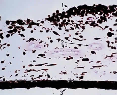
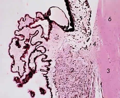
in each quadrant of the posterior sclera (Fig. 17).

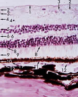
Fig. 16. Choroid at the macula: 1, internal limiting membrane of retina; 2, nerve fiber layer of retina; 3, ganglion cell layer of retina (multicell thickness); 4, inner plexiform layer; 5, bipolar cell layer; 6, outer plexiform layer; 7, nuclei of rods and cones; 8, outer limiting membrane; 9, cone (and rod) layer; 10, pigment epithelium of retina; 11, Bruch's membrane; 12, choriocapillaris (× 520, KEI 8982B).

Fig. 17. Venous drainage of the choroid.

In choroidal detachment, the vortex veins still adhere to the sclera, inducing loculations or grapelike mounds of fluid compartments in the edematous choroid.

MICROSCOPIC FEATURES

The choroid may be classified in layers, from the external to the internal surface. The suprachoroid is the space between the inner pigmented sclera (lamina fusca) and the large vessels of the choroid. The large-vessel layer (Haller's layer) is the outermost layer of the choroid. It is characterized by wide-caliber veins and arteries. Many melanocytes and ciliary nerve fibers are scattered throughout the vessel layers. The melanocytes and the Schwann cells encasing the nerve fibers, along with an occasional cluster of nevus cells, may be the precursors of the dreaded malignant melanoma of the choroid. The medium-vessel layer (Sattler's layer) is composed of medium-sized blood vessels and is located in the center of the choroid. The choriocapillaris is a layer of very large fenestrated capillaries (40 to 60 μm in diameter) lying in a single plane external to Bruch's membrane (Fig. 18; no fenestration of the choriocapillaris is visible at this level of magnification). The capillary lumina are large enough to pass several red blood cells simultaneously (Fig. 19). Unlike the larger vessels in the choroid, the fenestrated choriocapillaris lacks an internal elastic lamina and therefore leaks fluorescein during clinical angiography. Pericytes are found on the outer wall of the capillaries. Postmortem injection studies suggest that the choriocapillaris is continuous, but the choriocapillaris functions clinically like an end arteriole network. At the posterior pole, the choriocapillaris has a lobular pattern with a central precapillary arteriole and 
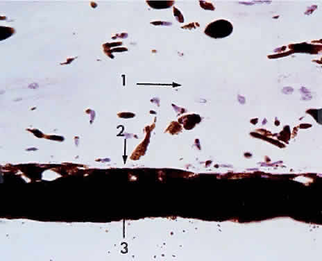
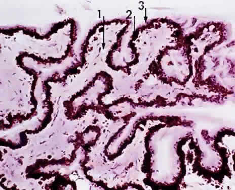
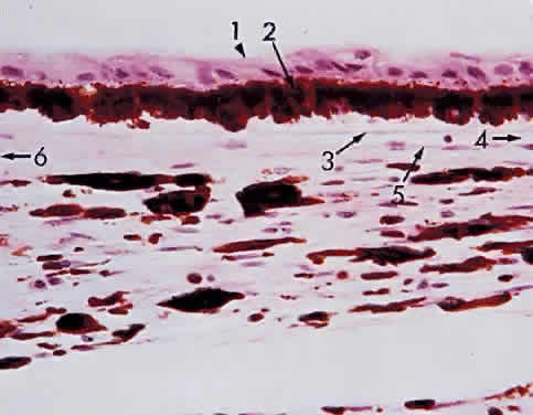
a peripheral postcapillary venule.

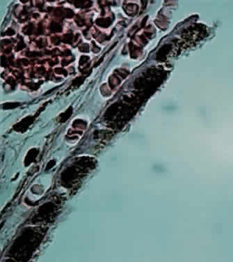
Fig. 18. Choriocapillaris at macula under Bruch's membrane and the retinal pigment epithelium (retina is detached) (× 800, KEI 71125).

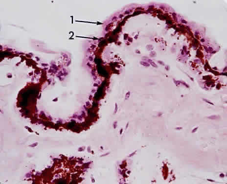
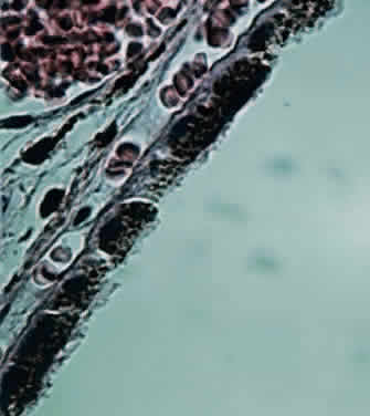
Fig. 19. Choriocapillaris at macula shows red blood cells in rouleaux pattern (retina is detached) (× 800, KEI 71125).

Scanning electron microscopy reveals three patterns of vascular structures in the choriocapillaris. At the posterior pole, the capillaries assume a lobular pattern. This architecture of the macular areas choriocapillaris makes it ideal for photodynamic therapy for “wet” macular retinal degeneration. At the equator, the pattern is spindle-shaped, and at the periphery, the capillaries have a ladder pattern. This structure explains the various degrees of flushing of fluorescein during angiography.

In the foveal zone, the sole vascular supply to the retina is the choriocapillaris. In central retinal artery occlusion, a major portion of the retinal circulation is blocked, and ischemia results. The cherry-red spot represents the intact choriocapillaris providing continued nutrition to the overlying area of macular photoreceptors. If a cilioretinal artery is present, this tiny vessel may be adequate to save the eye from blindness after a central retinal artery occlusion. The choriocapillaris circulation also acts as a heat diffuser to protect the macula from the heat generated by light rays striking the retina. This protection may be diminished by aging or disease, resulting in a reduction of central vision by heat phototoxicity, and may be a factor in age-related maculopathy.

Bruch's membrane (lamina vitrea) is a composite multilaminar hyaline PAS-positive layer featuring primarily an outer elastic lamina (derived from the mesodermal choroid) and an inner cuticular layer (basement membrane of the RPE neuroectoderm). it is usually difficult to demonstrate the multilaminar structure of Bruch's membrane, but hyaline nodular thickenings of the cuticular or basement membrane layer form the various drusen bodies (Figs. 20 and 21) seen frequently with advancing age on funduscopic examination. At the optic disc, Bruch's membrane forms the outer boundary of the disc. In high myopia, the scleral crescent denotes the stretched, distorted end of Bruch's membrane.

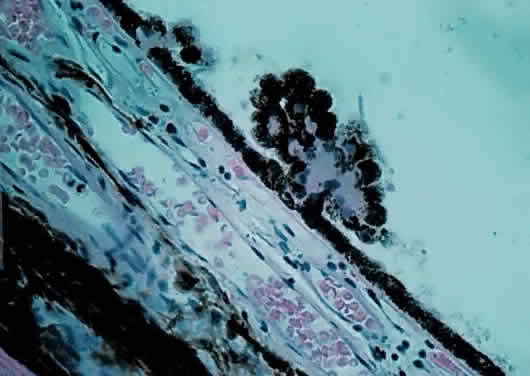
Fig. 20. Choriocapillaris near macula with small drusen on Bruch's membrane elevating retinal pigment epithelium (retina is detached) (× 800, KEI 71125).

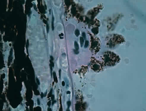
Fig. 21. Choriocapillaris with large drusen elevating retinal pigment epithelium and displacing tight retinal pigment epithelium junctions (retina is detached) (× 800, KEI 71125).

ULTRASTRUCTURE OF BRUCH'S MEMBRANE

Electron microscopy shows that Bruch's membrane is formed of five elements:

  • Basal lamina of the choriocapillaris
  • Outer collagenous layer
  • Porous band of elastic fibers
  • Collagenous inner layer
  • Basal lamina of the RPE

Bruch's membrane thus is permeable to fluorescein. Diseases that affect the choroid and RPE may therefore lead to subretinal neovascularization. Fluorescein may record the defect during angiography, thereby facilitating the site for photocoagulation to abort or minimize the degenerative process.

Back to Top
BIBLIOGRAPHY

Barsky D: Color Atlas of Pathology of the Eye. New York: McGraw-Hill, 1966

Duke-Elder S: System of Ophthalmology, Vol 11. The Anatomy of the Visual System. London: Kimpton, 1961

Blum M et al: Age-related changes of the ciliary sulcus:Implications for implanting sulcus-fixated lenses. J Cataract Refract Surg 23:91, 1997

Fine BS, Yanoff M: Ocular Histology. 2nd ed. Hagerstown: Harper & Row, 1979

Orgul SL et al. The diameter of the ciliary sulcus: A morphometric study. Graefes Arch Ophthalmol 231:487, 1993

Hogan MJ, Alvardo JAN, Weddell JE: Histology of the Human Eye. Philadelphia: WB Saunders, 1971

Hogan MJ, Zimmerman LE: Ophthalmic Pathology. 2nd ed. Philadelphia: WB Saunders, 1962

Wilson FM, Weingeist TA, Anderson RE: Ophthalmic Pathology and Intraocular Tumors, Section 4. San Francisco: American Academy of Ophthalmology, 1991-1992

Yanoff M, Fine BS: Ocular Pathology: A Color Atlas. 4th ed. Philadelphia: JB Lippincott, 1996

Back to Top