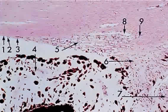
Fig. 4.
Anterior chamber angle and iris base: 1, Descemet's membrane; 2, endothelium; 3, Schwalbe's line; 4, iris; 5, corneoscleral trabecula; 6, uveal trabecula; 7, ciliary muscle; 8, canal of Schlemm; 9, scleral spur (80% of × 175, KEI 8982B).