Chapter 21
Diagnosis and Management of Fungal Keratitis
DAN B. JONES
Main Menu   Table Of Contents

Search

CLASSIFICATION
PATHOGENESIS
CLINICAL DIAGNOSIS
LABORATORY DIAGNOSIS
ANTIFUNGAL THERAPY
SURGICAL INTERVENTION
REFERENCES

Fungal keratitis is caused by invasion of the cornea by filamentous fungi, yeasts, and dimorphic fungi. The steps in management of fungal keratitis are identical to other forms of infectious microbial keratitis: Make the clinical diagnosis, perform proper laboratory studies, initiate antimicrobial therapy, modify therapy, and terminate therapy. The purpose of this chapter is to provide guidelines for prompt recognition, laboratory diagnosis, and treatment of fungal keratitis.
Back to Top
CLASSIFICATION
Medically important fungi are classified into three principal types based on macroscopic properties: filamentous fungi or molds, which produce cottony, feathery, or powdery aerial growth above culture media; yeasts, which produce creamy, opaque, pasty colonies on the surface of culture or media; and dimorphic fungi, which exhibit properties of yeasts when cultivated at 37°C and molds when grown at 25°C to 30°C.1 Filamentous fungi are multicellular microorganisms joined together to produce tubelike filaments or hyphae. Filamentous fungi can be subdivided into septate (hyphate) organisms, which produce cross-walls or septa behind the tip of the growing hyphae during elongation, and nonseptate organisms, which do not form cross-walls. Septate fungi are either nonpigmented (Moniliacea) or pigmented (Dematiaceae). Although fungi can be further classified by cellular and molecular features, these taxonomic schemes are complex and are not essential to the laboratory identification or therapy of fungi important in corneal infections.

At least 70 genera of filamentous fungi (56), yeasts (11), and dimorphic fungi (3) have been reported to cause human keratitis2, 3, 4, 5, 6, 7, 8, 9, 10, 11 (Table 1). The principal causes in the United States are the septate filamentous fungi, predominantly Alternaria, Aspergillus, Curvularia, and Fusarium and yeasts, predominantly Candida. Of importance to ophthalmologists are the differences in geographic prevalence, risk factors, pathogenesis, distinctive signs of keratitis, and antifungal susceptibility of filamentous fungi and yeasts. Fungi account for less than 5% of cases of microbial keratitis in developed countries and 10% to 15% or more in developing countries.12


TABLE 1. Reported Causes of Fungal Keratitis

  Filamentous Fungi
  Septate

  Moniliaceae (nonpigmented)
  Acremonium
  Acrostalagmus
  Arthrobotrys
  Arthrographis
  Aspergillus
  Aureobasidium
  Beauvaria
  Botryodiplodia
  Botrytis
  Calcarisporium
  Cladosporium
  Colletotrichum
  Cylindrocarpon
  Drechslera
  Epidermophyton
  Exserohilum
  Fonsecaea
  Fusarium
  Fusidium
  Geotrichum
  Glenospora
  Graphium
  Helminthosporium
  Hormodendrum
  Metarhizium
  Microsphaeropsis
  Microsporum
  Nigrospora
  Paecilomyces
  Penicillium
  Periconia
  Phaeoisaria
  Phoma
  Pithomyces
  Rhizoctonia
  Scedosporium
  Scopulariopsis
  Stachybotrys
  Syncephalastrum
  Tetraploa
  Trichoderma
  Trichophyton
  Trichosporon
  Verticillium
  Volutella
  Dematiaceae (pigmented)

  Alternaria
  Aureobasidium
  Bipolaris
  Curvularia
  Exserohilum
  Lasiodiplodia
  Phialophora
  Torula


  Nonseptate

  Absidia
  Mucor
  Rhizopus


  Yeasts

  Candida
  Monilia
  Oospora
  Pichia
  Rhodosporidium
  Rhodotorula
  Saccharomyces
  Sporotrichum
  Torulopsis
  Trichosporon
  Ustilago


  Dimorphic Fungi

  Blastomyces
  Cryptococcus
  Sporothrix


Compiled from multiple sources including the Sid W. Richardson Ocular Microbiology Laboratory.


Back to Top
PATHOGENESIS
Comparable to other forms of microbial keratitis, initiation of fungal infection requires the presence viable microorganisms and a mechanism of corneal inoculation. Fungi cannot penetrate the intact corneal epithelium and do not enter the cornea from episcleral limbal vessels. The principal routes of inoculation are introduction concurrent with a penetrating or perforating wound, either mechanical injury or surgery, and introduction through an epithelial defect. Posttraumatic infections are caused principally by filamentous fungi in the United States. Fungal keratitis following penetrating keratoplasty is uncommon but may result from contaminated donor material or late inoculation in the presence of epithelial defects, sutures, and topical corticosteroids. Fungal keratitis has also occurred following radial keratotomy,9,13,14 excimer laser photorefractive keratectomy,15 and laser-assisted in-situ keratomileusis (LASIK),16, 17, 18, 19, 20 either by intraoperative inoculation via contaminated instruments or subsequent introduction through an epithelial defect. Infection of pre-existing epithelial defects is caused primarily by the indigenous microflora of the conjunctiva and adnexa, predominantly Candida. Candida keratitis has also occurred in individuals abusing topical anesthetics,21 crack cocaine,22 and methamphetamine,23 presumably resulting from the toxic effect on the structure and function of the corneal epithelium by the agents. Other routes of corneal inoculation, such as contiguous spread from fungal scleritis or extension through the endothelium in endophthalmitis, are extremely rare.

Contact lens wear is an uncommon risk factor in fungal keratitis.24 Filamentous fungi cause most infections associated with cosmetic or aphakic refractive contact lens wear. Candida is the principal cause of keratitis secondary to therapeutic contact lens wear. Pathogenesis of these infections is probably enhanced by fungal contamination of contact lens cases and paraphernalia, by the ability of fungi to adhere to soft contact lenses, and by microtrauma to the corneal epithelium, which accompanies lens wear.

The precise events that follow adherence of fungi to the corneal epithelium or exposed deeper structures and replication of the organism to produce keratitis have not been defined. The intrinsic virulence of fungi relates to their ability to proliferate within corneal tissue, resist host defenses, and produce tissue damage.25 Filamentous fungi proliferate within the corneal stroma without release of chemotactic substances, thereby delaying the beneficial, inhibitory phases of the host response. Filamentous fungi multiply by apical hyphal extension, whereas yeast multiply by budding or transformation from the blastophore to the pseudohyphal phase. The large size of the true hyphae of filamentous fungi and pseudohyphae of yeasts precludes complete ingestion by neutrophils and macrophages. Certain fungi secrete enzymes that degrade organic substances into soluble nutrients, which are passively absorbed or actively transported into the cell. Candida albicans contains a phospholipase A and lysophospholipase on the surface of blastospores, which may aid invasion of tissue. Similar mechanisms probably account for the ability of Fusarium solani and other filamentous fungi to ramify within the corneal stroma and penetrate Descemet's membrane. Fungi also release various toxic substances, including proteases, hemolysins, and exotoxins. The role of mycotoxins in the pathogenesis of fungal keratitis has not been determined. Comparable to the pathogenesis of other forms of infectious microbial keratitis, the host response contributes to stromal destruction by release of lysosomal substances from polymorphonuclear leukocytes and other substances through a cascade of complex reactions.

Back to Top
CLINICAL DIAGNOSIS
The clinical diagnosis of fungal keratitis is based on analysis of risk factors and the distinctive signs of corneal infection. The principal risk factor for filamentous fungal keratitis is corneal trauma, most commonly in healthy individuals during some form of recreational or work activity outdoors. The frequency of injury with plant or dirt material, such as a tree branch or flying debris, is substantial evidence that the responsible organisms are inoculated into the cornea by the injuring material rather than by subsequent contamination of an epithelial defect by environmental organisms. Many infected individuals do not recall antecedent corneal injury, thereby suggesting that certain fungi invade the cornea through small asymptomatic breaks in the epithelial surface. Topical corticosteroids enhance the likelihood of replication of filamentous fungi but are not absolute requisites for infection.

The principal risk factors for yeast or Candida keratitis are protracted epithelial ulceration, penetrating keratoplasty, and therapeutic soft contact lens wear. Concurrent administration of topical corticosteroids enhances the replication of Candida through suppression of the host inflammatory response. Although polymicrobial keratitis is uncommon, risk factors for Candida are comparable to bacterial keratitis, and Candida is the most common genus among mixed bacterial and fungal keratitis.26,27 Polyfungal keratitis is rare.14

The distinct biomicroscopic signs of infectious microbial keratitis are epithelial and stromal ulceration (vs. intact epithelium), necrotizing stromal inflammation (vs. nonnecrotizing inflammation), and single location (vs multifocal sites) of the stromal inflammation. These distinctive signs are determined by the strain (virulence) of the responsible organism(s); the mechanism of inoculation into the cornea; the time interval from inoculation to examination; the antecedent status of the cornea; antimicrobial or corticosteroid therapy; and other host factors, such as an ocular surface abnormality, tear dysfunction state, lid closure dysfunction, and immunosuppression. Other biomicroscopic signs are helpful in judging the severity of the inflammatory process but not the presence or type of infection. These include epithelial edema, stromal edema, loss of endothelial reflectivity (pseudoguttata), endothelial plaque, aqueous flare and cells, and hypopyon.

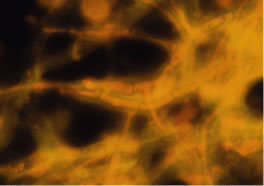
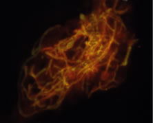
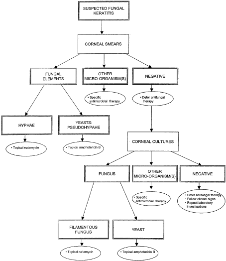
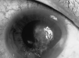
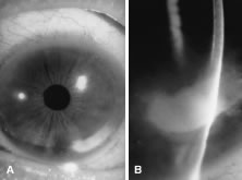
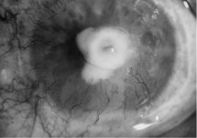
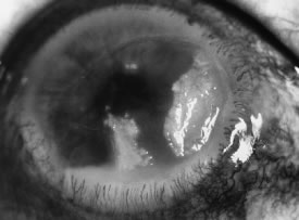
In the early stages of infection, filamentous fungi produce signs that are readily distinguishable from yeast or bacterial keratitis. The most distinctive sign is the presence of delicate, fine, feathery, opalescent, gray-white or yellow-white material in the anterior stroma, surrounded by scant cellular infiltrate or edema (Fig. 1). The epithelium may be intact. The overlying epithelium may be granular and the surface elevated and irregular in contour. Linear infiltrates typically extend into the adjacent stroma. Multiple discrete opacities may develop outside the perimeter of the principal focus of inflammation, either separated by clear stroma or linked by fine linear collections of inflammatory cells and material (Figs. 2 and 3). In the absence of inflammation in the adjacent stroma, branching hyphal fragments may be visualized by biomicroscopy (Figs. 4 and 5). Confocal microscopy may also detect hyphal elements within the stroma.28,29 Peripheral infection resembles noninfectious marginal infiltrative and ulcerative keratitis (Fig. 6). Multifocal keratitis may develop after contact lens wear or injury by multiple projectiles (Fig. 7). In the early stages, iritis is present and the intraocular pressure remains normal. Inappropriate, empirical therapy of fungal keratitis with topical fluoroquinolone or aminoglycoside antibiotics may suppress or eliminate the superficial elements but allow extension of the organisms into the stroma because these agents may possess selective antifungal activity.4,30,31

Fig. 1. Curvularia keratitis (slit-beam illumination). Note the fine feathery infiltrates extending from the central component, minimal cellular infiltrate in the adjacent stroma, and intact epithelium.

Fig. 2. F. solani keratitis. A. Full illumination. B. Slit illumination. Note the multifocal and linear infiltrates.

Fig. 3. F. solani keratitis in a farmer following injury. Note the delicate, feathery, infiltrates within the inferior component.

Fig. 4. Bipolaris rostrata keratitis (slit-beam-direct retroillumination). Note the linear hyphae within the corneal stroma. (Courtesy of A. Bron.)

Fig. 5. Microsphaeropsis olivacea keratitis in a 51-year-old man, 6 weeks following perforating corneal injury with a screwdriver. Note the fine, linear, hyphae within the corneal stroma (direct retroillumination). Nylon suture in place to close the corneal perforation.

Fig. 6. Bipolaris australiensis keratitis. Empirical antibacterial therapy for presumed bacterial keratitis for 4 weeks. A. Full illumination. B. Slit illumination. Note the localized infiltrate in the peripheral cornea and clear adjacent stroma.

Fig. 7. Paecilomyes keratitis following aphakic, extended-wear soft contact lens use. Note the separate areas of infiltration. (Light reflex to the right.) (Courtesy of M.B. Hamill.)

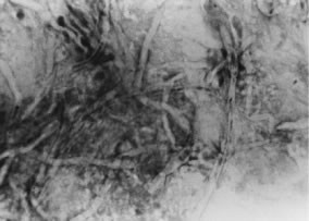
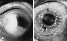
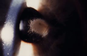
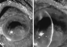
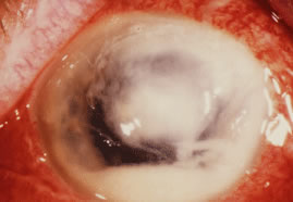
There is no distinguishing clinical sign by which to recognize the genus or species of the infectious filamentous fungus. F. solani is the most virulent organism and typically produces rapidly progressive infection characterized by epithelial and stromal ulceration, dense stromal necrosis, abundant cellular infiltrate, and edema in the adjacent stroma and hypopyon (Figs. 8 and 9). Delicate feathery components are transient. Individual hyphal fragments are rarely visualized. Infection by certain species of Aspergillus and Scedosporium (Figs. 10 and 11) resembles F. solani keratitis and progresses rapidly. Infection by less virulent organisms, such as Curvularia and Alternaria species, produces small, focal (less than 3-mm diameter) areas of nonnecrotizing stromal inflammation with delicate feathery borders (see Fig. 1 and Fig. 12). Macroscopic pigmentation may develop in keratitis caused by Alternaria, Curvularia, and other dematiaceous fungi (Fig. 13).4,11,14 The central component may progress to dense, opaque, gray-white suppuration in the deep stroma without enlargement in total area and may be accompanied by mild inflammation in the adjacent stroma. Iritis is minimal to moderate. Infection caused by other, relatively less virulent organisms resembles herpes simplex or noninfectious keratitis (Fig. 14).

Fig. 8. F. solani keratitis. Note the dense opaque central necrosis surrounded by fluffy, nonhomogeneous infiltrate.

Fig. 9. F. solani keratitis. Epithelial and stromal ulceration and necrotizing stromal keratitis.

Fig. 10. Scedosporium apiospermum keratitis in a 53-year-old woman, 10 days following unknown foreign body injury while gardening. Epithelial and stromal ulceration, endothelial plaque, and hypopyon. Note the corneal edema surrounding the central area of suppuration.

Fig. 11. Scedosporium apiospermum keratitis. Note the dense, suppurative stromal keratitis and hypopyon.

Fig. 12. Curvularia keratitis. A. Full illumination. B. Slit illumination. Focal, nonsuppurative infiltrate and intact epithelium.

Fig. 13. Curvularia keratitis in a 75-year-old man following sand injury. Note the macropigmentation in the area of the epithelial and stromal ulcer and surrounding, nonnecrotizing stromal keratitis.

Fig. 14. Bipolaris hawaiiensis keratitis. A. Full illumination. B. Slit illumination. Note the irregular area of epithelial ulceration, nonnecrotizing stromal inflammation, and stellate borders.

Keratitis caused by Nocardia, nontuberculous Mycobacterium, and other low-virulence bacteria resembles filamentous fungal keratitis by the presence of intact epithelium, multiple stromal infiltrates, and minimal inflammation in the adjacent stroma.

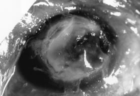
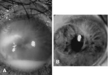
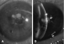
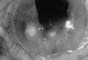
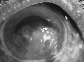
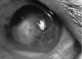
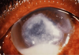
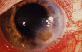
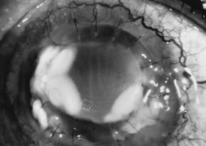
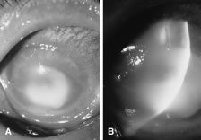
Candida infection typically produces epithelial ulceration, focal necrotizing stromal inflammation, moderate cellular infiltrate and edema in the adjacent stroma, and mild or moderate iritis in the early stages, indistinguishable from bacterial keratitis (Figs. 15, 16, and 17). Fungal elements cannot be detected by biomicroscopy. If untreated, the keratitis evolves to produce dense suppuration and necrosis of the deep stroma. Although multifocal suppuration may develop in polymicrobial keratitis, there is no distinctive sign of mixed Candida and bacterial infection (Fig. 18).

Fig. 15. C. albicans keratitis in an eye following chronic use of corticosteroid drops for herpes simplex keratitis. Note the central suppuration and perforation, endothelial plaques, and hypopyon.

Fig. 16. C. albicans following penetrating keratoplasty in a woman with severe rheumatoid arthritis. Chronic application of corticosteroid eye drop. Note the area of dense, opaque white suppuration that straddles the graft-host junction.

Fig. 17. C. albicans in a 21-year-old woman. Topical proparacaine hydrochloride abuse. Epithelial and stromal ulceration. Note the dense, white area of necrotizing stromal keratitis.

Fig. 18. C. albicans and Streptococcus keratitis in a woman following chronic application of corticosteroid eye drops. Note the separate area of suppurative keratitis. Both organisms were isolated from the separate foci.

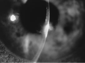
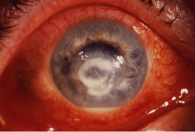
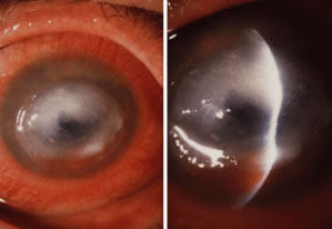
Advanced, severe filamentous fungal or yeast keratitis is indistinguishable from keratitis caused by virulent bacteria such as Staphylococcus aureus or Pseudomonas aeruginosa. The area of epithelial and stromal ulceration is large. Dense, opaque, homogenous, yellow-white stromal necrosis develop and is surrounded by confluent cellular infiltrate and full-thickness stromal edema (Figs. 19, 20, 21). Hyphal elements may penetrate Descemet's membrane and endothelium and be visualized in the anterior chamber. Fibrinous material accumulates over the endothelium, anterior chamber angle, and iris. Pain is typically severe. Secondary ocular hypertension may ensue. Progressive stromal necrosis leads to corneal perforation and, rarely, consecutive endophthalmitis.

Fig. 19. F. solani keratitis in an 81-year-old man, 3 weeks following corneal injury with a stick. Empirical antibacterial therapy for presumed bacterial keratitis. Note the area of dense, white suppurative stromal keratitis and hypopyon. Failure of antifungal therapy required therapeutic penetrating keratoplasty.

Fig. 20. C. albicans in a woman after chronic administration of proparacaine hydrochloride drops. A. Full illumination. B. Slit illumination. Dense, homogenous suppuration is noted.

Fig. 21. C. parapsilosis keratitis in a blind eye after orbital trauma and exposure. Note the liquefactive necrosis and purulent exudates.

Back to Top
LABORATORY DIAGNOSIS
The most important step in the initial management of suspected fungal keratitis is to obtain corneal material for direct smears and inoculation of media. Because of the invalidity and similarity of clinical signs of infectious microbial keratitis, the fundamental principle is to use the same method for collection of materials in all cases (Table 2). Ideally, all antimicrobial agents should be discontinued 24 to 48 hours before obtaining corneal scrapings or material in suspected infectious keratitis. Advanced, severe, suppurative keratitis requires immediate laboratory investigation. The time interval required for clearance of antimicrobial agents from the preocular tear film and cornea is unknown.


TABLE 2. Materials and Media Used


MaterialsMedia
Proparacaine hydrochloride 0.5%Blood agar plate (to be incubated at 35°C, 10% carbon dioxide)
Kimura platinum spatulaaChocolate agar plate (35°C, 10% carbon dioxide)
Alcohol lampThioglycollate broth (35°C)
Glass slides (frosted ends, etched circles)Schaedler's agar plate (35°C, anaerobic system)
Methyl alcohol 90%; Coplin jarSabouraud's agar plate (25°C)
Calcium alginate swabs (plastic shaft) 

aBausch & Lomb Pharmaceuticals, Inc, St Louis, MO.


The Kimura platinum spatula (number E1091)* is ideal for obtaining material from the cornea. The spatula tip cools rapidly after sterilization in the alcohol flame. Use of several spatulas expedites specimen collection. If a platinum spatula is not available, material may also be obtained with a disposable blade, bent needle, surgical knife, disposable cautery, or similar instrument. Proparacaine hydrochloride (0.5%) is less antiseptic than cocaine, tetracaine, and other topical anesthetics and is preferred for anesthetizing the cornea. Topical anesthetics do not interfere with cytologic techniques. Glass slides with frosted ends and etched circles facilitate labeling and recognition of smeared material. Slides should be dipped in methanol and wiped to remove dust and other material, which may also absorb stain to produce false-positive results. After transfer of material, slides should be immersed immediately in methanol (95%) in a Coplin jar and retained for 5 to 10 minutes for fixation before staining.


*Bausch & Lomb Pharmaceuticals, Inc, St Louis, MO.

Blood and chocolate agar are used in the standard procedure principally for primary isolation of aerobic bacteria; however, these media support the growth of most fungal pathogens at 35°C under increased carbon dioxide.32,33 Sabouraud's dextrose agar with gentamicin (50 μg/ml) is the primary isolation medium for fungi. The medium is nutritionally deficient but the low pH favors fungal growth. Cycloheximide should not be incorporated in Sabouraud's medium because it inhibits the saprophytic fungi principally responsible for keratitis. Thioglycollate broth is a semisolid medium that provides adequate redox potentials for the growth of aerobic fungi, as well as aerotolerant anaerobes, microaerophilic bacteria, facultative anaerobes, and aerobic bacteria. Schaedler's agar enhances the growth of anaerobic bacteria but does not support growth of filamentous fungi and yeasts.

Instillation of one or two drops of proparacaine hydrochloride (0.5%) provides adequate corneal anesthesia in adults. Sedation, akinesia, or general anesthesia may be required for children. The cooled Kimura spatula should be scraped over the surface of the area of suppuration with a series of short, moderately firm strokes in one direction, taking care not to scrape adjacent, normal intact epithelium or touch the eyelids. Each scraping should be used to inoculate only one medium or to prepare one smear. The agar plate should be inoculated by lightly streaking both sides of the spatula over the surface to produce a row of separate inoculation marks. The surface of the agar should not be penetrated because the recognition and isolation of fungi and other microorganisms in cut streaks of the agar is difficult. Thioglycollate broth should be inoculated by transferring the material from the spatula to a moistened calcium alginate swab and pushing the shaft to the bottom of the tube. Multiple samples should be obtained for each medium and for preparation of multiple slides.

The preferred stains for identification of hyphal fragments and yeasts in smears of corneal scrapings are acridine orange and calcofluor white.33, 34, 35, 36, 37, 38 Each of these techniques requires use of a fluorescent microscope. The optical activity of acridine orange is due to the binding of the dye to deoxyribonucleic acid (DNA) of bacteria, fungi, and amoebae to form a green-fluorescing complex. Acridine orange is more sensitive than the Gram stain in detecting microorganisms in clinical specimens36 and is, therefore, a rapid and sensitive method for screening all corneal smears in suspected infectious keratitis. If microorganisms are detected by acridine orange stain, the slide can be washed and stained with Gram or other specific stains. The preferred procedure is to fix the smears in 95% methanol for 5 to 10 minutes, apply the acridine orange dye (number 3336-75)* for 2 minutes, rinse the slide with tap water, blot dry, and examine by fluorescent microscopy. Hyphal fragments stain yellow-orange or green, and yeasts stain brilliant orange (Fig. 22).


*Difco Laboratories, Livonia, MI.

Fig. 22. Curvularia senagalensis keratitis. Fluorescent microscopy. Direct smear of corneal scraping stained with acridine orange. Note the distinction of the septate hyphal fragments. (× 630.)

Calcofluor white is a fabric whitener used in the textile industry. The material absorbs ultraviolet light at 340 to 400 nm and emits a blue light. Calcofluor white has an affinity for cellulose and chitin, which are common compounds of hyphal fragments and yeasts.37,38 The preferred procedure is to fix the smear in 95% methanol for 5 to 10 minutes, apply calcofluor white stain (number 17353)* to the surface of the slide, apply a coverslip over the slide, drain excessive fluid with an absorbent towel, and examine by fluorescent microscopy. Depending on the selection of filters, fungal elements stain apple green to steel gray against a rust or red background (Fig. 23).


*Polysciences, Warrington, PA.

Fig. 23. Curvularia senagalensis keratitis. Fluorescent microscopy. Direct smear of corneal scraping stained with calcofluor white stain. Prominent septate hyphal fragments are noted. (× 160.)

Other stains should be used if a fluorescent microscope is not available. The Gram stain does not stain the cell wall or septa of hyphal fragments but is absorbed by the protoplasm. The stain characteristics are inconsistent and nonspecific (Fig. 24). Yeasts typically stain dark blue and can be readily distinguished from bacteria, foreign material, precipitate, and other artifacts. The Giemsa stain is a simple, rapid method for identifying fungal elements, although not as sensitive as acridine orange or calcofluor white in detecting hyphal fragments and yeasts. The Giemsa stain has an affinity for DNA and ribonucleic acid (RNA) in microorganisms. The stain must be prepared fresh daily according to standards. The preferred method is to fix the smear in 95% methanol for 5 to 10 minutes, immerse the slide in Giemsa stain for 1 hour, rinse the slide briefly in 95% ethanol, blot dry, and examine by standard light microscopy. Hyphal fragments and yeasts appear dark blue or purple.

Fig. 24. F. solani keratitis. Light microscopy. Gram stain of direct smear of corneal scraping. Note the deposition of stain within the background material and channels of the hyphal fragments. (× 400.)

Fungal elements may also be identified by standard light microscopy in direct smears by trichrome, Gomori's methenamine silver, and periodic acid-Schiff stains, although these techniques require 1 to 2 hours. A saline or potassium hydroxide wet mount is a simple, rapid technique.10,39 Polymerase chain reaction may be further developed as a practical and sensitive method of detecting fungi in corneal material.14,40,41

Most fungi responsible for keratitis generally appear on solid media or in liquid media within 2 to 5 days after inoculation. Solid media should be examined daily by direct illumination and a dissecting microscope. All media should be kept for a minimum of 21 days because of the slow growth of certain organisms. Filamentous fungi and yeasts are identified according to standard procedures. Speciation may be helpful in predicting the severity of keratitis, such as F. solani or C. albicans, but often requires consultation with a diagnostic mycologist or reference laboratory.

Corneal material is difficult to obtain with a Kimura's spatula if the epithelium is intact or if the locus of infection is deep within the corneal stroma. The preferred method is corneal biopsy with the aid of topical anesthesia (proparacaine hydrochloride 0.5%) and slit lamp biomicroscopy. A simple method is to incise the cornea to the approximate depth of the suppuration with a disposable 2- or 3-mm punch trephine Baker's biopsy punch*, deepen the incision with an unguarded diamond or metal blade, grasp the tissue with a 0.12-mm forceps, and excise with straight or angled small (Vannas) scissors. The fragment should be placed into trypticase soy broth or saline without preservative for transportation to the microbiology laboratory. The fragment is emulsified and inoculated on the same media as used for corneal scrapings (see Table 2). Smears are prepared directly from the suspension. A second biopsy specimen should be submitted to the pathology for histologic sections. Corneal fragments can also be excised with blade or scissors and inoculated directly on media and emulsified for smears. Corneal tissue obtained by superficial keratectomy or penetrating keratoplasty should be bisected by the surgeon. One half should be submitted to the microbiology laboratory and one half for histologic section.


*Baker Cummins, Miami, FL.

Keratocentesis is reserved for suspected extension of organisms into the anterior chamber from deep stromal suppuration that cannot be sampled by corneal scraping or biopsy.

Back to Top
ANTIFUNGAL THERAPY
The decision to initiate antimicrobial therapy and the selection of specific agents in suspected infectious keratitis are determined by the severity of the corneal inflammation and interpretation of the corneal smears. In severe keratitis, typified by a large area of epithelial ulceration, extensive stromal suppuration, and marked anterior chamber reaction, the initial strategy is to anticipate the likelihood of a virulent bacterium. If one or more microorganismsare detected in corneal smears, specific antimicrobial therapy is based on the interpretation of the smears. If fungal elements alone are detected in corneal smears, initial therapy is one more antifungal agents without concurrent antibacterial therapy. If microorganisms are not detected, the preferred initial management of severe keratitis is broad antibacterial therapy, ideally ceftazidime (50 mg/ml) and tobramycin (14 mg/ml) drops. This strategy recognizes the possibility of false-negative corneal smears and the probability that severe necrotizing keratitis is most commonly caused by one of four groups of aerobic bacteria (Micrococcaceae, Streptococcus, Pseudomonas, or Enterobacteriaceae). In less severe keratitis, antimicrobial therapy should be deferred pending laboratory confirmation. One exception is the high probability of bacterial infection based on risk factors or biomicroscopic signs, such as in contact lens-related bacterial keratitis.

If fungal keratitis is suspected on the basis of the risk factors and distinctive biomicroscopic signs, antifungal therapy should be deferred until confirmation of fungal elements in smears of corneal or growth of an organism in one or more media (Fig. 25).

Fig. 25. Algorithm for initial management of suspected fungal keratitis based in the interpretation of the corneal smears and cultures. Antifungal therapy should be deferred until confirmation of fungal elements.

INITIAL THERAPY

The preferred initial therapy of early, nonsevere filamentous fungal keratitis, as suggested by the detection of hyphal fragments in corneal smears, is natamycin 5% drops administered hourly. Natamycin is a tetraene polyene antibiotic similar in mode of action to amphotericin B. Natamycin is effective in vitro against most filamentous fungi responsible for keratitis. Polyene antifungal agents achieve fungicidal activity by complexing with ergosterol in the plasma membrane, causing membrane disruption, increased permeability, leakage of cytoplasmic contents, and cell death. The compound is available as a 5% microsuspension (Natacyn*) in a 15-ml glass bottle and should be refrigerated or stored at room temperature with minimal light exposure. Corneal and intraocular penetration of natamycin following topical application is limited, although enhanced in the presence of epithelial ulceration and stromal inflammation. The compound is relatively nonirritating to the conjunctiva and cornea. It typically adheres to the stromal surface within the area of ulceration and accumulates in the inferior fornix. Natamycin is insoluble in water. Undiluted or diluted natamycin 5% suspension should not be administered by subconjunctival injectionbecause of the potential for producing severe conjunctival inflammation and necrosis. If natamycin is not immediately available, initial therapy should be amphotericin B drops (1 to 2.5 mg/ml) every 15 minutes for the initial 24 to 48 hours (see below).


*Alcon Laboratories, Fort Worth, TX.

The preferred initial therapy for early, nonsevere yeast keratitis as suggested by the detection of yeasts or pseudohyphae in corneal smears is topical amphotericin B drops (1 to 2.5 mg/ml) every 15 minutes for the initial 24 to 48 hours. Amphotericin B is a heptaene polyene antibiotic, similar in mode of action to natamycin and other polyenes. Amphotericin B is more active in vitro against C. albicans and other Candida species than natamycin and may be equally effective against various filamentous fungi. The initial selection of amphotericin B in yeast infections is based on comparative trials in experimental keratitis and clinical experience with keratitis in humans. Topical administration of amphotericin B drops produces low levels of the compound in the corneal stroma, which is enhanced by the absence of epithelium and the presence of stromal inflammation.

Amphotericin B drops are prepared by adding 10 ml of sterile distilled water without a bacteriostatic agent to the intravenous preparation (Fungizone Intravenous*) to produce a concentration of 5 mg/ml. This solution is further diluted by sterile distilled water to produce a final concentration of 1 to 2.5 mg/ml. Amphotericin B eye drops are irritating to the conjunctiva and cornea, presumably related to the sodium deoxycholate used as a solubilizing agent in the intravenous preparation. Amphotericin B eye drops as constituted should be protected from light and kept on ice or refrigerated. A new formulation should be prepared weekly. Topical amphotericin B ointment (5 mg/g) has been used as single or adjunctive therapy for A. fumigatus and F. solani keratitis.42 The safety and efficacy of topical or intravenous amphotericin B lipid complex in fungal keratitis has not been established.43


*E.R.Squibb & Sons, Princeton, NJ.

The consideration of additional topical, subconjunctival, or systemic antifungal therapy in filamentous fungi or yeast keratitis is problematic. The rationale for use of one or more additional agents is based on the availability of new compounds and their in vitro activity, safety, and efficacy in keratitis in humans and relative lack of toxicity. The most promising compounds are the azole class of antifungal agents.

Azoles are synthetic chemical antifungal agents that contain a five-member azole ring that is attached by a carbon-nitrogen bond to other aromatic rings. Older imidazole drugs, such as clotrimazole, econazole, miconazole, and ketoconazole, contain two nitrogen atoms in the azole ring. The newer triazole drugs, such as fluconazole, itraconazole, and voriconazole, contain three nitrogen atoms in the azole ring. The primary mechanism of action of the azole compounds is to interact with the cytochrome P-450 enzyme to inhibit C-14 demethylation of lanosterol, thereby causing ergosterol depletion and accumulation of aberrant sterols in the fungal cell membrane.44 Inhibition of ergosterol biosynthesis alters fungal cell membrane fluidity and modifies the function of other fungal cell membrane-associated enzymes. In high concentrations, the azoles directly damage the cell membrane by attacking phospholipids thereby exerting rapid fungicidal activity. The compounds may also inhibit other enzyme systems and block the morphogenetic transformation of Candida yeast form to pseudohyphae. Although similar in mechanism of action, the antifungal azoles differ extensively in antifungal spectrum, water solubility, tissue penetration, pharmacokinetics, and toxicity. Because systemic azole antifungal agents inhibit cytochrome P-450 dependent enzymes involved in biosynthesis of steroid hormones in mammalian cells, they may produce endocrine-related side effects such as depletion of testosterone and glucocorticoids, resulting in gynecomastia and adrenal insufficiency, respectively.44

For the initial treatment of severe filamentous fungal or yeast keratitis or for keratitis progressing on initial topical natamycin or amphotericin B therapy, fluconazole should be considered as adjunctive therapy by oral, topical, or subconjunctival administration.45,46 Fluconazole has broad in vitro antifungal activity but is most active against Candida and Aspergillus. Intravenous miconazole is no longer available for reconstitution for topical administration or subconjunctival injection. Oral ketoconazole is not indicated as adjunctive therapy with the availability of the newer triazoles.

Fluconazole, is a water-soluble triazole; has a relatively low molecular weight (305); and is available in oral 100-mg capsules (Diflucan oral) and intravenous solution (Diflucan IV), sterile solution in 0.9% sodium chloride, 2 mg/ml, 100 ml unit.47 Fluconazole is weakly protein bound (11%), has a high bioavailability (half-life of approximately 30 hours), is metabolically stable, penetrates well into cerebrospinal fluid, and is excreted unchanged in the urine. Adverse reactions following oral administration are uncommon, predominantly nausea, vomiting, diarrhea, abdominal pain, and headache. Oral fluconazole produces effective drug levels in the cornea, aqueous, vitreous, and retina/choroid in animals.48 Intraocular injection of fluconazole (100 μg) is well tolerated and has been associated with resolution of consecutive and exogenous fungal endophthalmitis. Although the minimal inhibitory concentrations of fluconazole are higher than other azoles in most susceptibility test systems, fluconazole achieves greater effectiveness in nonocular human infections on the basis of its more favorable pharmacokinetic properties.44,47 The recommended daily dosage of oral fluconazole is 200 to 400 mg, either by oral or intravenous administration. Oral absorption of the drug is rapid and almost complete, thereby reducing the requirement for intravenous administration. The efficacy and safety of higher doses have not been determined.

Itraconazole may also be a helpful adjunctive agent in fungal keratitis,49 based on in vitro activity, pharmacokinetic properties, and limited clinical trials in nonocular infections. Itraconazole is a triazole antifungal agent similar in spectrum of in vitro activity to that of fluconazole but is poorly soluble in water and highly protein bound, thereby limiting bioavailability in tissue. Food increases absorption of itraconazole. Following oral administration, tissue concentrations are often higher than plasma concentrations, even in patients with meningitis. Oral itraconazole has been most effective as alternate therapy to intravenous amphotericin B in invasive Aspergillus and has been superior to other azoles in the treatment of oral, esophageal, and vaginal candidiasis. Itraconazole is available in oral 100-mg capsules (Sporanox); the recommended daily dosage is 200 to 400 mg.

The most promising new triazole antifungal agent for treatment of fungal keratitis is voriconazole (UK-109,496), a derivative of fluconazole, currently in clinical trials by Pfizer Central Research. Its mechanism of action is similar to that of the other triazole agents, however, it is more selective to various mammalian cytochrome P-450 enzyme systems than other azoles. It is more highly active in vitro than fluconazole and itraconazole and equally effective as amphotericin B against Aspergillus sp., F. solani, F. oxysporum, Scedosporium apiospermum, S. prolificans, and Candida sp.50, 51, 52, 53, 54, 55, 56, 57, 58 Intravenous and oral administration of voriconazole in normal adults in dosages of 50 to 200 mg/day produces maximum plasma concentration between 1 and 2 hours.57 Adverse side effects are minimal and include fully reversible visual disturbances, gastrointestinal upset, and elevation of liver function enzymes. As an investigational agent, voriconazole is available as an intravenous preparation as a cyophile, containing 200 mg voriconazole with 3200 mg sulphobutyl ether-B-cyclodextrin sodium, and 50-mg tablets (An open label, noncomparative protocol for the emergency use of Voriconazole [UK 109.496] in patients with life-threatening, invasive mycoses who are failing on currently available antifungal agents, Pfizer Central Research, September, 1999).

F. solani endophthalmitis in a 16-year-old girl resolved following intravenous (4 mg/kg twice daily), oral (6 mg/kg twice daily), topical (10 mg/ml), and intraocular (10 μg/0.1 ml) administration of voriconazole.59 At the Cullen Eye Institute, oral voriconazole was successfully used (200 mg daily) to treat Scedosporium apiospermum keratitis in a 53-year-old woman failing on topical natamycin; topical, subconjunctival, and oral fluconazole; oral itraconazole; and oral terbinafine.

Supplementary initial therapy in fungal keratitis includes a cycloplegic-mydriatic agent, such as atropine 1% every 8 hours or scopolamine hydrobromide every 6 hours. A topical β-blocker or oral carbonic anhydrase inhibitor should be used to control secondary ocular hypertension. Topical or oral nonsteroidal anti-inflammatory agents and topical anticollagenolytic agents provide no additional benefit. Topical corticosteroids should not be used.

OTHER AGENTS

Terbinafine is an allylamine antifungal agent that inhibits the enzyme squalene epoxidase in the fungal membrane,60 thereby interfering with the biosynthesis of ergosterol. Terbinafine has broad in vitro antifungal activity against filamentous fungi and yeasts, including Aspergillus, Fusarium, Scedosporium, and Candida,61, 62, 63, 64, 65, 66 and acts synergistically in vitro with amphotericin B and selected azoles against certain fungi.65 Terbinafine is available for oral administration (Lamisil), 250 mg once daily, primarily for the treatment of onychomycosis. Adverse reactions are minimal, predominantly skin rash and gastrointestinal upset. Reports are lacking on the efficacy of oral terbinafine as adjunctive therapy of fungal keratitis, although in vitro activity, pharmacokinetic properties, and safety are promising.

Chlorhexidine gluconate is a biguanide germicide that has broad antimicrobial effect and is well tolerated by the ocular surface in concentrations less than 1%. Chlorhexidine gluconate 0.02% drops have been used extensively in single or combined therapy of Acanthamoeba keratitis in humans. Recent randomized clinical trials in human fungal keratitis suggested that chlorhexidine gluconate 0.2% drops may be as equally effective as natamycin 2.5% drops in Aspergillus and Fusarium keratitis.67

Polyhexamethylene biguanide is a polymeric biguanide that interacts with cytoplasmic membranes to cause leakage of cellular constituents and inhibition of respiratory enzymes. The agent is active in vitro against a variety of bacteria, fungi, and Acanthamoeba. In a rabbit model of F. solani keratitis, polyhexamethylene biguanide 0.02% drops were effective in reducing the number of colony-forming units as compared with placebo controls.68

MODIFICATION OF THERAPY

Refinement of initial therapy of fungal keratitis is hampered by multiple factors, including the slow evolution of clinical signs, uncertainty of the virulence and destructiveness of the organism based solely on the identification of genus or species, invalidity of in vitro antifungal susceptibility tests in predicting in vivo efficacy, and limited availability of safe and effective alternate antifungal agents. The rate of improvement and duration of antifungal therapy of individual genera or species of fungi cannot be predicted. If untreated, F. solani, Scedosporium, and C. albicans infections produce rapidly progressive stromal necrosis, fibrin accumulation in the anterior chamber, secondary glaucoma, and corneal perforation.

Improvement in clinical signs may be difficult to detect during the initial days of effective antifungal therapy. The most helpful biomicroscope signs are blunting of the perimeter and reduction of the density of the suppuration, reduction in cellular infiltrate and edema in the surrounding stroma, reduction in anterior chamber inflammation, and progressive re-epithelialization. The most encouraging sign of improvement is loss of the delicate, feathery perimeter of the stromal inflammation. Reduction in the area of suppuration may be difficult to detect. Frequent sketching of the cornea and measurement of the principal site of suppuration with the continuous variable slit on the Haag-Streit slit lamp or the eyepiece reticule on the Zeiss Slit lamp are helpful. The area of stromal suppuration can be estimated by measuring the greatest length and width in millimeters and multiplying the figures. Rate of epithelial healing is not a reliable sign, particularly because natamycin 5%, amphotericin B 0.1% to 0.25%, and other agents interfere with epithelial repair. Anterior chamber flare and cells diminish slowly.

For suspected filamentous fungal keratitis, natamycin 5% drops should be administered hourly for the initial 24 to 48 hours and at 2-hour intervals for the next 3 to 5 days if the corneal signs suggest stability or improvement of corneal inflammation. For yeast keratitis, amphotericin B 0.1% to 0.25% drops should be administered at 15-minute intervals for the first 24 to 48 hours and thereafter reduced during the next 4 to 6 days (e.g., 30-minute intervals for 2 to 3 days, hourly for 2 to 3 days). Cycloplegic-mydriatic drops should be continued. Antifungal therapy should be maintained for at least 4 to 6 weeks or longer if the inflammatory process is judged to be improved or inactive. Reduction in frequency of daily administration of drops should be at once- or twice-weekly intervals (i.e., eight times daily for 1 week, six times daily for 1 week, etc.). Exophytic fibroinflammatory fungal sequestration may require superficial keratectomy.14 Protracted epithelial ulceration, punctate corneal epithelial erosion, and diffuse stromal haze imply drug toxicity. Mild or moderate conjunctival hyperemia is an acceptable adverse reaction if other signs show improvement.

Identification of the fungal genus or species does not predict the clinical course and likely response to one or more antifungal agents. Decisions about alternate therapy must be based on the biomicroscopic signs and tolerance of the topical medications. The value of antifungal susceptibility testing for therapy of fungal keratitis has not been established. These procedures are not routinely performed by clinical laboratories but are available in some reference laboratories. The broth dilution method is the preferred technique for susceptibility tests of ocular yeasts and filamentous organisms and may suggest alternate therapy within a class of antifungal agents (e.g., polyenes, azoles). Antifungal susceptibility testing is highly dependent on various test conditions, including the size of the inoculum, type and pH of the medium, incubation temperature, time of reading results, and criteria for end points.

Progressive infection is characterized by an increase in the area and density of stromal suppuration, reduction in clarity of the adjacent stroma, and increase in anterior chamber flare and cells. If initial therapy was topical natamycin or amphotericin B drops alone and the stromal suppuration is judged to be progressing, topical (2 mg/ml) and oral fluconazole (200 mg twice daily) should be added for either filamentous fungal or yeast keratitis. In vitro susceptibility tests and limited animal models of nonocular infections imply that polyene and azole antibiotics may have a synergistic effect against certain fungi. If the suppuration progresses to the deep stroma with advanced corneal thinning and impending perforation, subconjunctival fluconazole (1 mg) may provide additional antifungal activity. In fungal keratitis following LASIK, management may require elevation of the flap, irrigation of the bed with amphotericin B or fluconazole, and, in some instances, excision of the flap. Consideration should also be given to acquiring voriconazole from Pfizer Central Research for compassionate use by intravenous and oral administration.

The empirical addition of a second topical antifungal agent is problematic. In vitro susceptible tests and trials in keratitis in animals judging the efficacy of combined topical antifungal agents are inconclusive. No evidence exists that combined topical natamycin and amphotericin B therapy is more efficacious than either drug alone. Administration of two polyene antibiotics may also enhance drug toxicity. Failure of topical natamycin therapy in filamentous fungal keratitis merits a trial of topical amphotericin B drops at the highest concentration tolerated (2.5 to 5 mg/ml). The possibility of polymicrobial keratitis and superinfection with other microorganisms should be considered. Interruption of antifungal therapy and repeat corneal scrapings and laboratory investigations may be warranted.

Back to Top
SURGICAL INTERVENTION
If corneal suppuration progresses despite maximum antifungal therapy, consideration must be given to surgical intervention by penetrating keratoplasty. Progression is characterized by increased area of stromal suppuration, advancement of suppuration into the deep stroma, stromal ulceration to the point of extreme corneal thinning, dense fibrinous exudate in the anterior chamber, increased intraocular pressure, and corneal perforation. Failure of medical therapy and progressive stromal necrosis occur most commonly in advanced, untreated fungal keratitis, particularly if enhanced by corticosteroid therapy; in F. solani and Scedosporium keratitis; in infection following traumatic inoculation of organisms into the deep corneal stroma; and in C. albicans infections in eyes with severe predisposing factors, such as atopic keratoconjunctivitis or cicatricial pemphigoid. Among 435 consecutive cases of microbial keratitis at the Bascom Palmer Eye Institute between 1983 and 1986, therapeutic penetrating keratoplasty was required seven times more frequently in fungal keratitis (14 of 59) than bacterial keratitis (13 of 376).69

To assess the effectiveness of medical therapy and determine the requirement for surgical intervention, a careful, detailed corneal drawing should be made and the area, depth, and density of the stromal suppuration should be frequently quantitated. The extent of progression of suppuration into the deep stroma is difficult to judge in the presence of an endothelial plaque or fibrinous exudate in the anterior chamber. Abrupt withdrawal of topical corticosteroids produces increased stromal and anterior chamber inflammation, which are difficult to distinguish from signs of progressive invasion of replicating organisms. Imminent perforation is suggested by reduction in the density of the stromal suppuration and improved visualization of the anterior segment secondary to loss of stromal substance.

Small corneal perforations (less than 2 mm in diameter) occurring early in the course of medical therapy should be managed by either a therapeutic soft contact lens or tissue adhesive. These measures prolong antifungal therapy and reduce intraocular inflammation, thereby allowing penetrating keratoplasty to be performed under more ideal conditions. Application of a tissue adhesive prevents full visualization of the area of stromal suppuration and may create a partial barrier to penetration of effective antifungal agents. A trial of a soft therapeutic contact lens may effectively seal the perforation and promote restoration of the anterior chamber. The principles are to use a tight-fitting lens, continue cycloplegic-mydriatic drops (atropine 1% or hyoscine 0.25%), and avoid external pressure on the globe. A Fox shield should be used. If the anterior chamber can be restored and maintained, medical therapy should be continued. Control of fungal replication may permit stromal healing and eliminate urgent penetrating keratoplasty. In larger perforations (greater than 3 mm in diameter) or if a soft therapeutic contact lens alone fails to maintain the anterior chamber, tissue adhesive should be applied. In the presence of large areas of stromal thinning and necrosis, the likelihood of corneal perforation should be anticipated and penetrating keratoplasty performed urgently.

The goals of therapeutic penetrating keratoplasty are to restore the structure of the cornea, maintain the anterior chamber, and eliminate replicating organisms. In phakic eyes, an additional goal is to preserve the integrity of the lens and avoid additional intraoperative procedures that may enhance the spread of organisms into the vitreous to produce consecutive fungal endophthalmitis. In eyes without impending or extant perforation, akinesia and anesthesia may be accomplished by standard lid and retrobulbar block. General anesthesia, possibly supplemented by direct infiltration of extraocular muscles, is preferred in the presence of corneal perforation. Cycloplegic-mydriatic drops should be discontinued 24 to 48 hours before surgery to facilitate protective miosis. A carbonic anhydrase inhibitor (e.g., acetazolamide, 500 mg, oral or intravenous) and intravenous mannitol (1 g/kg) should be administered.

Although the decision on the size and placement of graft must be made at surgery, careful preoperative slit lamp biomicroscopy is helpful to determine the requirement for donor tissue. The trephination should provide a margin of 1 to 1.5 mm around the area of suppuration to eliminate the replicating organisms. The exact extent of organisms within the stroma adjacent to the area of suppuration cannot be accurately determined by slit lamp biomicroscopy. If the required donor cornea is estimated to be less than 12 mm, use of corneal-scleral tissue stored in standard medium is preferred and the eye bank should be advised of the need for tissue with an adequately large scleral rim. If a larger graft is required, a whole globe is preferred to simplify excision of the donor tissue.

In an intact globe, external pressure should be applied by a balloon device or hand compression to soften the eye and reduce the orbital contents. The Schott speculum* is ideal to support the eyelids, expose the globe, and minimize extrinsic pressure. A partial or 360-degree peritomy should be done if the anticipated graft-host junction requires placement of corneal sutures outside the limbus. A scleral support ring should be sutured 3 to 4 mm posterior and concentric to the limbus. To facilitate vitreous aspiration if required to reform the anterior chamber, a partial-thickness sclerotomy incision should be made 3.5-mm posterior to the limbus in the inferotemporal quadrant and partially closed with a horizontal mattress suture.


*International Instruments, Weston, MA.

The size and placement of the graft are determined by the location and area of the stromal suppuration. The goal is to center the trephination with reference to the cornea rather than to the site of suppuration. If the area of suppuration is large and extends asymmetrically, decentration of the trephination may be required to obtain an adequate margin adjacent to the limbus (Fig. 26). If the suppuration is located in the peripheral cornea, a small, decentered graft may be preferred to provide a free margin of replicating organisms and avoid the complications of a large graft, despite the necessity of placement of the graft-host junction within the optical zone (Fig. 27). Circular trephinations are preferred; free-hand, half-circle, or annular grafts are technically difficult and unnecessary. If suppuration and necrosis extend beyond the limbus, a large 10- or 11-mm graft is required.

Fig. 26. F. solani keratitis in the right eye of a farmer. A. Dense suppurative necrosis and perforation. B. 9-mm penetrating keratoplasty decentered temporally to surround the area of suppuration and provide an adequate optical zone.

Fig. 27. Fungal keratitis. A. Suppuration and perforation of the peripheral cornea. B. 5-mm penetrating keratoplasty decentered to surround the area of suppuration. Note the graft-host junction within the optical zone. Subsequent 7.5-mm centered penetrating keratoplasty.

The size of the trephination is best determined in an intact globe. In corneal perforation, intraoperative application of tissue adhesive permits reformation of the anterior chamber through a corneal stab incision. The donor cornea should be oversized at least 0.25 mm in phakic eyes and 0.5 mm or 1.0 mm in phakic eyes for trephinations 9.5 mm or larger. The hollow trephine handle and disposable blade are ideal for trephination of the host cornea. The initial incision should be three-fourths to seven-eighths thickness. The anterior chamber is entered with an unguarded diamond or metal blade. In corneal perforation, the host cornea should be marked with the trephine and the cornea incised full thickness with the diamond or metal blade.

The excised tissue should be bisected. One half should be submitted for histopathologic study. One half should be transported immediately to the laboratory to be emulsified for inoculation on standard media as for corneal cultures. Fibrinous exudate should be removed from the anterior chamber and iris surface and inoculated directly on media or placed in tryptic soy broth for transportation to the laboratory. One or two midstromal or peripheral iridectomies should be done. Posterior synechiae should be lysed by gentle pressure on the iris surface at the pupillary margin or direct synechialysis with a blunt instrument to avoid damaging the lens. Acetylcholine should be instilled to reduce the pupil. A viscoelastic substance should be injected into the chamber angle and over the iris surface to separate the host tissue, reform the anterior chamber, and facilitate placement of the donor cornea. In corneal perforation or if extension of replicating organisms into the anterior chamber is suspected, 5 μg/ml of amphotericin B in 0.1 ml should be injected into the anterior chamber. Every effort should be made to retain the crystalline lens to reduce the likelihood of consecutive endophthalmitis. If the suppurative process involves the lens, posterior chamber, or vitreous, lensectomy, anterior vitrectomy, and intravitreal injection of amphotericin B (5 μg/ml in 0.1 ml) are required. Multiple interrupted 10-0 nylon sutures are preferred for wound closure. Inability to maintain or reform the anterior chamber necessitates aspiration of vitreous through the prepared sclerotomy site.

Postoperative medical therapy is determined by multiple factors, including the surgeon's estimate of the likelihood of excision of replicating fungi, degree of postoperative inflammation, intraocular pressure, status of other intraocular structures, results of cultures of corneal tissue and intraocular fluid, and histopathologic examination of the excised host or cornea. Topical antifungal agents may be withheld in the early postoperative period if corneal perforation did not occur, trephination adequately surrounded the area of suppuration, and organisms were not cultured from anterior chamber material. Oral fluconazole (400 mg daily) should be administered for the initial 7 to 10 days or longer. If excision of infected corneal tissue was incomplete and organisms were present in the anterior chamber based on clinical findings or culture, a topical antifungal agent (natamycin 5% or amphotericin B 0.25% to 0.5%) should be continued during the early postoperative period. Signs of corneal toxicity should be monitored carefully. Topical and oral corticosteriods should be avoided during the initial 48 to 72 hours and longer if possible. Cyclosporine A has been demonstrated to have an inhibitory effect against Fusarium oxysporum, F. solani, and Aspergillus fumigatus in vitro70 and may be an effective alternative to topical corticosteriods following therapeutic penetrating keratoplasty. Identification of fungal elements in the corneal sections or recovery of the organism in corneal cultures does not necessarily require antifungal therapy for prophylaxis of consecutive endophthalmitis. Fungal endophthalmitis secondary to therapeutic penetrating keratoplasty of fungal keratitis in phakic eyes is uncommon. Intraocular injection of antifungal agents and other surgical measures should be reserved for eyes with confirmed fungal endophthalmitis.

Conjunctival flap or lamellar keratoplasty is not indicated in the management of fungal keratitis unresponsive to medical therapy. These procedures do not eliminate replicating organisms and may create barriers to penetration of potentially effective antifungal agents.

Back to Top
REFERENCES

1. Dixon DM and Fromtling RA: Morphology, taxonomy, and classification of the fungi. In Murray PR, Baron EJ, Pfaller MA et al (eds): Manual of Clinical Microbiology. 6th ed. Washington DC: American Society of Microbiology, 1995, pp 699–708

2. Jones BR, Richards AB, Morgan G: Direct fungal infection of the eye in Britain. Trans Ophthalmol Soc UK 1969;89:727

3. DeVoe AG, Silva-Hutner M: Fungal infections of the eye. In Locatcher-Khorazo D, Seegal BC (eds): Microbiology of the Eye. St Louis: CV Mosby, 1972, p 208

4. Forster RK, Gerbert R, Wilson LA: Dematiaceous fungal keratitis. Br J Ophthalmol 1975;59:372

5. Srivastava OP, Lal B, Agrawal PK et al: Mycotic keratitis due to Rhizoctonia sp. Sabouraudia 1977;15:125

6. Sandhu DK, Rattan AS: Keratomycosis: A review. Mykosen 1981;24:503

7. Thomas PA, Kuriakose T: Keratitis due to Arthrobotrys oligospora Fres. 1850. J Med Vet Mycol 1990;28:51

8. Upadhyay MP, Karmacharya PCD, Koirala S et al: Epidemiologic characteristics, predisposing factors, and etiologic diagnosis of corneal ulceration in Nepal. Am J Ophthalmol 1991;111:92

9. Rosa RH Jr, Miller D, Alfonso EC: The changing spectrum of fungal keratitis in South Florida. Ophthalmology 1994;101:1005

10. Panda A, Sharma N, Das G et al: Mycotic keratitis in children: Epidemiologic and microbiologic evaluation. Cornea 1997;16:295

11. Garg P, Gopinathan U, Choudhary K et al: Keratomycosis: Clinical and microbiologic experience with dematiaceous fungi. Ophthalmology 2000;107:574

12. Wilhelmus KR. Epidemiology of ocular infections. In Tasman W, Jaeger EA (eds): Foundations of Clinical Ophthalmology. Vol 2. Philadelphia: Lippincott-Raven, 1998

13. Panda A, Das GK, Vanathi M et al: Corneal infection after radial keratotomy. J Cataract Refract Surg 1998;24:331

14. Wilhelmus KR, Jones DB: Curvularia keratitis. Trans Ophthalmol Soc 2001;99:111

15. Dunphy D, Andrews D, Seamone C et al. Fungal keratitis following excimer laser photorefractive keratectomy. Can J Ophthalmol 1999;34:286

16. Read RW, Chuck RS, Rao NA et al: Traumatic Acremonium atrogriseum keratitis following laser-assisted in situ keratomileusis. Arch Ophthalmol 2000;118:418

17. Sridhar MS, Garg P, Bansal AK et al: Aspergillus flavus keratitis after laser in situ keratomileusis. Am J Ophthalmol 2000;129:802

18. Sridhar MS, Garg P, Bansal AK et al: Fungal keratitis after laser in situ keratomileusis. J Cataract Refract Surg 2000;26:613

19. Chung MS, Goldstein MH, Driebe Jr WT et al: Fungal keratitis after laser in situ keratomileusis: A case report. Cornea 2000;19:236

20. Sridhar MS, Garg P, Bansal AK et al: Fungal keratitis after laser in situ keratomileusis, J Cataract Refract Surg 2000;26:613

21. Chern KC, Meisler DM, Wilhelmus KR et al: Corneal anesthetic abuse and Candida keratitis. Ophthalmology 1996;103:37

22. Sachs R, Zagelbaum BM, Hersh PS: Corneal complications associated with the use of crack cocaine. Ophthalmology 1993;100:187

23. Poulsen EJ, Mannis MJ, Chang SD: Keratitis in methamphetamine abusers. Cornea 1996;15:477

24. Wilhelmus KR, Robinson NM, Font RA et al: Fungal keratitis in contact lens wearers. Am J Ophthalmol 1988;106:708

25. Jones DB: Pathogenesis of bacterial and fungal keratitis. Trans Ophthalmol Soc UK 1978;98:367

26. Jones DB: Strategy for the initial management of suspected microbial keratitis. In Symposium on Medical and Surgical Diseases of the Cornea; Transactions of the New Orleans Academy of Ophthalmology. St. Louis: Mosby, 1980

27. Jones DB: Polymicrobial keratitis. Trans Am Ophthalmol Soc 1981;79:153

28. Florakis GJ, Moazami G, Schubert H et al: Scanning slit confocal microscopy of fungal keratitis. Arch Ophthalmol 1997;115:1461

29. Petroll WM, Cavanagh HD, Jester JV: Clinical confocal microscopy. Curr Opin Ophthalmol 1998;9:59

30. Nakajima R, Kitamura A, Someya K et al: In vitro and in vivo antifungal activities of DU-6859a, a fluoroquinolone, in combination with amphotericin B and fluconazole against pathogenic fungi. Antimicrob Agents Chemother 1995;39:1517

31. Chodosh J, Miller D, Tu E et al: Tobramycin-responsive Fusarium oxysporum keratitis. Can J Ophthalmol 2000;35:29

32. Waxman E, Chechelnitsky M, Mannis MJ et al: Single culture media in infectious keratitis. Cornea 1999;18:257

33. Wilhelmus KR, Liesegang TJ, Osato MS et al: Cumitech 13A. In Specter SC (coordinating ed): Laboratory Diagnosis of Ocular Infections. Washington DC: American Society of Microbiology, 1994

34. Kanungo R, Srinivasan R, Rao RS: Acridine orange staining in early diagnosis of mycotic keratitis. Acta Ophthalmol 1991;69:750

35. Chander J, Chakrabarti A, Sharma A et al: Evaluation of calcofluor staining in the diagnosis of fungal corneal ulcer. Mycosis 1993;36:243

36. Torres-Gomez J, Robinson NM, Wilhelmus KR et al: Comparison of acridine orange and Gram stain in bacterial keratitis. Am J Ophthalmol 1988;106:735

37. Sutphin JE, Robinson NM, Wilhelmus KR et al: Improved detection of oculomycoses using induced fluorescence with cellufluor. Ophthalmology 1986;93:416

38. Marines HM, Osato MS, Font RL: The value of calcofluor white in the diagnosis of mycotic and Acanthamoeba infections of the eye and ocular adnexa. Ophthalmology 1987;94:23

39. Vajpayee RB, Angra SK, Sandramouli S et al: Laboratory diagnosis of keratomycosis: comparative evaluation of direct microscopy and culture results. Ann Ophthalmol 1993;25:68

40. Alexandrakis G, Jalali S. Gloor P: Diagnosis of Fusarium keratitis in an animal model using the polymerase chain reaction. Br J Ophthalmol 1998;82:306

41. Okhravi N, Adamson P, Mant R et al: Polymerase chain reaction and restriction fragment length polymorphism mediated detection and speciation of Candida species causing intraocular infection. Invest Ophthalmol Vis Sci 1998;39:859

42. Hirose H, Terasaki H, Awaya S et al: Treatment of fungal corneal ulcers with amphotericin B ointment. Am J Ophthalmol 1997;124:836

43. Goldbum D, Frueh BE, Zimmerli S et al: Treatment of postkeratitis Fusarium endophthalmitis with amphotericin B lipid complex. Cornea 2000;19:853

44. Georgopapadakou NH, Walsh TJ: Antifungal agents: Chemotherapeutic targets and immunologic strategies. Antimicrob Agents Chemother 1996;40:279

45. Rao SK, Madhavan HN, Rao G et al: Fluconazole in filamentous fungal keratitis [Letter]. Cornea 1997;16:700

46. Thakar M: Oral fluconazole therapy for keratomycosis. Acta Ophthalmol 1994;72:765

47. Saag MS, Dismukes WE: Azole antifungal agents: emphasis on new triazoles. Antimicrob Agents Chemother 1988;32:1

48. O'Day DM, Foulds G, Williams TE et al: Ocular uptake of fluconazole following oral administration. Arch Ophthalmol 1990;108:1006

49. Thomas PA, Abraham DJ, Kalavathy CM et al: Oral itraconazole therapy for mycotic keratitis. Mycoses 1988;31:278

50. Radford SA, Johnson EM, Warnock DW: In vitro studies of activity of voriconazole (UK-109,496), a new triazole antifungal agent, against emerging and less-common mold pathogens. Antimicrob Agents Chemother 1997;41:841

51. Wildfeuer A, Seidl HP, Haberreiter P et al: In vitro evaluation of voriconazole against clinical isolates of yeasts, moulds and dermatophytes in comparison with itraconazole, ketoconazole, amphotericin B and griseofulvin. Mycosis 1998;41:309

52. Marco F, Pfaller MA, Messer SA et al: Antifungal activity of a new triazole, voriconazole (UK-109,496), compared with three other antifungal agents tested against clinical isolates of filamentous fungi. Medical Mycology 1998;36:433

53. Johnson EM, Szekely A, Warnock DW: In vivo activity of voriconazole, itraconazole and amphotericin B against filamentous fungi. J Antimicrob Chemotherapy 1998;42:741

54. McGinnis MR, Pasarell L, Sutton DA: In vitro activity of voriconazole against selected fungi. Med Mycol 1998;36:239

55. Marco F, Pfaller MA, Messer S et al: In vitro activities of voriconazole (UK-109,496) and four other antifungal agents against 394 clinical isolates of Candida spp. Antimicrob Agents Chemother 1998;42:161

56. Cuenca-Estrella M, Ruiz-Diez B, Martinez-Suarez JV et al: Comparative in-vitro activity of voriconazole (UK-109,496) and six other antifungal agents against clinical isolates of Scedosporium prolificans and Scedosporium apiospermum. J Antimicrob Chemother 1999;43:149

57. Kappe R: Antifungal activity of the new azole UK-109, 496 (voriconazole). Mycoses 1999;42:83

58. Lee JK, Peters D, Obias AA et al: Activity of voriconazole against Candida albicans and Candida krusei isolated since 1984. Int J Antimicrob Agents 2000;16:205

59. Reis A, Sundmacher R, Tintelnot K et al: Successful treatment of ocular invasive mould infection (fusariosis) with the new antifungal agent voriconazole [Letter]. Br J Ophthalmol 2000;84:932

60. Ryder NS, Favro B: Antifungal activity and mechanism of action of terbinafine. Rev Contemp Pharmacother 1997;8:275

61. Katz HI: Drug interactions of the newer oral antifungal agents. Br J Derm 1999;141:26

62. Ryder NS: Activity of terbinafine against serious fungal pathogens. Mycoses 1999;42:115

63. Hay RJ: Therapeutic potential of terbinafine in subcutaneous and systemic mycoses. Br J Derm 1999;141:36

64. Jessup CJ, Ryder NS, Ghannoum MA: An evaluation of the in vitro activity of terbinafine. Med Mycol 2000;38:155

65. Meletiadis J, Mouton JW, Rodriguez-Tudela JL et al: In vitro interaction of terbinafine with itraconazole against clinical isolates of Scedosporium prolificans. Antimicrob Agents Chemother 2000;44:470

66. Jessup CJ, Ryder NS, Ghannoum MA: An evaluation of the in vitro activity of terbinafine. Med Mycology 2000;38:155

67. Rahman MR, Johnson GJ, Husain R et al: Randomized trial of 0.2% chlorhexidine gluconate and 2.5% natamycin for fungal keratitis in Bangladesh. Br J Ophthalmol 1998;82:919

68. Fiscella RG, Moshiva M, Messick CR et al: Polyhexamethylene biguanide (PHMB) in the treatment of experimental Fusarium keratomycosis. Cornea 1997;16:447

69. Forster RK: The role of excisional keratoplasty in microbial keratitis. In Cavanagh HD (ed): The Cornea: Transactions of the World Congress on the Cornea III. New York: Raven Press, 1988, p 529

70. Bell NP, Karp CL, Alfonso EC et al: Effects of methylprednisolone and cyclosporine A on fungal growth in vitro. Cornea 1999;18:306

Back to Top