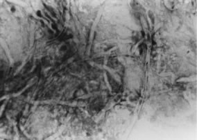
Fig. 24.
F. solani
keratitis. Light microscopy. Gram stain of direct smear of corneal scraping. Note the deposition of stain within the background material and channels of the hyphal fragments. (× 400.)