Chapter 18
Bacterial Corneal Ulcers
RICHARD L. ABBOTT, MICHAEL ZEGANS and PAUL A. KREMER
Main Menu   Table Of Contents

Search

PATHOGENESIS
CAUSATIVE ORGANISMS
GRAM-POSITIVE COCCI
OTHER GRAM-POSITIVE ORGANISMS
GRAM-NEGATIVE RODS
OTHER GRAM-NEGATIVE ORGANISMS
CLINICAL APPROACH
LABORATORY DIAGNOSIS
THERAPEUTIC APPROACH
ANTIBIOTIC THERAPY
SPECIFIC ANTIBIOTICS
ADJUNCTIVE THERAPY
REFERENCES

Ulceration of the cornea may be either infectious or sterile and requires immediate intervention to prevent complications. Widespread use of contact lenses and the increasing number of refractive corneal surgical procedures require ophthalmologists to be well versed in the diagnosis and treatment of bacterial keratitis. The accurate determination of an infectious agent and management of the ulceration can be among the most challenging of clinical problems. The causative organisms, laboratory diagnosis, therapeutic approach, and major antibiotics are reviewed.
Back to Top
PATHOGENESIS
Numerous microorganisms on the lid margin and in the normal uninfected conjunctival sac provide a constant source of potentially pathogenic bacteria to the cornea.1 The ability of the cornea to prevent invasion by most bacteria depends on an intact epithelial surface and a normal tear flow. Any impairment in the integrity of these natural barriers may permit entrance of microorganisms and produce ulceration.

The smooth surface of the cornea offers some protection from infection by combining the action of tears and blinking to mechanically remove organisms from the surface. In addition, lysozymes, beta lysins, and antibodies in the tears exert antibacterial action. Most cases of bacterial keratitis result from a disruption in the corneal epithelium. There are many compromising factors that can adversely affect the integrity of the corneal epithelium and tear film, causing breakdown and secondary infection (Table 1).1–9


TABLE 18-1. Predisposing Condition to Bacterial Keratitis

  Contact Lens Wear
  Overnight wear
  Soft contact lenses
  Homemade solutions
  Nonsurgical Trauma
  Corneal abrasion or wound
  Corneal foreign body
  Toxic medications (e.g., anesthetic abuse, idoxuridine)
  Surgical Trauma
  Suture abscess
  Dellen
  Postoperative epithelial defect
  Corneal Injury Caused by Lid Dysfunction
  Trichiasis
  Lagophthalmus (mechanical or neurologic)
  Altered mental status (neurologic disease, substance abuse)
  Corneal, Conjunctival, or Lacrimal Dysfunction
  Bullous keratopathy
  Neurotrophic cornea
  Ocular cicatricial pemphigoid
  Stevens-Johnson syndrome
  Graft versus host disease
  Tear insufficiency
  Mucin insufficiency


The association between contact lens wear and infectious ulcerative keratitis has been well documented.10–15 A 38-year study of the incidence of ulcerative keratitis in Olstead County, Minnesota found contact lens wear to be a predisposing factor in 56% of patients.16 This was followed by ocular trauma, which was a predisposing factor in 25% of patients. Extended-wear contact lens regimens have been associated with an increased risk of developing ulcerative keratitis compared with daily-wear regimens.17,18 The risk of ulcerative keratitis appears to be directly related to the extent of overnight wear.19 Disposable contact lenses used on an extended-wear basis may not provide significant protection against the development of corneal ulcers compared with conventional extended-wear lenses.20–22 Annual risks of ulcerative keratitis in cosmetic contact lens wearers have been estimated to be 13.3 to 20.9 cases per 10,000 for extended soft contact lens wear; 2.2 to 4.1 cases per 10,000 for daily soft contact lens wear; and 2 cases per 10,000 for rigid gas-permeable lens wear.23

Certain organisms, such as Neisseria gonorrhoeae, Listeria, Corynebacterium, and Haemophilus aegypticus may invade directly through intact corneal epithelium. These bacteria must first adhere to the epithelium or stroma before they can initiate penetration and infection.24,25 This biologic adhesion allows diffusion of toxins and bacterial by-products necessary to facilitate actual entrance of the bacteria into the stroma.26

Once bacteria have invaded the corneal stroma, the host response is initiated by the polymorphonuclear leukocyte (PMN). The PMN phagocytoses the bacteria and, by means of intracytoplasmic lysosomes, destroys the organism.3 Although the enzymes produced by the PMNs help destroy the invading bacteria, they also produce toxic metabolites, which may contribute to progressive corneal destruction.

Back to Top
CAUSATIVE ORGANISMS
Most of the organisms cultured from corneal infections are of the same species that are normally found on the lids and periocular skin, in the conjunctival sac, or in adjacent nasal passages1. The spectrum of microorganisms that produce bacterial corneal infection is influenced by preexisting disease or injury and the severity of any other compromising factors (Table 2).2,7 Although regional variations within the U.S. may exist, the most common organisms cultured from bacterial ulcers in the uncompromised, healthy cornea are Staphylococcus, Streptococcus, Pseudomonas, Enterobacteriaceae, Moraxella species, and Klebsiella pneumoniae.4,27 Pseudomonas has been reported to be the bacteria most commonly isolated from contact lensassociated corneal ulcers. In the compromised cornea, normally harmless bacteria can become opportunistic pathogens and may produce infected corneal ulcers. The most common organisms in this group are Staphylococcus aureus, the coagulase-negative staphylococci, alpha-hemolytic streptococcus, beta-hemolytic streptococcus, Pseudomonas, and Proteus.4,27 Ulcers in the pediatric age group are most commonly caused by Pseudomonas, Staphylococcus, and fungi.1,28 Among patients with AIDS, fungal ulcers also appear to be more common.29 Streptococcus pneumoniae seems to be a particularly important cause of bacterial keratitis in the developing world, where contact lens-associated corneal ulceration represents a smaller portion of cases of bacterial keratitis. In a large study of Indian patients with corneal ulceration, S. pneumoniae was the most commonly isolated bacterial pathogen.30


TABLE 18-2. Organisms Commonly Isolated From Corneal Ulcers


Healthy CorneaCompromised Cornea*Pediatric
StaphylococcusStaphylococcus aureusPseudomonas
StreptococcusStaphylococcus epidermidisStaphylococcus
Pseudomonasα-Hemolytic StreptococcusFungi
Enterobacteriaceaeβ-Hemolytic Streptococcus 
MoraxellaPseudomonas 
KlebsiellaProteus 


The severity of corneal infection usually depends on the underlying condition of the cornea and the pathogenicity of the infecting organism. Toxins produced by some organisms facilitate infection through cytotoxic destruction of the cell membrane or by inhibiting cellular protein synthesis.3 An understanding of the characteristics and pathogenicity of bacteria commonly known to cause infectious keratitis is helpful in the treatment of these infections.

The more common bacteria isolated from bacterial corneal ulcers, their clinical characteristics, and the laboratory techniques used in their identification are presented.

Back to Top
GRAM-POSITIVE COCCI

STAPHYLOCOCCI

Staphylococci are among the most common bacteria encountered in ocular infections. Staphylococcus aureus (coagulase-positive staphylococcus) is generally considered to be a pathogenic organism. The coagulase-negative staphylococci include Staphylococcus epidermidis, which is thought to be part of the normal body flora. There are at least 14 other species of coagulase-negative staphylococci that are rarely associated with infectious keratitis.31

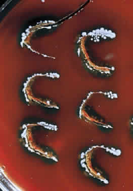
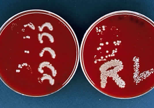
During microscopic examination, staphylococci almost always appear Gram-positive, are spherical with a mean diameter of approximately 1 mm, and are seen extracellularly. Old cultures or scrapings from antibiotic-treated cases may show Gramnegative staining characteristics. Although grape-like clusters are frequently seen in scrapings or smears from nonocular tissues, these are unusual in scrapings from corneal ulcers, in which single cocci or clusters of two to six cocci are observed. Two species, S. epidermidis and S. aureus, cannot be distinguished by Gram stain; however they show distinct characteristics in culture. In general the staphylococci are easily cultured on standard bacteriologic media (see laboratory section) and grow rapidly in incubation at 37°C. They do not have a distinctive odor and grow in the presence or absence of oxygen. Typical colonies are round, flat, smooth, and several millimeters in diameter by 48 hours (Fig. 1). S. aureus colonies show beta hemolysis (clear zones of hemolysis on blood agar) and some degree of pigmentation (light cream to golden yellow). In contrast, S. epidermidis colonies are usually nonhemolytic and rarely exhibit pigmentation.

Fig. 1. Typical colonies of Staphylococcus aureus growing on “C” streaks placed on blood agar at 37°C. Note the round, flat, smooth appearance of the colonies.

Because S. epidermidis is often a nonpathogenic component of normal conjunctival and eyelid flora, it is important to differentiate it from the often pathogenic S. aureus. The usual approach is to perform a catalase test, which is positive for staphylococcus but negative for streptococcus. This is followed by a coagulase test, which is positive for S. aureus and negative for S. epidermidis. Typing of S. aureus strains has indicated that the vast majority of S. aureus infections are caused by organisms normally carried by the patient.32,33 With the emergence of methicillin-resistant S. aureus (MRSA), antibiotic susceptibility testing of corneal S. aureus isolates has become essential. MRSA should be suspected in hospitalized patients. If MRSA infection is discovered, consultation with an infectious disease specialist or hospital epidemiologist may be warranted in order to determine whether the patient is a carrier of MRSA so that appropriate treatment may be initiated.

S. aureus and the coagulase-negative staphylococci produce corneal ulcerations that may appear similar; however, those caused by S. aureus are in general more severe and are associated with more complications. The production of extracellular proteins and enzymes by S. aureus helps facilitate the spread of infection by combating host defense mechanisms and destroying healthy corneal tissue.1 A staphylococcal ulcer may present as either a central, infected ulcer or a marginal, toxic (allergic) ulcer. Most commonly, S. aureus is the responsible organism; however, coagulase-negative staphylococci may be causative agents in eyes with prior corneal disease such as bullous keratopathy or herpes simplex, or with prolonged wearing of a bandage contact lens.27

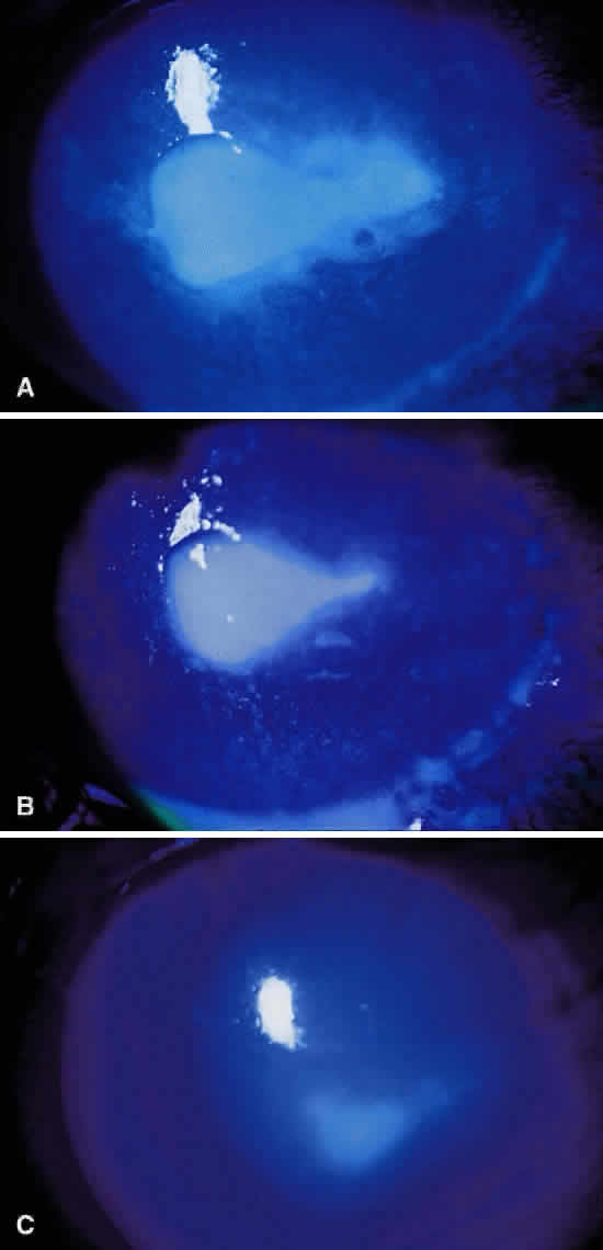
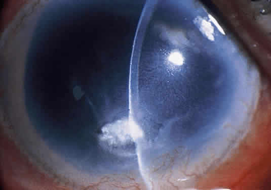
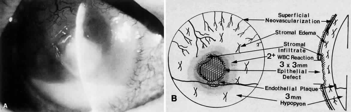
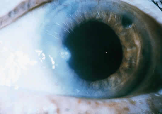
Clinically, the infected staphylococcal corneal ulcer presents with a yellow-white, well-demarcated area of infiltrate, which appears directly beneath an epithelial defect (Fig. 2). On occasion, multiple, small satellite lesions may develop. The infection may initially be superficial; however, if inadequately treated it can produce a mid to deep stromal abscess that may eventually lead to perforation. Stromal edema and white blood cell migration frequently surround the dense infiltrate and clear as the infection comes under control. Although there may be a marked anterior chamber reaction with hypopyon, the ulcer more frequently is indolent, with only a minimal inflammatory reaction.34

Fig. 2. Staphylococcus aureus corneal ulcer in a 70-year-old woman wearing an extended-wear aphakic soft contact lens. Note the dense white, well-demarcated stromal infiltrate beneath a larger epithelial defect (arrows).

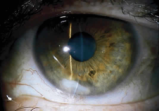
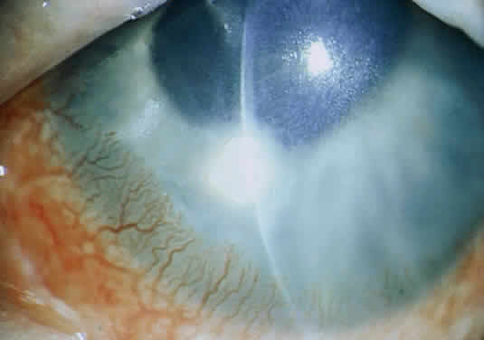
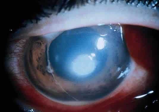
The more peripheral marginal infiltrate is sterile and is caused by a hypersensitivity reaction to the exotoxin or the bacterial antigens of S. aureus.35,36 The peripheral infiltrates usually are associated with blepharitis or conjunctivitis and frequently begin in the areas where the lid margins cross the limbus (at 2-, 4-, 8-, and 10-o'clock positions). The lesions often have a typical clinical appearance characterized by one or more small, well-circumscribed anterior stromal infiltrates, with a lucid interval between them and the limbus (Fig. 3). Although the epithelium is usually intact, it may break down, leading to ulceration, scarring, and neovascularization.

Fig. 3. Staphylococcus aureus marginal infiltrate (arrows) associated with chronic blepharitis. The lesion is well circumscribed, with a lucid interval between it and the limbus. The infiltrate is sterile and involves the anterior stroma; the overlying epithelium is intact.

STREPTOCOCCI

The streptococci consist of a diverse collection of organisms that have been classified into many different groups based on antigenic properties, types of hemolysis, and growth characteristics.31 The most common of these organisms, cultured from infected corneal ulcers, are Streptococcus pneumoniae (pneumococcus), S. viridans (alpha-hemolytic streptococcus), S. pyogenes (beta-hemolytic streptococcus), and the group D streptococci S. faecalis (nonhemolytic streptococcus).

Although S. pneumoniae was once the most common cause of bacterial keratitis, it has been surpassed by Staphylococcus and Pseudomonas7,8 in the United States. However, S. pneumoniae remains the most common cause of bacterial keratitis in the developing world.30 S. viridans ulcers occur more frequently than those caused by the more virulent S. pyogenes, presumably because the former may be part of the normal flora of the upper respiratory tract. The less common E. faecalis (enterococcus) corneal infections have been reported in either immunosuppressed hosts or after epithelial corneal injury and direct transmission from the gastrointestinal tract.25 E. faecalis is the third leading cause of nosocomial infection in the U.S., and recently cases of vancomycin-resistant enterococcus have been reported.37,38

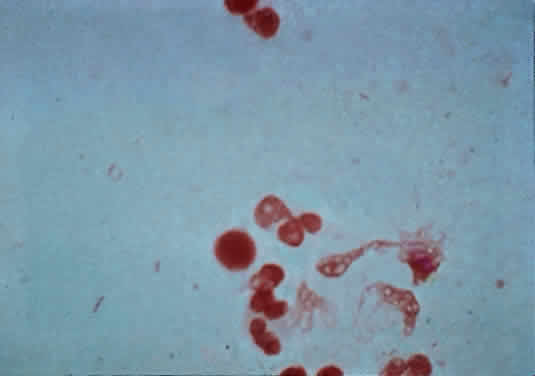
The microscopic appearance of all streptococci, except for pneumococcus, is similar. Smears taken from culture material show Gram-positive cocci in chains of varying lengths. Scrapings taken directly from infected ocular tissues may not demonstrate the chain effect, and differentiation from staphylococci may be difficult (Fig. 4). The morphologic characteristics of the Staphylococcus and Streptococcus organisms are outlined in Table 3.

Fig. 4. Gram-stained smear of Streptococcus organisms from a corneal ulcer. Note the fragmentation of the chains into smaller lengths, making differentiation from Staphylococcus difficult.


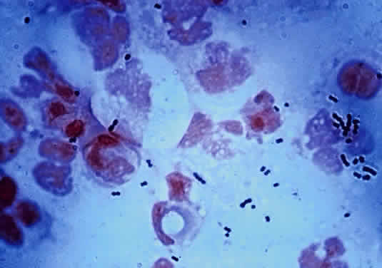
TABLE 18-3. Morphologic Characteristics of Staphylococcus and Nonpneumococcal Streptococcus on Gram-Stained Smear


FactorStaphylococcusStreptococcus
SizeLargerSmaller
ShapeSphericalMore oval
Associated with polymorphonuclear leukocytesYesNo (except for S. pyogenes)


The Gram stain of S. pneumoniae differs from that of other Streptococcus organisms by the presence of a polysaccharide capsule surrounding the lancet-shaped diplococci. Streptococci grow best on blood agar or nutritionally enriched media such as brain-heart infusion medium. Identification of the different species is aided by the hemolytic nature of the organism. With alpha hemolysis, the red blood cells are not completely lysed and a green color change is observed in the surrounding blood agar. With beta hemolysis, there is complete lysis of the red blood cells in the agar surrounding the Streptococcus colony. In general, the colonies of Streptococcus are elevated but quite small and measure between 1 and 2 mm in diameter. The pneumococci produce alpha hemolysis and may be differentiated in culture from S. viridans by the non-elevated, pitted appearance of the pneumococcal colony at 48 hours. In addition, the pneumococci produce capsules that impart a more translucent appearance to the colony.

Because the 24-hour culture of pneumococci may be easily confused with that of S. viridans (α-hemolytic streptococcus), laboratory tests are needed to make a definitive identification. Optochin susceptibility and bile solubility suggest S. pneumoniae.

Because all streptococci are fermentative organisms and lack the enzyme catalase, this characteristic may be used to help differentiate the nonhemolytic streptococci from similar-appearing colonies of Staphylococcus. The catalase test is useful in quickly determining whether a colony is a Streptococcus.

Although S. pneumoniae is an inhabitant of the upper respiratory tract in approximately 50% of normal human adults, it also is frequently found in the conjunctiva and lacrimal drainage apparatus. This close anatomical proximity to the cornea provides a source of organisms for possible infection. Any minor corneal trauma associated with disruption in the epithelium may lead to rapid invasion by this organism. The virulence of the S. pneumoniae organism is influenced by its polysaccharide capsule, which is serologically specific for more than 80 serotypes.4 Ocular isolates of these infections, however, do not seem to be caused by any particular pneumococcal serotype.39

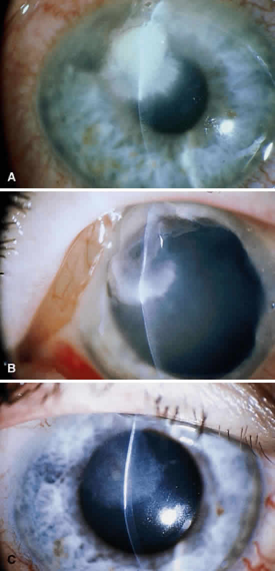
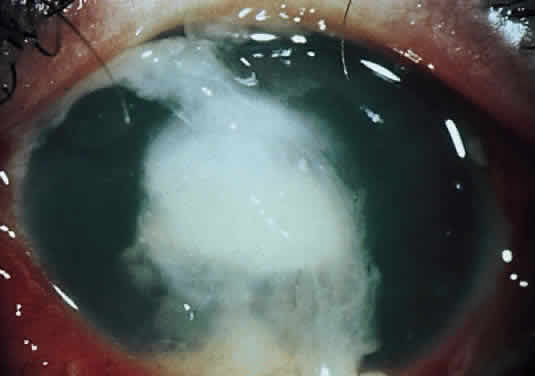
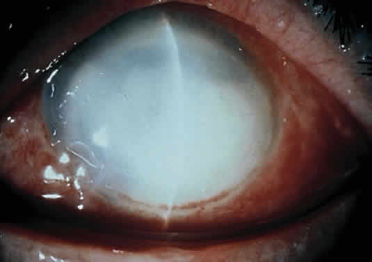
Corneal ulcers caused by S. pneumoniae are typically described as serpiginous or creeping and most often spread toward the center of the cornea. They are characterized by a gray-yellow disc-shaped ulcer with an overhanging margin at the advancing edge (Fig. 5). The ulcer usually progresses rapidly, extending into the deep stroma and often leading to corneal perforation. Extensive damage to corneal tissue, as well as a characteristic sterile hypopyon, are caused by the rapid production of bacterial exotoxin within the stroma.40,41

Fig. 5. Large, disk-shaped corneal ulcer caused by Streptococcus pneumoniae in an elderly woman. The presence of a sterile hypopyon is often associated with these ulcers.

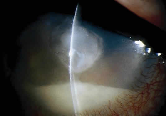
Streptococcus viridans (alpha streptococcus), although of low-grade pathogenicity, may be a cause of corneal ulceration, particularly in the locally immunosuppressed host.42 The organism produces a characteristically indolent, anterior stromal inflammatory reaction that is slow to progress and that may be accompanied by a minimal to moderate anterior chamber reaction. The ulcer is usually well circumscribed, gray-white, “dry” in appearance, and lying beneath a well-demarcated epithelial defect (Fig. 6). Because the organism is of relatively low-grade pathogenicity, the infection responds rapidly to specific antibiotic therapy.

Fig. 6. Streptococcus viridans corneal ulcer in a young woman wearing cosmetic soft contact lenses. The ulcer is well defined and dry in appearance with only a minimal anterior chamber reaction.

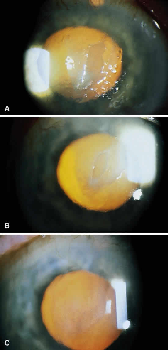
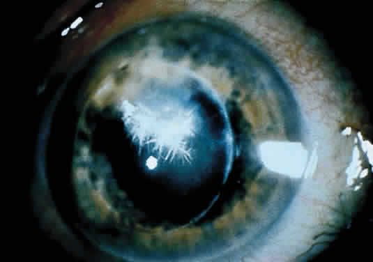
Infectious crystalline keratopathy is most commonly associated with S. viridans infection.43–45 This keratitis is characterized by discrete, white, crystalline, or fern-like stromal opacities, often under an intact epithelium, that are slowly progressive and have little associated corneal inflammation (Fig. 7). There is a strong association with prior topical corticosteroid therapy or penetrating keratoplasty. The condition has been termed infectious crystalline keratopathy and also has been reported with other organisms, including Haemophilus aphrophilus, Peptostreptococcus, and Staphylococcus epidermidis, as well as the fungal infections Alternaria and Candida tropicalis.46–49

Fig. 7. Infectious crystalline keratopathy caused by Streptococcus viridans. This infiltrate typically is slowly progressive, with little associated corneal inflammation.

A corneal biopsy may be necessary to gain access to the “crystalline” infiltrates for identification and culture of organisms. This may be accomplished using a disposable 2-mm dermatologic biopsy punch. These infections may be extremely resistant to therapy, requiring an extended course of antibiotics.

Streptococcus pyogenes (group A Streptococcus), although an important pathogen in other areas of medicine, is an infrequent cause of corneal infection. Marginal corneal ulcers associated with dacryocystitis have been reported with this organism.56 These marginal ulcers appear similar to the catarrhal ulcers associated with staphylococcal blepharitis. They may result from either a hypersensitivity or toxic response to the organism or from direct infection of the cornea.50

Enterococcus, a common organism found in the gastrointestinal tract, may produce a corneal ulcer in the presence of severely impaired host resistance or after epithelial injury. The organism produces a rapid and severe inflammatory response that can be devastating to the eye.

Back to Top
OTHER GRAM-POSITIVE ORGANISMS
Less common causes of infectious ulcerative keratitis that are classified as Gram-positive organisms include the aerobic, spore-forming bacilli Bacillus coagulans and B. brevis,51 as well as the non-spore-forming bacillus Corynebacterium diphtheriae.51 Bacillus infections of the cornea are associated with prior injury and produce severe local tissue damage. Corynebacterium diphtheriae corneal ulceration is a rare complication of conjunctivitis and can progress rapidly to perforation within 24 hours. The mechanism responsible for the rapid and massive dissolution of the cornea is presumably related to diphtheria toxin.52

Peptostreptococcus, an anaerobic Gram-positive coccus, may produce a corneal ulcer after injury to the cornea.53 Other rare causes of virulent corneal ulcers include the anaerobic spore-forming bacilli Clostridium perfringens and Clostridium tetani and the anaerobic non-spore-forming bacilli Actinomyces species and Propionibacterium acnes.54 Mixed aerobic and anaerobic bacterial infections also may occur and are frequently misdiagnosed and difficult to treat.

Other infrequent bacterial agents causing corneal ulcers include the nontuberculous mycobacteria55,56 and Nocardia species.3,4 These organisms are similar in appearance on scrapings and cultures and usually require 3 to 5 days to grow on blood agar. Stained smears show Gram-positive, slender rods that are variable in size. Mycobacterial ulcers occur after foreign body or surgical trauma.56 They are best isolated in Lowenstein-Jensen medium and require an aerobic atmosphere with an increased amount of carbon dioxide. Nocardia species are obligate aerobes and are best isolated on blood or Sabouraud's agar. Clinically, corneal ulcers caused by these organisms are indolent, progressing slowly for weeks to months, and are often recalcitrant to treatment.

Back to Top
GRAM-NEGATIVE RODS

PSEUDOMONAS

Pseudomonas is a Gram-negative rod that is ubiquitous in our environment and is a common cause of bacterial keratitis. The organism is a slender rod with parallel sides and rounded ends. There are 30 species within the genus; however, Pseudomonas aeruginosa is by far the one that is most often implicated in keratitis. Xanthamous P. maltophilia and P. cepacia are also well recognized corneal pathogens; P. stutzeri and P. acidovorans are blamed more rarely.57 The members of this genus are usually free-living bacteria and are considered opportunistic pathogens.

Pseudomonas aeruginosa is easily seen on Gram stain, although there tends to be a paucity of organisms (Fig. 8), in contrast to pneumococcal ulcers (see Fig. 4). The organism is easily grown on a variety of substances and can tolerate a wide temperature range, but is an obligate aerobe. The oxidase test is positive early in almost all pseudomonas infections and can be helpful for rapid identification. Pseudomonas aeruginosa has a distinctive “fruity” odor, is extremely active and motile on wet mounts, and elaborates two pigments (fluorescein and pyocyanin) with a striking blue-green color.

Fig. 8. Gram-stained smear of Pseudomonas organisms (arrows). Typically, there is a paucity of organisms present, and care must be taken to identify the slender gram-negative rods.

It has been demonstrated experimentally in mice that corneal injury is often a predisposing cause of Pseudomonas keratitis, because it allows bacteria to adhere to damaged epithelial cells and exposed stroma.24,58 Considerable biochemical and experimental evidence indicates that Pseudomonas can elaborate an extracellular protease (not a collagenase as suggested in early reports59) capable of destroying the corneal stroma.60–62 Several investigators have also demonstrated the ability of P. aeruginosa to produce exotoxin-A, which can inhibit protein synthesis and is similar to diphtheria toxin.63,64 The exotoxin is capable, when injected intrastromally into rabbit corneas, of producing loss of epithelium, endothelium, keratocytes, and opacification of the cornea.65 This effect is dose related and can be completely neutralized with a specific antitoxin. All of the above evidence helps explain why Pseudomonas corneal ulcers are potentially so devastating and destructive even after the active replicating bacteria have been killed with antibiotics.

Of the Gram-negative bacteria that cause keratitis in the U.S., Pseudomonas is the most common, and in some areas, such as Miami, it is the most common cause of corneal ulcers.7 These infections have commonly been traced to contaminated eye makeup,66,67 fluorescein solutions,68 and contact lens cases.10 They also have been associated with abnormal epithelium such as occurs with bullous keratopathy and after herpes simplex keratitis.26 Pseudomonas aeruginosa corneal ulcers often have been seen in patients using daily-wear contact lenses,11,12 extended-wear cosmetic lenses,12,14,15 and aphakic extended-wear lenses.11,15 Less common infective sources include the patient's own sputum and tracheostomy sites.69,70

Clinically, a bacterial corneal ulcer caused by Pseudomonas aeruginosa evolves quite rapidly and involves large areas of the cornea. It is frequently seen in a debilitated, elderly patient, and especially in those wearing extended-wear aphakic contact lenses.11,15 It generally begins centrally with a gray infiltrate that has an overlying epithelial defect. The adjacent cornea often has a hazy appearance secondary to epithelial and stromal edema. The ulcer commonly has a yellow green discharge and a “soupy” appearance (Fig. 9). The discharge contains both fluorescein and pyocyanin pigments and fluoresces with Wood's lamp (but not with the cobalt blue light of the slit lamp biomicroscope).71

Fig. 9. Rapidly progressive Pseudomonas aeruginosa corneal ulcer in a 65-year-old man. Note the characteristic soupy appearance of the infiltrate and the mucopurulent discharge adherent to the ulcer bed.

Ring abscesses can occur and may represent antigen-antibody reactions to exotoxin or other substances.72 A large, sterile hypopyon is frequently present and should not be mistaken for endophthalmitis. The infection spreads rapidly in all directions without treatment and can involve the sclera, with serious consequences, or can cause corneal destruction with perforation.73 Occasionally, the clinical course is much more indolent.71 In patients with HIV infection, the keratitis may be much more resistant to therapy.74

Treatment of these ulcers is often quite difficult; many resistant strains have been encountered. It is important to be aware of the optimal treatment for this common corneal ulcer and to modify therapy appropriately.

PROTEUS

Infections of the cornea with Proteus organisms are somewhat rare, but are very severe when they do occur. The organism is prevalent in the environment and can be cultured from many seemingly normal eyes.75 These organisms are members of the family Enterobacteriaceae and are commonly found in the intestinal tracts of humans and animals. There are four species in the genus: Proteus vulgaris, P. mirabilis, P. myxofaciens, and P. penneri. P. mirabilis is most commonly isolated from corneal ulcers. The other species are rarely found in eye cultures.

The organisms are Gram-negative rods that vary in length according to the amount of time in culture. In early cultures they appear very long, in snake-like forms, but become much shorter in older cultures and can resemble coccoid rods. Characteristic of the organism is its ability to “swarm” over a culture plate in hours, which produces a sense of wavelike growth. This ability is owing to the presence of many flagella. Like many enteric organisms, it also has many pili, or fine hairlike structures whose functions and purposes are unknown.

The Proteus organisms are nonfastidious and grow rapidly on almost any medium. They exhibit a characteristic, strong, offensive odor when grown on rich media. When identification is not obvious, the more specific tests, such as the phenylalanine deaminase test (only Proteus is positive for this enzyme), may be used. The urease test is occasionally helpful; however, other bacteria are also capable of producing this enzyme, so it is far less specific. The indole test is helpful to speciate Proteus, because only P. mirabilis does not produce indole.76 The ornithine decarboxylase test also is helpful to separate species: only P. mirabilis and P. morganii are associated with this enzyme.

The Proteus species elaborate a number of degradative enzymes such as ureases, deaminase, decarboxylase, gelatinase, and lipases.77 Like all enteric Gram-negative bacteria, Proteus also produces endotoxin, which may play a role in pathogenesis. Clinically, Proteus causes an intense keratitis, which is characteristic of most enteric bacteria. Dense stromal infiltration with occasional ring abscesses and even perforation has been seen.

SERRATIA

Serratia marcescens was considered to be a benign saprophyte found in water, soil, and food. It has since been reported to cause central corneal ulceration.78,79

Serratia is an aerobic, Gram-negative rod of the Enterobacteriaceae family. It often appears as sheets of Gram-negative rods on gram stain and grows easily on blood agar and enriched broth. There are many species; however, only S. marcescens has been implicated in human disease.

Two cornea-damaging proteases have been isolated and implicated in its pathogenesis. The organism is most commonly associated with contact lens wear and locally compromising ocular conditions, such as bullous keratopathy, neurotrophic keratitis, or keratitis sicca.78 There is a report of three postkeratoplasty patients who developed Serratia corneal ulcers that were traced to the outer grooves of medication bottle tops and the inner surface of eye-drop caps.79 The solution inside these bottles did not yield the organism; the moisture that collected between the cap and the bottle was apparently a culture medium for Serratia.

Clinically, the organism often produces a large yellowish or grayish infiltrate beneath an irregular epithelial defect. There is often significant tissue destruction and necrosis associated with a large hypopyon. Like Pseudomonas, the acute ulcer is associated with stromal edema and swelling, followed by thinning, and occasionally, perforation. As with most bacteria, Serratia has no characteristic that allows certain identification on clinical grounds alone.

MORAXELLA

Moraxella species were originally discovered as ocular pathogens and were arbitrarily divided into groups that are not well founded on clinical or bacteriological grounds.80,81 The organism is a noncapsulated, short, plump diplobacillus that can be confused clinically with Neisseria. It stains Gram-negative but occasionally may retain crystal violet and appear Gram-positive, resembling Streptococcus pneumoniae. The species that most commonly cause corneal ulceration are Moraxella liquifaciens, M. nonliquifaciens, M. lacunata, and rarely M. osloensis. M. lacunata was formerly believed to be the only cause of angular blepharitis and conjunctivitis; however, it also has been well documented as a cause of central corneal ulcers with hypopyon.82,83

The Moraxella organisms are fastidious and often difficult to grow in culture, especially M. lacunata. The organism needs a humid environment to grow, and fresh blood or chocolate agar with a moist surface is essential. Loeffler coagulated serum is an excellent medium for speciating Moraxella. All species are coagulase positive. The organisms are more commonly encountered in hot, dry areas of the world, which seems contradictory to their growth needs in the laboratory. The reservoir of the organism seems to be the human respiratory tract. Moraxella corneal ulcers are often found in alcoholic or debilitated patients with a compromised immune function.84 The pathogenesis of these ulcers is related to the production of proteases. Endotoxin activity also has been demonstrated,85 and may also contribute to morbidity.

Clinically, the ulcer often starts centrally or inferiorly in areas of exposure. There is initially a gray-white, dense, anterior stromal abscess with an overlying well-delineated epithelial defect. The infiltrate spreads deeply into the cornea, forming a stromal abscess. The ulcers are often painless, but almost invariably cause a hypopyon and occasionally a hyphema. The organism tends to be sensitive to most antibiotics and is readily sterilized, however complications such as descemetocele or perforation may occur despite appropriate antibiotic therapy.86 These ulcers are very slow to heal; it may take weeks before the epithelium is intact.

Back to Top
OTHER GRAM-NEGATIVE ORGANISMS
Certain other Gram-negative organisms also have been known to cause corneal ulcers. Both Neisseria gonorrhoeae and Neisseria meningitidis are infrequent causes of ulcerative keratitis. Either may invade intact corneal epithelium after untreated or inadequately treated conjunctivitis. Most enteric bacteria are aerobic, non-spore-forming, gramnegative rods that may rarely cause keratitis. Klebsiella pneumoniae, Escherichia coli, and Enterobacter aerogenes can produce indolent corneal ulcers that are usually seen in debilitated or immunocompromised patients or in corneas with an underlying pathologic condition.4 Haemophilus aegyptus and Haemophilus influenzae are rarely isolated causes of more virulent corneal ulcers.4 These organisms often grow at the border of colonies of Staphylococcus (satellitism) and have become increasingly resistant to ampicillin. Aeromonas hydrophilia is a Gram-negative rod commonly found in water, soil, and food, and it also has been reported to be a causative organism in ulcerative keratitis.87 Shigella and Salmonella also are rare causes of microbial keratitis in the United States.88
Back to Top
CLINICAL APPROACH
Bacterial identification in suppurative microbial keratitis is crucial for appropriate therapy and the prevention of potentially severe complications. A thorough history, detailed clinical examination, and meticulous microbiological technique are essential steps in successful management. The clinician should include each step routinely in the work-up of patients presenting with a corneal ulcer and avoid the temptation to eliminate portions of the diagnostic plan.

The symptoms of corneal ulcers are similar in most patients, but may differ depending on the severity of the infection. They include decreased visual acuity, photophobia, pain, redness, swelling, and discharge. The severity may vary in relation to the underlying causative organism, the condition of the host, and the duration of the symptoms before the patient is examined.

The initial clinical examination should be thorough and should include evaluation of the lids and conjunctiva as well as the cornea, anterior chamber, and posterior segment of the eye. All periocular and ocular abnormalities, including intraocular pressure, should be accurately documented at this examination.

In general, patients presenting with bacterial corneal ulcers develop moderate to severe lid and conjunctival edema and inflammation as well as a purulent discharge. A papillary conjunctival response predominates, but may be masked by chemosis. The corneal epithelium becomes ulcerated, and the underlying stroma shows a dense, gray-white, necrotic-appearing infiltrate. The borders of the epithelial defect are sharply defined, but the edges of the stromal infiltrate are indistinct and often extend beyond the epithelial defect. White blood cell infiltration and edema of the cornea surround the ulcer, and fibrin plaques may be present on the endothelium. The anterior chamber reaction may range from mild to severe, but is considered to be sterile so long as Descemet's membrane remains intact. A hypopyon may be present and is the result of the toxic effects of the organism on the vessels of the iris and ciliary body. The severe inflammatory reaction from virulent bacterial corneal ulcers may lead to the development of cataracts, anterior and posterior synechiae, elevated intraocular pressure, descemetocele, perforation, and permanent scarring and edema of the cornea.

Although these clinical findings are widely observed in most bacterial corneal ulcers, clinical features that frequently characterize certain groups of infecting bacteria are worth noting. The recognition of these features may be helpful in attempting to determine the responsible causative organism early in the management of the ulcer. In general, Gram-positive cocci such as Staphylococcus aureus and Streptococcus pneumoniae produce round or oval ulcers that are gray-white and dry in appearance, with distinct borders (see Fig. 5). These organisms almost always produce a severe anterior chamber reaction, particularly in the case of pneumococcal ulcers, in which a sterile hypopyon frequently occurs. The Gram-negative rods, however, usually produce a more profuse, wet or soupy appearing infiltrate that is rapidly progressive and spreads to involve the entire cornea (see Fig. 9). The mucopurulent discharge produced by Pseudomonas aeruginosa typically is yellowish-green and clings tenaciously to the ulcer's surface.25 The anterior chamber reaction produced by the Gram-negative rods also is quite intense and includes hypopyon formation.

During the initial clinical examination of the patient, a detailed drawing should be made of the corneal ulceration (Fig. 10). This drawing should include accurate measurements of the size and shape of the epithelial defect and stromal infiltration using the slit beam ruler (Fig. 11). An estimation should be made of the relative density of the stromal infiltration and the surrounding white blood cell reaction, as well as the depth of the stromal infiltration and appearance of the infiltrate borders. In addition, careful attention should be paid to the anterior chamber reaction, including keratic precipitates on the endothelium. Documenting these specific findings at the initial examination is important for providing a “starting point” or basis against which daily comparisons can be made once treatment has commenced (see Therapeutic Approach). The use of slit-lamp photography also can be extremely helpful in documentation and comparison of the changing characteristics of the ulcer.

Fig. 10. A. Slit-lamp photograph of the initial appearance of a Streptococcus pneumoniae corneal ulcer before the microbiologic workup. B. Clinical drawing of the same ulcer documenting important signs key to the therapeutic management of this problem. Daily drawings should be performed by the clinician to document these changes in the course of the ulcer.

Fig. 11. Using the slit-beam ruler to measure the size of the epithelial defect, stromal infiltrate, and hypopyon in the management of a corneal ulcer.

Because the patient's history and clinical appearance are inadequate for making a specific bacterial diagnosis, laboratory investigation is mandatory for serious corneal ulcers. Although most laboratories have appropriate stains, incubators, media, and technicians to aid in the interpretation of this data, it is the clinician's responsibility to collect this data in an efficient and purposeful manner (see Laboratory Diagnosis). On many occasions, patients present with corneal ulceration when laboratory personnel are not available, requiring the ophthalmologist to be ready and able to perform the initial work independently. It is most helpful to have a prepared corneal ulcer diagnostic tray that is easily accessible and contains the necessary instrumentation for securing specimens. This tray should include the following (Fig. 12): alcohol lamp and matches, platinum (Kimura or Lindner) spatula, sterile calcium alginate swabs, clean glass slides, topical anesthetic drops, grease marking pen, and laboratory forms for bacteriology.

Fig. 12. Contents of the corneal ulcer tray containing the necessary items for securing specimens for laboratory study: (1) alcohol lamp and matches, (2) platinum (Kimura) spatula, (3) sterile swabs, (4) glass slides, (5) topical anesthetic drops, (6) grease marking pen, (7) laboratory forms, (8) culture plates (must be fresh from laboratory).

Prior discussions with the bacteriology laboratory regarding the location of culture media, stains, and a microscope help avoid frustrating situations when laboratory personnel are not in attendance. Once the microbiologic work-up has been completed and the stained smears have been carefully examined, the therapeutic approach should be formulated and treatment begun.

Back to Top
LABORATORY DIAGNOSIS
Some organisms may have a fairly characteristic clinical appearance (e.g, Pseudomonas), which may allow the clinician to strongly suspect a specific causative agent in the case of ulcerative keratitis (see Clinical Approach). However, the variable response of the cornea and the potentially diverse clinical appearance mandate laboratory work-up for accurate diagnosis and rational antimicrobial therapy.

The initial procedure should be obtaining culture material from the conjunctivae and lid margins of both eyes. The use of anesthesia in this procedure should be avoided because preservatives in most topical anesthetics may cause a decreased yield of live organisms. Calcium alginate swabs are made of inert material (alginic acid) and are preferred to cotton swabs, which frequently contain fatty acids that inhibit bacterial growth.89,90 The swab should be moistened in a liquid medium (brain-heart infusion or thioglycolate broth) and the entire lower cul-de-sac should be wiped. The upper and lower tarsal conjunctivae and all of the material obtained is placed directly into culture. All conjunctival specimens should be plated directly onto blood agar, chocolate agar, Sabouraud's dextrose plates, and thioglycolate broth. The upper and lower lid margins also should be cultured and placed on the same plates used for the conjunctival specimens (Fig. 13).

Fig. 13. Conjunctival and lid specimens are plated onto appropriate media for culture. A separate swab is used for each streaking and is represented as follows: R, right lid margin; L, left lid margin; vertical streak, right conjunctiva; horizontal streak, left conjunctiva.

Next, the cornea in the affected eye is anesthetized using proparacaine hydrochloride (0.5%) because it is the least bacteriocidal of the topical anesthetic agents. Corneal specimens are taken under biomicroscopic guidance. A flame-sterilized platinum spatula (Kimura or Lidner) or calcium alginate swab may be used. Calcium alginate swabs moistened in a nutrient broth may yield a higher number of positive bacterial cultures compared with the platinum (Kimura) spatula technique.91 If the surface of the ulcer has mucus or necrotic debris on it, this must be removed (and cultured) before material that is more likely to contain live organisms can be obtained. Some organisms (e.g., Streptococcus pneumoniae) are more often found at the leading edge of an active ulcer; others (e.g., Moraxella) are more frequently found deep in the base of the ulcer. Therefore it is imperative to obtain material from both areas to optimize the yield. An ulcer may require multiple scrapings before live organisms are obtained. Each corneal scraping may be inoculated onto the surface of the media in a row of C streaks (Fig. 14). Each additional scraping should be plated as a new row of streaks. Penetration of the surface of the agar should be avoided because this can result in deep deposition of organisms, which hinders isolation. It is essential when culturing a corneal ulcer not to touch the lids or conjunctiva with the sterile spatula.

Fig. 14. Inoculation of corneal scrapings onto the surface of the culture medium with a flame-sterilized spatula.

Superficial infiltrates and copious mucopurulent material make culturing relatively easy. However, there may be only scanty amounts of material, very deep ulcers, or even an impending descemetocele; these circumstances limit the number of scrapings as well as the total amount of the specimen. Ideally, all of the various media should be used, but this is not always practical. Therefore, the ophthalmologist should have an order of priority, which may be adjusted according to clinical suspicions. The following list of the various culture materials available are given in the order in which we routinely use them:

  1. Blood agar
  2. Clean glass slides (2)
  3. Chocolate agar
  4. Sabouraud's agar
  5. Thioglycolate broth
  6. Brain-heart infusion broth

The work-up for a possible fungal ulcer is necessary because these ulcers can easily mimic bacterial ulcers.

In most cases of ulcerative keratitis in which a bacterial infection is strongly suspected, blood agar should be inoculated first. The medium readily supports the growth of most corneal pathogens and is used extensively, but it must be made “fresh” at least every 2 weeks. The plates should be incubated at 37°C and also at 25°C, if enough material is available.

Next, material should be spread onto a clean (nonsterile) glass slide in a thin, even manner to allow for Gram and Giemsa staining and microscopic examination.92,93 The Gram stain demonstrates certain characteristics of most bacteria; however, caution must be used in the interpretation of results. An inability to see the organisms on the slide may be caused by insufficient material, failure to examine the entire slide, or prior antibiotic therapy. Acridine orange is an alternate stain for bacterial identification. Although data are limited, acridine orange appears to have an equivalent specificity and possibly a higher sensitivity than Gram stain for cases of bacterial keratitis.94,95

Factors that can alter the staining characteristics of bacteria include excessive heat fixation, prior antibiotic therapy, long standing infections, and improper staining techniques. If enough material is available, Giemsa stain should be performed on all ulcers when there is any possibility of fungi being present. This stain is also excellent for discerning cellular details and response, and for demonstrating intracytoplasmic inclusions, multinucleated giant cells, and the crosswalls of fungal hyphae.96 Methenamine silver is the best stain for fungi,97 however, and Papanicolaou's stain is the best for demonstrating intranuclear inclusions. Acid-fast stains should be used when Mycobacterium fortuitum or Nocardia are suspected in cases of indolent corneal ulceration. Additional stains that may be helpful in identifying fungi are the potassium hydroxide preparation and the periodic-acid-Schiff (PAS) stain. However, only the Gram stain should be performed if the material is limited.

The next medium that should be used is chocolate agar. The material should be plated as described earlier and incubated at 37°C. This medium is used to enhance the isolation of Moraxella, Neisseria, and Haemophilus species, none of which grow well on blood agar. Although most bacteria grow on chocolate agar as well as on blood agar, the evaluation of the hemolysis pattern often permits certain differentiations to be made at an earlier stage with blood plates. Both Haemophilus and Neisseria grow much better in an enriched carbon dioxide atmosphere.

Sabouraud's agar is excellent for isolating fungi and should be used routinely at room temperature if enough material is present. The plates contain B vitamins, which are required for most fungi to grow, and a broad spectrum antibiotic (e.g., gentamicin) is added to minimize bacterial contamination. Cycloheximide should be avoided because it is an inhibitor of saprophytic fungi that are frequently the cause of fungal keratitis.

It may be helpful to use thioglycolate broth at 37°C, which promotes the growth of anaerobic and microaerophilic bacteria. An alternative, and possibly better, technique for these organisms is to place a blood agar plate in an anaerobic candle jar (with a high carbon dioxide atmosphere).

The brain-heart infusion broth is used at 25°C. This medium is helpful in the isolation of fastidious bacteria and fungi, and also for cultures taken from eyes previously treated with antibiotics. In cases of long-standing indolent ulceration, Loewnstein-Jensen agar is useful for Mycobacterium and Nocardia. Thayer-Martin agar is especially designed for the isolation of Neisseria. These plates also must be incubated in a high carbon dioxide atmosphere (e.g., candle jar).

Bacterial growth on two or more C streaks on any of these plates generally signifies that the isolate is the true causative agent of the ulcer (Fig. 15). Growth off the streaks is generally considered a contaminant. Growth from the lids and conjunctiva is helpful only when the corneal cultures are all negative. These adnexal cultures are especially of use when the one ipsilateral to the ulcer grows a pathogen not found in the other eye. The results of the various stains are helpful if they demonstrate Gram-negative rods, when they correlate with the growth of the corneal cultures, or when the cultures are negative. The clinician must be careful when using the staining results to guide the initial antibiotic therapy (see Therapeutic Approach).

Fig. 15. Blood agar culture plates showing growth of bacteria on the “C” streaks from the involved cornea (left). The lid margins and conjunctiva (right) show significantly more bacterial growth from the right eye.

The bacteria of most corneal ulcers begin to grow on some media within 24 to 48 hours. Some laboratories begin antibiotic sensitivity testing before the final species identification is made. This may allow rational adjustments in therapy when correlated with the clinical response. In the future, the use of polymerase chain reaction may allow the rapid identification of bacterial pathogens from corneal ulcers.

Back to Top
THERAPEUTIC APPROACH
After completion of the clinical and microbiologic work-up, a therapeutic plan is developed to most effectively manage the infected corneal ulcer. The goals of therapy include the eradication of viable bacteria from the cornea and rapid suppression of the inflammatory response elicited by the causative microorganisms. These goals are best achieved by prompt initiation of specific antimicrobial therapy. Finally, after resolution of the ulcer, attention should be given to correction of any predisposing conditions that contributed to the development of the infection (see Table 1).

In cases of infectious corneal ulceration, many authorities advocate an initial therapy of broad spectrum antibiotics, with modifications of this therapy based on the clinical impression and Gram stain. Several authors have reported an approximately 60% correlation between the Gram-stained corneal smear and the cultured bacterial organisms that are later isolated.7,9 Data from the Gram stain must be interpreted with these figures in mind, and probably should not be used alone for selection of a narrow spectrum antibiotic therapy. In cases in which inadequate material is obtained for smear or in which no organisms are identified, one must rely on treatment with broad spectrum antibiotics until organisms are identified on culture.

It is important to asses the severity of the microbial keratitis at the initial examination to determine whether hospitalization is indicated and the relative level of antibiotic therapy that may be required. Using a modification of Jones's grading for the severity of microbial keratitis (Figs. 16 to 18),9 the following general guidelines are offered (Table 4).

Fig. 16. Nonaxial, small anterior stromal corneal infiltrate with minimal anterior chamber reaction.

Fig. 17. Central, moderately sized corneal ulcer involving the mid stroma and producing a significant anterior chamber reaction.

Fig. 18. Large, necrotic-appearing corneal ulcer extending into the deep stroma and causing severe anterior segment inflammation.


TABLE 18-4. Keratitis Severity and Initial Therapy


FactorGrade IGrade IIGrade III
LocationNonaxialCentral or peripheralCentral or peripheral
Area2 mm2–6 mm6 mm
DepthSuperficial one thirdSuperficial two thirdsExtending to inner one third
Anterior segment inflammationMildModerate or severe; fibrinous exudateSevere; hypopyon
Hospitalization*NoPossiblyPossibly
Initial antibiotic therapy†Topical fortified dropsTopical fortified dropsTopical fortified drops
   Consider intravenous antibiotics‡

*The need for hospitalization is based not only on the severity of the corneal ulcer but also on the patient's ability to comply with the rigorous medication schedule and reliability in returning for frequent follow-up visits.
†Subconjunctival antibiotics may be considered in cases in which there may be a delay or inability to administer topical medications.
‡If perforation or scleral extension present.


The rationale for topical, subconjunctival, and intravenous antibiotic therapy is discussed later in this chapter. Modification of initial antibiotic therapy based on the morphology of the bacteria seen on Gram stain is summarized in Table 5. Fluoroquinolones may represent a single-drug broad spectrum therapy for infectious keratitis. They may be particularly useful as the first line of therapy for dry-appearing, small corneal infiltrates with minimal epithelial defects (see Fig. 16).


TABLE 18-5. Modification of Initial Antibiotic Therapy Based on Gram-Stained Smear Morphology


Smear MorphologyTopical*SubconjunctivalIntravenous†
No organismCefazolin (50 mg/ml) and gentamicin (13.6 mg/ml) or tobramycin (13.6 mg/ml)Cefazolin (100 mg) and gentamicin (20 mg) or tobramycin (20 mg)Cefazolin (0.5–1 g/8 hr) and gentamicin (3–7 mg/kg/day) or tobramycin (3–5 mg/kg/day)
Gram-positive cocciCefazolin (50 mg/ml) or bacitracin (10,000 units/ml)Cefazolin (100 mg) or methicillin (100 mg)Cefazolin (0.5–1 g/8 hr) or methicillin (200 mg/kg/day)
Gram-positive rodsGentamicin (13.6 mg/ml) or tobramycin (13.6 mg/ml)Gentamicin (20 mg) or tobramycin (20 mg)Gentamicin (3–7 mg/kg/day) or tobramycin (3–5 mg/kg/day)
Gram-negative cocci Penicillin G (100,000 units/ml) or bacitracin (10,000 units/ml) Penicillin G (500,000 units/ml)Penicillin G (2–6 million units/4 hr)
Gram-negative rodsTobramycin (13.6 mg/ml) and ticarcillin (6.7 mg/ml)Tobramycin (20 mg) and ticarcillin (20 mg)Tobramycin (3–5 mg/kg/day) or ticarcillin (200–300 mg/kg/day)

*See discussion on fluoroquinolones in therapeutic approach.
†Choice of drug or dosage must be adjusted in patients with impaired renal function. Intravenous antibiotics should only be considered if perforation or scleral extension is present.


If no organisms are seen on the initial Gram stain smear, a decision must be made as to whether or not the ulcer is infectious. If the ulcer is judged to be noninfectious, treatment may be deferred for 12 to 24 hours, with re-examination at that time. It is important that the clinician be familiar with the signs of an active corneal infection so that treatment is not unduly delayed. When in doubt, it is safest to initiate broad spectrum treatment in an attempt to control the infectious process at an early stage, rather than waiting for culture results or clinical progression.

Once antibiotic treatment has been instituted, management of the microbial keratitis is based on the careful modification of therapy and the judicious use of adjunctive measures to promote rapid healing. The decision to modify therapy is based on several factors, including clinical response to initial therapy, preliminary culture results, and tolerance of the antimicrobial agents.

The clinician must examine the patient on a daily basis after the initiation of antibiotic therapy and compare subsequent findings with the original appearance of the ulcer. As stated earlier, it is essential in the work-up of the patient to document with a clinical drawing or photograph the extent of involvement and the main characteristics of the infectious process (see Fig. 10).

The corneal ulcer at 18 to 24 hours may begin to show subtle signs of improvement if appropriate antibiotic therapy has been instituted. Specifically, evidence of a decreasing epithelial defect (Fig. 19); reduction in the size, density, and edges of the stromal infiltrate (Fig. 20); and a decrease in the surrounding white blood cell reaction (Fig. 21), as well as the anterior chamber reaction, indicate clinical improvement of the corneal infection (Table 6). Recognition of these clinical signs is crucial when beginning the modification of therapy and should be used to reach a therapeutic decision in combination with the preliminary culture results. The clinician should check with the laboratory within 18 hours of obtaining the cultures because tentative bacterial identification is often possible at that time. It is important to note, however, that the initial antibiotics employed should be continued for at least 36 to 48 hours if clinical improvement is observed, despite preliminary results from the laboratory. If, on the other hand, there has been deterioration despite 24 to 48 hours of antibiotic therapy, therapeutic changes should be made based on culture results. In some cases the stromal infiltrate may improve but the epithelium and conjunctiva worsen because of the toxicity of the fortified topical antibiotics. Thus, a patient may have an improved stromal infiltrate in the setting of an enlarged epithelial defect and increased conjunctival chemosis after 24 hours of fortified topical antibiotics.98 This problem may be remedied by reducing the frequency and concentration of the applied antibiotics. The clinician should be aware that many of the concentrated topical antibiotics are potentially toxic to the ocular surface, and a worsening of this aspect of the clinical picture may not reflect a true progression of the microbial keratitis.

Fig. 19. Clinical signs of improvement in the healing of a corneal ulcer: decrease in corneal epithelial defect during treatment of a Serratia marcescens corneal ulcer. A. Initial defect. B. Three days of therapy. C. Ten days of therapy.

Fig. 20. Clinical signs of improvement in the healing of a corneal ulcer: reduction in size and density of the stromal infiltrate and “blunting” of the infiltrate edges with treatment of a Pseudomonas aeruginosa corneal ulcer. A. Initial appearance. B. Two days of therapy. C. Seven days of therapy.

Fig. 21. Clinical signs of improvement in the healing of a corneal ulcer: decrease in surrounding stromal white blood cell reaction during treatment of a Serratia marcescens corneal ulcer. Note also the decreasing epithelial defect. A. Initial appearance. B. Three days of therapy. C. Fourteen days of therapy.


TABLE 18-6. Important Clinical Signs in Microbial Keratitis


SignImprovementWorsening
Epithelial defect (size)No change or smallerLarger
Stromal infiltrate  
 DensityDecreasedIncreased
 BordersMore distinctLess distinct
 DepthNo changeDeeper
 SizeNo change or smallerLarger
Stromal white blood cell reactionDecreased (localized)Increased
Anterior chamber reactionDecreasedIncreased


Antimicrobial sensitivities using quantitative and standardized disc-agar diffusion techniques indicate relative efficacies of the various antibiotics for the causative bacteria. The results of sensitivity testing are usually available within 36 to 48 hours after inoculation of the culture media and often prove to be quite helpful in the modification of antibiotic therapy. In most cases, the drugs chosen initially are effective, but adjustments of antibiotic therapy should be based on the results of sensitivity testing.27 If two antibacterial agents were initially chosen to provide broad spectrum coverage, the less effective drug may be discontinued. In a similar fashion, a change in antibacterial therapy should be considered if progression of corneal suppuration occurs despite 48 hours of intensive antibiotic therapy.2

Antibacterial sensitivity testing is also helpful in determining an alternative medication when toxicity or allergic reactions to a specific drug develop.

The tapering or reduction of antibiotic therapy after 24 to 36 hours of frequent fortified topical medications is probably the most poorly handled step in the management of infected corneal ulcers. Careful attention must be paid to clinical signs of healing, and appropriate adjustments must be made to avoid unnecessary side effects. A suggested schedule for tapering topical and adjunctive medications is offered; the patient's response to therapy, however, is the ultimate guide in adjusting medication schedules (Table 7).


TABLE 18-7. Suggested Schedule for Tapering Topical and Adjunctive Medications


ScheduleTreatment
Initial treatmentTopical Fortified Antibiotic A Every Hour (on the Hour)
 Topical fortified antibiotic B every hour (on the half hour)
 First doses given as a “loading dose”: 1 drop every minute for 5 min
 Possible subconjunctival or IV therapy (see Table 3)
36–48 hrReduction in therapy if clinical improvement occurs
 Topical fortified antibiotic A every 2 hr
 Topical antibiotic B: either discontinued or used every 2 hr, 5 min after antibiotic A
 Discontinue subconjunctival antibiotic injections
48–72 hrTopical fortified antibiotic A every 3–4 hr
 Use antibiotic ointment at bedtime; discontinue medication after bedtime
96+ hrChange to regular strength antibiotic drops and slowly taper off this medication
 Continue use of antibiotic ointment at night for approximately 1 wk
 Consider addition of a topical steroid after organism is identified and treated with an antibiotic to which the organism is sensitive and the epithelium has healed


Extreme care must be taken in the reduction of the medication for all corneal ulcers that are caused by Gram-negative rods because of their marked virulence and potential for rapid progression. It is often helpful in these cases to repeat the corneal smears and cultures after 48 to 72 hours, before changing the initial dosage regimen. If there is no growth on the repeat cultures, the subconjunctival injections may be discontinued, with continuation of the concentrated topical antibiotics at the same frequency for an additional 48 to 72 hours, depending on the clinical response. Once definite signs of clinical improvement are observed, the topical drops may be tapered as previously suggested.

In cases in which cultures show no evidence of aerobic or anaerobic growth by 48 to 60 hours, the physician must re-evaluate the clinical situation. If there are signs of improvement (see Table 6), the antibiotic treatment schedule should be tapered in accordance. In the event of further worsening of the corneal ulcer, the possibility of diseases mimicking bacterial keratitis should be considered. These include HSV keratitis, staphylococcal marginal infiltrates, acanthamoeba keratitis, fungal keratitis, corneal phlyctenule, and topical anesthetic abuse. Repeat scrapings and cultures should be obtained for bacteria, fungi, amebae, and herpes and withdrawal of antibiotic therapy for 24 to 48 hours should be considered until a new diagnosis can be made. As mentioned earlier, the possibility of toxicity from concentrated antibiotics also must be considered as a causative factor in a deteriorating clinical picture.

Back to Top
ANTIBIOTIC THERAPY
The primary treatment for bacterial ulcerative keratitis is intensive administration of antibiotics. For the clinician to treat these infections optimally and safely, it is necessary to be familiar with current antibiotic therapies. This includes an understanding of each drug's mechanism of action, spectrum of activity, appropriate dosage, and potential for toxicity.

The choice of the initial antibiotic in suspected cases of infectious corneal ulcers is usually made according to one of two basic schemes. One method is to use the results of the Gram and Giemsa stains to help guide the initial selection of drugs. The rationale for this scheme is that by using the most specific antibiotics possible, one achieves the greatest effect while minimizing toxicity. The other approach is to use the so-called shotgun therapy, which may be varied slightly according to specific staining results and clinical data. The rationale of this method is to use a combination of drugs that have an extremely broad antibacterial spectrum while awaiting a laboratory diagnosis from cultures. This avoids the potential pitfalls of inaccurate staining results and varied clinical presentations.

As stated earlier, for cases of ulcerative keratitis in which bacteria are suspected, most authorities advocate the initiation of broad spectrum therapy after obtaining material for culture and stains. The choice of drugs for coverage may be influenced by the clinical picture and results of the stains to some degree, but corneal infections can be varied in appearance and staining results do not always reflect the true causative organism. Infectious corneal ulcers can progress so rapidly without treatment that it may be dangerous to wait for culture results (24 to 48 hours) before initiating therapy.

ROUTES OF ADMINISTRATION

Numerous reports have shown that systemic administration of antibiotics is ineffective in treating bacterial corneal ulcers.26,98–102 This is because of the very small proportion of administered drug that is actually delivered to the cornea after the systemic dose is diluted in the total blood volume and tissue water.101 This lack of efficacy, in the face of potential systemic toxicity, makes parenteral administration a poor choice. This route of drug delivery is reserved for corneo-scleral ulcers, perforated ulcers (or those with impending perforation), post-perforating injury ulcers, and infections caused by bacteria known to require systemic therapy (e.g., Neisseria, Haemophilus).

The current mainstay of therapy for infectious bacterial ulceration is frequent administration of topical fortified antibiotics. In cases of corneal infiltration without significant epithelial defect, commercially available topical fluoroquinolones are an alternative therapy. Some controversy exists regarding the use of ointments versus solutions as the delivery vehicle. While it is true that an ointment mechanically remains in contact with the eye longer, the drug/ointment solubility relationship may lead to a slower diffusion of drug into the eye than would be desired. Because of this uncertainty, and more importantly because of the ability of pharmacies to easily make high concentrations of antibiotics in solution, the standard medications used are drops.

Several authors have shown that commercial preparations of aminoglycosides (3 mg/ml) are ineffective in many cases of clinical and experimentally induced Pseudomonas keratitis, whereas fortified preparations (9 to 40 mg/ml) are effective.103–106 The concentrations and specifics of preparation of commonly used topical fortified antibiotics are given in Table 8.


TABLE 18-8. Preparation of Commonly Used Fortified Topical Antibiotics


AntibioticsRemove*AddReplace*ContentsFinal Contents
Tobramycin gentamicin) 2 ml parenteral (40 mg/ml) to 5 ml commercial ophthalmic bottle of antibiotic (3 mg/ml) 55 mg drug in 6-ml solution13.6 mg/ml
Cephazolin7 ml tears2 ml tears to vial of parenteral cefazolin (500 mg)2 ml reconstituted drug into tear bottle500 mg drug in 10-ml solution50 mg/ml
Penicillin G5 ml tears5 ml tears to vial of penicillin G (5 million units)5 ml reconstituted drug into tear bottle5 million units in 15-ml solution333,000 units/ml
Ticarcillin15 ml tears10 ml sterile water to 1 vial carbenicillin (1 g)1 ml reconstituted drug into tear bottle100 mg drug in 15-ml solution6.7 mg/ml
Bacitracin9 ml tears3 ml tears to each of 3 vials bacitracin powder(IM) (50,000 units/vial)All 3 ml reconstituted drugs from each vial (9 ml total) into tear bottle150,000 units drug in 15-ml solution10,000 units/ml
Erythromycin1.5 ml tears5 ml sterile water to 1 vial erythromycin lactobionate (500 mg)1.5 ml reconstituted drug into tear bottle150 mg drug in 15-ml solution10 mg/ml
Vancomycin2 ml tears2 ml tears to vial vancomycin hydrochloride (500 mg)2 ml reconstituted drug into tear bottle500 mg drug in 15-ml solution33 mg/ml
Polymyxin B2 ml tears2 ml sterile saline to 1 vial polymyxin B sulfate (500,000 units)2 ml reconstituted drug into tear bottle500,000 units in 15-ml solution33,000 units/ml

*The amount removed is from the same 15-ml bottle of tear substitute to which the reconstituted drug is added. Be sure that the initial volume is 15 ml. It is generally safe to store fortified antibiotics for 1 week if refrigerated. Check package inserts for specific antibiotics.


The optimal frequency and intervals of administration of topical antibiotic drops also has been a subject of some controversy. Most authors have suggested giving drops every 30 to 60 minutes.107 It has been shown experimentally with topical prednisolone acetate that giving a drop every 15 minutes is significantly more effective than a drop every 60 minutes in reducing the number of invading polymorphonuclear leukocytes.108 This same study also showed that if five doses were given, 1 minute apart and every hour, the therapeutic effect was equal to drops every 15 minutes.

During the last few decades, bacterial keratitis was frequently treated with a combination of topical medication and adjuvant subconjunctival antibiotic injections. More recently, studies have shown that topical therapy alone is at least as effective as the combination therapy and reduces the morbidity associated with repeated subconjunctival injections.104,111,112

Subconjunctival antibiotic injections for bacterial ulcerative keratitis still may be considered in cases with impending perforation or when there is some question of the patient's ability to receive frequent administration of topical medication reliably (e.g., children or poorly compliant patients). Collagen shields (discussed later) are another alternative delivery method for these patients. Because some authors continue to recommend the use of periocular injections, the technique is described. There are no preparations of antibiotics specifically made for this administration, but any formulation suitable for either intramuscular or (preferably) intravenous use is acceptable. Subconjunctival injections may be extremely painful, particularly in an inflamed eye. Topical proparacaine 0.5% should be used liberally before injecting. Some patients may require the injection of lidocaine 2% into the same site before or in addition to the antibiotic dose. Despite these measures, patients may have considerable pain and anxiety regarding repeated injections and may benefit from systemic analgesia, such as meperidine. The antibiotics should be injected with a 30-gauge needle to balloon up the conjunctiva near the limbus as close to the ulcer as possible. In central ulcerations, the injection is placed wherever technically easiest. If additional injections are necessary, they may be performed at 12- to 24-hour intervals. The dosage of antibiotics for subconjunctival injection is given in Table 9.


TABLE 18-9. Dose of Subconjunctival Antibiotics


AntibioticDose
Gentamicin20 mg
Tobramycin20 mg
Cefazolin100 mg
Penicillin G500,000 units
Bacitracin*10,000 units
Erythromycin50 mg
Vancomycin25 mg
Polymyxin B10 mg

*Rarely used secondary to toxicity


Collagen shields have been advocated as a drug delivery method to treat infectious corneal ulcers and as prophylaxis against post-operative infections. They may provide an additional or alternative therapeutic route for the treatment of infectious corneal ulcers, particularly in unreliable or uncooperative patients who cannot effectively use conventional therapy.

Collagen shields are shaped like contact lenses and are cross-linked with either bovine or porcine scleral collagen. They biodegrade over varying time periods and are packaged in a dehydrated form. Prior to placement in the eye they are soaked in a solution containing a water-soluble drug, thereby allowing the drug to be trapped in the collagen matrix. Drug delivery by collagen shields depends on the absorption and subsequent release of the drug by the shield. Collagen shields impregnated with antibiotics, such as gentamicin and vancomycin, have been shown to have drug delivery comparable to eyedrop therapy or subconjunctival injections.111 In addition, gentamicin and vancomycin combined in a collagen shield did not decrease the delivery of either drug when compared with delivery of each drug separately.

The advantages of using collagen shields for drug therapy include avoiding frequent topical eyedrop installations and subconjunctival injections, which may be painful and risk globe perforation. The disadvantages may include corneal toxicity, incompatible combinations of drugs resulting in precipitation or inactivation, and cost. Gentamicin and methylprednisolone (Solumedrol) are not compatible for use in a collagen shield; when the two are mixed, the solution precipitates and corneal epithelial sloughing may result.112

The recommended therapy for the initial treatment of bacterial ulcerative keratitis should be a loading dose of broad spectrum antibiotics followed by hourly or half-uhourly administration of the same medications; more frequent administration is not usually feasible. One standard therapy includes cefazolin (50 mg/ml) and tobramycin (14 mg/ml), according to the schedule outlined in Table 8. These drugs should be maintained around the clock until either specific bacteriologic data from the laboratory or clinical response indicates a change in dosage or medication.

Back to Top
SPECIFIC ANTIBIOTICS
The following descriptions of the major antibiotics used in ophthalmology include information on the toxicities associated with their systemic use. Unless specifically indicated, few if any of the systemic side effects have been reported with topical administration.

AMINOGLYCOSIDES

The aminoglycosides are antibiotics commonly used in ophthalmology. These drugs are poorly absorbed orally and thus are used topically or parenterally. Once absorbed systemically, they are approximately 30% protein bound and are excreted by the urinary system. Patients with impaired renal function must have their parenteral dosage adjusted based on creatinine clearance. These drugs are bacteriocidal, primarily because of their role in the inhibition of protein synthesis and misreading of the genetic code.113 In general they are effective against most Gram-negative organisms (e.g., Pseudomonas, Proteus, Klebsiella, E. coli, Serratia) and some Gram-positive organisms.114 Staphylococcal resistance to aminoglycosides may be increasing, and corneal concentrations from hourly fortified drops may fall below the minimum inhibitory concentration of some species.115 Aminoglycosides are characteristically ineffective against Streptococcus.

Gentamicin is commonly used in ophthalmology because most Pseudomonas species are sensitive to it and this organism is responsible for a high percentage of corneal ulcers, as well as cases of endophthalmitis.106 Resistance to gentamicin has occurred from acquired microbial enzymatic inactivation in the bacterial membrane or near the site of drug transport.

Tobramycin is similar to gentamicin in spectrum and toxicity and has been shown to be effective against up to 50% of Pseudomonas species that are resistant to gentamicin.116–118 Unfortunately, there are some strains that are resistant to both drugs and when these species are isolated, amikacin (a semisynthetic aminoglycoside) is often an effective alternative.109 Kanamycin is another aminoglycoside that is similar to gentamicin in action, but is currently not available in a commercial ophthalmic preparation. All of these drugs have approximately equal degrees of epithelial toxicity when administered in fortified concentrations. The most commonly used aminoglycosides among ophthalmologists are probably gentamicin and tobramycin.

Aminoglycoside nephrotoxicity in proximal tubular cells is seen in about 8% of patients on systemic therapy and persists in 2% to 3% despite discontinuation of the drug. Ototoxicity occurs in 2% of patients, and in half of these the hearing loss is permanent. Topically fortified solutions are associated with punctate keratitis, inhibition of epithelial mitosis, contact dermatitis, and pseudomembranous conjunctivitis.

The dosage of subconjunctival aminoglycosides is 20 mg and the concentration of fortified drops is 9 to 14 mg/ml. The 9-mg/ml dose has been shown to be effective at sterilizing ulcers and likely is less toxic to the epithelium than higher concentrations.

CEPHALOSPORINS

Cephalosporins have become increasingly important in all areas of medicine, including ophthalmology, because of their broad spectrum of action and relatively low toxicity.119,120 These drugs are bactericidal and act by interfering with cell wall synthesis and binding to cell membrane proteins. Like penicillin, they are beta-lactam antibiotics, but they have a different parent nucleus, aminocephalosporanic acid. They generally resist beta-lactamases and for this reason are usually effective against penicillinase-producing organisms (e.g., staphylococci). These drugs are highly protein bound, and their renal excretion may be partially blocked with probenecid.

Cephalosporins are generally classified into three categories: first, second, and third generation. The first generation drugs were the first to be developed and generally have a narrower spectrum of action than the subsequent generation drugs, but the best coverage of the Gram-positive organisms. Table 10 is a partial list of the cephalosporins.


TABLE 18-10. Selected Cephalosporins


First GenerationSecond GenerationThird Generation
CefadroxilCefaclorCefixime
CefazolinCefonicidCeftizoxime
CephalexinCeforanideCeftriaxone
CephalothinCefamandoleMoxalactam
CefapirinCefotetanCefotaxime
CefradineCefoxitinCeftazidime*
 CefuroximeCefpiramide*
 CefotiamCefoperazone*

*Enhanced antipseudomonal activity


First generation cephalosporins are usually active against Gram-positive cocci, including penicillinase-producing and nonpenicillinase-producing Staphylococcus aureus and S. epidermidis; group A beta-hemolytic streptococci (S. pyogenes); group B streptococci; and S. pneumoniae. They have limited activity against Gram-negative bacteria and are not effective for treatment of enterococci, methicillinresistant staphylococci, B. fragilis, Proteus, L. monocytogenes, Pseudomonas, and Serratia.

First generation cephalosporins may be considered as alternative antibiotics for penicillin-allergic patients.120 Adverse reactions to systemic cephalosporins occur in 1% to 10% of patients. Anaphylaxis is uncommon, occurring in less that 0.02% of patients.120 A history of penicillin allergy is associated with a small increase in the incidence of cephalosporin allergy but it is generally considered safe to treat penicillin-allergic patients with systemic cephalosporins.120 We are not aware of any large studies reporting the incidence of adverse reactions to topical cephalosporins in penicillin-allergic patients. The risk of allergic reaction to cephalosporins may be related to the severity of a prior reaction to penicillin, and therefore caution should be used when treating a patient with anaphylactic or other generalized allergic reactions to penicillin.120

Cefazolin, a first generation cephalosporin, is commonly used for the initial treatment of bacterial keratitis because of its excellent coverage for streptococcus and has excellent activity against penicillinase-producing organisms. It is prepared as a fortified topical medication (33 to 50 mg/ml) and can also be delivered subconjunctivally (100 mg), but is painful when injected.

Second generation cephalosporins are generally more effective against Gram-negative organisms than first generation drugs, particularly against enteric bacteria. They also have an excellent action against Haemophilus and may be considered for this corneal pathogen. They are ineffective against the methicillin-resistant staphylococci, enterococci, Pseudomonas, and L. monocytogenes.

Third generation cephalosporins generally have the broadest spectrum of action of the three classes, although their activity against Gram-positive organisms may not be as good as that of the first generation drugs. They offer the best coverage against Gram-negative organisms, with good to excellent activity against the Enterobacteriaceae, including those resistant to first and second generation drugs. This class of cephalosporins has good activity against Citrobacter, Enterobacter, E. coli, Klebsiella, Neisseria, Proteus, and Serratia, and has some activity against B. fragilis and Pseudomonas. Their activity against Pseudomonas is variable, however, and aminoglycosides remain the mainstay of therapy for this organism in ophthalmology. Third generation cephalosporins are inactive against methicillin-resistant staphylococci, enterococci, and L. monocytogenes. Intravenous ceftriaxone is the drug of choice for the systemic treatment of gonococcal keratitis, in combination with topical antibiotics.

Second and third generation cephalosporins are generally not used as topical medications for the treatment of bacterial keratitis. They have not been shown to be advantageous over the first generation cephalosporins for Gram-positive coverage, nor are they advantageous over other antibiotics (aminoglycosides, fluoroquinolones, polymyxin B) currently being used for Gram-negative coverage for bacterial keratitis.

Gastrointestinal symptoms are the most common side effects associated with the systemic use of cephalosporins. Patients may complain of nausea, anorexia, vomiting, or diarrhea. Pseudomembranous colitis caused by Clostridium difficile can occur with any of the cephalosporins. This potentially life-threatening complication warrants immediate discontinuation of the drug and hospitalization of the patient for therapy. Reversible renal impairment has been observed with cephalothin; an additive effect may occur when it is used concomitantly with an aminoglycoside. Other potential side effects include liver function abnormalities, bone marrow suppression, phlebitis at the site of injection, and overgrowth of resistant organisms with prolonged therapy.

FLUOROQUINOLONES

The fluoroquinolones are a relatively new, commercially prepared topical ophthalmic antibiotic medication. These drugs are fluorinated derivatives of nalidixic acid, an antimalarial agent. They are bactericidal through their inhibition of DNA gyrase and are highly effective against a broad spectrum of Gram-positive (Staphylococcus and methicillinresistant Staphylococcus125), Gram-negative (Haemophilus, Pseudomonas, Moraxella, Neisseria, Proteus, E. coli, Klebsiella), and chlamydial organisms. The flouroquinolones are less active against streptococcal species.121

Ciprofloxacin (Ciloxan) and ofloxacin (Ocuflox) are the most commonly used topical fluoroquinolones. These preparations have become an increasingly popular choice for the empiric treatment of bacterial keratitis because they are broad spectrum antibiotics that are well tolerated and because they do not require special preparation and storage, as do fortified antibiotics. Two large multi-center studies have compared fluoroquinolone monotherapy to fortified cefazolin and tobramycin in the treatment of bacterial keratitis. Both studies concluded that fluoroquinolone monotherapy was equivalent to fortified antibiotics.122,123 For mild to moderate peripheral ulcers, monotherapy with a fluoroquinolone is appropriate. However, it is important to recognize that dosing every 30 minutes was the initial treatment used in these studies. Less frequent administration may be associated with less successful outcomes. A wide spectrum of ulcers were included in these studies and the results may or may not apply to patients with large, vision-threatening central ulcers. In another study, the in vitro antibiotic susceptibilities of corneal bacterial isolates to ofloxacin and ciprofloxacin were 88% and 82%, respectively. In contrast, when a fluoroquinolone was used in combination with fortified cefazolin, the in vitro susceptibility was 98%.124 Therefore, when treating a large ulcer in the visual axis, the use of fluoroquinolone in combination with cefazolin is prudent.

Systemic use of fluoroquinolones may have a role in the treatment and prevention of enophthalmitis because of their excellent penetration into the aqueous and vitreous. Even oral ciprofloxacin is capable of reaching therapeutic levels in the eye.125 However, systemic use of fluoroquinolones has been shown to interfere with limb bud growth and lead to cartilage degeneration in juvenile animals, and for this reason these drugs are generally not used systemically in children. Topical administration of fluoroquinolones in immature animals has not been associated with these abnormalities.

Adverse reactions to the fluoroquinolones are limited. The most common reactions to systemic treatment have been gastrointestinal upset and diarrhea. A smaller number of patients have developed central nervous system complaints, the most common being headache. Topical reactions to these drugs include discomfort, conjunctival hyperemia and chemosis, eyelid edema, and superficial punctate keratitis. A white crystalline precipitate of the drug may deposit in the superficial cornea with frequent use but usually resolves after therapy is discontinued. The presence of the white precipitate does not warrant a change in the frequency or nature of the therapy. Systemic absorption from topical use is negligible, and systemic reactions have not been reported.

PENICILLINS

The penicillins are capable of penetration through the cornea into the aqueous humor of the normal eye and of even greater penetration through inflamed tissues.126 The lack of a commercially available topical preparation has limited their use in ophthalmology, however they remain the drug of choice for pneumococci, alpha-hemolytic streptococci, group A beta-hemolytic streptococci, and some anaerobes (but not Bacteriodes fragilis).108 These drugs have a common structure of a thiazolidine ring connected to a beta-lactam ring with a side chain. These side chains determine the characteristics of the individual penicillins. They are bactericidal by interfering with bacterial cell wall synthesis. The major drawback to these drugs is a high rate of allergic reactions, with approximately 10% of patients developing a reaction. Most reactions are mild (skin rash, contact dermatitis, fever); however, a small number of patients have a generalized reaction with diffuse urticaria or even anaphylaxis.

Penicillinase-Resistant Penicillins

The bactericidal penicillinase-resistant penicillins include methicillin, nafcillin, oxacillin, cloxacillin, and dicloxacillin.126 The main indication for these drugs is the treatment of penicillinase-producing Staphylococcus. However, they are much less effective on a gram for gram basis than natural penicillins when treating non-penicillinase-producing bacteria. These semisynthetic drugs vary in amount absorbed and protein binding, but generally are considered clinically equivalent. Methicillin-resistant strains have been isolated with increasing frequency, particularly in hospital-acquired infections. These cases should be treated with alternative drugs, such as vancomycin or a fluoroquinolone.

Penicillinase-resistant penicillins should not be used in penicillin-allergic patients. Their use may be associated with the development of interstitial nephritis and agranulocytosis.

Antipseudomonal and Extended-Spectrum Penicillins

The antipseudomonal penicillins include carbenicillin, ticarcillin and azlocillin.127 The extendedspectrum penicillins include mezlocillin and piperacillin. The chief advantage of all of these drugs is their activity against Pseudomonas aeruginosa. They also have good activity against most Proteus, Enterobacter, and Acinetobacter species not susceptible to other penicillins and many cephalosporins. Serratia and Klebsiella are generally resistant to the anti-pseudomonal penicillins. These classes of penicillins are inactivated by beta-lactamases and have less adequate coverage of Gram-positive bacteria than the natural penicillins. These medications may cause skin rash, urticaria, and bone marrow depression. When renal function is impaired, hemorrhagic manifestations may occur.

ERYTHROMYCIN

Erythromycin is a macrolide antibiotic that is bacteriostatic at normal therapeutic concentrations but is bactericidal at higher concentrations. It acts by inhibiting bacterial protein synthesis. It is absorbed well orally and is tolerated well topically, but is extremely painful when injected subconjunctivally.

The drug is effective against some staphylococci, alpha-streptococcus, pneumococci, Neisseria species, Haemophilus, Actinomyces, and Chlamydia. The major use for erythromycin is the treatment of pneumococcal or other streptococcal infections. It should not be used routinely for Staphylococcus aureus because many resistant strains have developed. Erythromycin is the least toxic to the corneal epithelium of any of the major antibiotics, but its penetration into the cornea is poor. It can be associated with cholestatic hepatitis when given systemically.

BACITRACIN AND THE POLYMYXINS

The polypeptide antibiotics bacitracin and the polymyxins are bactericidal by interfering with cell wall synthesis and by binding to cell membranes to produce false pores and flux of ions. Polymyxins B and E and gramicidin are similar in structure, action, and spectrum. Because of their marked nephrotoxicity, the use of these drugs is limited to topicalapplication.

These drugs are effective against most Grampositive cocci, some Gram-positive bacilli (e.g., Corynebacterium diphtheriae, Clostridium, and Actinomyces), and Neisseria. Bacitracin is resistant to penicillinase and is useful against almost all strains of Staphylococcus aureus.109 Subconjunctival therapy is extremely painful and toxic. The commercial ointment for topical use (500 units/g) is well tolerated, as is the fortified solution (10,000 units/ml). The solution is rapidly inactivated at room temperature, but remains stable for 5 to 7 days if refrigerated.

CHLORAMPHENICOL

Chloramphenicol is bacteriostatic in a manner similar to erythromycin, by inhibiting bacterial protein synthesis. It may be given orally or intravenously and is available commercially as an ointment (1%) or topical solution (0.5%). It is well tolerated subconjunctivally (50 to 75 mg, reconstituting the powder for intravenous injection). Chloramphenicol has a broad spectrum of action and excellent penetration into the ocular tissue because of its lipid solubility. It has good action against E. coli, Klebsiella, Moraxella, Salmonella, Shigella, Proteus, pneumococcus, Streptococcus, Haemophilus influenzae, and some anaerobes (Bacteroides fragilis). It is moderately effective against Staphylococcus.

The major limitation to the use of this antibiotic in ophthalmology is its well known idiosyncratic, irreversible bone marrow depression. This has usually occurred after oral administration, but at least two cases have been reported after the use of topical ophthalmic solution.128,130 Because of the risk of this fatal complication, most authorities reserve this drug for infections that are resistant to medications such as penicillins, cephalosporins, and fluoroquinolones. Chloramphenicol also has been associated with skin rash and optic neuritis.

VANCOMYCIN

Vancomycin is a bactericidal glycopeptide that acts by inhibiting cell wall synthesis. It is quite nephrotoxic and serum drug levels must be closely monitored with parenteral use. Its spectrum of action includes many Gram-positive cocci including alpha-hemolytic streptococci, Streptococcus faecalis, and resistant strains of Staphylococcus, which is its main ophthalmic use. Occasionally, it may be used in combination with gentamicin in treating enterococcal infections.

TETRACYCLINE

Tetracycline inhibits both bacterial and mammalian protein synthesis.129 Its oral absorption is decreased by food, alkaline environment (e.g., milk, calcium, antacids), iron, and chelation. It is effective against Chlamydia (and lymphogranuloma venereum), Mycoplasma pneumoniae, Treponema pallidum, Rickettsia, and Actinomyces.131 It is also effective against some Gram-positive organisms, but many resistant strains limit this application.

Tetracycline should not be used in children under 8 years of age or in pregnant women because it permanently discolors developing teeth and may lead to growth retardation. The drug also has been known to cause pseudotumor cerebri, gastrointestinal upset, hepatic dysfunction, and Candida superinfection.

SULFONAMIDES

The sulfonamides are bacteriostatic and act by inhibiting the uptake of para-aminobenzoic acid, used in folic acid synthesis.129 They are effective against many Gram-positive organisms, Haemophilus influenza, and Nocardia. They are also used to treat chlamydial infections and can be used to treat ocular toxoplasmosis when used in conjunction with pyrimethamine.

The major side effects of the sulfonamides are sensitization, erythema mulitforme (StevensJohnson syndrome), and folate deficiency anemia.

RIFAMPIN

Rifampin interferes with RNA synthesis by binding the DNA-dependent RNA polymerase and has been used primarily in treating tuberculosis. It is absorbed both orally and topically and has been useful in treating ulcers caused by Mycobacterium fortuitum. It also has some effect against Gram-positive organisms, as well as E. coli, Proteus, Pseudomonas, and Klebsiella.132 The main toxic side effects are intestinal upset and bone marrow depression.

Back to Top
ADJUNCTIVE THERAPY
The successful management of bacterial ulcerative keratitis includes not only the appropriate use of antibiotics to control the infection, but also the use of other medications and techniques to prevent or ameliorate possible complications. Problems such as anterior-chamber inflammation, elevated intraocular pressure, or melting of the corneal stroma must be recognized and promptly treated by the clinician. Despite the correct use of antibiotic therapy, the healing of the corneal ulcer may be hindered by these undetected or misinterpreted findings. The rationale for the use of specific therapy to help alleviate these problems is discussed.

CORTICOSTERIODS

The use of corticosteroids in the management of bacterial keratitis has been a subject of some controversy.1,4 The rationale for their use in this condition is to control the damage produced by invading polymorphonuclear leukocytes and their destructive enzymes and to decrease the visual loss from post-inflammatory corneal scarring. The reasons given for avoiding topical corticosteroids include the potential for interfering with host-defense mechanisms by impairing the phagocytosis of bacteria,1 the inhibition of corneal wound healing,133 and the increased risk of corneal perforation.2 Avoiding the use of topical coticosteroids has specifically been recommended by some authors for cases involving Pseudomonas aeruginosa, in which bacterial enhancement or recurrence has occurred with this treatment.12,134 Others have demonstrated that in experimentally induced Pseudomonas keratitis the combination of optimum topical fortified aminoglycoside therapy and intensive topical prednisolone acetate did not adversely affect the results of antibiotic therapy.135

When they are indicated, it is suggested that topical corticosteroids be used in most cases after a 72- to 96-hour period of treatment with fortified topical antibiotics if no evidence of clinical progression has been seen. In corneal ulcers caused by Gramnegative rods, topical corticosteroids should be withheld until 96 hours or more of antibiotic treatment has been completed and no signs of progression are apparent. Topical steroid therapy most commonly consists of 1% prednisolone acetate suspension applied with the same frequency as the topical antibiotics.1 As the inflammatory process subsides, the steroids are tapered on a dose-for-dose basis with the antibiotics.

CYCLOPLEGICS

Cycloplegic drugs should be used in all cases to prevent the formation of posterior synechia and to relieve ciliary spasm. Topical 0.25% scopolamine, 5% homatropine, or 1% atropine used two or three times daily is generally adequate.

ENZYME INHIBITORS

The use of collagenase inhibitors as adjunctive therapy in the management of progressive corneal melting has been clinically disappointing. Both disodium EDTA and acetylcysteine have been employed to inhibit collagenase activity, particularly in Pseudomonas corneal infections. Other enzyme inhibitors, such as the metalloproteases, are currently under investigation and may be of clinical value in the future. The rationale for their use is to prevent corneal tissue destruction, but there has been no clear evidence that they have been of clinical benefit.27

THERAPEUTIC SOFT CONTACT LENSES

Bandage lenses may be helpful during the course of a bacterial corneal ulcer when there is significant loss of the anterior stromal tissue or poor re-epithelialization of the ulcer bed. The lens should not be used during the active infectious stage, because it may interfere with penetration of the topical drugs and provide a more conducive environment for bacterial replication. If a bandage lens is inserted, coverage with topical antibiotic solution should be continued to prevent a superinfection from developing.

PREVENTION OF RECURRENCES

After an episode of bacterial keratitis has been successfully treated, it is important for the physician to address the conditions that predisposed the patient to develop the ulcer (see Table 1). Modified or discontinued use of contact lenses may be appropriate. Protective eyewear should be recommended for patients who developed keratitis secondary to ocular trauma. Surgical repair of eyelid pathology such as trichiasis or lagophthalmus may be indicated. Increased use of artificial tears or punctal occlusion may be required in patients with dry eyes.

Back to Top
REFERENCES

1. Leibowitz HM: Bacterial keratitis. In Leibowitz HM (ed): Corneal Disorders: Clinical Diagnosis and Management, p 353. Philadelphia, WB Saunders, 1984

2. Wilhelmus KR: Bacterial corneal ulcers. In Olson RJ (ed): Common Corneal Problems, p 2. Boston, Little, Brown & Co, 1984

3. Hyndiuk RA, Nassif KF, Burd EM: Bacterial diseases. In Smolin G, Thoft RA (eds): The Cornea, p 148. Boston, Little, Brown & Co, 1983

4. Arfa RC: Infectious ulcerative keratitis. In Grayson's Diseases of the Cornea, pp 163–164. St. Louis, CV Mosby, 1991

5. Smolin G, Tabbara K, Whitcher J: Infectious Diseases of the Eye, p 85. Baltimore, Williams & Wilkins, 1984

6. Musch DC, Sugar A, Meyer RF: Demographic and predisposing factors in corneal ulceration. Arch Ophthalmol 101:1545, 1983

7. Liesegang TJ, Forster RK: Spectrum of microbial keratitis in south Florida. Am J Ophthalmol 90:38, 1980

8. Asbell P, Stenson S: Ulcerative keratitis: Survey of 30 years' laboratory experience. Arch Ophthalmol 100:77, 1982

9. Jones DB: Decision making in the management of microbial keratitis. Ophthalmology 88:814, 1981

10. Wilson LA, Schlitzer RL, Ahern DG: Pseudomonas corneal ulcers associated with soft contact lens wear. Am J Ophthalmol 92:546, 1981

11. Galentine PG, Cohen EJ, Laibson PR et al: Corneal ulcers associated with contact lens wear. Arch Ophthalmol 102:891, 1984

12. Krachmer JH, Purcell JJ: Bacterial corneal ulcers and cosmetic soft contact lens wearers. Arch Ophthalmol 96:57, 1978

13. Hassman G, Sugar J: Pseudomonas corneal ulcer with extended-wear soft contact lenses for myopia. Arch Ophthalmol 101:1549, 1983

14. Adams CP Jr, Cohen EJ, Laibson PR et al: Corneal ulcers in patients with cosmetic extended-wear lenses. Am J Ophthalmol 96:705, 1983

15. Lemp MA, Blackman HJ, Wilson LA et al: Gram negative corneal ulcers in elderly aphakic eyes with extended-wear lenses. Ophthalmology 91:60, 1984

16. Erie JC, Nevitt MP, Hodge DO, Ballard, DJ: Incidence of ulcerative keratitis in a defined population from 1950 through 1988. Arch Ophthalmol 111:1665, 1993

17. Dart JK, Stapleton F, Minassian D: Contact lenses and other factors in microbial keratitis. Lancet 338:650, 1991

18. Dart JK: Predisposing factors in microbial keratitis. Br J Ophthalmol 72:926, 1988

19. Schein OD, Glynn RJ, Poggio EC et al. The relative risk of ulcerative keratitis among users of daily-wear and extended-wear soft contact lenses. N Engl J Med 321:773, 1989

20. Buehler PO, Schein OD, Stamler JF: The increased risk of ulcerative keratitis among disposable soft contact lens users. Arch Ophthalmol 110:1555, 1992

21. Mathews TD, Frazer DG, Minassian DC et al: Risk of keratitis and patterns of use with disposable contact lenses. Arch Ophthalmol 110:1559, 1992

22. Poggio EC, Abelson M: Complications and symptoms in disposable extended wear lenses compared with conventional soft daily wear and soft extended wear lenses. CLAO J 19:31, 1993

23. Leisegang TJ: Contact lens-related microbial keratitis. Part I: Epidemiology. Cornea 16:125, 1997

24. Ramphal R, McNiece MT, Polack FM: Adherence of Pseudomonas aeruginosa to the injured cornea: A step in the pathogenesis of corneal infections. Ann Ophthalmol 13:421, 1981

25. Reed WP, Williams RC: Bacterial adherence: First step in pathogenesis of certain infections. J Chronic Dis 31:67, 1978

26. Hyndiuk RA: Experimental Pseudomonas keratitis (thesis). Trans Am Ophthalmol Soc 79:541, 1982

27. Wilson L: Bacterial corneal ulcers. In Duane TD (ed): Clinical Ophthalmology, vol 4, chap 18. Hagerstown, MD, Harper & Row, 1976

28. Cruz OA, Sabin SM, Capo H, Alfonso EC: Microbial keratitis in childhood. Ophthalmology 100:192, 1993

29. Hemady RK: Microbial keratitis in patients infected with the human immunodeficiency virus. Ophthalmology 102:1026.

30. Srinivasan M, Gonzales CA, George C et al: Epidemiology and aetiological diagnosis of corneal ulceration in Madurai, South India. Br J Opthalmol 81:965, 1997

31. Buchanan RE, Gibbons NE: Bergey's Manual of Determinative Bacteriology, 8th ed, pp 483–489. Baltimore, Williams & Wilkins, 1974

32. Lochatcher-Khorazo D, Sullivan N, Gutierrez E: Staphylococcus aureus isolated from normal and infected eyes. Arch Ophthalmol 77:370, 1967

33. Lochatcher-Khorazo D, Gutierrez E: Bacteriophage typing of Staphylococcus aureus: A study of normal, infected eyes, and environment. Arch Ophthalmol 63:774, 1960

34. Webb RM, Tabbara KF: Indolent bacterial corneal ulcers. Cornea 1:337, 1982

35. Thygeson P: Marginal corneal infiltrates and ulcers. Trans Am Acad Ophthalmol Otolaryngol 51:198, 1947

36. Chignell AH, Easty DL, Chesterton JR et al: Marginal ulceration of the cornea. Br J Ophthalmol 54:433, 1970

37. Barron EJ, Peterson LR, Finegold SM (eds): Bailey and Scott's Diagnostic Microbiology, p 338. 9th ed. St. Louis: Mosby, 1994

38. Mortensen JE, LaRocco M: Enterococci: An old bug has learned new tricks. Clin Microbiol. Newslett 14.57, 1992

39. Okumoto M, Smolin G: Pneumococcal infections of the eye. Am J Ophthalmol 72:175, 1971

40. Johnson MK, Allen JH: Ocular toxin of the pneumococcus. Am J Ophthalmol 72:175, 1971

41. Jones S, Cohen EJ, Arentsen JJ et al: Ocular streptococcal infections. Cornea 7:295, 1988

42. Friedlander MH, Masi RJ, Okumoto R et al: Ocular microbial flora in immunodeficient patients. Arch Ophthalmol 98:1211, 1980

43. Meisler DM, Langston RHS, Naab TJ: Infectious crystal-line keratopathy. Am J Ophthalmol 97:337, 1984

44. Gorovoy MS, Stern GA, Hood IC et al: Intrastromal noninflammatory bacterial colonization of a corneal graft. Arch Ophthalmol 101:1749, 1983

45. Groden LR, Pascucci SE, Brinser JH: Haemophilus aphrophilus as a cause of crystalline keratopathy. Am J Ophthalmol 89:104, 1987

46. Eiferman RA, Ogden LL, Snyder J: Anaerobic peptostreptococcal keratitis. Am J Ophthalmol 100:335, 1985

47. Weisenthal RW, Krachmer JH et al: Postkeratoplasty crystalline deposits mimicking bacerial infectious crystalline keratopathy. Am J Ophthalmol 105:70, 1988

48. Lubniewski AJ, Houchin KW, Holland EJ et al: Posterior infectious crystalline keratopathy with Staphylococcus epidermidis. Ophthalmology 97:1454, 1990

49. Cohn H, Mondino BJ, Brown SI et al: Intrastromal noninflammatory bacterial colonization of a corneal graft. Arch Ophthalmol 101:1749, 1983

50. Kim HB, Ostler HB: Marginal corneal ulcer due to beta-Streptococcus. Arch Ophthalmol 95:454, 1977

51. Van Bijsterveld OP, Richards RD: Bacillus infections of the cornea. Arch Ophthalmol 74:91, 1965

52. Chandler JW, Milam DF: Diphtheria corneal ulcers. Arch Ophthalmol 96:53, 1978

53. Jones DB, Robinson NM: Anaerobic ocular infections. Trans Am Acad Ophthalmol Otolaryngol 83:309, 1977

54. Ostle HB, Okumoto M, Wilkey C: The changing pattern of the etiology of central bacterial corneal (hypopyon) ulcer. Trans Pac Coast Otoophthalmol Soc 57:235, 1976

55. Turner L, Stinson I: Mycobacterium fortuitum as a cause of corneal ulcer. AM J Ophthalmol 60:329, 1965

56. Bullington RH Jr, Lanier JD, Font RL: Non-tuberculous mycobacterial keratitis: Report of two cases and review of the literature. Arch Ophthalmol 110:519, 1992

57. Brinser JH, Torcynski E: Unusual Pseudomonas corneal ulcers. Am J Ophthalmol 84:462, 1977

58. Stern GA, Weitzenkorn D, Valenti J: Adherence of Pseudomonas aeruginosa to the mouse cornea. Arch Ophthalmol 100:1956, 1982

59. Schoellmann G, Fisher E Jr: A collagenase from Pseudo-monas aeruginosa. Biochem Biophys Acta 122:557, 1966

60. Kessler E, Mondino BJ, Brown SI: The corneal response to Pseudomonas aeruginosa: Histopathologic and enzymatic characterization. Invest Ophthalmol Vis Sci 16:116, 1977

61. Brown SI, Bloomfield SE, Tam WI: Cornea destroying enzyme of Pseudomonas aeruginosa. Invest Ophthalmol Vis Sci 13:174, 1974

62. Kessler E, Kennah HE, Brown SI: Pseudomonas protease purification, partial characterization, and its effect on collagen, proteoglycan, and rabbit corneas. Invest Ophthalmol Vis Sci 16:488, 1977

63. Pollack M, Taylor HS, Callahan CT III: Exotoxin production by clinical isolates of Pseudomonas aeruginosa. Infect Immun 15:776, 1977

64. Pavlorskis OR, Iglewsti BH, Pollack M: Mechanism of action of P. aeruginosa exotoxin A in experimental mouse infections: Adenosine diphosphate ribosylation of elongation factor 2. Infect Immun 19:29, 1978

65. Igleswsti BH, Burns RP, Gibson IK: Pathogenesis of corneal damage from Pseudomonas exotoxin A. Invest Ophthalmol Vis Sci 16:73, 1977

66. Wilson LA, Ahern DG: Pseudomonas-induced corneal ulcers associated with contaminated eye mascara. Am J Ophthalmol 84:112, 1977

67. Reed FR, Wood TO: Pseudomonas corneal ulcers, the causative role of contaminated eye cosmetics. Arch Ophthalmol 97:1640, 1971

68. Vaugh DG Jr: The contamination of fluorescein solutions (with special reference to Pseudomonas aeruginosa). Am J Ophthalmol 39:55, 1955

69. Rosenkranz L: Pseudomonas keratitis. Am J Ophthalmol 94:556, 1983

70. Hutton WL, Sexton RR: Atypical Pseudomonas corneal ulcers in semi-comatose patients. Am J Ophthalmol 73:37, 1972

71. Burns RP: Pseudomonas aeruginosa keratitis: Mixed infections of the eye. Am J Ophthalmol 67:257, 1969

72. Belmont JB, Ostler HB, Dawson CR et al: Noninfectious ring-shaped keratitis associated with Pseudomonas aeruginosa. Am J Ophthalmol 92:338, 1982

73. Raber IM, Laibson PR, Kurz GH et al: Pseudomonas corneal-scleral ulcers. Am J Ophthalmol 92:353, 1982

74. Nanda M, Pflugfelder SC, Holland S: Fulminant pseudomonal keratitis and scleritis in human immunodeficiency virus-infected patients. Arch Ophthalmol 109:503, 1991

75. Okumoto M, Smolin G, Belfort R Jr et al:Proteus species isolated from human eyes. Am J Ophthalmol 81:495, 1976

76. Masten JM, Blazzevic DJ, Ryan JA et al: Characterization of indole-positive Proteus mirabilis. Appl Microbiol 23:592, 1972

77. Wilson GS, Miles A: Topley & Wilson's Principles of Bacteriology, Virology & Immunity, p 887. Baltimore, Williams & Wilkins, 1975

78. Lass JH, Haaf J, Foster CS et al: Visual outcome in eight cases of Serratia marchescens keratitis. Am J Ophthalmol 92:384, 1981

79. Templeton WC III, Eiferman RA, Snyder JW et al: Serratia keratitis transmitted by contaminated eye dropper. Am J Ophthalmol 93:723, 1982

80. Morax V: Note sur un diplobacille pathogene pour la conjunctivité. Ann Inst Pasteur 10:337, 1896

81. Petit P: Sur une forme particulière d'infection cornéene à type serpigneux. Ann Ocul 141:166, 1899

82. Fedulowicz H, Horwich H: Gram negative diplobacillus in hypopyon keratitis. Arch Ophthalmol 49:202, 1953

83. McKee H: Ulceration of the cornea from the diplobacillus of Morax-Axenfeld. Ophthalmic Rec 16:183, 1907

84. Baum J, Fedukowicz HB, Jordan A: A survey of Morax corneal ulcers in the derelict population. Am J Ophthalmol 90:476, 1975

85. Davis RH, Palazzolo AM: Evidence for the existence of endotoxic activity in three strains of Moraxella. Can J Microbiol 21:688, 1975

86. Marioneaux SJ, Cohen EJ, Arentsen JJ et al: Moraxella Keratitis. Cornea 10:21, 1991

87. Feaster FT, Niesbet RM, Barber JC: Aeronomas hydrophilia corneal ulcer. Am J Ophthalmol 85:114, 1978

88. Hyman BN: Shigella sonnei: Corneal ulcer. Am J Ophthalmol 69:873, 1970

89. Pollack MR: Unsaturated fatty acids in cotton plugs. Nature 161:853, 1948

90. Rubbo SD, Benjamin M: Some observations on the survival of pathogenic bacteria on cotton wool swabs. Br Med J 1:983, 1951

91. Benson WH, Lanier JD: Comparison of techniques for culturing corneal ulcers. Ophthalmology 99:800, 1992

92. Hyndiuk RA, Seideman S: Clinical and laboratory techniques in external ocular disease and endophthalmitis. In Fedukowicz HB (ed): External infections of the Eye, 2nd ed, chap 5. New York: Appleton-Century-Crofts, 1978

93. Jones DB: Initial therapy of suspected microbial corneal ulcers: II. Specific antibiotic therapy based on corneal scrapings. Surv Ophthalmol 24:97, 1979

94. Gomez JT, Robinson NM, Osato MS et al: Comparison of acridine orange and Gram stains in bacterial keratitis Am J Ophthalmol 106:735, 1988

95. Groden LR, Rodnite J, Brisnor JH et al: Acridine orange and Gram stains in infectious keratitis. Cornea 9:122, 1990.

96. Kimura SJ, Thygeson P: The cytology of external ocular disease. Am J Ophthalmol 39:127, 1955

97. Forster RK, Worta JG, Solis M et al: Methenamine-silver-stained corneal scrapings in keratomycosis. Am J Ophthalmol 82:261, 1976

98. Stern GA, Schemmer GB, Farber RD et al: Effect of topical antibiotic solutions on corneal epithelial wound healing. Arch Ophthalmol 101:644, 1983

99. Furgiuele FP, Smith JP, Baron JG: Tobramycin levels in human eyes. Am J Ophthalmol 85:121, 1978

100. Town AE, Hunt ME: Concentration of penicillin in the aqueous humor following systemic administration. Am J Ophthalmol 29:171, 1946

101. Woo FL, Johnson AP, Insler MS et al: Gentamicin, tobramycin, amikacin and netilmicin levels in tears following intravenous administration. Arch Ophthalmol 103:216, 1985.

102. Davis SD, Sarff LD, Hyndiuk RA: Comparison of therapeutic routes in experimental Pseudomonas keratitis. Am J Ophthalmol 87:710, 1970

103. David SD, Sarff LD, Hyndiuk RA: Topical tobramycin therapy of experimental Pseudomonas keratitis. Arch Ophthalmol 96:123, 1978

104. Davis SD, Sarff LD, Hyndiuk RA: Relative efficacy of the topical use of amikacin, gentamicin and tobramycin in experimental Pseudomonas keratitis. Can J Ophthalmol 15:28, 1980

105. Kupferman A, Leibowitz HM: Topical antibiotic therapy of Pseudomonas aeruginosa keratitis. Arch Ophthalmol 97:1699, 1979

106. Nassif KF, Davis SD, Hyndiuk RA et al: Factors that influence the efficacy of topical gentamicin prophylaxis for experimental Pseudomonas keratitis. Am j Ophthalmol 94:216, 1982

107. Jones DB: A plan for antimicrobial therapy in bacterial keratitis. Trans Am Acad Ophthalmol Otolaryngol 79:95, 1973

108. Leibowitz HM, Kupferman A: Optimal frequency of topical prednisolone administration. Arch Ophthalmol 97:2154, 1979

109. Leibowitz HM, Ryan WJ, Kupferman A: Route of antibiotic administration in bacterial keratitis. Arch Ophthalmol 99:1420, 1981

110. Baum J, Barza M: Toptical vs. subconjunctival treatment of bacterial corneal ulcers. Ophthalmology 90:161, 1983

111. Phinney RB, Schwartz SD, Lee DA, Mondino BJ: Collagen shield delivery of gentamicin and vancomycin. Arch Ophthalmol 106:1599, 1988

112. American Academy of Ophthalmology: Clinical Alert 11/1: Corneal toxicity with antibiotic-steroid soaked collagen shields. San Francisco, Public Health Committee, Clinical Alert Program, American Academy of Ophthalmology, November 1990

113. Hesburg PC: Treatment of Pseudomonas keratitis in humans. Am J Ophthalmol 61:896, 1966

114. Richards F, McCall D, Cox C: Gentamicin treatment of staphylococcal infections. JAMA 215:1297, 1971

115. Mader TH, Maher KL, Stulting RD: Gentamicin resistance in staphylococcal corneal ulcers. Cornea 10:408, 1991

116. Smolin G, Okumoto MA, Wilson FM: Effect of tobramycin on gentamicin resistant strains of Pseudomonas keratitis. Am J Ophthalmol 77:585, 1974

117. Stewart RH, Smith RE, Cagle GD et al: Tobramycin and the treatment of external ocular infections: A clinical study. Ocul Ther Surg, January-February 1982, p 72

118. Wilhelmus KR, Gilbert ML, Osalo MS: Tobramycin in ophthalmology. Surv Ophthalmol 32:111, 1987

119. Moellering RC JR, Swartz MN: The newer cephalosporins. N Engl J Med 294:24, 1976

120. Thompson RL: The cephalosporins. Mayo Clin Proc 52:625, 1977

121. Neu HC: Microbiologica aspects of the fluoroquinolones. Am J Ophthalmol 112:15S, 1991

122. Hyndiuk RA et al: Comparison of ciprofloxacin ophthalmic solution 0.3% of fortified tobramycin-cefazolin in treating bacterial corneal ulcers. Ophthalmology 103:1854, 1996

123. O'Brien TP, Maguire MG, Fink NE et al: Efficacy of ofloxacin vs cefazolin and tobramycin in the therapy of bacterial keratitis. Arch Ophthalmol 113:1257, 1995

124. Bower KS, Kowalski RP, Gordon MS, Gordon YJ: Fluoroquinolones in the treatment of bacterial keratitis. AJO 121:712, 1996

125. Lesk MR, Ammann H, Marcil G et al: The penetration of oral ciprofloxacin in the aqueous humor, vitreous, and subretinal fluid of humans. Am J Ophthal 115:623, 1993

126. American Medical Association: AMA Drug Evaluations, 6th ed, chap 69. Chicago, American Medical Association, 1985

127. Rosenthal RL, Blackman A: Bone marrow hypoplasia following the use of chloramphenicol eye drops. JAMA 191:136, 1965

128. Carpenter G: Letter: Chloramphenicol eye drops and marrow aplasia. Lancet 2:326, 1975

129. Salaman SM: Tetracyclines in ophthalmology. Surv Ophthalmol 29:265, 1985

130. Herman PE: General principles of antimicrobial therapy. Mayo Clin Proc 52:603, 1977

131. Wilkie J, Smolin G, Okumoto M: The effect of rifampicin on Pseudomonas keratitis. Can J Ophthalmol 7:309, 1972

132. Phillips K, Arffa R, Cintron C et al: Effects of prednisolone and medroxyprogesterone on corneal wound healing, ulceration and neovascularization. Arch Ophthalmol 101:640, 1983

133. Harbin T: Recurrence of corneal Pseudomonas keratitis infection after topical steroid therapy: Report of a case. Am J Ophthalmol 58:640, 1964

134. Davis SD, Sarff LD, Hyndiuk RA: Corticosteroid in experimentally induced Pseudomonas keratitis. Arch Ophthalmol 96:126, 1978

135. Leibowitz HM, Kupferman A: The effect of topically administered corticosteroids on antibiotic-treated bacterial keratitis. Arch Ophthalmol 98:1287, 1980

Back to Top