|
|
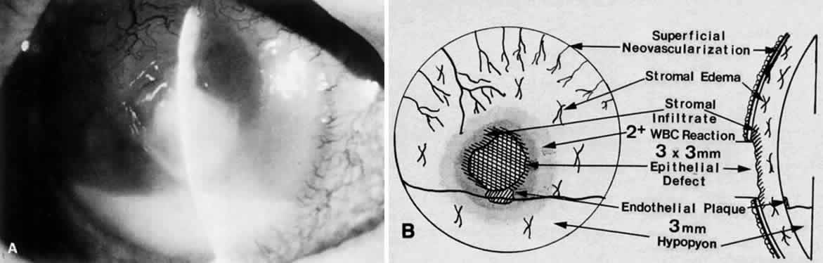
| Fig. 10. A. Slit-lamp photograph of the initial appearance of a Streptococcus pneumoniae corneal ulcer before the microbiologic workup. B. Clinical drawing of the same ulcer documenting important signs key to the therapeutic management of this problem. Daily drawings should be performed by the clinician to document these changes in the course of the ulcer. |