Chapter 13
Lacrimal Drainage System
MYRON TANENBAUM and CLINTON D. McCORD JR
Main Menu   Table Of Contents

Search

ANATOMY
PHYSIOLOGY OF LACRIMAL ELIMINATION
EMBRYOLOGY
CONGENITAL ANOMALIES
LACRIMAL PROBLEMS IN NEONATES
DISEASES OF THE PUNCTA
DISORDERS OF THE CANALICULI
DISORDERS OF THE LACRIMAL SAC
DISORDERS OF THE NASOLACRIMAL DUCT
LACRIMAL PUMP FAILURE: MISCELLANEOUS DISORDERS
DIAGNOSIS IN PATIENTS WITH EPIPHORA
TREATMENT OF DISORDERS OF LACRIMAL DRAINAGE
REFERENCES

A clinician's approach to lacrimal disorders should be logical and organized. Just as neurologic disorders must be carefully localized, so too should lacrimal problems be correctly localized and diagnosed before treatment is implemented. A thorough evaluation of the lacrimal drainage system should begin with the eyes, eyelids, and puncta and terminate with the distal nasolacrimal duct and intranasal passages. Haphazard trial-and-error therapies are to be avoided.

This chapter is organized into basic sections on anatomy, physiology, pathology/pathophysiology, diagnosis, and treatment. The content of these sections emphasizes clinically relevant material to aid in understanding, diagnosing, and treating lacrimal disorders.

“Watery eyes” are among the most common lacrimal symptoms. Patients with this symptom have one of two problems: either they produce too many tears (hypersecretion) or the tears that are produced cannot properly drain (epiphora). Through the understanding of relevant lacrimal anatomy, physiology, pathology/pathophysiology, and diagnostic techniques (none more important than history), a clinician can make such very basic and very important distinctions in patients with watery eyes. The material presented in this chapter thoroughly addresses disorders of the lacrimal drainage system, such as the watery eyes example, and offers clinicians a logical, orderly approach.

Back to Top
ANATOMY
An understanding of the anatomic elements of the lacrimal drainage system is necessary to appreciate the operation of the system and how different diseases can affect its function. These elements include the bony conduit, the membranous conduit, and the surrounding soft tissues, tendons, and muscles.

BONY CONDUIT

Within the nasofrontal process of the maxilla at the anterior nasal portion of the orbit is the lacrimal fossa, in which is found the lacrimal sac. From there, the nasolacrimal canal, which contains the nasolacrimal duct, extends down inside the lateral wall of the nose and opens under the inferior turbinate or concha in the vault of the meatus of the turbinate. The nasolacrimal canal is approximately 16 mm posterior from the anterior tip of the turbinate and is located between the anterior third and the middle third of the meatus of the turbinate. It exits in the vault of the meatus of the turbinate 17 mm above the floor of the nose laterally. There are, however, normal variations in which the end of the nasolacrimal canal can extend farther down and open at various positions in the lateral wall of the nose. The lacrimal sac fossa is present in the anterior-inferior nasal portion of the bony orbit and is delineated anteriorly by the anterior lacrimal crest and posteriorly by the posterior lacrimal crest. If viewed from the inside of the nose, it would be outlined on the lateral wall of the nose in front of the anterior tip of the middle turbinate (Fig. 1). The nasolacrimal canal is not vertical in direction but angulates posteriorly 15 degrees and slightly inward almost 5 degrees to reach its final destination under the inferior turbinate (Fig. 2). Two sinuses are in intimate relationship with the lacrimal bony conduit, the maxillary sinus or antrum, which forms the lateral wall of the nose in that area, and the ethmoidal sinus, which borders the posterior edge of the lacrimal sac fossa and the superior nasolacrimal duct. The ethmoidal sinus is a system of mucous membrane-lined air cells that are positioned immediately posterior to the lacrimal sac fossa and the lacrimal bone. It is not uncommon (as many as 80% to 90% of patients),1 however, for the ethmoidal air cells to encroach in various degrees into the bone at the posterior lacrimal crest; in some cases, they actually extend as far as the anterior lacrimal crest area separating the lacrimal sac from the lateral wall of the nose and intranasal cavity (Fig. 3). Also, the anterior tip of the middle turbinate may extend various degrees anteriorly into the intranasal cavity. These anatomic variations become important considerations when lacrimal surgical procedures are necessary.

Fig. 1. Outline of the lacrimal sac fossa and nasolacrimal duct in its course under the inferior turbinate as viewed inside the nose looking at the lateral wall of the nose.

Fig. 2. Frontal view of the nasolacrimal duct and its course.

Fig. 3. Axial view of the normal relationship of the lacrimal sac fossa, the ethmoidal air cells, and the tip of the middle turbinate, with variations that may be encountered.

MEMBRANOUS CONDUIT

Dimensions

Contained within the bony conduit and the nasal portion of the eyelids are the epithelium-lined tear ducts through which tears pass from the eyelids into the nose. The openings of this membranous conduit in the eyelids are the upper and lower puncta, which are located 6 mm from the inner canthus. The punctal openings in the eyelid are 0.3 mm in diameter. The canalicular portion just beyond the punctum is called the ampulla. It is vertically oriented for 2 mm and balloons out to a diameter of 2.5 mm (Fig. 4). The canaliculi then narrow to about 1 mm in diameter and extend nasally 8 mm to enter the lacrimal sac. In more than 90% of people, the canaliculi join before entering the sac at the level of the lower border of the medial canthal tendon. The tear sac itself is 12 to 15 mm in vertical length, and in most instances there is a portion above the entrance of the common canaliculus 3 to 4 mm in height called the fundus (dome), which is usually compressed by the medial canthal tendon. The nasolacrimal duct extends from the inferior portion of the sac through the nasolacrimal canal or bony conduit and travels 12 mm to the position where it opens underneath the inferior turbinate in a space called the meatus (see Fig. 4). The membranous conduit in most cases extends 5 mm farther downward and usually is located in the vault portion underneath the inferior turbinate. However, again, there is some variability in the nasolacrimal canal and duct, and it may open farther down on the lateral wall of the nose in a slitlike manner (Fig. 5).

Fig. 4. Dimensions and internal structure of the membranous conduit for lacrimal elimination (puncta, lacrimal sac, nasolacrimal duct). Valvelike folds are diagrammatically represented: 1, valve of Rosenmüller; 2, valve of Krause; 3, spiral valve of Hyrtl; 4, valve of Taillefer; 5, valve of Hasner or plica lacrimalis.

Fig. 5. Variations in position and shape of the opening of the end of the nasolacrimal duct under the inferior turbinate. (Adapted from Shaffer JP: Types of ostia nasolacrimalia in man and their genetic significance. Am J Anat 13:183, 1912.)

Structure The lining of the lacrimal canaliculi, the sac, and the nasolacrimal duct is pseudostratified columnar epithelium similar to that found in the upper respiratory system. The walls contain much elastic tissue. The canaliculi, in particular, contain large amounts of elastic tissue. In addition, the lacrimal sac and the nasolacrimal duct have collagen, elastic fibers, and amounts of lymphoid tissue in the walls.2,3 The mucous membrane within the sac and nasolacrimal duct is arranged into membranous folds that act as valves. The two most important folds, as best as can be determined clinically, are the valve of Rosenmuller, where the common canaliculus enters into the sac, and the valve of Hasner (plica lacrimalis) at the end of the nasolacrimal duct under the inferior turbinate. Other valvelike folds have been identified, as have valvelike constrictions at the junction of the sac in the nasolacrimal duct (valve of Krause, sinus of Arlt) and within the nasolacrimal duct (spiral valve of Hyrtl and the valve of Taillefer; see Fig. 4).

ENCOMPASSING SOFT TISSUE

The muscles within the eyelid in front of the tarsus (pretarsal orbicularis), the protractor muscles, are anchored at the lateral canthal tendon and the lateral palpebral raphe. They travel horizontally across the surface of the upper and lower tarsus, and as they reach the medial canthal area they split into a superficial and deep portion or head. The deep head of the pretarsal orbicularis muscle from the upper and lower lid inserts on the lacrimal bone at the posterior lacrimal crest behind the sac and in some cases has been referred to as Horner's muscle. An additional strand of orbicularis muscle from the preseptal area in the lower lid inserts on the periosteum, which covers the lacrimal sac and its fossa, which extends from the posterior to the anterior lacrimal crest described by Jones.4 The superficial head of the pretarsal orbicularis and preseptal orbicularis fibers insert in a dense conjoined medial canthal tendon anterior to the fundus of the lacrimal sac (Fig. 6).

Fig. 6. Diagram of the tendon and insertion of the eyelid muscles in and around the lacrimal sac and canaliculus. (Adapted from Jones LT: An anatomic approach to problems of the eyelids and lacrimal apparatus. Arch Ophthalmol 66:137, 1961.)

Back to Top
PHYSIOLOGY OF LACRIMAL ELIMINATION
Much has been written and hypothesized about the mechanism of lacrimal elimination. The abundance of theories and conflicting information in the literature often serves to dissuade practicing clinicians from investigating this topic. A basic understanding of lacrimal physiology, however, is a useful foundation for making treatment decisions about many lacrimal disorders.

The tear film travels across the surface of the globe and eyelids, enters the puncta/ampulla, passes through the canaliculi, and enters the lacrimal sac/nasolacrimal duct/nasal passages. This schema is of course oversimplified, because much of the tear film is likely “eliminated” by direct evaporation or absorption at the level of the lacrimal sac. The first key physiologic point to understand, however, is that the lacrimal outflow system is based on an active, dynamic pumping mechanism. It has long been noted that the blinking mechanism readily drains tears even with the head held in an inverted position. When the palpebral blink mechanism is impaired, however, epiphora is common, such as in patients with facial paralysis.

Although multiple mechanisms may contribute to lacrimal outflow, present evidence suggests that the most important factor is the active palpebral-canalicular pump. This theory is based on the cumulative works of Doane,5 Rosengren,6 Frieberg,7 Chavis and colleagues,8 and Maurice.9 the blink mechanism with high-speed photography. Carbon particles were used to photograph the actual flow of the precorneal tear meniscus. The important findings of this work are summarized as follows. With the eyelids open before the start of a blink, the canaliculi are already filled with tears. As the upper lid descends at the start of a blink, the medial portion of the eyelids around the puncta elevates. The upper and lower puncta come into forceful contact by the time the eyelids are only halfway closed. This important event occludes the puncta such that the remaining portion of the blink acts to compress the canaliculi and lacrimal sac, thus forcing the contained fluid into the nasolacrimal ducts and nasal passages. The volume of fluid within the lacrimal outflow system is at its minimum at the point of maximum lid closure during a blink. As the eyelids begin to open, the muscular compressive force terminates and the elastic walls of the canaliculi and lacrimal sac attempt to restore their original shape. The puncta remain occluded such that a partial vacuum forms within the membranous lacrimal conduit. As the eyelid-opening phase of the blink continues, the two lacrimal puncta pop open and expose the adjacent lacrimal lake to this partial vacuum. Tears rapidly flow into the canaliculi during the 1- to 3-second interval immediately after the blink. Once again, the canaliculi fill with fluid so that the pumping action of the next blink can continue the lacrimal elimination cycle.

In addition to the important role of the active palpebral-canalicular pump, other factors contributing to lacrimal elimination may include physical forces such as gravity and capillary attraction of the tears, reservoir drainage into the lacrimal sac (so-called Krehbiel flow), microcilliation in the nasolacrimal duct, and finally evaporation of tears from the ocular surface and absorption of tears by the lacrimal sac mucosa.

The “lacrimal pump” theory is widely noted in the ophthalmic literature and is based on classic anatomic studies by Jones,10 describing tendinous and muscular insertions exerting their action on and around the lacrimal sac. The Jones theory holds that with closure of the eyelid margin, the eyelid fissure moves nasally and forces tears toward the area of the puncta and interface between the lids, conjunctiva, and caruncle in the area of the lacrimal lake. With relaxation of the eyelids on opening, the canaliculi and ampulla, which have been compressed by the pretarsal deep and superficial muscles because of their elasticity, create a negative pressure in the ampulla-canalicular system, causing tears to be sucked into the puncta. When the eyelids are closed again, the tears, which previously entered the ampulla-canalicular system when the eyelids were opened, are squeezed into the lacrimal sac. It is also theorized that the muscular pull of the preseptal orbicularis muscles on the lacrimal sac creates a negative pressure within the sac. With the eyelids open, the sac is normally collapsed. The valves within the sac and nasolacrimal duct prevent retrograde passage of tear flow in normal situations. This proposed lacrimal sac pumping mechanism is based on anatomic studies and likely does not have a large role in normal lacrimal elimination, because the system functions quite well with the lacrimal sac slit completely open, as is the case after dacryocystorhinostomy (DCR).

Traditional teachings have previously held that the lower canalicular drainage system was far more important than the upper system. This old wives' tale is completely incorrect. Studies by White and colleagues11 and Daubert and associates12 have demonstrated equal tear flow between the upper and lower canalicular systems using radioactive dacryoscintigraphy flow studies. Linberg and Moore13 evaluated the clinical symptoms associated with alternate monocanalicular occlusion of the upper and lower puncta. They found that approximately 50% of patients experience mild intermittent symptoms of epiphora associated with experimental monocanalicular obstruction. The symptoms were identical whether patients' upper canalicular system or lower canalicular system was occluded. Meyer and associates14 studied fluorescein dye disappearance in 20 subjects and found that 90% of patients showed minimal or no impairment with monocanalicular (either upper or lower) obstruction.

A number of important clinical principles can be derived from the physiologic information just provided. Present evidence supports the crucial role of the palpebral-canalicular pump mechanism in lacrimal elimination. All efforts should be made to preserve the lacrimal canaliculi. Repeated instrumentation of the lacrimal system or nasolacrimal duct probings are unlikely to help the underlying pathology and may in and of themselves injure the canaliculi and thus permanently impair lacrimal elimination. Very little can be done to restore scarred fibrosed canaliculi. Frieberg has used a manometer to measure a pressure gradient within the canaliculi and lacrimal sac.7 This pressure gradient cannot be produced if the canaliculus is slit open. This information should caution clinicians against performing overly aggressive procedures on the lacrimal outflow system, such as aggressive “three-snip” punctoplasties or aggressive surgical treatment of canaliculitis. Experimental and clinical studies show that tear elimination is equivalent through the upper and lower canalicular systems.11–14 Surgeons should thus give equal consideration to a patient with lacerations of either the upper or lower canaliculus. Traditional teachings that upper eyelid canalicular lacerations are unimportant are simply not true.

Back to Top
EMBRYOLOGY
The anlage of the membranous lacrimal conduit is an ectodermal thickening of a groove between the lateral, nasal, and maxillary process in the 12-mm-stage embryo. After this stage, the anlage detaches and becomes buried in the mesoderm. Solid cords of epithelial cells form the anlage of the canalicular system in the eyelids, with a projection downward that will form the nasolacrimal sac and the nasolacrimal duct at the 16-mm stage. Canalization of these epithelial cords starts at the 50-mm stage, or 4 months of gestation, beginning as scattered patches throughout the system and creating a lumen through the system. This lumen finally breaks through its latest stage in the nasolacrimal duct to form a continuous opening just before birth. The lower end of the lacrimal duct is the last to canalize, and in more than half of infants the last portion of this nasolacrimal stem may not completely finalize its patency at birth.15,16 During embryonic development, migrations of epithelial cords can cause various anomalies within the lacrimal system.
Back to Top
CONGENITAL ANOMALIES

DACRYOSTENOSIS

Dacryostenosis is a common condition in which the extreme end of the nasolacrimal duct underneath the inferior turbinate fails to complete its canalization in the newborn period; it produces clinical symptoms in 2% to 4% of newborns. In most instances, the obstruction is a small membrane at the end of the nasolacrimal duct that persists because of failure of complete canalization of the nasolacrimal duct. Other rarer types of congenital occlusion of the nasolacrimal duct and canal may occur if the epithelial cords have migrated within the mesoderm and do not open immediately under the inferior turbinate. Blind pouches can occur within the turbinate itself. A bony obstruction is often found under the inferior turbinate, and in some cases the epithelial cord migrates laterally so that the nasolacrimal canal ends in a blind pouch in the lateral wall of the nose.17,18 There are normal variations in the position of the opening of the nasolacrimal duct under the inferior turbinate. The nasal ostium of the duct, at the highest portion of the inferior meatus within the vault beneath the inferior turbinate, is usually wide open. It may be puckered, slitlike, or grooved down the lateral wall of the nose or have a puncturelike appearance in the vault of the turbinate (see Fig. 5).

ABSENCE OF VALVES

The fold normally present at the end of the nasolacrimal duct or valve of Hasner may be absent, in which case pneumatoceles of the sac may occur and nose blowing may cause retrograde passage of air. If the valve of Rosenmuller is also absent, it is possible to blow air from the nose into the eye, and nosebleeds may produce bloody tears.19

ANOMALIES OF THE SAC

Although diverticuli of the lacrimal sac may occur, a congenital fistula of the lacrimal sac, which has been termed lacrimal anlage duct by Jones, is more common (Fig. 7).19 This anomaly has been described by others and in some cases is found to be autosomal dominant and may coexist with thalassemia. It undoubtedly is the result of canalization of a strand of epithelial cords that extends from the sac to the skin surface. The fistulas often have to be completely excised to prevent drainage of tears externally on the skin.

Fig. 7. Lacrimal duct anlage. Close-up photograph demonstrates a congenital lacrimal drainage fistula inferonasal to the medial canthal angle (arrow).

ANOMALIES OF THE PUNCTA

Congenital atresia, supernumerary or double puncta, and congenital slits of the puncta all may occur from aberrations in the location of the epithelial cord and its opening to the surface epithelium. Lateral displacement of the puncta may occur in some congenital syndromes such as blepharophimosis.

ANOMALIES OF THE CANALICULI

Atresia or failure of canalization of the lacrimal canaliculi may occur in conjunction with punctal atresia. In many cases, particularly in patients with mesodermal dysplasia, the lacrimal canaliculi and puncta may be absent and a normal tear sac and nasolacrimal duct may be present but not connected to the eyelid surface.

Back to Top
LACRIMAL PROBLEMS IN NEONATES
Neonates have tear secretion at birth, and 96% to 98% have a totally patent and functional lacrimal drainage system.20 The 2% to 4% who do not have a lacrimal drainage system intact have a thin residual membrane at the distal end of the nasolacrimal duct. This membrane spontaneously dissolves in 80% to 90% of patients within the first few months of life.21 Clinical manifestations of congenital nasolacrimal duct obstruction are the following.

AMNIOTOCELE

This occurs in neonates as a distention in the lacrimal sac. Amniotic fluid enters the sac, is retained by a nonpatent nasolacrimal duct, and is trapped in the sac by the valve at the common canaliculus, the valve of Rosenmuller. Probing the nasolacrimal duct as an office procedure is usually curative.

DACRYOCYSTITIS (ACUTE MUCOCELE OR PYOCELE)

This condition also exhibits acute distention and inflammation in the lacrimal sac region and may occur in the neonatal period. Probing is necessary in newborns with acute dacryocystitis to establish drainage as soon as possible. This procedure is performed with topical local anesthesia only.

TEARING AND MATTERING

Newborns who have congenital dacryostenosis may not develop acute dacryocystitis with a mucocele or pyocele of the sac in the early neonatal period but may simply have tearing with a chronic mucopurulent discharge, which usually becomes manifest at 2 weeks of age. Topical antibiotics should be administered, and the parents must be instructed in the proper technique of lacrimal sac compression and massage. More than 90% of these cases clear and become asymptomatic with conservative management.22,23 Under normal circumstances, these children with mild to moderate symptoms of epiphora and lid crusting can be monitored for the first year of life without serious consequence or sequela (Fig. 8). There is rarely any imperative reason to make probing mandatory at an early age (e.g., before 6 months of age). A number of studies have confirmed that probing or silicone tube intubation in children after 12 months of age still has a very high success rate. These techniques are discussed later in the treatment section in this chapter.

Fig. 8. This 15-month-old child had congenital left nasolacrimal duct obstruction. Chronic mattering of the left eyelashes and excess tearing are evident in the left eye.

Back to Top
DISEASES OF THE PUNCTA

MALPOSITION OR EVERSION

The normal position of the punctum is pointing inward toward the lacrimal lake. It cannot be seen by an observer looking directly toward the eyelid. The punctum can be displaced in a congenital anomaly with eyelid syndromes such as blepharophimosis or congenital ectropion. Malposition more commonly is an acquired anomaly in older individuals with eyelid laxity and senile ectropion (Fig. 9). It also may occur after injury or burns with skin shrinkage, which results in turning the punctum outward.

Fig. 9. Medial ectropions. This 78-year-old man had laxity-related bilateral lower eyelid medial punctal ectropions with epiphora in both eyes.

ATRESIA

Isolated stenosis of the punctum with an intact and patent canalicular system distally may occur congenitally or as a result of cicatrizing inflammations, trauma, and senile atrophy associated with long-term drying in patients with ectropion.

TUMORS

Basal cell carcinomas occur most frequently in the inner canthal area and often involve the punctum, as may benign growths in this area.

Back to Top
DISORDERS OF THE CANALICULI

OBSTRUCTION

Obstruction or atresia of the canalicular system may follow any of the conditions discussed next.

Cicatrizing Conjunctivitis

Obstruction or atresia of the canaliculi may follow infections such as herpes simplex, herpes zoster, vaccinia, trachoma, infectious mononucleosis,24–26 or other inflammations such as the Stevens-Johnson syndrome or ocular pemphigoid.

Trauma

Chemical or thermal burns, dog bites, and other lacerations may also cause obstruction or atresia of the canaliculi. At one time or another, every ophthalmologist is called on to repair a canalicular laceration. Acute lacerations of the canaliculi may occur after sharp penetrating wounds or as a result of shearing or ripping wounds of the eyelid caused by hooklike objects or teeth. The location of the lid laceration medial to the lacrimal punctum heightens the examiner's suspicion of the possibility of a canalicular laceration. If a patient is cooperative, the canaliculus can be gently probed with a lubricated 00 Bowman probe. Visualizing the shiny metal probe within the wound confirms the presence of a canalicular laceration. In children and some adults, the diagnosis of a canalicular laceration can be established only in the operating room or during examination under anesthesia. As with any adnexal injury, the ophthalmologist must perform a complete examination, including dilated retinal examination, to rule out serious ocular or intraocular injury.

Irradiation

Occlusion of the canaliculi and puncta occurs after irradiation for basal cell carcinoma almost 100% of the time, although intubation with silicone tubing may prevent this problem in some cases.27

Tumors

Skin cancer may involve the canalicular system, but intrinsic canalicular tumors such as papillomas may occur, producing occlusion and secondary inflammation.28

Use of Eye Drops

Echothiophate (Phospholine) iodide has been incriminated as a cause of canalicular stenosis as well as ocular pemphigoid syndrome, and idoxuridine toxicity may cause temporary occlusion of the punctum and canaliculus.29

Repeated Probing

One of the most common causes of stenosis of the lacrimal canalicular system is repeated and traumatic probing of the canalicular system for whatever reason.

Canaliculitis

Inflammation of the canalicular system can occur secondary to dacryocystitis, but isolated bacterial infections of the canaliculus are rare. Perhaps the most common infections are caused by Streptomyces, Actinomyces israelii, and Arachnia propionica (previously mislabeled as Streptothrix). Fungal infections with organisms such as Candida albicans, Aspergillus niger, and Nocardia have been reported. However, actinomycotic infection is by far the most common.30 In the clinical presentation, the lower canaliculus is usually involved and the patient reports epiphora. Swelling and inflammation of the lid medially are noted (Fig. 10). The punctum is swollen and red, sometimes described as pouting, and a mucoid and mucopurulent discharge is present. Irrigation may or may not be possible through the canaliculus, and a small probe may encounter gritty resistance. Diagnosis is made on expressing yellow-tinged concretions from the canaliculus. On cytologic examination, they show gram-positive branching filaments.

Fig. 10. Left upper lid canaliculitis with diverticuli formation of the canaliculus.

Allergy

Wojno31 has demonstrated intermittent allergic obstruction at the level of the canaliculus or lacrimal sac. This phenomenon is associated with allergic conjunctivitis and is noted in patients who chronically rub their eyes.

Back to Top
DISORDERS OF THE LACRIMAL SAC

INFECTIONS

Infections of the lacrimal sac are clinically manifested as either dacryocystitis, marked by a tender and swollen lacrimal sac, rather severe pain, redness, and tearing; or chronic dacryocystitis, characterized by a smoldering infection within the lacrimal sac without distention of the sac but producing tearing and, in many cases, a chronic unilateral conjunctivitis (Fig. 11). Both conditions have as their underlying cause an obstruction to the normal tear passage within the nasolacrimal duct and sac, causing stasis and stagnation of tears and mucus and subsequent infection. The most common organisms in the ensuing infections are pneumococci. Other organisms include streptococci, diphtheroids, Klebsiella pneumoniae, Haemophilus influenzae, Pseudomonas aeruginosa, and mixed organisms. Actinomyces and fungi, such as Candida, are not infrequent, and granules and casts of the nasolacrimal duct and sac may be present in these cases.32 Sarcoidosis producing a chronic recurrent dacryocystitis has been reported in conjunction with systemic sarcoid.33,34

Fig. 11. Wegener's granulomatosis in a 71-year-old man. Acute dacryocystitis with lacrimal sac rupture is present in his left eye. Chronic conjunctivitis due to right nasolacrimal duct obstruction with chronic low-grade infection is noted in his right eye.

LACRIMAL STONES OR CASTS

Casts or plugs may form within the lacrimal sac or nasolacrimal duct from fungus infections, organized blood clots, or inspissated mucous plugs. The most common organisms causing these seem to be A. israelii and Candida species. The clinical signs are intermittent epiphora and lacrimal obstruction, lacrimal conjunctivitis, recurrent or intermittent dacryocystitis, and variable localized tenderness. The underlying causes of the formation of stones are unclear. However, they occur more frequently in younger (under age 50) individuals and in heavy smokers.32 In many cases, it is possible to express, irrigate, and probe casts through the nasolacrimal duct, but DCR may be needed. A syndrome of acute noninfectious dacryocystic retention may occur when a cast, which may be mobile, plugs the nasolacrimal duct.35 Fluid buildup in the lacrimal sac causes closure of the valve of Rosenmuller at the common canaliculus, and the sac becomes acutely swollen and tender. This can be treated by percutaneous aspiration of the sac, followed by probing and irrigation. Casts may occur from chronic use of epinephrine drops.36

DACRYOCYSTITIS

The differential diagnosis of dacryocystitis includes a number of different clinical entities. The presentation of a mass in the lacrimal sac may be caused by a noninfectious amniotocele in a neonate or acute dacryocystic retention in an adult. Lacrimal sac neoplasms, unless attended by infection, are generally not tender and have a slower onset. In neoplastic disease, the mass in many cases extends above the level of the medial canthal tendon, which does not occur in dacryocystitis because of the compression of the fundus of the sac by the medial canthal tendon. Neoplasms that arise extrinsic to the lacrimal sac area include nasopharyngeal carcinomas, orbital rhabdomyosarcomas, and tumors of the antrum. A congenital midline meningoencephalocele may present as a mass in the lacrimal sac area and cause a secondary dacryocystitis. This anomaly may or may not communicate with the intracranial cavity. Dermoid cysts occur quite commonly nasally, although they are more often located in the supra-nasal quadrant and most usually are located at the lateral brow. Ethmoidal and frontoethmoidal mucoceles can occur and produce a firm masslike swell-ing at the inner canthal area and also a secondarydacryocystitis. Skin cysts and inclusion cysts maysimulate a mass in the lacrimal sac area. A chronicunilateral conjunctivitis with tearing and purulentdischarge from the lower canaliculus that occurs with dacryocystitis also can be encountered in isolated canaliculitis.

TUMORS

Clinical signs of a lacrimal sac tumor are tearing, painless irreducible swelling in the lacrimal sac area, and secondary dacryocystitis. Bleeding on probing of the lacrimal system is not an infrequent finding in sac tumors. It may be possible to irrigate through the lacrimal sac containing a tumor before the tumor completely occludes the lacrimal drainage system. Extension of tumors outside the sac area may cause intranasal symptoms, and radiographic changes are noted with erosion in the bone. Injection contrast radiography (dacryocystography) of the lacrimal sac may also demonstrate a nonfilling mass but may add little to the diagnosis, which may already be suspected. Historically, series of primary lacrimal tumors have been reviewed by Ashton and colleagues,37 Jones,38 and Radnot and Gall.39 Additional cases have been reported by Ryan and Font,40 Schenck and associates,41 and Stokes and Flanagan.42 In one series, about a third of the “tumors” were pseudotumors and were described as nonspecific granulomas, rhinoscleroma, lymphomatous lesions, sarcoid, syphilis, and fungi.39 In most series of true neoplasms of the lacrimal sac, 50% to 60% arise from the epithelial lining of the sac and are of the papillary carcinoma group, with histologic pictures identical with solid cylindric cell tumors arising from the respiratory epithelium.40

The well-differentiated papilloma group should be treated by local excision and have a favorable prognosis, although the rate of recurrence is high. It has been recommended that all of the membranous lacrimal drainage apparatus, including the canaliculi and nasolacrimal duct, should be excised. The papillomas are classified into three histologic types: squamous cell, transitional cell, and mixed cell. They are all resistant to radiation therapy. The carcinomas are histologically squamous and transitional cell carcinomas, and they also may arise from the degeneration of a pre-existing papilloma. In Ryan and Font's series, the survival rate of these patients after a wide local excision with frozen-section control was “reasonably good.”40

The wall of the lacrimal sac contains lymphoid tissue, and lymphomas are probably the second most common intrinsic lacrimal sac tumor. They are similar to lymphomas occurring in the orbit in that they may be forerunners of lymphoid disease elsewhere in the body or the head and neck. The ultimate prognosis may be improved by noting the presence or absence of follicles, the anaplasia of the cells, and cell surface immunology.43 After biopsy and systemic evaluation, radiotherapy is indicated. Intubation of the lacrimal passages with silicone tubing before irradiation is indicated, and normal lacrimal drainage function for the patient after treatment may be retained.

Other primary lacrimal sac tumors that have been reported are malignant melanoma,44 oncocytic adenocarcinoma,45 neurilemmoma,46 adenocanthoma,47 hemangiopericytoma,48 and fibrous histiocytoma.49

The differential diagnosis should include invasive pharyngeal or sinus carcinoma and mucoceles of the ethmoidal sinus, which can be demonstrated on computed tomography scans. Rarely, orbital rhabdomyosarcomas present in this manner, and other mass lesions, previously mentioned, should be considered.

Back to Top
DISORDERS OF THE NASOLACRIMAL DUCT

PRIMARY ACQUIRED NASOLACRIMAL DUCT OBSTRUCTION

Primary acquired nasolacrimal duct obstruction (PANDO) is the most common clinical syndrome of acquired nasolacrimal duct obstruction in adults. Patients may present with symptoms of chronic epiphora, conjunctivitis, and low-grade infections or with acute dacryocystitis. This clinical syndrome is most common in elderly white women.

Clinical pathologic studies by Linberg and McCormick50 examined the histopathology of the entire membranous nasolacrimal duct in patients with the clinical syndrome of PANDO. These studies have revealed inflammation, vascular congestion, and edema of the nasolacrimal duct in the early phases and, ultimately, fibrosis with complete occlusion of the nasolacrimal duct's lumen in the late phases.

The specific trigger of this sequence of events is not known. Nonetheless, it is reasonable to postulate that inflammation with partial ductal obstruction leads to accumulation of cellular debris, which aggravates the ongoing inflammation and creates a vicious cycle that leads to permanent cicatrization of the nasolacrimal duct lumen.

Linberg's studies have demonstrated that in patients with symptoms of relatively short duration (less than 1 year), the inflammation and edema “functionally” occlude the nasolacrimal duct. A potential space does remain within the lumen, however. This pathologic finding lends credence to the hope that PANDO may be reversible in patients with symptoms of short duration. For this reason, as discussed later in the treatment section, this subset of patients may be candidates for either medical therapy with antiinflammatory drugs or nasolacrimal duct intubation with silicone tubes to maintain patency of the duct until the inflammation subsides or has been treated.

Patients with PANDO and chronic symptoms (greater than 2 to 3 years' duration) demonstrate dense fibrous scar tissue and cicatrization of the nasolacrimal duct as a sequela of chronic inflammation, edema, and stasis of cellular debris. In these patients, the lumen of the nasolacrimal duct is permanently obliterated by scar tissue. This histopathologic finding correlates with the very poor success rate of nasolacrimal duct probing or intubation with silicone lacrimal tubes in adult patients with chronic PANDO symptoms. As discussed later, DCR remains the treatment of choice for this group of patients.

Although most adult nasolacrimal duct obstructions represent the syndrome of PANDO, noninflammatory infiltrative disorders can occlude the nasolacrimal duct. Clinicians must maintain a high index of suspicion in patients with known systemic disorders such as sarcoidosis, lymphoma, or leukemia. In these situations, distal nasolacrimal sac or nasolacrimal duct biopsy is an important part of the DCR surgery. Linberg and McCormick recommend their nasolacrimal duct biopsy technique as a routine part of all DCRs.50 In this manner, important infiltrative causes of nasolacrimal duct obstructions would not be overlooked.

TRAUMA

Nasolacrimal duct obstructions may occur as a sequela of midfacial fractures involving the bony nasolacrimal canal. Immediate disruption of the nasolacrimal drainage system occurs in some patients. This is typical of severe crushing nasal orbital fractures and the Lefort II and LeFort III fractures, in which extensive damage is sustained by the entire lacrimal drainage apparatus, including the canthal tendons. In other patients, bony fractures initiate an inflammatory, cicatrizing process that results in symptomatic nasolacrimal duct obstructions many years after the original injury. Some investigators have advocated early silicone tube intubation of the lacrimal outflow system in patients with complex midfacial fractures.51 The efficacy of this treatment is not yet fully established.

A number of cases of dacryostenosis have been reported after cosmetic rhinoplasty. It is believed that in most instances, the damage to the membranous nasolacrimal duct occurs during the lateral osteotomy.52 Other sinus and nasal operations may also injure the nasolacrimal duct. This is especially true of Caldwell-Luc procedures and nasoantral window formation. Nasoantral windows are usually created in the most anterior-inferior portion of the maxillary sinus. If they are placed too high or too posterior, however, or if the nasolacrimal canal is in an anomalous position, excision of a portion of the nasolacrimal duct may occur. The Ogura procedure (orbital decompression) with excision of the floor of the orbit and ethmoidal air cells through an antrostomy may also be associated with postoperative epiphora. Again, the likely cause may be the nasoantral window formation.53,54

Prior midfacial or nasal radiation therapy may result in nasolacrimal duct obstructions. In prior generations, radiation treatment was common for conditions such as facial acne and chronic sinusitis. As with some traumas, this occlusive radiation fibrosis effect may be delayed for many years.

INTRANASAL DISORDERS

The distal aspect of the nasolacrimal duct may be obstructed as a result of intranasal pathology. Intranasal scarring with inferior turbinate adhesions may occur as a sequela of trauma, radiation therapy, or surgical procedures (e.g., nasoantral window formation). Allergic rhinitis may be associated with nasal mucosal hypertrophy. In some individuals, an abnormally wide nasal vestibule is associated with compensatory hypertrophy of the inferior turbinate that occludes the valve of Hasner (open nasal space syndrome with epiphora).55 Tumors are uncommon and can be benign, such as granulomas or nasal polyps, or malignant, such as squamous cell carcinoma.

Back to Top
LACRIMAL PUMP FAILURE: MISCELLANEOUS DISORDERS
As indicated previously, the lacrimal outflow system may be anatomically patent to irrigation yet be functionally inadequate in terms of normal lacrimal elimination. Facial nerve paresis is among the most common situations in which lacrimal pump failure is present despite patency of the membranous lacrimal conduit. Any condition that impairs the normal contractile and elastic properties of the palpebral-canalicular pump mechanism can cause epiphora. These conditions include scleroderma, radiation fibrosis of the eyelids, and cutaneous burns or trauma of the periocular region. Chronic or recurrent canaliculitis may leave the canaliculi anatomically patent yet functionally impaired.
Back to Top
DIAGNOSIS IN PATIENTS WITH EPIPHORA
The most common symptom of disorder in the function or anatomy of the lacrimal drainage system is tearing. The blockage or malfunction may occur at many places along the tear elimination route, and the appropriate therapy, surgical and nonsurgical, may be necessary to correct it. Some patients with tearing symptoms truly produce more tears than the normal drainage system can handle, and some people may have conditions that cause more of a sensation of tearing than actual overloading of the drainage system. Table 1 shows a classification of the various physiologic and anatomic causes of tearing.


The steps necessary in evaluating a patient with tearing are listed below in the usual sequence. In many cases, not all the steps are needed because the diagnosis may be apparent with some of the more simple tests alone.

  1. History
  2. Slit-lamp examination (noting lid movement and position and integrity of punctum)
  3. Pressure over lacrimal sac
  4. Irrigation of lower canaliculus
  5. Intracanalicular probing (diagnostic)
  6. Dye drainage tests (fluorescein test) for functional blocks
  7. Schirmer's test
  8. Intranasal examination
  9. Conventional x-ray films and computed tomography
  10. Dacryocystography
  11. Dacryoscintigraphy

A patient presenting to an ophthalmologist with tearing should be evaluated in a systematic manner, and the proper sequence should be observed to promote efficient use of time and to avoid confusion.

HISTORY

In evaluating a patient with tearing symptoms, no diagnostic technique is more valuable than taking a careful history. A unilateral watering eye is more commonly a sign of an obstructive process, although bilateral nasolacrimal duct obstructions can occur. Is there actual excess tearing in which tears run down the patient's cheek(s) and require frequent dabbing, or does the patient have diminished visual acuity and simply refer to the sensation as “my eye is tearing”—a common complaint of patients with macular degeneration.

The primary consideration for clinicians is to distinguish epiphora from hyperlacrimation. Epiphora denotes symptoms of excess tearing due to lacrimal outflow deficiency. Hyperlacrimation denotes that an excess production of tears accounts for the patient's symptoms. Table 1 shows a classification scheme for tearing disorders, including the differential diagnosis of epiphora and hyperlacrimation.

Patients with hyperlacrimation commonly have ocular discomfort. A chronic gritty foreign body sensation associated with excess tearing is typical of patients with keratitis sicca (“dry eye” syndrome). Patients with entropion or trichiasis, blepharitis, corneal abrasions, chronic conjunctivitis, or photophobia and iritis also frequently have hyperlacrimation symptoms. In addition to the comfort of their eyes, patients should be questioned about prior seventh nerve palsy or increased tearing while eating.

Patients with epiphora commonly have “excess tears” as their only symptom. The examiner should carefully question the patient about intermittent redness of the eyes, mucous production or heavy lid crusting in the morning, pain or swelling in the region of the lacrimal sac, or prior episodes of acute dacryocystitis. These symptoms may indicate chronic or intermittent dacryocystitis. Severe, intermittent symptoms of tearing or dacryocystitis that are interrupted by completely normal periods may suggest a “ball valve” disorder such as dacryolithiasis. The clinician should question the patient with epiphora to focus on the key anatomic areas of interest. A history of facial nerve paresis, scleroderma, or lid scarring may indicate a dysfunctional lacrimal pump mechanism. The chronic use of phospholine iodide, idoxuridine, or prior severe conjunctivitis can point toward punctal stenosis as the problem. A history of previous lacrimal therapy may be significant. Repeated probing or instrumentation of the lacrimal canaliculi can result in severe canalicular stenosis. Prior “overaggressive” punctoplasties may actually impair tear elimination. Patients should be questioned about a history of chronic allergies or sinusitis, previous nasal or sinus surgery, and prior midfacial fractures or radiation therapy. Any of these would be pertinent in considering a possible nasolacrimal duct obstruction.

The age of the patient (child, adult, elderly) and the duration of the symptoms (acute, chronic) are relevant. These variables have an important bearing on the likely cause of the problem and thus greatly influence the treatment that is implemented. Childhood lacrimal obstructions are usually due to an imperforate membrane at the distal nasolacrimal duct. Adults, once hyperlacrimation has been ruled out, commonly have the syndrome of PANDO. This type of adult lacrimal obstruction is potentially reversible if the blockage is functional or if symptoms have been of a relatively short duration (less than 6 months). Chronic epiphora symptoms (greater than 12 months) are rarely treatable with any method other than definitive DCR. Specific management of these epiphora problems is expanded on in the treatment section.

EXTERNAL EXAMINATION

The puncta should be examined with the use of the slit lamp. They should be positioned in the lacrimal lake and should not be visible without mild eversion of the lid. The eyelid movement, with each blink, is normally a nasalward compression in which the lids are completely in contact with the globe and move toward the inner canthal area. There should be no mechanical or contour obstruction of the margin impeding the movement of the tear film across the lower lid into the lacrimal lake. The punctal opening should be patent without stenosis or external occlusion. External diseases of the eyelids that may be irritating, or keratitis that may be producing a hypersecretion of tears, must be detected. The flow of fluorescein into the punctum can be observed. Swelling and redness of the punctum in the canaliculus may indicate canaliculitis.

PRESSURE OVER LACRIMAL SAC

A simple, quick confirmatory test for sac and nasolacrimal duct infection and probable obstruction, particularly when a patient has a history of dacryocystitis, is massaging of the tear sac. The sac may or may not be distended; however, regurgitation of mucus or pus through the canaliculus and puncta is indicative of dacryocystitis and obstruction, either intermittent or permanent, in the nasolacrimal duct or sac.

IRRIGATION OF CANALICULUS

Syringing saline into the lower punctum demonstrates complete or severe obstruction in the nasolacrimal duct or sac if saline regurgitates through the upper punctum. This regurgitated substance may be clear or accompanied by mucopurulent material and is diagnostic of obstruction in the nasolacrimal duct or sac. Saline that irrigates into the nose with syringing indicates only that there is not a complete obstruction in the membranous conduit of the lacrimal system. It does not rule out an incomplete block (partial block) or what has been termed a functional block, which may prevent tear passage into the nose under normal tear flow pressure. With irrigation of the lower canaliculus, if the saline neither goes into the nose nor regurgitates from the upper punctum and pressure is encountered, the obstruction is in the canalicular system. The proper irrigating technique with a 5-mL syringe and irrigating cannula or a blunted 26-gauge needle is to flush the solution through the canalicular system into the nose so that it may be recovered (i.e., the patient leans forward over a small emesis basin). This is necessary so that the fluid may be examined for casts.

PROBING

Probing is used only as a diagnostic measure in adults to determine the location of a stricture in the canalicular system. No larger than a 00 probe should be used. The residual length of patency in a canaliculus can be determined in this manner, and thus the best possible corrective procedure can be selected.

DYE (FLUORESCEIN) TESTS

Dye (fluorescein) tests are mainly useful in the differential diagnosis of epiphora occurring in patients with incomplete or functional blocks of the sac or nasolacrimal duct (i.e., patients with clinical tearing and a narrowing in the membranous conduit but in whom saline can be syringed into the nose with pressure applied on the syringe). Even though there is some opening in the membranous conduit, the normal pressure of tear flow under physiologic conditions is inadequate to eliminate the tears, and in these patients the symptoms of tearing are usually just as severe in most cases as in patients with complete obstruction.

To determine whether the tears are passing into the nose under normal physiologic pumping conditions, the precorneal tear film is stained by instilling 2% fluorescein solution. The inferior turbinate in the floor of the nose laterally is then sprayed with decongestant and topical anesthetic. A dry cotton roll fluff, about one third the diameter of a cigarette, is wrapped around a nasal wire (or a straightened paper clip) and placed under the anterior half of the inferior turbinate (cotton applicator sticks are too large to find adequate placement under the turbinate). Within 5 minutes, the cotton is removed and examined for staining. If the cotton is stained with fluorescein, then the tear flow through the lacrimal system is normal. If no dye is present on the cotton at the end of this time, then a functional nasolacrimal block is highly possible. This is the primary dye test as described by Jones and Linn.56 (They originally called the test “negative” if no dye was present, creating a source of much confusion because in medical thinking the word negative generally means no pathology.) If dye can be obtained from under the turbinate in the described manner in a patient with true tearing, then the problem is hypersecretion and not deficient lacrimal drainage or elimination. In a tearing patient in whom the dye does not flow through under the turbinate, a second step is needed. The lower canaliculus is then irrigated with saline, in a manner in which it can be recovered (i.e., having the patient lean forward and expectorate into an emesis basin) and examined for staining. If the fluid recovered in this manner does have fluorescein staining, this finding indicates that the dye enters the sac normally but does not pass through the duct into the nose, thus confirming the diagnosis of incomplete or functional block of the nasolacrimal duct. DCR is then indicated. This second step is the secondary dye test of Jones. If the irrigated saline is not stained with fluorescein as it is syringed through, this finding indicates that the dye is not even entering the canaliculus and the sac, and previous examinations and tests must be repeated for obstructions that have been missed higher up in the lacrimal drainage system.

Other tests for functional or incomplete blocks of the nasolacrimal duct are the dye disappearance tests57 and the taste test with saccharin.58 Examination of intranasal cotton to determine the presence of fluorescein was also believed to be enhanced by the use of ultraviolet light. The disappearance of dye from the conjunctival sac is not specific for nasolacrimal duct obstruction and may be influenced by other factors. A sweet or bitter fluid, if instilled in the conjunctival cul-de-sac, may never reach the taste buds for reasons other than nasolacrimal duct obstruction, and any subjective tests are objectionable. The use of ultraviolet light to detect the faintest trace of fluorescein may show some dye getting through the drainage system despite the clinical situation of a functional block.

The dye tests as proposed by Jones and others56,59 are the most valuable tools to diagnose functional nasolacrimal blocks. However, they are certainly not quantitative in that the time sequence of the appearance of the dye and the absolute amount of the dye that is actually passed are not measured. It is obvious that all functional blocks are not of the same severity, and more sophisticated tests are needed to quantitate the severity of functional blocks.

Dye Disappearance Test

The fluorescein dye disappearance test is a safe, simple, physiologic indicator of a patient's lacrimal outflow system.60 This objective test can be very helpful in diagnosing (or verifying) lacrimal drainage insufficiency.

This test relies on one drop of 2% sodium fluorescein instilled in the lower conjunctival cul-de-sac. It is most helpful when both sides are tested simultaneously. In this manner, dye disappearance is evaluated over a 5-minute period. Clinically, the epibulbar surface appears intensely yellow as the fluorescein drop is instilled. Patients commonly report that “things appear yellow.” The examiner must take note of any asymmetric overflow of fluorescein over the lid margin. This can give false and misleading results. Patients must be kept from dabbing or wiping their eyes.

The dye disappearance test is graded at 5 minutes on a scale from 0 to 4+ ; 0 represents no dye remaining and 4+ indicates that virtually all of the dye remains. Normal eyes exhibit a faint yellow fluorescein color (e.g., ½ to 1+ ) at the end of 5 minutes. Attempting to grade or quantitate the remaining fluorescein dye in an eye is a highly subjective maneuver. In evaluating unilateral tearing symptoms, it is often more helpful simply to compare the dye disappearance results of the patient's two eyes.

Elderly patients with intermittent epiphora symptoms may not always have a grossly exuberant precorneal tear film. The dye disappearance test is a simple, objective means of assessing lacrimal outflow function in these patients. The dye disappearance test is also quite useful in ascertaining the tear outflow function in patients who have undergone DCR.

SCHIRMER'S TEST

Patients who have normal lacrimal drainage as diagnosed by the tests described may still have tearing symptoms due to “pseudoepiphora.” This condition is characterized by an actual deficiency of basic tear secretion with overcompensation of tear production from the main lacrimal gland, causing a watery eye.

Rough quantitation of tear production is aided by the use of Schirmer's test strips (Whatman number 41 filter paper, a 35-mm-long and 5-mm-wide strip). The filter paper strip is folded at the notched indentation, and the short end is draped over the lower lid margin. The first Schirmer's test is performed without anesthesia during a 5-minute period. Filter paper wetting between 10 and 30 mm is considered normal. This test measures basic secretion and reflex tearing. The second Schirmer's test measures wetting caused by irritative nasal stimulation (e.g., cotton-tipped applicator) and is a test for possible fatigue block in the reflex arc between trigeminal sensory nerve fibers and the facial nerve.

The traditional test for dry eye syndrome is the Schirmer's test with anesthesia, or so-called basic secretory test (BST). In this test, topical anesthetic drops are instilled in the eye, and the excess is blotted from the inferior conjunctival cul-de-sac. The Schirmer's test strips are then draped over the lid margin for 5 minutes. Although this test is still widely used, it is highly unlikely that it accurately measures basal tear secretion. There is considerable doubt, in fact, about whether a steady basal rate of tear production even exists.61,62 In all likelihood, the BST measures both basic secretion and some reflex tearing. To label all patients with wetting of less than 10 mm as having dry eye syndrome is an oversimplification. The BST is a roughly quantitative test only. We do attach significance to severely limited amounts of filter paper wetting (0 to 2 mm of wetting), and these patients may have symptoms of pseudoepiphora.

INTRANASAL EXAMINATION

An intranasal examination is necessary in patients in whom an obstruction in the sac or nasolacrimal duct is demonstrated. After the intranasal cavity is sprayed with anesthetic and decongestant, a nasal speculum is introduced with the blades oriented vertically to avoid pressing on the intranasal septum when the blades are separated. With a headlight (an indirect ophthalmoscope may be used), the intranasal cavity is examined, To see the middle turbinate and the area in front of the turbinate, the examiner must look upward into the nose with the patient's head tilted backward and note any septal deviation, turbinate disease, or polyposis that may produce technical problems with DCR or Jones' tube procedure. To examine the inferior turbinate area, the inferolateral corner of the nasal cavity, the examiner must look straight into the nose and slightly downward to be able to see the tip of the inferior turbinate and examine it for evidence of disease or an obstructive process that could be the cause of the tear drainage. An intranasal neoplastic process must always be kept in mind.

CONVENTIONAL X-RAY FILMS

Plain films or computed tomography scans are helpful when paranasal sinus disease or tumors are suspected. Ethmoidal sinus enlargement such as ethmoidal mucocele or anterior encroachment of the ethmoidal mucocele area may be detected, as well as any erosion that may be caused by a neoplastic process.

DACRYOCYSTOGRAPHY (INJECTION CONTRAST RADIOGRAPHY)

Dacryocystography, a technique of anatomically displaying the lacrimal sac and ducts by radiopaque dye, was popularized by Milder and Demorest.63 Radiopaque dye is forcibly injected into the lower canaliculus with a syringe, using a lacrimal cannula or a polyethylene tube. Radiographs in the Caldwell and lateral views are taken. Preliminary syringing of the lacrimal sac with saline before injection of the dye should be performed to cleanse the sac and make room for the dye. Dye should be wiped from the lids before x-ray films are taken to avoid obscuring details, and oblique views instead of lateral views should be obtained radiologically if both sacs are to be x-rayed simultaneously. Dacryocystography may be helpful in showing the size of the sac, the relationship of the ethmoidal air cells to the lacrimal sac, filling defects in the sac such as lacrimal casts or lacrimal sac tumors, diverticula and fistulas of the sac, and possibly the exact level of the stricture within the lacrimal sac or nasolacrimal duct (Fig. 12). Dacryocystography is not a test of function, because the dye is forcibly injected, and it is of no value in diagnosing a functional block. It does not demonstrate the canaliculi and, in fact, in most cases bypasses them completely. Most surgeons believe that conventional dacryocystography does not alter the clinical approach to patients or affect the therapeutic decisions that can be made at the time of surgery, because a high index of suspicion for unusual conditions may already be present from other clinical signs. Dacryocystography can, however, be a useful adjunct for confirmation of a problem.

Fig. 12. A. Normal dacryocystogram (Waters' view roentgenogram). The contrast dye fills the normal left lacrimal sac and nasolacrimal duct. Contrast dye collects along the floor of the nose as it exits from the distal nasolacrimal duct. B. Abnormal dacryocystogram. Contrast dye fills a grossly enlarged lacrimal sac with ectasias in a patient with a functional nasolacrimal duct obstruction.

More sophisticated techniques using x-ray cinematography may be helpful.64 A more precise instillation of the dye into the lacrimal system with x-ray subtraction techniques has been introduced, entitled intubation microdacryocystography. It has been suggested that more subtle abnormalities can be seen, and indeed, those that may produce the functional block can also be demonstrated.65

DACRYOSCINTIGRAPHY

In 1972, Rossomondo and colleagues66 introduced a test in which aqueous radioactive tracer (sodium pertechnetate) is introduced into the tear film by a dropper. By scanning the lacrimal area with a gamma camera with a 3-mm pinhole collimator, an examiner can follow the progress of the radioactive tracer in the tears into the canaliculi, the lacrimal sac, the nasolacrimal duct, and the nose. Because the material is not injected, it identifies the tear progress and elimination under physiologic or normal circumstances. Hurwitz and associates65 introduced quantitative lacrimal scintigraphy using a gamma camera interfaced with a computer in which the transit time of the tracer through the canaliculi, the sac, and the nasolacrimal duct in a tearing patient is compared with that of a normal individual. Subsequent studies have shown it to be a very sensitive test of canalicular function and of the adequacy of the lacrimal canaliculi pumping mechanism, but it is not as sensitive a test for the elimination of tears from the sac and nasolacrimal duct.67 These researchers suggest that the elimination of tears from the sac and the nasolacrimal duct is more passive, dependent on gravity, influenced by head position, and influenced by the volume of tears that has accumulated in the sac. Further standardization of testing conditions, however, may make this quantitative dacryoscintigraphy meaningful in evaluating a tearing patient's entire lacrimal drainage pathway. At present, dacryoscintigraphy appears to be a useful adjunct to conventional tests but may increase in practicality in the future.

SUMMARY OF DIAGNOSTIC TECHNIQUES

This section has presented many of the available diagnostic techniques. In most patients, a few of the simple tests alone will solve the problem.

A clinician's first priority should be to separate hyperlacrimation from true epiphora. History and slit-lamp examination commonly reveal signs and symptoms of ocular irritation with associated reflex hyperlacrimation. The fluorescein dye disappearance test is a simple, objective means of demonstrating a lacrimal outflow deficiency to help confirm the diagnosis of epiphora. The next maneuver is to irrigate fluid through the canaliculus. If 100% of the fluid refluxes from the opposing punctum, the diagnosis is a nasolacrimal duct obstruction and the workup is almost complete. If fluid irrigates into the pharynx (nasal passages), an examiner must consider more subtle means of testing for a functional nasolacrimal duct obstruction or other causes of epiphora (see Table 1).

Lid laxity and orbicularis strength must be carefully re-examined. Correct punctal position is often best verified by slit-lamp examination. Patients should be closely examined for subtle evidence of facial nerve paresis. In patients with horizontal laxity, it may be helpful to “tighten” their lower lids with tape. This office maneuver can help identify patients in whom surgical horizontal lower lid tightening will alleviate epiphora. The Jones I and II dye tests can further define a patient's lacrimal outflow system capability. These dye studies should be performed under physiologic conditions. They should generally not be performed on the same day after vigorous irrigation or extensive manipulation of the lacrimal drainage system. The Jones I and II dye tests are useful in verifying a lacrimal outflow deficiency and localize the disorder to the upper system (lids, puncta, ampulla, canaliculi, or common canaliculus) or the lower system (lacrimal sac, nasolacrimal duct, or intranasal passages).

In select patients, radiographic studies may be helpful in confirming the clinical evaluation. The 30-minute dacryocystogram dye retention study is useful in confirming a nasolacrimal duct obstruction. In this test, water-soluble contrast dye is irrigated through the lacrimal outflow system. A simple Waters' view roentgenogram is taken 30 minutes later. The presence of a significant amount of retained contrast dye in the lacrimal sac or nasolacrimal duct indicates a functional obstruction of the nasolacrimal duct. In other patients, radionuclide dacryoscintigraphy may yield physiologic information about the canalicular pump and the entire lacrimal outflow system.

Back to Top
TREATMENT OF DISORDERS OF LACRIMAL DRAINAGE
As has been emphasized, a proper diagnosis is the prerequisite for implementing the correct therapy.

EYELID/PERIOCULAR SURGERY

In most patients with lower eyelid punctal malposition, the cause is excessive horizontal lower eyelid laxity. These malpositions are corrected with a simple lower eyelid horizontal tightening procedure performed at the outer canthus.68,69 These tightening procedures restore adequate horizontal tension to the lower eyelid and often correct any punctal ectropion. The tightening effect of surgery can be simulated with a piece of tape used to tighten the lids. This maneuver is helpful in predicting which patients may be helped by definitive surgery. In some patients, punctal ectropion can be corrected by medial transconjunctival tightening and rotation procedures.70,71 These medial rotational procedures rely on the excision of a diamond-shaped ellipse of conjunctiva and lower eyelid retractors from the retropunctal region. Suture closure of the cut edges of conjunctiva and lower eyelid retractors can be brought full thickness through the lid to enhance the rotational effect, as advocated by Tse.71 These medial spindle procedures for correction of punctal ectropion can be combined with lateral horizontal tightening procedures.

Conjunctivochalasis is the term used to describe redundant epibulbar conjunctiva that is interposed between the globe and lower eyelid and protrudes over the lower lid margin.72 This redundant fold of conjunctiva interferes with the normal tear meniscus and can be seen to be draped over the lower eyelid punctum in affected patients. The treatment is surgical excision of a strip of redundant conjunctiva. Serrano and Mora have recommended that an inferior limbal peritomy be used to excise the redundant conjunctiva.73 This incision heals quite well and avoids scarring or retraction of the inferior conjunctival fornix. Other investigators have reported success with excision of a redundant crescent of epibulbar conjunctiva located 3 to 4 mm below the inferior corneal limbus.74,75 With either method, the surgeon must avoid overzealous excision of conjunctiva.

PUNCTAL SURGERY

Punctal stenosis is an underdiagnosed and relatively simple-to-treat cause of epiphora. The one-snip punctoplasty is successful in most cases.76 The follow-up treatment of patients undergoing punctoplasty is as important as the actual procedure itself.

A one-snip punctoplasty may be performed with topical anesthesia, although we prefer a small local infiltration with lidocaine 2%. If the punctum is completely stenosed, a sharp probe may be necessary to initiate a small opening. This small punctal opening should then be gently dilated. A one-snip punctoplasty is performed with sharp Westcott's scissors. The Westcott's scissors are held perpendicular to the lid margin, and one blade of the scissors is introduced into the dilated punctum and ampulla. A vertically oriented snip opening approximately 2 to 3 mm long is made in the punctum along the tarsal conjunctival surface. The punctoplasty itself is a relatively simple procedure to perform. Proper postoperative care is important in maintaining the patency of the punctoplasty. It is often necessary to have a patient return two or three times during the first postoperative week to prevent reocclusion of the fresh punctoplasty incision. Gentle redilation of the punctoplasty with a lubricated dilator ensures that it heals in an open position. Patients are instructed to stretch their eyelid laterally several times a day to stretch open the recent punctoplasty. This simple one-snip technique with proper postoperative care for 1 week has a very high success rate.

Traditional two-snip techniques rely on the use of two connecting snips made along the conjunctival side of the punctum, excising a triangular wedge of tissue. These two-snip techniques offer little advantage over a one-snip procedure combined with watchful postoperative care and redilatation.

Three-snip procedures are potentially destructive to the lacrimal outflow apparatus and rely on two vertically oriented snips that are connected at their base by a third snip. We recognize the tendency for the three-snip procedure to be overaggressive and at present do not recommend this technique.

In some patients, punctal stenosis may recur despite the implementation of proper punctoplasty techniques. In these cases, a repeat one-snip punctoplasty should be combined with silicone tube intubation of the lacrimal outflow system. The silicone lacrimal tubes serve as internal splints to maintain patency of the lacrimal punctum. The silicone tubes are relatively inert and may be left in place several months until the punctoplasty openings are deemed stable. This combination punctoplasty technique with silicone tube intubation is also useful for reconstructing punctal injuries or for lacrimal puncta that have been occluded surgically and have resulted in symptomatic epiphora.

CANALICULAR SURGERY

Lacerations

As has been emphasized, it is important to repair upper as well as lower canalicular lacerations. Repair can be delayed for as long as 48 hours with the use of ice compresses, although prompt repair of these injuries is ideal.77 Local injections of small quantities of lidocaine with epinephrine and hyaluronidase (Wydase) at the time of repair can assist in reducing local tissue edema and maintaining hemostasis. For ease of examination of the tissues, especially with extensive injuries, general anesthesia may be preferable. Excessive amounts of local anesthetic should be avoided because they may distort the tissues. In most cases, surgical loupes (e.g., 3.0 to 3.5× ) magnification are quite adequate to perform the repair. In selected cases, an operating microscope can be used to help find the severed ends of the canaliculus.

Internal splinting of the canaliculus with a soft, pliable material is mandatory to repair the laceration. End-to-end anastomosis of the canaliculi is ideal with 7-0 or 8-0 Vicryl sutures. It may, however, be quite difficult or impossible to suture the cut edges for 360 degrees (Fig. 13). The following are our materials of choice for intracanalicular stenting:

Fig. 13. Proper placement of sutures and an internal splint in a lacerated canaliculus.

  • Crawford lacrimal intubation set with suture78 (JEDMED Instrument Co., 5416 Jedmed Ct., St. Louis, MO 63129-2217; phone 314-845-3770): This is our canalicular stenting material of choice for lacerations of the ipsilateral superior and inferior canaliculi. Crawford probes are threaded through the upper and lower puncta and are sequentially withdrawn from the distal cut end of each canaliculus. These olive-tipped probes are then inserted, one at a time, into the proximal cut end of each respective canaliculus and sequentially threaded through the lacrimal sac and nasolacrimal duct. This single silicone loop is the most stable method of repairing simultaneous ipsilateral upper and lower canalicular laceration injuries.79
  • Monoka monocanalicular lacrimal intubation system (FCI Ophthalmics, P.O. Box 465, Marshfield Hills, MA 02051; phone 800-932-4202): These are our stents of choice for monocanalicular injuries.80 The Mini Monoka stents are recommended for midcanalicular laceration injuries, or canalicular lacerations located relatively close to the punctum. The Mini Monoka silicone stent is 40 mm long. The full-length Monoka stents are recommended for canalicular lacerations located closer to the lacrimal sac, where our longer Silastic tubing will offer greater stability. A useful example is the Medium Collarette Monoka stent, which is 260 mm long. The Monoka stents have an attached, self-retaining punctal plug that securely holds the distal end of the silicone tubing. The proximal end of the stent gets placed into the proximal canalicular laceration. The Mini Monoka is only 40 mm long; thus, it does not enter the nasolacrimal duct. The full-length Monoka stent is on a pliable metal probe that gets threaded through the lacrimal sac and nasolacrimal duct.
  • The Monoka system has the compelling advantage of applying instrumentation only to the injured canaliculus. Both the Crawford tubes and the regular-length Monoka tubes have attached metal probes; these stents require nasolacrimal duct intubation skills and knowledge of intranasal anatomy on the part of the surgeon.

In monocanalicular lacerations, pigtail probe instrumentation poses a significant risk to the opposite canaliculus. This instrumentation system is recommended for experts only, or under closely supervised conditions.81

Identifying the proximal cut end of the canaliculus (or canaliculi) is perhaps the most trying experience in canalicular repair. If the laceration is distal and swelling is minimal, identification generally poses no problem because the canalicular diameter is fairly large (1 to 1.5 mm). If the laceration is close to or into the lacrimal sac and swelling is present, identification may be very difficult because the tissues are distorted and the membranous conduit is compressed. Allowing tissue swelling to subside with time, applying ice compresses, and injecting hyaluronidase solution with massage may restore normal contour and alignment so that the lacrimal laceration may be identified. If the opening cannot be identified, irrigation of the opposite canaliculus with air and flooding the field with water can demonstrate air bubbles emerging from the laceration site.82 Milky corticosteroid suspensions can also be irrigated through the opposite canaliculus and subsequently visualized at the laceration opening. Injection of methylene blue is not advised because it stains the tissues and may further obscure the anatomy.

Canaliculitis

Most cases of canaliculitis can be cured with vigorous curettage and topical antibiotic eye drops. The canaliculus is anesthetized with a local anesthetic infiltrated in the medial portion of the affected eyelid. The punctum is dilated if needed. Vigorous, repeated curettage of the canaliculus is performed with small and medium-sized chalazia curettes, which are advanced through the canaliculus up to the junction of the lacrimal sac. The repeated curettage maneuvers will withdraw yellow, granular “cementous” material that can be sent for culture and histopathologic examination. The dilated canaliculus often has multiple diverticula, thus the need for thorough curettage. Once the canaliculus has been cleared of all formed material, lavage with 1% tincture of iodine is performed, followed by treatment with antibiotic eye drops (e.g., sulfacetamide eye drops). Most cases of canaliculitis can be cured with this approach. Treatment failure is often due to incomplete curettage. Difficult, refractory cases of canaliculitis can be treated with complete surgical marsupialization (i.e., cut open the entire length of the posterior surface of the canaliculus and openly débride the canaliculus83).

Lacrimal Duct Anlage

The lacrimal anlage is typically located inferonasal to the medial canthal angle. Treatment is surgical excision of this congenital fistulous drainage tract of the lacrimal sac.84 During surgery, a Bowman's lacrimal probe may be placed within the lacrimal anlage to guide the surgeon. Sharp dissection is performed around the entire anomalous lacrimal fistula until the base is reached. The entire lacrimal anlage is then excised. Vicryl sutures (7-0) are used to close the excised base of the anlage. The remaining subcutaneous tissues and skin are meticulously closed in layers. In young children, with patients still under general anesthesia, it is very important to determine the patency of the lacrimal outflow system and nasolacrimal duct in its entirety. Irrigation or probing should be used to confirm its patency. If a distal nasolacrimal duct obstruction exists, intubation with silicone lacrimal tubes should be performed in young children. It is not necessary to perform DCR in the treatment of these patients.

Canalicular Tumors

Tumors of the canaliculi are rare and are best treated by complete surgical excision. Surgeons should consider full-thickness resection of the eyelid margin along with the affected portion of the canaliculus. Frozen-section monitoring of the margins should be performed during surgery to ensure complete excision of any lesion. Full-thickness canalicular defects can usually be closed by mobilizing the cut edges and internally splinting the reconstructed canaliculus with silicone lacrimal tubes.

Common Canalicular Stenosis

A localized stricture of the common internal punctum and segmental closure of the common canaliculus may follow repeated probing, may occur after DCR, and may follow inflammations such as herpes simplex and other inflammatory agents. If the problem is a very localized narrowing of the common internal punctum, silicone tube intubation can be attempted without the need for an open surgical procedure. It is a relatively favorable prognostic sign if the olive tip of a Crawford silicone tube can be passed through the localized stricture. Silicone lacrimal tubes are commonly left in position for at least 6 months. They act as an internal splint to dilate and maintain patency of the common internal punctum.

In patients with common canalicular obstructions, it is important to determine whether an underlying nasolacrimal duct obstruction is present as well. Putterman has described a clever technique of transcutaneous dacryocystography.85 In this technique, a 25-gauge needle is used percutaneously to introduce water-soluble contrast dye directly into the lacrimal sac. A conventional roentgenogram is used to visualize the lacrimal sac and nasolacrimal duct for pathology. This diagnostic technique bypasses the problem of an occluded common canaliculus and allows the surgeon to determine whether concomitant DCR is necessary along with treatment of the common canalicular obstruction.

If attempted probing of the canaliculi reveals that the segment of occlusion is quite wide, an open surgical procedure is necessary to attempt to reconstruct this region. A typical DCR skin incision is used to expose the lacrimal sac region. The lacrimal sac should be slit open carefully to expose the occluded common internal punctum. An 00 lacrimal probe is maintained in the involved canaliculus to help localize the lateral extent of the problem. In some patients, using this open exposure, it is possible to core out the area of stenosis; in other patients, a wider excision of the canaliculus and common internal punctum is required. After excision, the lateral cut edges of the canaliculus should be brought medially into the region of the lacrimal sac. Careful microsurgical closure combined with silicone lacrimal tube intubation of the system maximizes the chances for ultimate reepithelialization of an intact membranous lacrimal conduit. The lacrimal sac can be closed with interrupted 6-0 Vicryl sutures and the skin incision closed according to the surgeon's usual routine. The silicone tubes remain in place for approximately 6 months.

As has been mentioned, the wider the area of common canalicular occlusion, the lower the success rate of the previous surgical procedures. The prognosis is quite guarded in many of these patients, many of whom ultimately require a Jones tube procedure for adequate tear elimination.

LACRIMAL SAC SURGERY

Medical Treatment of Acute Dacryocystitis

Only a very localized stricture, such as that which occurs with congenital dacryostenosis, or a plug or a cast within the membranous conduit can be cured by probing the lacrimal system. Repeated probing for other situations is to be condemned and is a common cause of cicatricial canalicular stenosis. In most situations of acute or chronic dacryocystitis, medical therapy is initiated before definitive DCR. With dacryocystitis, pressure over the lacrimal sac may result in reflux of mucopurulent material from the punctum. Gram stain and culture and sensitivity testing should be performed on this material to direct the clinician in the choice of antibiotics.

Acute dacryocystitis is often painful and may be associated with regional soft tissue cellulitis. Initial therapy should include warm compresses, topical broad-spectrum antibiotic eye drops, and oralpenicillinase-resistant antibiotics. For broad-spectrum coverage, we commonly use Ocuflox eye drops and oral cephalosporin. Elderly or frail patients may become toxic as a result of acute dacryocystitis. Hospitalization and intravenous antibiotic treatment (gram-positive, gram-negative, and anaerobic organism coverage) should be implemented in seriously ill patients with acute dacryocystitis. As a general rule, acute dacryocystitis is managed medically without cutaneous drainage of the lacrimal sac. If the painful lacrimal sac abscess is pointing to a head (impending spontaneous rupture), however, patients may benefit if the physician “pops” the lacrimal sac with a #11 blade. This relieves the pain and pressure and allows expression of purulent material using a gentle rolling action of cotton-tipped applicators. Broad-spectrum ophthalmic antibiotic ointment (e.g., Ciloxan) can then be introduced into the blade drainage slit and used to refill the lacrimal sac with ointment. Oral antibiotics are used as adjunct treatment for the soft tissue cellulitis. This maneuver is virtually always curative of the infection in such cases of acute dacryocystitis. After definitive DCR, it is distinctly rare for this group of patients to be adversely affected by a fistula with chronic cutaneous drainage.

We have encountered patients with their fifth or sixth episode of painful acute dacryocystitis, having refused prior recommendations for DCR. The lacrimal sac is fibrotic and shrunken and contains sequestered pockets of purulent material. To supplement oral antibiotic treatment, we have used regional intramuscular and intralacrimal sac antibiotic injections (e.g., gentamicin, 40 to 80 mg injectable). The injection itself is quite painful; thus, lidocaine 2% should be infiltrated before injection of the antibiotic. This local use of intramuscular antibiotics is very helpful in these difficult-to-treat cases and may spare patients the need for intravenous therapy.

Both the physician and patient should view the medical management of acute or chronic dacryocystitis as a temporary, palliative treatment. DCR is the definitive curative treatment of choice.

Dacryocystectomy for Lacrimal Sac Tumors

Complete excision is the treatment of choice for epithelial tumors of the lacrimal sac. This excision should include the canaliculi, lacrimal sac with mass, and an appropriate amount of the nasolacrimal duct. This surgery can be performed with local anesthesia through a standard DCR incision. It is mandatory to sever the anterior limb of the medial canthal tendon to expose fully the fundus of the lacrimal sac. The lacrimal sac is reflected laterally, and the bone of the lacrimal sac fossa is inspected for pitting or evidence of tumor invasion. With the surgical field isolated and exposed, a confirmatory biopsy with frozen-section analysis is performed. If the biopsy sample indicates a lymphocytic process (e.g., lymphoma), then no further surgical excision is necessary. Silicone lacrimal tubes should be intubated into the system in anticipation of possible postoperative radiation treatment. If the biopsy reveals inflammation only, then the surgeon proceeds with standard DCR. Complete dacryocystectomy is indicated for epithelial lacrimal sac tumors or other nonlymphocytic malignancies. Lacrimal probes are placed in the canaliculi to guide complete removal of both canaliculi and the common canaliculus. After free dissection of the canaliculi and lacrimal sac, the surgeon next exposes and dissects the nasolacrimal duct. The Linberg “biopsy” technique is useful for removing the nasolacrimal duct.50 Frozen sections should again be used to verify that the distal nasolacrimal duct margin is free and clear of tumor. Alternatively, a Caldwell-Luc surgical exposure can be used to remove larger portions of the nasolacrimal duct and surrounding bony nasolacrimal canal.

Congenital Nasolacrimal Duct Obstruction

The initial treatment of congenital nasolacrimal duct obstructions is medical. The parents must be instructed on the proper method of lacrimal sac massage.86 An index finger is pressed over the common canaliculus to prevent regurgitation of material from the puncta. The finger is then stroked downward firmly to increase hydrostatic pressure within the lacrimal sac and nasolacrimal duct. The parents should perform approximately 10 to 15 strokes two or three times per day. Significant mucopurulent discharge is treated with a topical antibiotic ointment such as erythromycin or broad-spectrum antibiotic eye drops. Medical management can be used under most circumstances for the first 12 months of life.

Controversy exists concerning the indications and optimal timing for interventional lacrimal procedures for this problem.87,88 In part, this controversy regarding invasive procedures results from the high percentage, greater than 90%, of congenital nasolacrimal duct obstructions that will resolve with conservative medical management during the first 12 months of life. This controversy also exists due to the relative ease of office-based probing in 3- to 6-month-old infants versus the frequent need for hospital-based, general anesthesia probing in patients greater than 12 months of age versus considerations of cost effectiveness.88–90 We generally defer probing or silicone tube intubation until a case has proved refractory to conservative management and the child is 12 months of age.90 We defer interventional procedures until this age because the success rate of lacrimal probing or intubation remains extremely high, and we avoid performing potentially unnecessary procedures prematurely. Most lacrimal probings in children 12 months of age or older are performed as an outpatient, hospital-based/ambulatory center procedure using general anesthesia. Although select 12-month-old children can be restrained and probed in an office setting, modern general anesthetic techniques are very safe and a more suitable choice for most of these older children.

Early intervention (i.e., office-based lacrimal probing in children less than 6 to 8 months old) should be performed in patients with chronic conjunctivitis, or acute or chronic dacryocystitis. Special social circumstances that may warrant lacrimal probing at an early age include chronic mucopurulent discharge (a day-care center may refuse to care for a child) or inability to afford a hospital-based procedure with general anesthesia (a 6- to 8-month-old child could be treated with a more cost-contained probing in an office setting).

Appropriate decisions should be individualized for each patient and his or her circumstances; the above recommendations are general guidelines only.

The technique of probing consists of two equally important maneuvers: passage of the lacrimal probe, and intranasal manipulation and irrigation. A #1 or smaller Bowman's probe is introduced into the upper or lower canaliculus after the probe has been adequately lubricated with an ophthalmic antibiotic ointment. The punctum may require dilation. The probe is first introduced vertically into the punctum and ampulla and then rotated horizontally 90 degrees in the same plane, conforming to the bend in the first portion of the canaliculus. With lateral tension placed on the lid to prevent kinking of the canaliculus, the probe is then advanced until it touches against bony firmness, indicating that it has reached the nasal wall of the lacrimal sac. The probe is then slightly withdrawn and rotated upward 90 degrees, in the same plane, and then angled to point 15 degrees posteriorly. It is then advanced down the nasolacrimal duct, through which it should slide easily (Fig. 14). The probe then meets some resistance at the membranous obstruction at the distal end of the nasolacrimal duct and may suddenly be felt to “give way” as it pops the membrane.

Fig. 14. Proper technique of probing. Initial insertion of the probe into the ampulla (1), rotation to advance into the sac (2), and direction of probe down the nasolacrimal canal (3).

Attention is then directed to the undersurface of the inferior turbinate, which has previously been vasoconstricted with combined pediatric oxymetazoline hydrochloride (Afrin) and pediatric Neo-Synephrine 1/8% nasal sprays. A thin periosteal elevator is slid under the turbinate and is rubbed against the probe, which has been passed down through the nasolacrimal duct. It should be noted that the probe in the lacrimal duct moves as it is touched, and a metal-on-metal grating is felt and also heard (“see it, hear it, feel it”). If the turbinate is compressed over the probe, a twist of the periosteal elevator fractures the turbinate inward and opens the nasolacrimal duct (Fig. 15). The probe and the periosteal elevator are then withdrawn, and fluorescein-stained saline is irrigated into the punctum. It should easily travel down the canaliculus, lacrimal sac, and nasolacrimal duct and can be aspirated from the nasal cavity with suction apparatus. The patency of the membranous lacrimal conduit is thus confirmed.

Fig. 15. Intranasal manipulation and fracture inward of the turbinate, if needed, in probing of congenital dacryostenosis.

In children older than 18 months, surgeons should strongly consider intubation of the lacrimal system with silicone tubes at the time of probing. The usually high success rate of nasolacrimal duct probing is diminished in these older children,22,89 and the use of silicone lacrimal tubes will increase the overall success rate of the procedure.91,92 The potential drawbacks of silicone tube intubation are considerably less than the risks and cost of a second procedure under general anesthesia should the initial probing be unsuccessful. Balloon catheter dilation93 is also a viable treatment option in this older group of children. Silicone tube lacrimal intubation and balloon catheter dilation both have an increased success rate in older children compared with lacrimal probing alone.

INTUBATION WITH SILICONE TUBING

Intubation of the membranous lacrimal outflow system is performed using soft silicone tubing guided by pliable lacrimal probes. The Crawford lacrimal intubation set has three distinct advantages. First, the silicone tubes are factory-glued to metal probes and do not pull apart during intubation. Second, an olive tip is present on the end of the metal probe for easy intranasal retrieval with the Crawford hook. Third, an intraluminal 6-0 silk suture is present for tying the ends of the silicone tubing together (Fig. 16).94 An alternate choice is Quickert-Dryden tubing, which features a 0.5-mm-diameter silicone tube that is swaged onto a lacrimal probe.95

Fig. 16. Crawford lacrimal intubation set: (1) olive-tipped Crawford probes and attached silicone tubes with intraluminal 8-0 silk suture, (2) Crawford retrieval hook, and (3) Crawford stripper.

Silicone tube intubation can be performed under local or general anesthesia. In either case, the nasal mucosa and inferior turbinate are vasoconstricted with combined pediatric oxymetazoline hydrochloride (Afrin) and pediatric Neo-Synephrine 1/8% nasal sprays. The lacrimal punctum is gently dilated to allow passage of the Crawford probe and olive tip. In some patients, a small one-snip procedure is performed to allow easy entry and passage of the lacrimal probe and silicone tubing. The probe is lubricated with an ophthalmic antibiotic ointment and passed first vertically through the lower punctum into the lower canalicular system. With the lower lid held on stretch to avoid kinking of the canaliculus, the probe is then rotated 90 degrees so that it is in the horizontal plane. The probe is then advanced nasally until it enters the lacrimal sac and encounters the medial orbital wall. It is then rotated 90 degrees vertically, angled 15 degrees posteriorly, and advanced down through the nasolacrimal duct. The probe exits from underneath the inferior turbinate, normally at the junction of the anterior and middle thirds of the undersurface of the turbinate (Fig. 17). With the aid of a nasal speculum and a fiberoptic headlight, the surgeon should visualize the olive-tipped probe exiting from under the inferior turbinate. It is helpful for the surgeon to stand on the patient's left when visualizing the extreme lateral inferior portion of the right nasal cavity to locate the lacrimal probe. The surgeon should engage the Crawford probe with the Crawford hook. Withdrawing the lacrimal probe slightly should allow the olive tip to engage snugly within the hook. The entire Crawford probe is then withdrawn from the nose. A similar maneuver is used to intubate the upper eyelid canaliculus, leading into the lacrimal sac and nasolacrimal duct. Direct visualization of the lacrimal probe avoids unnecessary manipulation or “raking” maneuvers along the nasal mucosa.

Fig. 17. Composite view showing the passage of the two probes.

Visualization of the lacrimal probes is also very helpful when using the Quickert-Dryden intubation set. With this type of probe, a grooved director may be slid under the inferior turbinate to allow the probe to be advanced out of the nose (Fig. 18A). Alternatively, a hemostat can be used to grasp the tip of the probe and withdraw it from the nose (see Fig. 18B).

Fig. 18. Technique of retrieval of the lacrimal probe from under the inferior turbinate when using the Quickert-Dryden technique of silicone intubation. A. Use of a grooved director to slide out the tip of the probe. B. Grasping the probe tip with a straight hemostat to pull the probe out.

Crawford tubing has an intraluminal 6-0 silk suture. The metal probes are cut loose from the distal ends of the silicone tubing. A Crawford stripper is used to cut the excess silicone tubing that extends beyond the external naris. The two ends of 6-0 silk suture are then tied together in multiple knots to secure the silicone tube as a single loop. The surgeon may suture this loop of silicone tubing to the nasal septum or lateral alar cartilage internally.96 It is important to avoid tying the silicone tubing under excessive tension, which could cause erosion through the lacrimal puncta and canaliculi. Silicone lacrimal tubes are typically left in place for 3 to 6 months97 and can be removed via the interpalpebral fissure in an office setting.98

Experimental studies have shown that the silicone tubing material is inert and well tolerated by the canaliculi.99 Reports of allergic reactions are rare.100 In most patients, tear flow and lacrimal elimination occur readily around the silicone tubes down through the lacrimal outflow system. There is some tendency, however, for mucus to accumulate.

APPROACH TO ACUTE VERSUS CHRONIC EPIPHORA

As emphasized previously, it is critical to rule out causes of hyperlacrimation. Acute epiphora symp-toms (duration of symptoms less than 6 months) may be due to a potentially reversible inflammatory blockage of the nasolacrimal duct (early PANDO). Medical anti-inflammatory treatment or silicone tube intubation or both may be considered in these cases. Careful examination for punctal stenosis is also important in patients with acute onset of symptoms. Patients with chronic epiphora symptoms (duration of symptoms longer than 12 months) usually have permanent nasolacrimal duct obstructions (late PANDO). DCR remains the treatment of choice. Treatment of patients whose epiphora symptoms have persisted for 6 to 12 months is ambiguous. Specific treatment is always individualized for each patient.

SILICONE LACRIMAL TUBES: SELECT PATIENTS ONLY

Angrist and Dortzbach have evaluated the role of silicone tube intubation in adults with PANDO.101 This study compared the treatment results in patients with partial (functional) blockage of the nasolacrimal duct and total nasolacrimal duct obstructions.

On attempted irrigation, patients with functional nasolacrimal duct obstructions typically show reflux of fluid from the opposing punctum and simultaneous entrance of fluid into the posterior pharynx (i.e., there is only a “partial” blockage of the nasolacrimal duct). These patients present with symptoms of chronic epiphora, although they usually do not have symptoms of chronic infection. In these patients, the fluorescein dye disappearance test shows delayed dye disappearance. The Jones I test is negative (no dye retrieved); the Jones II test is positive (dye is retrieved). A dacryocystogram may confirm anatomic and functional pathology in the nasolacrimal duct.

Angrist and Dortzbach studied silicone tube intubation in 23 cases of functional nasolacrimal duct obstruction. Silicone tubes were left in place approximately 5 months in most cases. With mean follow-up of almost 3 years, the patients' symptoms of epiphora were successfully alleviated in 70% to 80% of cases. Early relief of the patients' symptoms with the silicone tubes in place was a favorable prognostic sign.

By comparison, silicone tube intubation in patients with total anatomic obstruction of the nasolacrimal duct yielded poor results. Based on pathologic studies of the nasolacrimal duct, the late fibrotic stages of PANDO result in total cicatricial obstruction of the nasolacrimal duct. This pathologic finding would explain the poor treatment results of simple silicone tube intubation in patients with total nasolacrimal duct obstructions.

Alternatively, patients who present with functional nasolacrimal duct obstructions may have inflammatory swelling of the nasolacrimal duct (histopathologically the early stages of PANDO). This stage of nasolacrimal duct obstruction is potentially reversible, thus explaining the relatively high success rate of silicone tube intubation. In these patients, the silicone tubes are used as an internal splint to prevent permanent cicatricial closure of the nasolacrimal duct while corticosteroid eye drops and nasal sprays are used to treat the inflammatory component.

In summary, patients with functional nasolacrimal duct obstructions may have potentially reversible pathology of the nasolacrimal duct. Treatment with silicone tube intubation (approximately 6 months) and topical corticosteroids may achieve a 70% to 80% success rate in alleviating symptoms of epiphora. Although much experience with silicone tube intubation in adults is still lacking, this procedure has less morbidity than conventional DCR. Patients who have functional nasolacrimal duct obstructions and who do not respond to silicone tube intubation should be considered for DCR. For patients with total anatomic nasolacrimal duct obstructions, DCR remains the initial treatment of choice.

DACRYOCYSTORHINOSTOMY

The time-honored surgical treatment for obstruction in the lacrimal sac and nasolacrimal duct is DCR. The most well-known technique was originally described by Dupuy-Dutemps and Bourguet102 and popularized in this country by Hallum.103 The operation consists of an external cutaneous incision at the base of the nose inferior to the canthus, opening of a bony window into the nasal cavity just anterior to the middle turbinate, and anastomosis of the lacrimal sac to flaps of nasal mucosa.104 Variations in technique are discussed next and certain steps in the procedure emphasized. At present, most DCR surgeries are performed as an outpatient procedure using local anesthesia with intravenous sedation.

Skin Incision

The skin incision is calculated to avoid the angular vessels and provide adequate exposure. It may be placed close to the canthus, 3 mm from the inner canthus, over the anterior lacrimal crest; or farther on the nose, 11 mm from the inner canthus on the other side of the angular vessels (Fig. 19). In reality, angular vessel bleeding is the least troublesome bleeding encountered and often cannot be avoided regardless of placement of the incision; it is easily stopped with proper exposure of the vessels. The incision should be as straight as possible to avoid a bowstring of the wound.

Fig. 19. Optional skin incision placement for dacryocystorhinostomy or Jones tube procedure. We favor the one closer to the canthus.

Bony Opening

The bony opening historically was first made with an osteotome and mallet; however, more precise instruments are now used to make the initial osteotomy, such as a high-speed rotary drill. After either method of initial osteotomy, the bony opening must be enlarged from 1.5 to 2 cm with side-biting rongeurs. The bony opening straddles the anterior lacrimal crest and should be anteriorly and inferiorly placed and large. Closure of the intranasal ostium is an important cause of failure of DCR.105 This opening should be made with preservation of nasal mucosa (Fig. 20).

Fig. 20. Placement of dacryocystorhinostomy anterior to the tip of the middle turbinate as viewed inside of nose (A) and as viewed externally straddling the anterior lacrimal crest (B). The configuration of the sac and nasal mucosal flaps used by the authors is shown. A large posteriorly hinged nasal mucous membrane flap that is not sutured but splinted with a catheter and an anteriorly hinged lacrimal sac flap is used.

Sac and Nasal Mucosal Flaps

The anastomosis between the sac and nasal mucosa has been performed in a wide variety of ways with H-shaped incisions in the sac and nasal mucosa, making posterior or anterior flaps. A nasal flap along at least one area of the ostium seems advisable to cover it with continuous mucous membrane and prevent fibrous overgrowth of the DCR opening. Anterior flaps alone with cautery of the posterior edge of the nasal mucous membrane seem to be the most popular.106,107 Our personal preference is an anterior flap of lacrimal sac mucosa (see Fig. 20A) and a posterior flap of nasal mucosa (see Fig. 20B) with internal splinting. In children, the surgeon must not occlude the DCR ostium with too much nasal mucous membrane, which would predispose to closure of the ostium.

Splinting of the Ostium and Canaliculi

Various materials have been proposed to serve as a temporary internal splint of the surgically created DCR fistula during the healing process to ensure its patency. The most popular are gauze or umbilical tape saturated with antibiotic cortisone ointment or a French rubber catheter.108 Either seems satisfactory, but the catheter is easier to remove in 2 weeks than the packing. Neither the packing nor the catheter ensures the proper alignment of the common internal punctum with the newly created fistula into the nose. Intubation of the canalicular system with silicone tubing using the Crawford tubes in addition to the other splinting material has been our practice.109 This is performed in an identical manner as described previously, except that the probes, after being introduced through the canalicular system and common internal punctum, are brought out through the DCR skin incision. The probes are excised from the tubing, and the tubing is then reinserted into the nasal cavity. A straight hemostat inserted through the nostril grasps the tubes and extracts them. The tubes are tied into a single loop with the intraluminal 6-0 silk suture. The silicone tubes are left in place 3 to 6 months after surgery.

Complications

INTRANASAL PATHOLOGY.

An enlarged or diseased turbinate may extend and impede the DCR opening, which is usually just anterior to the tip of the turbinate; in such cases, the tip must be excised. Large intranasal polyps may also present an obstructive problem and must be removed. A high deviation of the intranasal septum may reduce the working space. However, only if the septum is against the lateral wall of the nose, as may occur in posttraumatic cases, does it preclude the success of the DCR. Biopsy samples should be taken of unusual tissue and sent for histologic examination to rule out a neoplastic problem.

ANTERIORLY PLACED ETHMOIDAL AIR CELLS.

In many patients who undergo DCR, the ethmoidal air cells have eroded into the lacrimal bone between the sac and the intranasal cavity.110 They are a source of intraoperative bleeding and may be a cause of postoperative closure if the patient develops ethmoiditis. If encountered, they should be completely removed from the area and stripped of their mucosal lining. Some cases of unusually large air cells between the sac and the intranasal cavity have been encountered; these have required a double osteotomy to reach the intranasal cavity, one into the air cells and the second from the air cells into the nose. The fact that the intranasal cavity has indeed been reached should be verified by passage of a probe or catheter through the nostril into the DCR opening into the sac area.

CONTRACTURE OF THE LACRIMAL SAC.

After long-standing infection or trauma, the sac may be severely contracted so that when it is opened, it is little more than a rim of mucous membrane around the entrance of the canaliculi at the common internal punctum. The chance of establishing a permanent DCR fistula into the nose is obviously lower in these cases. However, success may be obtained in many cases with internal splinting with silicone tubes, as previously described, and a rubber catheter, which is left in position for 3 weeks. The silicone tubing may be left in place for several months in these cases.

INTRAOPERATIVE HEMORRHAGE.

Minor but persistent hemorrhage that can be easily controlled may occur from the angular vessels. More severe, frustrating, and not as easily controlled hemorrhage can occur from the ethmoidal air cells and the turbinates. Proper positioning and an adequate amount of intranasal packing for vasoconstriction with 4% cocaine111 on umbilical tape in the intranasal cavity before the operation begins are mandatory. The packing should be placed to cover the lateral wall of the nose anterior to the middle turbinate and the turbinate area itself. All aspirin should be discontinued for 2 weeks before surgery because of its effect on platelet function. If general anesthesia is used, enflurane in the anesthesia is preferable to halothane because of its lack of vasodilation. Injection of the lacrimal sac area with 1:100,000 epinephrine and hyaluronidase is also helpful.

Endonasal Dacryocystorhinostomy

Intranasal approaches to DCR surgery were described more than 100 years ago. The more recent use of nasal endoscopy techniques combined with endonasal laser technology over the past 10 to 15 years has stimulated a renewed interest in this surgical approach to dacryocystorhinostomy.112–115

The modern endonasal DCR procedure may be performed using local anesthesia with intravenous sedation, or general anesthesia. A lubricated, 20-gauge fiberoptic light pipe is passed through either the superior or inferior canaliculus. Previously placed nasal packing is removed. The transillumination site of the fiberoptic light source is localized endonasally at the intended rhinostomy site. Under video endoscopic guidance, a high-energy (e.g., 5 to 15 W) argon surgical laser is used to ablate the nasal mucosa overlying the lacrimal sac and proximal nasolacrimal duct. The endocanalicular fiberoptic light pipe is advanced so as to break the thin bones of the nasolacrimal canal and to tent the lacrimal sac from within. Further laser ablation of these remaining tissues is performed to complete the DCR opening. Silicone lacrimal intubation is then performed, with the silicone tubes being secured intranasally.

The advantages of the endonasal DCR procedure include avoidance of a cutaneous incision and scar, less postoperative soft tissue bruising and swelling, and possibly a more ready patient acceptance of the procedure due to the absence of a skin incision, the use of “laser technology,” and so forth.116

The main disadvantage of the endonasal DCR procedure is the approximately 75% success rate compared with an established success rate of 90% or greater with external DCR surgery.117 Additionally, the endonasal procedure would not be appropriate for patients with lacrimal sac neoplasms, dacryolithiasis, or a severely cicatrized lacrimal sac.118 Endonasal DCR would not be feasible in patients with nasal severed altered nasolacrimal anatomy (e.g., posttraumatic). Intraoperative difficulty with nasal or regional bony anatomy can necessitate intraoperative conversion from the endonasal technique to an external DCR (13% of the time in a study of 53 attempted endonasal DCR procedures119).

At present, the standard, external DCR procedure is favored because it offers the highest success rate and the cutaneous scar is rarely a problem.

Transcanalicular Dacryocystorhinostomy

Transcanalicular DCR surgery has been performed experimentally and for revision of failed DCR surgeries. Experimental studies120,121 in human cadavers used a transcanalicular YAG laser technique to create a DCR fistula between the lacrimal sac and nasal cavity. The clinical study of DCR revisions in 11 patients used a transcanalicular neodymium:YAG laser with a success rate of 46%, results that compare relatively poorly to more conventional external DCR revision techniques.122 Transcanalicular laser techniques for the treatment of canalicular stenosis, PANDO, and DCR reoperations remain experimental at this time.

Repeat Dacryocystorhinostomy

The two most common causes of DCR failure are common canalicular obstruction and obstruction at the rhinostomy site.123 Lacrimal outflow irrigation often can differentiate between these two problems. A common canalicular blockage typically elicits pain on attempted irrigation, as well as direct regurgitation of fluid from the canaliculus being irrigated. If the common canaliculus is patent and the rhinostomy site is blocked, fluid irrigated through the lower lid canaliculus collects in the residual lacrimal sac and refluxes from the upper lid punctum. A dacryocystogram is often helpful in the evaluation of DCR failures.124 This contrast roentgenogram localizes the obstruction and possibly reveals other pathology such as dacryoliths or sequestered ectasias of the lacrimal sac.

During repeat DCR, the anterior crus of the medial canthal tendon is incised to gain full exposure of the fundus of the lacrimal sac. The surgeon should dissect anterior to the prior rhinostomy site to expose virgin nasal mucosa. This virgin nasal mucosa is then incised in a way that affords inspection of the internal aspects of the previous rhinostomy site. Bone, scar tissue, ethmoidal air cells, dacryoliths, or an adherent turbinate may be revealed as the cause of the initial DCR failure. In some patients, the intranasal ostium may simply have closed. Ina series of 22 DCRs, Linberg and colleaguesdocumented that surgically created ostia (average11.84 mm in diameter) undergo dramatic narrowing during the first few months of healing (average1.80 mm in diameter after surgery).105 Thus, complete ostium closure remains a frequent concern among DCR failures.

Surgical treatment is directed toward the cause of initial failure. It may include enlargement of the rhinostomy (ostium) site, extranasal ethmoidectomy, partial middle turbinectomy, and complete removal and curettage of dacryoliths. The lacrimal sac should be slit open from its fundus to the beginning of the nasolacrimal duct. Lubricated 00 Bowman's probes can identify the location of a common canalicular obstruction when present. In these cases, with a probe in position, an incision is made lateral to the site of the obstruction. Intervening canalicular scar tissue is excised, and healthy canalicular tissue is meticulously sutured to the lacrimal sac mucosa. Silicone stents are used in all cases involving common canalicular reconstruction.

Mitomycin C inhibits fibroblast activity during wound healing and has well-established uses in ophthalmic surgery.125–127 More recently, its use has been investigated in DCR surgery128,129 and for its effect on nasal mucosal fibroblasts.130 More long-term studies are needed, but adjunctive treatment with mitomycin C offers the potential of increasing DCR and reoperation DCR success rates in select cases by combating the mechanism of distal canalicular and osteotomy fibrosis and scarring. The effect of fibrovascular suppression appears to be dependent on the concentration of the drug, the duration of topical application, and other factors such as surrounding tissue inflammation and vascularity. More studies are needed in this area.

Finally, anterior and posterior flaps of lacrimal sac and nasal mucosa are carefully anastomosed. By incision of the anterior crus of the medial canthal tendon (performed earlier in the reoperation), good flaps can be fashioned both superior and inferior to the common canaliculus. In reoperation DCRs, the lacrimal sac or nasal mucosa or both may be deficient. Full-thickness buccal mucous membrane grafts may be used to create flaps in these especially difficult cases.

In DCR failures due to distal rhinostomy site obstructions, Berlin and associates have reported success in reoperations guided by nasal endoscopy.131 Post-DCR nasal endoscopy may reveal adhesions between the rhinostomy site and nasal septum or turbinates. These cases are treated by intranasal lysis of synechiae. Postoperative intranasal endoscopy may reveal pyogenic granulomas occluding the rhinostomy site; these can be treated with corticosteroid nasal sprays.

Evaluation of the DCR failure requires the same degree of thoroughness that one would use to evaluate any challenging lacrimal problem. As always, clinicians must correctly localize the obstruction before implementing treatment. A dacryocystogram is a very useful diagnostic tool in evaluating DCR failures. Reoperations should adhere to sound surgical principles such as opening the lacrimal sac completely from fundus to nasolacrimal duct and forming meticulously sutured anterior and posterior flaps. Nasal endoscopy has great promise as a valuable diagnostic and therapeutic instrument in certain DCR failures.131,132

JONES TUBE PROCEDURE (CONJUNCTIVODACRYOCYSTORHINOSTOMY)

A bypass procedure for tear elimination is indicated when the lacrimal canaliculi have been destroyed or the remnants of canaliculi cannot be anastomosed satisfactorily with the intranasal cavity to establish tear drainage. It is also indicated in some cases in which the eyelids are totally paralyzed or tethered by scar tissue so that the pumping action of the canaliculus is absent. A small Pyrex glass tube is placed to drain the tears from the lacrimal lake into the nose, and its mechanism is strictly that of gravity drainage, although some lid movements must be present to move the tears into the lake area. Materials other than glass have been used as the bypass conduit; however, these are not as effective because they are hydrophobic and repel a smooth flow of tears.133 Because no canalicular system has ever been successfully reconstructed even on a short-term basis using skin grafts, mucous membrane grafts, or vein grafts, this is the best available procedure. If one follows Jones' original technique, as described elsewhere,134 the success rate is quite high in adults135,136 and acceptable in children.

A DCR-type exposure with a bony opening is made. The opening may be smaller and inferiorly placed. In general, flaps are not required and in many cases are impossible because of destruction of the anatomy in that area from the disease process. We prefer to omit flaps to create more stability in the tube. After careful excision of the caruncle, a soft tissue tract is made from an area of the excised caruncle to the bony opening at an angle so that the flange of the Pyrex tube will be properly placed behind the eyelids in the canthal angle. The opening of the Pyrex tube in the lacrimal lake should be placed so that it is rotated slightly outward and the tube itself angled downward 45 degrees so that the tear flow enters the tube properly. A series of tubes with different lengths and curvature are then placed through the tract to ensure that the position of the flange at the canthal angle is proper and placed well within the lacrimal lake area and that the tube is angled downward, not horizontally, so that gravity removes the tears. A wide variety of lengths and configurations of Pyrex tubes can be obtained. Evaluation of the length of the tube is important, and the proper length is one in which the tip of the tube extends far enough into the nasal cavity to clear the lateral wall but not far enough to abut the intranasal septum. Placing the glass tube at the initial surgery and fixating it with a suture allow more precise placement and eliminate the need for the temporary polyethylene tube used in the original technique. Various lengths and shapes of tubes must be available to a surgeon in the operating room to do this. We use a tube configuration that is angled forward midway down the tube to avoid the tip of the middle turbinate. Fixation of the glass tube is accomplished with a suture that is left in position for 2 weeks. This is a nonabsorbable suture that is threaded through and looped around the tube. The suture is knotted at the edge of the flange and is then introduced into the eyelid to fixate the tube.137,138 Special tubes may be obtained with a hole in the flange for a fixating suture.

The intranasal anatomy is far more critical with a Jones tube procedure than with DCR. Adequate space must be available in the upper nasal cavity without significant deviation of the intranasal septum. Polyps or an enlarged middle turbinate tip may need excision. After an initial period of 2 to 3 months, the passageway may loosen and a different length of glass tube may be inserted if encroachment of the intranasal septum or turbinate occurs. Careful intranasal monitoring of the end of the glass tube is necessary to ensure drainage of tears and to avoid intranasal irritation and bleeding. We have been unable to get our patients to remove the tube entirely at the end of several months and keep the soft tissue fistula patent by home dilation with a probe, although Jones has had success with this. Most patients can retain the tubes quite successfully for an indefinite period of time.

Complications of Jones tubes include early and late tube migration or extrusion,136 obstruction with failure of tear drainage,139 periocular discomfort, and overall patient dissatisfaction with the procedure.140 Other less common reports of complications include tube breakage,141 systemic toxicity from phospholine iodine,142 and episcleral irritation and lid inflammation associated with Jones tube migration.143

Back to Top
REFERENCES

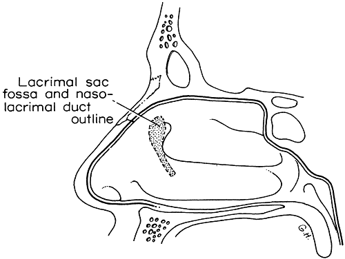
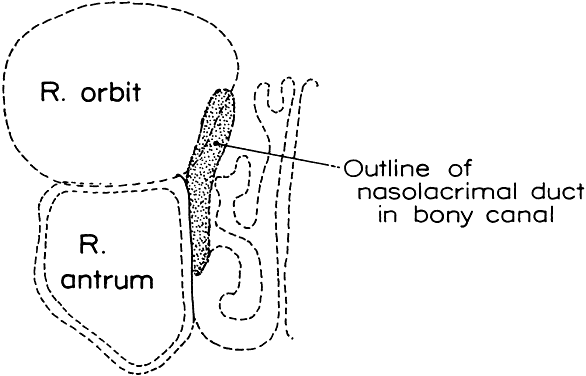
1. Blaylock WK, Moore CA, Lindberg JV: Anterior ethmoid anatomy facilitates dacryocystorhinostomy. Arch Ophthalmol 108:1774, 1990

2. Duke-Elder S: Textbook of Ophthalmology. Vol 1. St Louis: CV Mosby, 1940:238

3. Kestenbaum A: Applied Anatomy of the Eye. New York: Grune & Stratton, 1963:280–282

4. Jones LT: An anatomic approach to problems of the eyelids and lacrimal apparatus. Arch Ophthalmol 66:137, 1961

5. Doane MG: Blinking and the mechanics of lacrimal drainage system. Ophthalmology 88:844, 1981

6. Rosengren B: On lacrimal drainage. Ophthalmologica 164:409, 1972

7. Frieberg T: Wietere Untersuchungen uber die Mechanik Tranenableitung. Z Augenheilkd 39:266, 1918

8. Chavis RM, Welham RAW, Masey MW: Quantitative lacrimal scintillography. Arch Ophthalmol 96:2066, 1978

9. Maurice DM: The dynamics and drainage of tears. Int Ophthalmol Clin 13:103, 1973

10. Jones LT: Anatomy of the tear system. Int Ophthalmol Clin 13:3, 1973

11. White WL, Clover AT, Buckner AB et al: Relative canalicular tear flow as assessed by dacryoscintigraphy. Ophthalmotogy 96:167, 1989

12. Daubert J, Nik N, Chandeyssoun PA et al: Tear flow analysis through the upper and lower systems. Ophthalmol Plast Reconstr Surg 6:193, 1990

13. Linberg JV, Moore CA: Symptoms of canalicular obstruction. Ophthalmology 95:1077, 1988

14. Meyer D, Antonello A, Linberg J: Assessment of tear drainage after canalicular obstruction using fluorescein dye disappearance. Ophthalmol 97:1370, 1990

15. Mann I: The development of the human eye. Br J Ophthalmol 260, 1949

16. Duke-Elder S: Textbook of Ophthalmology. Vol 5. St Louis: CV Mosby, 1952:5348–5358

17. Cassidy JV: Developmental anatomy of the nasolacrimal duct. Arch Ophthalmol 47:141, 1952

18. Jones LT, Wobig JL: Newer concepts of tear duct and eyelid anatomy and treatment. Trans Am Acad Ophthalmol Otolaryngol 82:603, 1977

19. Levitt JM, Kravitz D: Lacrimal anomalies. Arch Ophthalmol 61:9, 1959

20. Cassidy JV: Dacryocystitis of infancy. Am J Ophthalmol 21:773, 1948

21. Ffooks OO: Dacryocystitis in infancy. Br J Ophthalmol 46:422, 1962

22. Nelson LB, Calhoun JH, Menduke H: Medical management of congenital nasolacrimal duct obstruction. Ophthalmology 92:1187, 1985

23. Katowitz JA, Welsh MG: Timing of initial probing and irrigation in congenital nasolacrimal duct obstruction. Ophthalmology 94:698, 1987

24. Bouzas A: Canalicular inflammation in ophthalmic cases of herpes zoster and simplex. Am J Ophthalmol 60:713, 1965

25. Harris G J, Hyndiuk RA, Fox MJ et al: Herpetic canalicular obstruction. Arch Ophthalmol 99:282, 1981

26. Harley RD, Stefanyszyn MA, Apt L et al: Herpetic canalicular obstruction. Ophthalmic Surg 18:367, 1987

27. Call NB, Welham RAN: Epiphora after irradiation of medial eyelid tumors. Am J Ophthalmol 92:842, 1981

28. Anderson DR: Unilateral epiphora caused by a papilloma of the lower canaliculus. Arch Ophthalmol 78:618, 1967

29. Wilson LA: Punctal occlusion in IDU toxicity. Personal communication, 1979

30. Ellis PP, Bausor SC, Fulmer JM: Streptothrix canaliculitis. Am J Ophthalmol 52:36, 1960

31. Wojno TH: Allergic lacrimal obstruction. Am J Ophthalmol 106:48, 1988

32. Jones LT: Tear sac foreign bodies. Am J Ophthalmol 60:111, 1965

33. Coleman SL, Brull S, Green WR: Sarcoid of the lacrimal sac and surrounding area. Arch Ophthalmol 88:645, 1972

34. Harris GJ, Williams GA, Clarke GP: Sarcoidosis of the lacrimal sac. Arch Ophthalmol 99:1198, 1981

35. Smith B, Tenzel RR, Buffam FV et at: Acute dacryocystic retention. Arch Ophthalmol 94:1903, 1976

36. Spaeth GL: Nasolacrimal duct obstruction caused by topical epinephrine. Arch Ophthalmol 77:355, 1967

37. Ashton N, Choyce DP, Fison LG: Carcinoma of the lacrimal sac. Br J Ophthalmol 35:366, 1951

38. Jones IS: Tumors of the lacrimal sac. Am J Ophthalmol 42:561, 1956

39. Radnot M, Gall J: Tumoran des Tranensackes. Ophthalmologica 151:2, 1966

40. Ryan SJ, Font RL: Primary epithelial neoplasms of the lacrimal sac. Am J Ophthalmol 76:73, 1973

41. Schenck NI, Ogura JH, Pratt LL: Cancer of the lacrimal sac: Presentation of five cases and review of the literature. Ann Otol Rhinol Laryngol 82:153, 1973

42. Stokes DP, Flanagan JC: Dacryocystectomy for tumors of the lacrimal sac. Ophthalmic Surg 8:85, 1977

43. Jakobiec FA, Iwamoto I, Knowles DM: Ocular adnexal lymphoid tumors: Correlative ultrastructural and immunologic marker studies. Arch Ophthalmol 100:84, 1982

44. Farkas RG, Lamberson RE: Malignant melanoma of the lacrimal sac. Am J Ophthalmol 66:45, 1968

45. Peretz WL, Ettinghausen SE, Gray GF: Oncocytic adenocarcinoma of the lacrimal sac. Am J Ophthalmol 66:45, 1968

46. Sen DK, Mohan H, Chatterjee PK: Neurilemmoma of the lacrimal sac. Eye Ear Nose Throat Monthly 50:56, 1971

47. Singh K, Mersol V, Mastny V et al: Adenoacanthoma of the lacrimal sac. Ann Ophthalmol 9:1027, 1977

48. Gurney N, Chakley T, O'Grady R: Lacrimal sac hemangiopericytoma. Am J Ophthalmol 71:757, 1971

49. Cole SH, Ferry AP: Fibrous histiocytoma of the lacrimal sac. Arch Ophthalmol 96:1647, 1978

50. Linberg JV, McCormick SA: Primary acquired nasolacrimal duct obstruction: A clinicopathologic report and biopsy technique. Ophthalmology 93:1055, 1986

51. Harris GJ, Furste FH: Lacrimal intubation in the primary repair of mid facial fractures. Ophthalmology 94:242, 1987

52. Cies WA, Baylis HI: Epiphora following rhinoplasty and Caldwell-Luc procedures. Ophthalmic Surg 7:77, 1976

53. Colvard DM, Waller RR, Neault RW et al: Nasolacrimal duct obstruction following transantral-ethmoidal orbital decompression. Ophthalmic Surg 10:25, 1979

54. Seiff SR, Shorr N: Nasolacrimal drainage system obstruction after orbital decompression. Am J Ophthalmol 160:204, 1988

55. Gaynon IE: Lacrimal insufficiency: Keratoconjunctivitis sicca and malfunction of the inferior turbinate. Am J Ophthalmol 53:614, 1960

56. Jones LT, Linn ML: The diagnosis of the causes of epiphora. Am J Ophthalmol 67:751, 1969

57. Zappia RJ, Milder B: Lacrimal drainage function, Part I. The Jones fluorescein test. Am J Ophthalmol 74:154, 1972

58. Hornblass A: A simple taste test for lacrimal obstruction. Arch Ophthalmol 90:435, 1973

59. Jones LT, Wobig JL: Surgery of the Eyelids and Lacrimal System, pp 141, 221. Birmingham, AL: Aesculapius, 1976

60. Zappia RJ, Milder B: Lacrimal drainage function. Part II: The fluorescein dye disappearance test. Am J Ophthalmol 74:160, 1972

61. Jordan A, Baum J: Basic tear flow: Does it exist? Ophthalmology 87:920, 1980

62. Clinch TE, Benedetto DA, Felberg NT et at: Schirmer's test: A closer look. Arch Ophthalmol 101:1383, 1983

63. Milder B, Demorest BH: Dacryocystography: The normal lacrimal apparatus. Arch Ophthalmol 51:180, 1954

64. Trokel SL, Potter GD: Kinetic dacryocystography: The normal lacrimal apparatus. Arch Ophthalmol 51:180, 1954

65. Hurwitz JJ, Welham RAN, Maisey MN: Intubation macrodacryocystography and quantitative scintillography: The “complete” lacrimal assessment. Trans Am Acad Ophthamol Otolaryngology 81:575, 1976

66. Rossomondo RM, Carlton WH, Trueblood JH et al: A new method of evaluating lacrimal drainage. Arch Ophthalmol 88:523, 1972

67. Chavis RM, Welham RAN, Maisey M: Quantitative lacrimal scintillography. Arch Ophthalmol 96:2066, 1978

68. Tenzel RR, Buffam FV, Miller GR: The use of the “lateral canthal sling” in ectropion repair. Can J Ophthalmol 12:199, 1977

69. Anderson RL, Gordy DD: The tarsal strip procedure. Arch Ophthalmol 97:2192, 1979

70. Nowinski TS, Anderson RL: The medial spindle procedure for involutional medial ectropion. Arch Ophthalmol 103:1750, 1985

71. Tse DT: Surgical correction of punctal malposition. Am J Ophthalmol 100:339, 1985

72. Hughes WL: Conjunctivochalasis. Am J Ophthalmol 25:48, 1942

73. Serrano F, Mora LM: Conjunctival chalasis: A surgical technique. Ophthalmic Surg 20:883, 1989

74. Bosniak SL, Smith BC: Conjunctivochalasis. Adv Ophthalmic Plast Reconstr Surg 3:153, 1984

75. Liu D: Conjunctivochalasis: A cause of tearing and its management. Ophthalmic Plast Reconstr Surg 2:25, 1986

76. Jones LT: Anatomic approach to problems of the eyelids and lacrimal apparatus. Arch Ophthalmol 66:111, 1961

77. Hanselmayer H: Prognosis of injured canaliculi in relation to elapsed time until primary operation. Ophthalmologica 166:175, 1973

78. Crawford JS: Intubation of obstructions in the lacrimal system. Can J Ophthalmol 12:289, 1977

79. Hawes MJ, Segrest DR: Effectiveness of bicanalicular silicone intubation in the repair of canalicular lacerations. Ophthalmic Plast Reconstr Surg 1:185, 1985

80. Kuchar A, Huber E, Steinkogler FJ: Monoka-Silikonintubation der Tranenwege nach retrograder Dilation. Ophthalmologe 92:43, 1995

81. McLeish WM, Bowman B, Anderson RL: The pigtail probe protected by silicone intubation: A combined approach to canalicular reconstruction. Ophthalmic Surg 23:281, 1992

82. Morrison FD: An aid to repair of lacerated tear ducts. Arch Ophthalmol 72:341, 1964

83. Demant E, Hurwitz JJ: Canaliculitis: Review of 12 cases. Can J Ophthalmol 15:73, 1980

84. Birchansky LD, Nerad JA, Kersten RC et al: Management of congenital lacrimal sac fistula. Arch Ophthalmol 108:388, 1990

85. Putterman AM: Dacryocystography with occluded common canaliculus. Am J Ophthalmol 76:1010, 1973

86. Crigler LW: The treatment of congenital dacryocystitis. JAMA 81:23, 1923

87. Kassoff J, Meyer DR: Early office-based versus late hospital-based nasolacrimal duct probing. Arch Ophthalmol 113:1168, 1995

88. Kuschner BJ: Early office-based versus late hospital-based nasolacrimal duct probing [editorial]. Arch Ophthalmol 113:1103, 1995

89. Stager D, Baker JD, Frey T et al: Office probing of congenital nasolacrimal duct obstruction. Ophthalmic Surg 23:482, 1992

90. EI-Mansoury J, Calhoun JH, Nelson LB et al: Results of late probing for congenital nasolacrimal duct obstruction. Ophthalmology 93:1052, 1986

91. Dortzbach RK, France TD, Kushner BJ et al: Silicone intubation for obstruction of the nasolacrimal duct in children. Am J Ophthalmol 94:585, 1982

92. Leone CR, Van Gemert JV: The success rate of silicone intubation in congenital lacrimal obstruction. Ophthalmic Surg 21:90, 1990

93. Becker BB, Berry FD, Koller H: Balloon catheter dilation for treatment of congenital nasolacrimal duct obstruction. Am J Ophthalmol 121:304, 1996

94. Crawford JS: Lacrimal intubation set with suture in the lumen. Ophthalmic Plast Reconstr Surg 4:249, 1988

95. Quickert MH, Dryden RM: Probes for intubation in lacrimal drainage. Trans Am Acad Ophthalmol Otolaryngology 74:431, 1970

96. Hunter LR: Crawford tubes (with suture) secured with absorbable suture. J Pediatr Ophthalmol Strabismus 32:197, 1995

97. Welsh M, Katowitz J: Timing of Silastic tubing removal after intubation for congenital nasolacrimal duct obstruction. Ophthalmic Plast Reconstr Surg 5:43, 1989

98. Kathuria SS, Harvey JT: Transcanalicular removal of silastic nasolacrimal tubes. Ophthalmic Plast Reconstr Surg 11:221, 1995

99. Snead JW, Rathbun JE, Crawford JB: Effects of the silicone tube on the canaliculus: An animal experiment. Ophthalmology 87:1031, 1980

100. Jordan DR, Nerad JA: An active inflammatory reaction to silicone stents. Ophthalmic Plast Reconstr Surg 3:147, 1987

101. Angrist RC, Dortzbach RK: Silicone intubation for partial and total nasolacrimal duct obstruction in adults. Adv Ophthalmic Plast Reconstr Surg 1:51, 1985

102. Hallus AV (trans): Procédé Plastique de dacryocystorhinostomie et ses résultats. Ann Ocul 158:241, 1921

103. Hallum AV: The Dupuy-Dutemps dacryocystorhinostomy. Trans Am Ophthalmol Soc 46:243, 1948

104. Patrinely JR, Anderson RL: A review of lacrimal drainage surgery. Ophthalmic Plast Reconstr Surg 2:97, 1986

105. Linberg JV, Anderson RL, Bumstead RM et al: Study of intranasal ostium external dacryocystorhinostomy. Arch Ophthalmol 100:1758, 1982

106. Viers ER: The use of cautery in external dacryocystorhinostomy. Am J Ophthalmol 72:679, 1971

107. Pico G: A modified technique of external dacryocystorhinostomy. Am J Ophthalmol 72:679, 1971

108. Iliff CD: A simplified dacryocystorhinostomy. Arch Ophthalmol 85:586, 1971

109. Older JJ: Routine use of a silicone stent in a dacryocystorhinostomy. Ophthalmic Surg 13:911, 1982

110. Blaylock WK, Moore CA, Lindberg JV: Anterior ethmoid anatomy facilitates dacryocystorhinostomy. Arch Ophthalmol 108:1774, 1990

111. Cruz O, Patrinely J, Reyna G et al: Urine drug screening for cocaine after lacrimal surgery. Am J Ophthalmol 111:703, 1991

112. Massaro BM, Gonnering RS, Harris GJ: Endonasal laser dacryocystorhinostomy: A new approach to nasolacrimal duct obstruction. Arch Ophthalmol 108:1172, 1990

113. Gonnering RS, Lyon DB, Fisher JC: Endoscopic laser-assisted lacrimal surgery. Am J Ophthalmol 111:152, 1991

114. Kong YT, Kim TI, Kong BW: A report of 131 cases of endoscopic lacrimal surgery. Ophthalmol 101:1793, 1994

115. Woog JJ, Metson R, Puliafito CA: Holmium:YAG endonasal laser dacryocystorhinostomy. Am J Ophthalmol 116:1, 1993

116. Bartley GB: The pros and cons of laser dacryocystorhinostomy. Am J Ophthalmol 117:103, 1994

117. Tarbet KJ, Custer PL: External dacryocystorhinostomy: Surgical success, patient satisfaction, and economic cost. Ophthalmology 102:1065, 1995

118. Mannor GE, Millman AL: The prognostic value of preoperative dacryocystography in endoscopic intranasal dacryocystorhinostomy. Am J Ophthalmol 113:134, 1992

119. Boush GA, Lemke BN, Dortzbach RK: Results of endonasal laser-assisted dacryocystorhinostomy. Ophthalmology 101:955, 1994

120. Levin PS, Stormo-Gipson J: Endocanalicular laser-assisted dacryocystorhinostomy: An anatomic study. Arch Ophthalmol 110:1488, 1992

121. Silkiss RZ, Axelrod RN, Iwach AG et al: Transcanalicular THC:YAG dacryocystorhinostomy. Ophthalmic Surg 23:351, 1992

122. Patel BCK, Phillips B, McLeish WM et al: Transcanalicular Neodymium:YAG laser for revision of dacryocystorhinostomy. Ophthalmology 104:1191, 1997

123. Welham RAN, Henderson PH: Results of dacryocystorhinostomy analysis of causes for failure. Trans Ophthalmol Soc UK 93:601, 1973

124. Welham RAN, Wulc AE: Management of unsuccessful lacrimal surgery. Br J Ophthalmol 71:152, 1987

125. Singh G, Wilson MR, Foster CS: Mitomycin eye drops as treatment for pterygium. Ophthalmology 95:813, 1988

126. Yamamoto T, Varani J, Soong HK et al: Effect of 5-fluorouracil and mitomycin C on cultured rabbit subconjunctival fibroblasts. Ophthalmol 97:1204, 1990

127. Krupin TH, Juzych MS, Shin DH et al: Adjunctive mitomycin C in primary trabeculectomy in phakic eyes. Am J Ophthalmol 119:30, 1995

128. Kao S, Liao CL, Tseng JH et al: Dacryocystorhinostomy with intraoperative mitomycin C. Ophthalmology 104:86, 1997

129. Camara JG, Bengzon AU, Henson RD: The safety and efficacy of Mitomycin C in endonasal endoscopic laser-assisted dacryocystorhinostomy. Ophthalmic Plast Reconstr Surg 16:114, 2000

130. Hu D, Sires BS, Tong DC et al: Effect of brief exposure to mitomycin C on cultured human nasal mucosa fibroblasts. Ophthalmic Plast Reconstr Surg 16:119, 2000

131. Allen KM, Berlin AJ, Levine HL: Intranasal endoscopic analysis of dacryocystorhinostomy failure. Ophthalmic Plastic Reconstr Surg 4:143, 1988

132. Orcutt JC, Hillel A, Weymuller EA: Endoscopic repair of failed dacryocystorhinostomy. Ophthalmic Plastic Reconstr Surg 6:197, 1990

133. Reinicke RD, Carroll JM: Silicone lacrimal tube implantation. Trans Am Acad Ophthalmol Otolaryngol 73:85, 1969

134. Jones LT: The cure of epiphora due to canalicular disorders, trauma, and surgical failures on the lacrimal passages. Trans Am Acad Ophthalmol Otolaryngol 66:506, 1962

135. Steinsapir KD, Glatt HJ, Putterman AM: A 16-year study of conjunctival dacryocystorhinostomy. Am J Ophthalmol 109:387, 1990

136. Rose G, Welham RAN: Jones' lacrimal canalicular bypass tubes: Twenty-five years' experience. Eye 5:13, 1991

137. Putterman AM: Fixation of Pyrex tubes and conjunctivo-dacryocystorhinostomy. Am J Ophthalmol 78:1024, 1974

138. Gladstone GJ, Putterman AM: A modified glass tube for conjunctivodacryocystorhinostomy. Arch Ophthalmol 103:1229, 1985

139. Sekhar GC, Dortzbach RK, Gonnering RS et al: Problems associated with conjunctivodacryocystorhinostomy. Am J Ophthalmol 112:502, 1991

140. Rosen N, Ashkenazi I, Rosner M: Patient dissatisfaction after functionally successful conjunctivodacryocystorhinostomy with Jones' tube. Am J Ophthalmol 117:636, 1994

141. Doucet TW, Hurwitz JJ: The broken Lester Jones tube. Can J Ophthalmol 17:32, 1982

142. Wood JR, Anderson RL, Edwards JJ: Phospholine iodide toxicity and Jones' tubes. Ophthalmology 87:346, 1980

143. Bartley GB, Gustafson RO: Complications of malpositioned Jones' tubes. Am J Ophthalmol 109:66, 1990

Back to Top