|
|
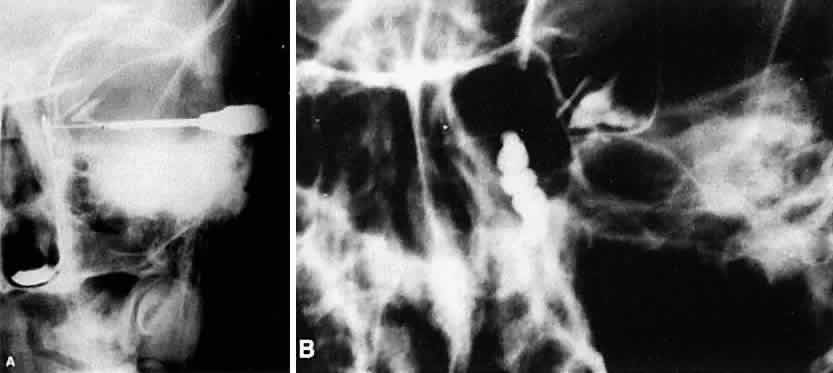
| Fig. 12. A. Normal dacryocystogram (Waters' view roentgenogram). The contrast dye fills the normal left lacrimal sac and nasolacrimal duct. Contrast dye collects along the floor of the nose as it exits from the distal nasolacrimal duct. B. Abnormal dacryocystogram. Contrast dye fills a grossly enlarged lacrimal sac with ectasias in a patient with a functional nasolacrimal duct obstruction. |