Chapter 1
Laboratory Diagnosis of Ocular Infectious Disease
RUSSELL N. VAN GELDER
Main Menu   Table Of Contents

Search

DIRECT OBSERVATION
CULTURE
SEROLOGY
POLYMERASE CHAIN REACTION
REFERENCES

Infectious disease remains a major cause of ocular morbidity throughout the world. Identification of the pathogenic organism responsible for a given ocular infection is critical to making the correct diagnosis, formulating a treatment plan, and providing an appropriate prognosis for the patient. The ophthalmologist must therefore be adept with the techniques of sample acquisition from a variety of ocular tissues, and have a working knowledge of the appropriate laboratory tests necessary for correct identification of pathogenic organisms.

A good working relationship with the clinical pathology laboratory is essential to ensure that samples are handled correctly. Many hospital and outpatient laboratories receive ophthalmic samples infrequently, which can lead to incorrect processing or even loss of valuable samples. For outpatient procedures, the laboratory should be contacted directly to ensure that the sample is processed correctly. For surgical biopsies, it is often advantageous to have the pathologist in the operating room at the time of sample acquisition to ensure that the often-miniscule biopsy tissue is handled appropriately.

There are four major means of laboratory identification of infectious organisms: direct observation, culture, serology, and molecular diagnostics. There are also four broad categories of pathogenic organisms affecting the eye: bacteria, fungi, viruses, and parasites. In this chapter, we consider each laboratory method in turn, highlighting the specifics of its application to each of the four categories of pathogens.

Back to Top
DIRECT OBSERVATION
Nearly all ophthalmic pathogens are microscopic; it is exceedingly rare to directly observe pathogenic organisms during the ophthalmic examination, although occasionally lice (i.e., Pediculus humanus spp. or Phthirus pubis) can be directly observed under the slit lamp in patients with chronic blepharitis. Far more frequently, direct observation requires obtaining tissue by methods such as scraping of a corneal ulcer, swabbing of acute purulent conjunctivitis, or vitreous aspiration in endophthalmitis, followed by light or electron microscopy with appropriate staining. Material from acute conjunctivitis can be obtained by swabbing the anesthetized conjunctiva with an alginate swab, whereas corneal samples are generally obtained with a sterilized Kimura spatula or needle. When sampling infected tissue, one should attempt to sample portions with active infection but without excessive necrosis. Care must be taken to identify samples from each swab carefully (e.g., right eye, left eye, lids, ulcer). For larger corneal ulcers, samples should be taken from several regions. Material can be applied directly to a glass slide as a thin smear and allowed to air dry.

BACTERIA

Most bacteria are not visible in unstained tissue. The standard initial staining method for identifying bacteria is Gram's stain. To perform a Gram stain, the sample is spread on a slide, dried, and briefly fixed in methanol. It is then stained with a cresyl violet solution (i.e., Hucker's solution) for 1 minute, rinsed in water, and then stained in Gram's iodide for 1 minute. Following decolorization in 95% ethyl alcohol and a brief water rinse, the sample may be counterstained with safranin O. The entire procedure can be completed in less than 5 minutes. Gram-positive organisms resist the decolorization step and remain stained dark purple, whereas gram-negative bacteria have their cell wall permeability increased by the alcohol wash and thus decolorize; they are visualized by the pink counterstain. Table 1 lists common gram-positive and -negative organisms causing ophthalmic disease.


Table 1. Common Gram-Positive and Gram-Negative Bacteria Causing Ocular Disease


GenusRepresentative SpeciesGram's StainCommon Clinical Context
Staphylococcusaureus, epidermidisPositive cocci (clusters)Cellulitis, keratitis, endophthalmitis
Streptococcus pneumoniae, viridansPositive cocci (chains)Conjunctivitis, keratitis, blebitis, endophthalmitis
BacilluscereusPositive rodTraumatic endophthalmitis, keratitis
ClostridiumperfringensPositive rodKeratitis
CorynebacteriumulceransPositive rod (club shaped)Conjunctivitis, keratitis
ListeriamonocytogenesPositive rodKeratitis
PropionibacteriumacnesPositive rodLate onset endophthalmitis
ActinomycesisraeliiPositive rod (branched)Keratitis, caniliculitis
NocardiaasteroidesPositive rodKeratitis, choroiditis
NeisseriagonorrhoeaeNegative cocciConjunctivitis, keratitis
BranhameliacatarrhalisNegative cocciKeratitis
EscherichiacoliNegative rodKeratitis
HaemophilusinfluenzaeNegative rodBlebitis, keratitis
KlebsiellapneumoniaeNegative rodKeratitis
MoraxellacatarrhalisNegative rodKeratitis
ProteusmirabilisNegative rodKeratitis
PseudomonasaeruginosaNegative rodKeratitis
SerratiamarcescensNegative rodKeratitis


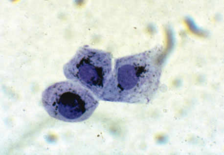
Certain bacteria are not readily visualized by Gram's stain, and may require other stains or types of microscopy for visualization. Mycobacteria are most easily visible using acid-fast stains such as the Ziehl-Neelsen stain. Intracellular Chlamydia are not directly visible with Gram's stain, and are generally visualized following Giemsa staining of intracellular inclusion bodies (Fig. 1). It is important to note that this Giemsa stain is the traditional 60-minute staining protocol; the brief Wright-Giemsa stain used for blood stains will not identify inclusion bodies. The Papanicolaou stain can also be used for the identification of Clamydia inclusions. Giemsa stain is also useful for the demonstration of Actinomyces and Nocardia species (which can also be seen on Gram's staining). Darkfield microscopy can be used to demonstrate the Treponema pallidum spirochete of syphilis, although serologic testing is more commonly performed to confirm infection. Serologic testing is more commonly performed to confirm the traditional 60-minute Giemsa staining protocol; the brief Wright-Giemsa stain used for blood stains will not identify inclusion bodies. The Papanicolaou stain can also be used for the identification of Chlamydia inclusions.

Fig. 1. Typical perinuclear intracytoplasmic inclusion bodies of Chlamydia in conjunctival cytologic preparation: Giemsa stain. (Photomicrograph courtesy of Dr. Morton Smith.)

Certain direct fluorescent stains may be used in lieu of Gram's strain for detection of bacteria. In particular, acridine orange has a much higher sensitivity for detection of bacteria than Gram's stain, and has the additional advantage of staining fungi more reliably than Gram's stain. The drawback of these direct fluorescent stains is the necessity of a fluorescent microscope for detection of the fluorescent signal. Positive specimens should be secondarily strained with Gram's stain to identify the detected pathogen.

Commercial monoclonal or polyclonal antisera can be used to identify specific organisms by indirect immunofluorescence. In ophthalmology, this is commonly used for Chlamydia species.1 Because a specific antiserum must be used for each suspected organism, the requesting physician must communicate with the clinical microbiology or pathology laboratory in order to request these tests.

FUNGI

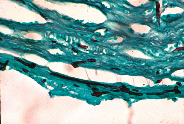
The most rapid technique for visualizing yeast or filamentous fungi is the potassium hydroxide (KOH) preparation. The scraping or swab is treated with 10% KOH in dimethyl sulfoxide. The strong base dissolves keratinized tissue (such as cornea), but the cell walls of fungi are resistant to dissolution. Both budding yeast and filamentous fungi can be visualized. Other staining methods also have high sensitivity for detection of yeast and fungi, including Giemsa stain, Gomori's methenamine silver (GMS), India ink staining, or periodic acid–Schiff (PAS) stain (Fig. 2). Fungi can also be identified on some Gram-stained samples.

Fig. 2. Fungal keratitis with branching septate hyphae, stained with Gomori's methenamine silver. (Photomicrograph courtesy of Dr. Morton Smith.)

VIRUSES

Viruses are typically submicrometer in size and cannot be directly visualized by light microscopy. Characteristic cellular changes can sometimes be identified as, for example, the cytoplasmic inclusion bodies of herpes infection on hematoxylin-eosin–fixed sections or on the Tzanck smear (in which the fluid from a suspected herpetic vesicle is stained with Giemsa, Papanicolaou, and Wright's stain; multinucleated giant cells indicate herpetic infection). Viruses can be directly observed by electron microscopy, but this is rarely used clinically because of the difficulty in specimen preparation and the low yield of positive results. Specific antisera for many viruses are available, and can be used to identify virally infected cells; however, this technique is typically used in research settings because it generally requires fixed biopsy tissue with preserved cytoarchitecture.

PARASITES

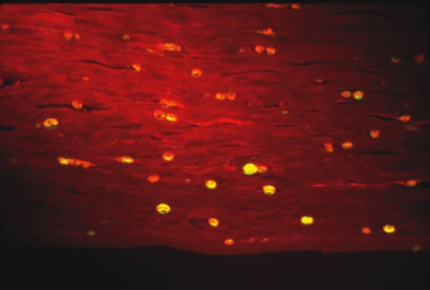
Some parasites can be seen free living under the microscope. Onchocerca volvulus, the causative agent of river blindness, can be directly observed swimming out of skin or corneal biopsies incubated in saline or culture medium. Modern imaging techniques are increasing the range of organisms that can be directly detected in situ. In particular, the confocal microscope has been successfully used to visualize Acanthamoeba in infected corneas.2 Certain parasites causing ophthalmic disease can be visualized with special stains. Acanthamoeba species can be visualized on corneal scrapings with PAS or GMS stains, or with the fluorescent dyes calcifluor white or acridine orange (Fig. 3). Both techniques require a fluorescent microscope to visualize the dye. Calcifluor white (a whitening agent used in laundry detergents) stains Acanthamoeba cell walls and fluoresces green under ultraviolet illumination. Toxoplasma gondii can be visualized with PAS or GMS stains from biopsy material. Specific antisera can be used in stains of biopsy specimens.

Fig. 3. Acanthamoeba in cornea stained with calcifluor white and imaged with ultraviolet fluorescence microscopy. (Photomicrograph courtesy of Dr. Morton Smith.)

Back to Top
CULTURE
The ability to grow organisms in vitro is the mainstay of pathogen identification for bacteria and some fungi. In addition to allowing a variety of metabolic tests to be run for precise identification of microbial organisms, antibiotic resistance can also be assayed.

Correct handling of material to be cultured is critical to achieving good yields on these diagnostic tests. Ocular surface or intraocular samples suspected of bacterial or fungal infection should be plated or inoculated on multiple media. Blood agar is useful for isolation of aerobic bacteria when grown at 37°C and may yield cultures of saprophytic fungi when grown at room temperature. Chocolate agar is useful for growing Haemophilus, Neisseria, and Moraxella cultures, and must be grown under 5% to 10% carbon dioxide. For isolation of most fungi, either Sabouraud's dextrose agar or brain-heart infusion media should be used and grown at room temperature. Finally, anaerobic bacteria may be isolated from thioglycolate broth. Specific media for other indications are discussed below.

BACTERIA

Many bacteria can be grown on the media described above. Some may be normal commensal organisms of the periocular region, such as Staphylococcus epidermidis, Streptococcus viridans, or Propionibacterium acnes. It should be noted that the intraocular contents are expected to be sterile at all times; any bacteria (even normal periocular commensal organisms) recovered from an aqueous or vitreous tap are potentially pathogenic.

Recovery of organisms from culture medium generally requires a minimum of 24 to 48 hours, with biochemical typing and sensitivity testing taking several additional days. Positive cultures typically are subjected to a panel of biochemical tests that allow definitive identification of the growing organism. Examples include coagulase testing of Staphylococcus species and oxidase testing for Enterobacteriaceae.

The traditional initial technique for assessing antibiotic resistance is the Kirby-Bauer disk diffusion technique. A defined inoculum of bacteria is seeded onto a large plate, and antibiotic-impregnated disks are placed onto the agar surface. As the bacteria on the lawn grow, they are inhibited to varying degrees by the antibiotic diffusing from the disk. Inhibition zones of given size surrounding the disks correspond to sensitivity or resistance to the antibiotic tested. Variants of this test (using defined gradient-sticks of antibiotic that provide data analogous to the minimal inhibitory concentration [MIC]) are entering into common use. Antibiotic resistance testing for some of the more common causes of resistance, such as β-lactamase testing, can be done more rapidly by chromogenic assay. When precise measurement of antibiotic resistance is required, bacterial growth against a dilution series of antibiotic concentrations can be used to determine the MIC.

VIRUSES

Viral culture is used infrequently in ophthalmology. Viral cultures are most commonly used for herpes family viruses, including cytomegalovirus (CMV), herpes simplex types 1 and 2, and varicella-zoster virus. The standard culture method is the “shell vial” technique, in which centrifugated virus is grown for 24 to 48 hours and then detected by an enzyme-conjugated antiviral antiserum. Although the method is relatively rapid, sensitivity is not perfect, and more traditional, tube-based viral cultures with plaque assays are generally also performed. Because of the slow speed and marginal sensitivity of viral culture techniques, polymerase chain reaction (PCR) diagnostics (see below) are steadily supplanting viral cultures.

PARASITES

Acanthamoeba may be difficult to culture and are typically grown on a plate seeded with an Escherichia coli lawn. It is the only common ocular parasite that is routinely detected by culture. Other parasites are cultured only infrequently, usually in a research setting.

Back to Top
SEROLOGY
There are two uses for serology in the diagnosis of ocular infectious disease. First, specific antibodies may be used for detection of pathogens (as discussed in the first section of this chapter). In addition to histologic analysis, such antibodies can also be used in solution-based enzyme-linked immunosorbant assays (ELISA) to determine pathogen load (Fig. 4). This is the basis of the Adenoclone test (Meridian Bioscience, Inc., Cincinnati, OH) for adenoviral-mediated conjunctivitis.3 These tests have high specificity but do not yet have the sensitivity of culture or PCR-based diagnostic tests.

Fig. 4. Enzyme-linked immunosorbant assay (ELISA). Red indicates purified pathogen antigen, which is fixed to the floor of the microtiter dish. The green antibody represents the host IgG, whereas the purple antibody is an enzyme-conjugated antibody that recognizes the fixed Ig portion of host antibody. After washing, the presence of the antibody sandwich is assayed by addition of a chromogenic substrate that changes color (here, clear → blue) via the enzyme linked to the antibody. The final colored substrate is measured with a densitometer or spectrophotometer.

More commonly, serology refers to the measurement of host-derived antibodies to the pathogen of interest. Typically, two classes of antibodies are measured: IgG and IgM. The latter represents acute antibody responses to a novel antigen, usually peaking within a few weeks of exposure and generally disappearing by 6 weeks after infection. Positive IgG titers typically persist for many years. In most cases, serologies are measured by ELISA; samples are usually diluted twofold and the lowest dilution producing a positive reaction is taken as the titer. Two types of serology are in use for ophthalmic disease. Serum serologies are useful primarily in ruling out a diagnosis or supporting a rare diagnosis, for example, ruling out ocular toxoplasmosis on the basis of negative IgG and IgM serologies or confirming the diagnosis of Toxocara canis infection, which is incidentally seropositive in 5% to 10% of the population.4 (Thus, given a pre-test probability of approximately 10%, a positive result on serology strongly supports, but does not definitively demonstrate, active infection). Positive serologies can also be used to confirm diagnoses of other organisms, including Treponema pallidum,Bartonellahenselae, and Borrelia burgdorferi.

BACTERIA

Because the treponemal bacterium T. pallidum cannot be readily cultured and is difficult to identify by darkfield microscopy from ocular tissues, serologic diagnosis has become the standard method of documentation of infection. Two types of tests are in widespread use. The fluorescent treponemal antibody absorption (FTA) and microhemagglutination (MHA-TP) remain positive for life once the patient is positive, whereas the titers of the Venereal Disease Research Laboratory (VDRL) and rapid plasmin reagent (RPR) tests are indicative of current infective load. The latter two tests are not fully specific for syphilis, and positive results must be confirmed by FTA or MHA-TP. Serologies are commonly used to confirm diagnosis of other unusual bacterial infections that can have ocular consequences, including the causative agents of cat-scratch disease (responsible for many cases of acute neuroretinitis), B. henselae, and Bartonella quintana, as well as the spirochetes responsible for Lyme disease: B. burgdorferi.

Serologies rely on documentation of B cell responses to previously encountered pathogenic antigens. Previous T cell encounters can be determined by skin testing (a measure of delayed-type hypersensitivity). The most commonly applied skin test for documentation of infection is the tuberculin skin test, purified protein derivative (PPD) test, or Mantoux test. In this test, 0.1 mL of PPD is injected subcutaneously and the injected area is examined 48 to 72 hours later for induration. A reading of greater than 15 mm is considered positive in all individuals. In many patients, including foreign-born individuals from Asia, Africa, or Latin America; intravenous drug users; residents of long-term care facilities; and patients with chronic disease, a 10-mm reading is considered positive. For patients with acquired immunodeficiency syndrome, radiographic evidence of tuberculous disease, or household contact with patients known to have tuberculosis, a reading of 5 mm is considered positive.5

FUNGI

Serology is typically not used for identification of acute fungal infections; the ubiquitous nature of fungal pathogens ensures nearly universal seropositivity to many common pathogens. Indeed, seropositivity to Candidaalbicans is sufficiently universal that the antigen is used as a positive control for other skin testing.

Historically, serology for Histoplasma capsulatum has been used to aid in diagnosis of the presumed ocular histoplasmosis syndrome.6 However, reports of reactivation of ocular disease following skin testing and later reports of disease occurring in regions devoid of H. capsulatum7 have limited use of this technique.

VIRUSES

Viral titers are occasionally of value in identifying a pathogen. In suspected acute retinal necrosis syndrome, for example, negative IgG and IgM titers for varicella-zoster virus or herpes simplex virus can effectively rule out these pathogens. Human immunodeficiency virus (HIV) can be detected by serology testing, which provides important adjunctive data for further work-up of many patients. Antiviral IgG titers are not very useful for confirmation of CMV or Epstein-Barr virus because exposure to these pathogens by adulthood is nearly universal.

Intraocular antibody titers may be very useful for determining that a positive serum titer is associated with acute intraocular infection.8 The means for determining the significance of an intraocular titer is called the Goldmann-Witmer coefficient and is calculated by: R3 = R1/R2, where R1 = Abx(eye)/Abx(serum), and R2 = albumineye/albuminserum, and Abx refers to the reciprocal of the titer in the respective tissue. Thus, the Goldmann-Witmer coefficient measures the ratio of ocular antibody concentration to serum, accounting for the diffusion of large macromolecules across the blood–eye barrier by measurement of an abundant protein such as albumin. Alternatively, serum and ocular titers to a ubiquitously seropositive viral titer (such as adenovirus) or total IgG can be substituted for the albumin measurements. As an arbitrary cutoff, Goldmann-Witmer coefficients greater than 3 are considered strongly suggestive of ocular infection.

PARASITES

Negative serum serologies are useful for ruling out T. gondii infection, and particularly high IgG titers (greater than 1:2048) or documentation of IgA titers can be suggestive of acute infection. Goldmann-Witmer coefficient testing is a very effective means for diagnosing intraocular toxoplasmosis.9 A presumptive diagnosis of T. canis can be supported by positive titers, whereas negative titers effectively rule out this disease.

Back to Top
POLYMERASE CHAIN REACTION
The PCR is an enzymatic method for amplifying small amounts of nucleic acids (deoxyribonucleic acid or ribonucleic acid). The technique is a relatively recent invention that was first described in 1985.10 A schematic of the PCR is shown in Figure 5. In order to perform PCR one must have the following:

Fig. 5. Polymerase chain reaction (PCR). Pathogen DNA (blue) is detected by repeated performance of PCR. An oligonucleotide primer (red) is annealed to the DNA in the sample, and extended with a thermostable DNA polymerase in the presence of appropriate buffers and nucleotide triphosphates. The parent and daughter strands are heat denatured, and oligonucleotides re-annealed. By repeated performance, exponential amplification of starting material can be achieved.

  1. tissue material harboring the suspected pathogen, usually an aqueous or vitreous tap
  2. oligonucleotide DNA primer sequences specific for the pathogens being detected
  3. appropriate enzymes for amplifying DNA (e.g., a thermostable DNA polymerase)
  4. buffers and nucleotide triphosphate “building blocks”
  5. a thermal cycling machine

Products—short pieces of double-stranded DNA—can be visualized by acrylamide or agarose gel electrophoresis, or subjected to DNA sequencing for confirmation of identity.

BACTERIA

Specific bacteria can be detected by PCR. This test is perhaps most useful for detection of P. acnes endophthalmitis, for which culture has a very poor yield. Culture has a yield for P. acnes of about 25%; PCR, by contrast, has a yield in suspected cases of greater than 90%.11 Other difficult-to-culture bacteria, including Mycobacterium tuberculosis, T. pallidum, and B. burgdorferi can be detected by PCR.12 PCR is also capable of detecting and identifying other unculturable bacteria; the most spectacular example of this is detection of Tropheryma whipelli, the causative agent of Whipple's disease.13

In addition to detection of single pathogens, PCR can be used to screen for multiple bacteria. Bacterial ribosomal genes are highly conserved. Specific primers are thus able to detect a wide range of bacteria; individual products may then be sequenced in order to positively identify the offending pathogen. By this means, new bacterial strains, such as those causing culture-negative endophthalmitis, have been identified.14

FUNGUS

As with bacteria, fungi can be detected by individual species, or by pan-amplification of conserved ribosomal sequences (in this case the 18S and 28S subunits).15 Identification of individual fungal species can then be accomplished through sequencing of the PCR product.

VIRUSES

Perhaps the most common use of PCR diagnostics is for the confirmation of viral pathogens in cases of acute retinal necrosis.16 PCR has a very high sensitivity and specificity for detection of the common viral pathogens herpes simplex types 1 and 2, and varicella-zoster. The technique is also very sensitive for the detection of CMV DNA. CMV retinitis can sometimes be confused with acute retinal necrosis syndrome in immunocompromised patients.

HIV is readily detected by reverse-transcription PCR (RT-PCR), which can be used quantitatively to determine viral load. This indicator has proven to be very useful for determining response to highly active antiretroviral therapy.17

PARASITES

T. gondii is readily detected by PCR amplification of its highly repeated B1 gene. However, because of the low numbers of tachyzoites in the aqueous, the yield on PCR for T.gondii is low from this fluid. The yield is somewhat better from vitreous fluid but still is in the range of 50% to 60%.18 It may be that a combination of Goldmann-Witmer coefficient and PCR testing gives the highest sensitivity for detection of ocular toxoplasmosis.9 PCR may be more useful in the acute stages of infection, and Witmer coefficients more useful in the convalescent phase.

ADVANTAGES AND PITFALLS OF POLYMERASE CHAIN REACTION DIAGNOSTICS

Polymerase chain reaction diagnostics offer several advantages over conventional culture techniques. Because of the exponential amplification of nucleic acids, the sensitivity of PCR for detection approaches theoretic bounds of a single DNA molecule (e.g., 0.1 zeptomolar). Thus, PCR is typically one to two orders of magnitude more sensitive for detection of pathogens than most culture techniques. PCR is also phenomenally specific; PCR primers can distinguish between pathogens differing by only a single base pair in their DNA. It is also a very rapid test, with results generally ready within several hours.

These same strengths can also become potential pitfalls of the technique. The extremely high sensitivity makes PCR prone to false-positive results, which may stem from two possible sources. First, “bystander” or commensal organism DNA can create false-positive results. Most herpes family DNA viruses remain latent in a subset of host cells. When the sensitivity of PCR is increased to near maximal levels (e.g., by employing “nested” PCR primers), positive signals from latent DNA can be confused with pathogen.19 Recent advances in “real-time” PCR, which allows derivation of an estimate of numbers of detected pathogenic DNA molecules, may help distinguish between background and real signals.20 The high specificity of PCR can also be a hindrance. If, for example, a base-pair mismatch is generated in a primer sequence because of a naturally occurring polymorphism in the gene being amplified, this can create false-negative results. Thus, negative results from PCR testing have more validity when at least two pathogen DNA targets are tested.

The range of applications of PCR diagnostics continues to expand. PCR can now be used to detect mutations conferring resistance to ganciclovir in CMV retinitis.21,22 PCR can also distinguish between strains of microorganisms that are not distinguishable by any other means. It has recently become apparent, for example, that ocular toxoplasmosis is caused by at least three strains of microorganisms, distinguishable by PCR, that respond differentially to antibiotic therapy in vitro.23 In the research domain, PCR has been used to amplify foreign DNA from diseases suspected of having infectious etiology. Perhaps the most spectacular example of this was the identification of a novel herpes-family virus as the causal agent of Kaposi's sarcoma.24

Back to Top
REFERENCES

1. Athanasiu P, Anghelescu S, Predescu E, et al: Rapid detection by immunofluorescence of multiple viral infections in patients with keratitis. Virologie 35:83, 1984

2. Winchester K, Mathers WD, Sutphin JE, Daley TE: Diagnosis of Acanthamoeba keratitis in vivo with confocal microscopy. Cornea 14:10, 1995

3. Wiley LA, Roba LA, Kowalski RP, et al: A 5-year evaluation of the Adenoclone test for the rapid diagnosis of adenovirus from conjunctival swabs. Cornea 15:363, 1996

4. Taylor MR: The epidemiology of ocular toxocariasis. J Helminthol 75:109, 2001

5. Rodnick JE: New guidelines for tuberculin skin test interpretation. West J Med 154:322, 1991

6. Smith RE, Ganley JP: Presumed ocular histoplasmosis. I. Histoplasmin skin test sensitivity in cases identified during a community survey. Arch Ophthalmol 87:245, 1972

7. Suttorp-Schulten MS, Bollemeijer JG, Bos PJ, Rothova A: Presumed ocular histoplasmosis in the Netherlands—an area without histoplasmosis. Br J Ophthalmol 81:7, 1997

8. Baarsma GS, Luyendijk L, Kijlstra A, et al: Analysis of local antibody production in the vitreous humor of patients with severe uveitis. Am J Ophthalmol 112:147, 1991

9. Fardeau C, Romand S, Rao NA, et al: Diagnosis of toxoplasmic retinochoroiditis with atypical clinical features. Am J Ophthalmol 134:196, 2002

10. Saiki R, Scharf S, Faloona F, et al: Enzymatic amplification of B globin genomic sequences and restriction site analysis for the diagnosis of sickle cell anemia. Science 230:1350, 1985

11. Lohmann CP, Linde HJ, Reischl U: Improved detection of microorganisms by polymerase chain reaction in delayed endophthalmitis after cataract surgery. Ophthalmology 107:1047, 2000

12. Van Gelder RN: Applications of the polymerase chain reaction to diagnosis of ophthalmic disease. Surv Ophthalmol 46:248, 2001

13. Richman L, Freeman W, Green W, et al: Brief report: uveitis caused by Tropheryma whippelii (Whipple's bacillus). N Engl J Med 332:363, 1995

14. Okhravi N, Adamson P, Lightman S: Use of PCR in endophthalmitis. Ocul Immunol Inflamm 8:189, 2000

15. Kercher L, Wardwell SA, Wilhelmus KR, Mitchell BM: Molecular screening of donor corneas for fungi before excision. Invest Ophthalmol Vis Sci 42:2578, 2001

16. Ganatra JB, Chandler D, Santos C, et al: Viral causes of acute retinal necrosis syndrome. Am J Ophthalmol 129:166, 2000

17. Hodinka RL: The clinical utility of viral quantitation using molecular methods. Clin Diag Virol 10:25, 1998

18. Montoya JG, Parmley S, Liesenfeld O, et al: Use of the polymerase chain reaction for diagnosis of ocular toxoplasmosis. Ophthalmology 106:1554, 1999

19. Short GA, Margolis TP, Kuppermann BD, et al: A polymerase chain reaction–based assay for diagnosing varicella-zoster virus retinitis in patients with acquired immunodeficiency syndrome. Am J Ophthalmol 123:157, 1997

20. Dworkin LL, Gibler TM, Van Gelder RN: Real-time quantitative polymerase chain reaction diagnosis of infectious posterior uveitis. Arch Ophthalmol 120:1534, 2002

21. Liu W, Kuppermann BD, Martin DF, et al: Mutations in the cytomegalovirus UL97 gene associated with ganciclovir-resistant retinitis. J Infect Dis 177:1176, 1998

22. Jabs DA, Martin BK, Forman MS, et al: Mutations conferring ganciclovir resistance in a cohort of patients with acquired immunodeficiency syndrome and cytomegalovirus retinitis. J Infect Dis 183:333, 2001

23. Grigg ME, Ganatra J, Boothroyd JC, Margolis TP: Unusual abundance of atypical strains associated with human ocular toxoplasmosis. J Infect Dis 184:633, 2001

24. Chang Y, Cesarman E, Pessin MS, et al: Identification of herpesvirus-like DNA sequences in AIDS-associated Kaposi's sarcoma. Science 266:1865, 1994

Back to Top