Chapter 54F
Glaucoma Associated with Corneal Disorders
SID MANDELBAUM
Main Menu   Table Of Contents

Search

GLAUCOMA ASSOCIATED WITH PRIMARY CORNEAL ENDOTHELIAL DISORDERS
GLAUCOMA ASSOCIATED WITH CORNEAL INFECTION AND INFLAMMATION
GLAUCOMA OCCURRING AFTER PENETRATING KERATOPLASTY
REFERENCES

Glaucoma may be associated with corneal diseases by a variety of mechanisms. Developmental abnormalities of the anterior segment may affect both the cornea and chamber angle, as in patients with Rieger's syndrome or Peters' anomaly: these conditions are discussed elsewhere in this series. Glaucoma may result from a primary corneal disorder, as is thought to be the case in patients with the iridocorneal endothelial syndrome. Elevated intraocular pressure may accompany corneal infectious or inflammatory processes, generally mediated by intraocular inflammation. Finally, eyes requiring penetrating keratoplasty may already have glaucoma, or may develop glaucoma as a result of structural changes to the anterior segment from the corneal transplant procedure.
Back to Top
GLAUCOMA ASSOCIATED WITH PRIMARY CORNEAL ENDOTHELIAL DISORDERS

IRIDOCORNEAL ENDOTHELIAL SYNDROME

The iridocorneal endothelial (ICE) syndrome consists of three rare clinical entities and their variations that, although separately described, are now considered as a spectrum of disease with a common pathogenetic mechanism. Patients with the ICE syndrome usually present in the third to fifth decades of life having noticed unilateral iris abnormalities or complaining of visual disturbances or ocular discomfort due to corneal edema or elevated intraocular pressure. The syndrome is more common in women, appears to occur sporadically, and has been recognized almost exclusively in whites. There are no known systemic associations.1,2

Clinical Features

The iris, cornea, and anterior chamber angle are affected in patients with the ICE syndrome. The major clinical variations within this syndrome are described below and are distinguished by particular patterns of involvement of these structures.

PROGRESSIVE (ESSENTIAL) IRIS ATROPHY. Progressive iris atrophy is characterized by prominent atrophy of the iris, with iris stromal thinning progressing to full-thickness iris holes.1,2 Peripheral anterior synechiae develop early, and progress both circumferentially and onto the cornea.1,2 Pupillary distortion and ectropion uveae occur, typically toward the most prominent peripheral anterior synechiae.1,3 Large and sometimes bizarrely shaped iris “stretch” holes occur, apparently from traction of the iris between peripheral anterior synechiae on opposite sides of the globe (Fig. 1).1,3,4 Less common, smaller oval holes may develop adjacent to peripheral anterior synechiae. These “melting” holes are presumed to be caused by iris ischemia resulting from obstruction of iris vessels within the synechiae.1,3 Multiple, fine, pedunculated iris nodules may occur, generally appearing late in the disease process.1,5

Fig. 1. Clinically normal right eye (A) and affected left eye (B) of a 41-year-old man with ICE syndrome who presented complaining of slight decrease in visual acuity and photophobia of the left eye. Note left conjunctival injection, iris stromal thinning, pupil irregularity, and two full-thickness iris holes. There was mild left epithelial edema with an applanation tension of 45 compared with 18 mm Hg in the right eye. Gonioscopic view (C) of the inferior angle of the left eye, showing prominent peripheral anterior synechiae adherent anterior to Schwalbe's line. Trabeculectomy was required for control of the glaucoma in the left eye.

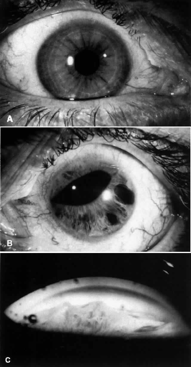
The corneal endothelium in involved eyes is described as showing a fine, beaten metal irregularity, sometimes in focal areas. Stromal and/or epithelial edema may be present. In some cases, no definite corneal abnormality is evident.1

CHANDLER'S SYNDROME. The dominant feature in this variant of the ICE syndrome is usually corneal edema, often at normal or only moderate levels of intraocular pressure elevation. Chandler described the corneal endothelium as having a “fine hammered silver appearance.” ,6 Iris atrophy is less prominent in Chandler's syndrome than in progressive iris atrophy and, when detectable, is often limited to the anterior iris stroma. The pupil is usually round or slightly oval.6

COGAN-REESE (IRIS NEVUS) SYNDROME. Patients with Cogan-Reese syndrome are characterized by iris pigmented lesions that range from multiple, pedunculated, nodular lesions to a more diffuse, smooth, velvety change of the iris. The iris surface tends to lose its normal pattern, and it usually appears darker than that of the fellow eye. Ectropion uveae, breaks in the iris stroma, and an ectopic pupil are often present. Peripheral anterior synechiae, corneal edema, and labile glaucoma are characteristic features.7

Pathogenesis

On the basis of careful clinical observations and histopathologic study, Campbell and co-workers3 and Eagle and co-workers8 have advanced the now generally accepted hypothesis that the primary abnormality in the ICE syndrome is proliferation of abnormal corneal endothelium. This abnormal endothelium correlates with the beaten metal appearance visible on slit lamp biomicroscopy. If the patient is examined in the early stages of the disease, demarcations between normal and abnormal corneal endothelium may be visible biomicroscopically.9,10 With time, the areas of abnormal endothelium enlarge, so that eventually the entire corneal endothelium is involved.9 The stroma and epithelium overlying the regions of abnormal endothelium may be clear or may become edematous.

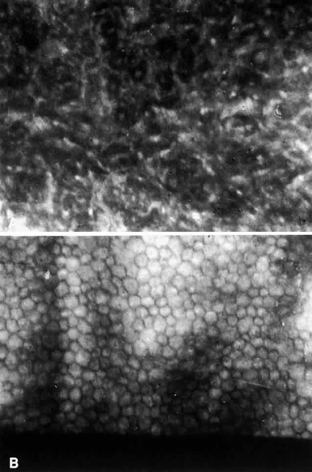
The specular microscopic changes of the corneal endothelium are sufficiently specific that a masked observer can usually differentiate the ICE syndrome from other endothelial conditions.11 The earliest visible changes are a loss of the uniform hexagonal shape of the endothelial cells. Dark areas begin to appear in individual cells. In more affected corneas there is increased cellular pleomorphism and the dark areas within individual cells become larger, eventually reaching the point at which the endothelial mosaic can no longer be recognized (Fig. 2).9,11 The darker areas within the abnormal endothelial cells may be accompanied by a brighter than normal reflection from the cell borders.12 In clinically normal-appearing areas in some affected eyes, specular microscopy has shown a normal endothelial mosaic but cells that are much smaller than usual9,10; the significance of this is unknown. In some cases, the uninvolved fellow eyes of patients with the ICE syndrome have shown abnormal endothelial cell pleomorphism with lower than expected cell counts.11,13

Fig. 2. A. Specular microscopy of the corneal endothelium in ICE syndrome. Cell borders are obscured, resulting in loss of the normal endothelial mosaic. Note dark areas within endothelial cells. Brighter reflections are believed to be from cell borders. B. Specular microscopy of fellow eye showing normal endothelial mosaic. (Courtesy of Ira J. Udell, MD)

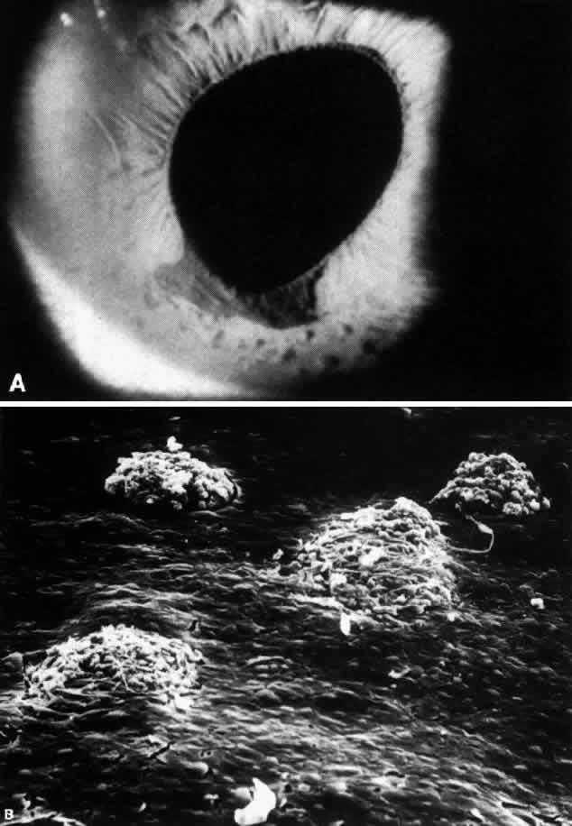
The abnormal endothelium and the basement membrane that it elaborates eventually spread from the cornea onto the trabecular meshwork and the surface of the iris.3,8 Contraction of this membrane results in the development of peripheral anterior synechiae in areas of previously open angle and may also result in ectropion uveae.3,8 Iris atrophy and full-thickness holes develop as a result of stretching of the iris between synechiae. Since the iris atrophy is actually a secondary phenomenon,8 the term progressive iris atrophy is preferred to the historically used term essential iris atrophy.2Iris nodules may develop in areas involved with the endothelium-basement membrane complex (Fig. 3); it has been suggested that the nodules form as a result of encircling and “pinching off” portions of the iris by the cellular membrane.3,8,14 The nodules are thus a marker for areas of iris endothelialization.14

Fig. 3. Iris nodules in ICE syndrome. A. Multiple nodules stud flattened area of iris stroma inferiorly, adjoining distorted pupil and iris pigment epithelial ectropion. Stromal effacement and iris nodules are a clinical marker for iris endothelialization. B. Scanning electron microscopy (SEM) of anterior iris surface in area of clinically effaced stroma shows multiple iris nodules surrounded by confluent sheet of corneal endothelial cells. Characteristic iris nodules appear to be formed by the encirclement of knuckles of iris stroma by the proliferating corneal endothelium. (SEM, x200) (Courtesy of Ralph C. Eagle Jr, MD)

The various clinical manifestations of progressive iris atrophy, Chandler's syndrome, and the Cogan-Reese syndrome can thus all be explained by the proliferation of this abnormal endothelium.2,3,8 The location, extent and specific pattern of proliferation account for the different clinical variations seen from patient to patient. Continued growth of the abnormal endothelium and basement membrane and its contraction results in the progressive changes that occur in a patient over years.

The cause of the endothelial proliferation is unknown. Ultrastructural study of Descemet's membrane in cases of the ICE syndrome shows normal anterior banded and posterior nonbanded zones, posterior to which is an abnormal collagenous layer.15 The abrupt transition from normal Descemets membrane to an abnormal posterior collagenous layer suggests that endothelial function was altered suddenly after having functioned normally for some time after birth. This damaged endothelium responds by proliferation.15 The nature of the event precipitating this presumed change in the endothelium is unknown. An alternate hypothesis has been proposed by Bahn and co-workers, who suggest that a primary neural crest abnormality could explain endothelial proliferation in these patients.16

Glaucoma in the ICE syndrome results from progressive loss of the chamber angle. Formation of peripheral anterior synechiae increases with time. Intraocular pressure may, however, be higher than expected based on the areas affected by peripheral anterior synechiae. Histologic study has confirmed that abnormal endothelium and basement membrane often overlie the trabecular meshwork even when the angle appears gonioscopically open.3,8,17 This membrane probably interferes with aqueous outflow even prior to the development of synechiae, contributing to elevated intraocular pressure.

Management

In the early stages, glaucoma may be controlled medically. Since by the time intraocular pressure is elevated the angle is often largely closed by synechiae or covered by the abnormal membrane, drugs decreasing aqueous production are more useful than are miotics. Laser trabeculoplasty is generally not effective for the same reason. Thus, if intraocular pressure cannot be controlled medically, then filtering surgery is required. The success rate for trabeculectomy in the ICE syndrome is comparable to that for primary open-angle glaucoma.18 Some of the late failures have been attributed to proliferation of abnormal endothelium into the filtering bleb.8 In these cases, another filtering procedure performed in a different location is appropriate. The success rates for repeated trabeculectomies are comparable to those of the initial procedure and similar to repeat procedures in patients with primary open-angle glaucoma.18

Patients with corneal edema may benefit from lowering intraocular pressure even if it is already within normal limits.6,19 Filtering surgery cannot be recommended solely in an attempt to resolve corneal edema by reducing intraocular pressure, however, since the cornea may remain edematous even at the lowest achievable levels of pressure.19 Hypertonic saline drops may be helpful for mild epithelial edema. If visually significant corneal edema is present after intraocular pressure has been maximally lowered medically, then penetrating keratoplasty is usually required. Provided intraocular pressure remains controlled, the prognosis for the corneal graft is very good.20 Recurrences of the endothelial abnormalities that characterize the ICE syndrome have not been noted to develop on the donor cornea.11,20

POSTERIOR POLYMORPHOUS DYSTROPHY

Posterior polymorphous dystrophy (PPMD) is now believed to be a congenital condition, usually with an autosomal dominant inheritance pattern. The cornea, iris, and anterior chamber angle are involved, but there is a wide variation in expression of this dystrophy both within and between affected families. It can be stationary or slowly progressive. Although the condition is bilateral, involvement may be so asymmetric as to be clinically evident in only one eye.21,22

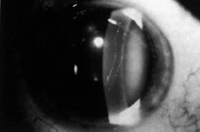
The corneal abnormalities are the most characteristic and the most varied. Lesions at Descemets membrane may take the form of individual or groups of vesicular-appearing lesions; bandlike lesions with scalloped, irregular edges (Fig. 4); or islands of abnormal-appearing endothelial cells. By specular microscopy, these areas consist of abnormal enlarged or irregularly shaped cells or black acellular zones.23 Areas of thickening of Descemet's membrane may occur, with involvement ranging from small areas to the entire posterior corneal surface.21,22

Fig. 4. Typical bandlike lesion with irregular, thickened margins at the level of Descemet's membrane and endothelium in a patient with posterior polymorphous corneal dystrophy.

In the majority of patients, only limited regions of Descemet's and the endothelium are involved.21,22 These patients are usually asymptomatic and may be unaware of their condition. Even areas of cornea that appear normal on biomicroscopy may have specular microscopic abnormalities, including abnormally small endothelial cells or enlarged pleomorphic endothelial cells.23 In cases with large areas of endothelial involvement, there may be focal or diffuse overlying corneal edema that can be severe enough to require penetrating keratoplasty. The edema can be so advanced as to preclude slit lamp evaluation of the posterior cornea, in which case the diagnosis can sometimes be made by recognizing typical lesions in family members. Corneal edema has been noted more commonly in older patients, presumably due to progressive corneal changes, but can be present in childhood or even at birth.21,22

Histopathologically, the most unusual finding in PPMD is that some areas of damaged endothelial cells have epithelial-like characteristics. These cells contain extensive microvilli and keratofibrils and have desmosomal attachments.22,24 The epithelial character of these cells has been confirmed by the demonstration that they contain epithelial keratins.25 In other areas, endothelial cells are degenerated, attenuated, and vacuolated. Fibroblastic proliferation has also been noted.22,26 Abnormalities in Descemet's membrane include abnormal lamination, deposition of abnormal fibrillar collagenous material on the posterior surface with areas of irregular guttate excrescences,22,26 and pits.26,27 These changes are probably secondary to the endothelial abnormalities.22,26

The abnormal endothelial cells, both those with and without epithelial characteristics, have been found to extend across the trabecular meshwork and onto the surface of the iris.22,24 It is likely that this extension results in the iridocorneal adhesions that occur in some patients with PPMD. These adhesions vary from peripheral anterior synechiae visible only on gonioscopy to prominent adhesions easily visible by slit lamp examination. In some cases, translucent membranes apparently extending from the posterior cornea are visible on the surface of the iris; these cause ectropion uveae and corectopia in occasional patients.22,28

The incidence of glaucoma in PPMD appears to be less than in the ICE syndrome. Krachmer reported that 14% of the referred patients he had examined with PPMD had elevated intraocular pressure.22 Since so many patients with PPMD are asymptomatic, the true incidence of elevated intraocular pressure is probably much lower. Most of the patients in Krachmer's group with elevated intraocular pressure had iridocorneal adhesions that closed portions of the filtration angle. A second group of PPMD patients with open angles and without iridocorneal adhesions have been recognized.21,22 The cause of the pressure increase in these patients is uncertain; in one such case examined histopathologically, an abnormally high insertion of the iris into the posterior trabecular meshwork with some collapse of the intertrabecular spaces was noted.29

Management

When present, glaucoma is best initially managed with medications that decrease aqueous secretion. Miotics may be helpful if areas of open angle are present. There are the same concerns with the use of laser trabeculoplasty in this condition as in the ICE syndrome: because of the growth of a cellular membrane across the chamber angle, trabeculoplasty might accelerate synechiae formation and thus aggravate rather than help the situation. In those patients whose intraocular pressure cannot be controlled medically, filtering surgery is probably the safest option.

Penetrating keratoplasty is required in only a minority of patients with PPMD. In those patients without iridocorneal adhesions or glaucoma, the prognosis for maintaining a clear graft is excellent. Patients with iridocorneal adhesions have a much lower success rate: only 31% with adhesions achieved 20/40 (6/12)* or better acuity compared with 78% without adhesions achieving this vision in Krachmer's series.22 Preoperative glaucoma, even though controlled at the time of surgery, was also found to be a poor prognostic factor for keratoplasty: 69% of eyes with glaucoma had final visual acuities of 20/400 (6/120) or less. In comparison, all eyes with normal intraocular pressure had better than 20/400 visual acuity after transplantation.22 All patients with preoperative glaucoma continued to have difficulties with glaucoma postoperatively.22 There was an overlap between these two groups since all eyes with glaucoma also had iridocorneal adhesions. Thus the presence of iridocorneal adhesions and preoperative glaucoma are poor prognostic factors for successful keratoplasty in PPMD and are more important to the outcome than is the severity of the corneal disease itself.22


* Metric equivalent given in parentheses following Snellen notation

FUCHS' ENDOTHELIAL DYSTROPHY

The terms Fuchs' endothelial dystrophy is applied to patients with extensive corneal guttata (often accompanied by flecks of endothelial pigment) who have developed corneal edema. Those patients with extensive guttata who have not yet developed corneal edema are said to have “endothelial dystrophy,”30 although in practice this strict distinction in terminology is not always followed. Although this dystrophy appears to be inherited in an autosomal dominant fashion, women are more frequently affected than men.30 The condition is bilateral but usually somewhat asymmetric. Once visually significant corneal edema develops, penetrating keratoplasty is generally required.

It has been taught that open-angle glaucoma is more common in patients with endothelial dystrophy and with Fuchs' dystrophy than in the general population.31 Data supporting this contention are difficult to locate, however. In a study of 64 families with endothelial dystrophy, only 1 of 71 patients with Fuchs' dystrophy (1.4%) was found to have open-angle glaucoma,32 which was considerably less than the 10% to 15% prevalence that had been previously estimated.31 There have been conflicting reports regarding the tonographic facility of aqueous outflow in patients with corneal guttata. Although a preliminary report in 1967 found that a high percentage of patients with corneal guttata had an abnormally low facility of aqueous outflow,33 Roberts and co-workers were unable to reproduce this finding in their 1984 study.34 They also graded the extent of endothelial guttata by slit lamp examination and by wide-field specular microscopy but could not demonstrate any relationship between outflow facility and the extent of guttata.34 Thus it is uncertain whether there is any increased likelihood of open-angle glaucoma in patients with Fuchs' dystrophy or its precursor states. When present, open-angle glaucoma in these patients should be treated with standard measures.

Occasionally, patients with Fuchs' dystrophy who have shallow anterior chambers may develop angle-closure glaucoma. In addition to thickening of the crystalline lens that occurs with aging, the angles in these patients may be further compromised by progressive edema of the peripheral cornea. Peripheral iridectomy is then required.

Back to Top
GLAUCOMA ASSOCIATED WITH CORNEAL INFECTION AND INFLAMMATION
Elevated intraocular pressure may accompany any active corneal inflammatory process. The clinical settings where this is most frequently encountered are in bacterial and fungal keratitis, in herpetic involvement of the cornea, and in patients with interstitial keratitis (most commonly luetic).

BACTERIAL AND FUNGAL KERATITIS

Several different mechanisms may be responsible for increased intraocular pressure in patients with bacterial or fungal keratitis. As a consequence of the anterior uveitis accompanying these infections, inflammatory cells and debris may become trapped within the trabecular meshwork, compromising aqueous outflow. In the case of fungal keratitis, organisms may penetrate Descemet's membrane35 and contribute to occlusion of the meshwork. Pupillary block may develop as a result of a mass of inflammatory material and/or posterior synechiae secluding the pupil. Intense anterior chamber inflammation may result in the formation of peripheral anterior synechiae that, if extensive, may contribute not only to acute pressure elevation but also to persistent glaucoma after the inflammatory process subsides.

HERPES SIMPLEX

In a report from Moorfield's Eye Hospital, 28% of patients seen with herpes simplex keratouveitis had elevated intraocular pressure.36 None of the patients with elevated pressure had primary ocular herpes. Of those with elevated pressures, 54% had disciform keratitis and 40% had stromal keratitis, in both cases usually accompanied by anterior uveitis.36 Although gonioscopy was difficult acutely, none of these patients was found to have an anatomically abnormal angle when examined later in the disease course.36 Mechanisms of pressure elevation include inflammation and edema of the trabecular meshwork itself, inflammatory cells and fibrin obstructing outflow channels, and increased aqueous viscosity.36-38

Corticosteroids are usually required in the treatment of stromal or disciform keratouveitis and are helpful for control of both the corneal and anterior chamber inflammation. Concomitant topical antiviral agents are generally administered to minimize the likelihood of epithelial recurrence of herpes. ß-Adrenergic blockers and carbonic anhydrase inhibitors are the best choices to decrease intraocular pressure until control of the inflammation is achieved. Ten percent of the patients in the above series eventually developed glaucomatous field defects, and 4% ultimately required filtering surgery.36

HERPES ZOSTER

Elevated intraocular pressures were noted in 16% of patients with ocular manifestations of herpes zoster ophthalmicus.39 All of these patients also had uveitis. Those patients treated promptly with topical corticosteroids had normalization of intraocular pressure. Mechanisms of pressure elevation are similar to cases of herpes simplex keratouveitis and relate to inflammatory cells and debris obstructing an already inflamed trabecular meshwork.40,41 In zoster, there may also be extensive loss of pigment granules from the iris that contributes to occlusion of the meshwork.40 The pressure elevation often responds to suppression of intraocular inflammation with frequently administered topical corticosteroids, which may be required for a long period and often must be tapered slowly to avoid rebound inflammation and pressure elevation. Contrary to the case with herpes simplex, there is no concern of corticosteroid-induced viral reactivation with herpes zoster. Otherwise unexplained late pressure elevations while using corticosteroids should raise the possibility of corticosteroid-induced glaucoma.

In patients with severe ocular herpes zoster there may be decreased aqueous production as a result of inflammation and/or necrosis of the ciliary body.40,41 Intraocular pressure in an individual case will depend on the balance between aqueous production and the limitation to outflow resulting from intraocular inflammation.

SYPHILITIC INTERSTITIAL KERATITIS

Interstitial keratitis occurring as a manifestation of congenital syphilis most commonly occurs from ages 5 to 20.42 The acute phase of interstitial keratitis is almost invariably accompanied by iridocyclitis,42 which may cause an elevation of intraocular pressure. It has been recognized that patients who previously had interstitial keratitis are prone to develop glaucoma many years later; 16% of patients in one series developed late glaucoma.43 The average period between the interstitial keratitis and presentation with this late form of glaucoma was found to be 27 years.44

Two subgroups of patients with late glaucoma have been identified.45 One group presents with chronic pressure elevation and normal depth chambers. Gonioscopically, these patients have postinflammatory peripheral anterior synechiae, with the open portions of the angle having a “dirty” appearance. Patients with this variety of glaucoma have generally responded poorly to antiglaucoma medications but have done well after filtering surgery.45 These patients have probably sustained postinflammatory damage to the trabecular meshwork. The second group of patients presents with angle closure, which is occasionally acute but more commonly subacute. These patients have smaller anterior segments and shallow anterior chambers. Iridectomy has resolved the glaucoma in some cases, although others have required additional medical treatment or filtering surgery. It is uncertain whether the anterior segment size was affected by the congenital syphilis or by the interstitial keratitis.45

Back to Top
GLAUCOMA OCCURRING AFTER PENETRATING KERATOPLASTY
Elevated intraocular pressure is a potentially serious complication of penetrating keratoplasty. The term post-keratoplasty glaucoma is frequently (if somewhat inaccurately) used to describe this pressure elevation, without regard to the presence of optic nerve damage. Because of the difficulties and inaccuracies of measuring the intraocular pressure of the recently grafted eye with Goldmann or Schiotz tonometry, the marked pressure rises that could occur shortly after keratoplasty were not recognized until electronic applanating devices were used in the postoperative period.46 Both the pathogenesis and management of elevated intraocular pressure after corneal transplantation are best discussed by dividing this entity into pressure elevations occurring in the immediate postoperative period and those that occur later and become chronic.

ACUTE PRESSURE ELEVATION AFTER PENETRATING KERATOPLASTY

Shallow Anterior Chamber

In acute post-keratoplasty pressure elevations, it is important to recognize those cases in which the anterior chamber has become shallow, since the mechanisms and management are different than if the chamber remains deep. A shallow anterior chamber may be caused by pupillary block, “ciliary block” (malignant glaucoma), or iris-cornea apposition that developed as a result of a wound leak. Pupillary block may occur due to posterior synechiae or to formation of a fibrin clot in the pupillary space, restricting aqueous flow from the posterior to the anterior chamber. The latter is particularly likely after therapeutic keratoplasty for a corneal perforation or for suppurative keratitis but may occasionally occur if the surgical procedure itself caused extensive intraocular inflammation. If patent peripheral iridectomies are not present, they should be promptly created either by laser if possible, or surgically. The Nd:YAG laser is preferable to the argon laser in this situation because it causes less inflammation when used for an iridectomy. If intraocular inflammation is contributing to the process, intensive administration of topical corticosteroids is indicated, to the extent allowed by the underlying condition. If despite the presence of patent peripheral iridectomies the chamber remains shallow, particularly centrally, then malignant (“ciliary block”) glaucoma must be considered. Although the pathogenesis of this condition has not been definitely established, the misdirection of aqueous into the posterior segment is believed to play an important role. Treatment options for this condition include either the combination of cycloplegics, hyperosmotics, and carbonic anhydrase inhibitors or surgical intervention.

A shallow anterior chamber may also develop as a result of a leak at the donor-host junction. Although an immediate postoperative leak will often be sealed by the development of a focal iris adhesion to the donor-host junction, occasionally the chamber may become shallow to the point where virtually the entire iris and peripheral cornea become appositional and adherent. Once this occurs, intraocular pressure will rise because aqueous can no longer reach the filtration angle. To avoid development of permanent synechial angle closure, the patient must be returned to the operating room, the iris separated from the peripheral cornea, and the donor-host junction carefully checked for leaks and reinforced as required.

Normal Anterior Chamber Depth

Substantial post-keratoplasty pressure elevations with normal anterior chamber depth occur almost exclusively in aphakic or pseudophakic patients.46-48 Zimmerman and co-workers have theorized that the weakening of both anterior and posterior support of angle structures by the keratoplasty incision and by loss of lens-zonular support, respectively, may result in partial collapse of the trabecular meshwork and/or Schlemm's canal, thus decreasing the outflow facility.49,50 They suggested oversizing the corneal donor button by 0.5 mm relative to the host opening and demonstrated that outflow facility in eye bank eyes was unchanged after combined keratoplasty and cataract extraction using the oversized donor.50 In a prospective study, significantly lower postoperative intraocular pressures were found in aphakic patients who underwent keratoplasty with 0.5-mm larger donor buttons compared with those who had the same size donor button and recipient opening.49 Other surgeons have subsequently examined the relationship between donor button size and aphakic post-keratoplasty glaucoma with conflicting results: some series have suggested51 or statistically shown52 a lower incidence of pressure elevation with oversized donor buttons, while others have not.47,53 It appears that the problem of high pressure after aphakic keratoplasty is at least partially iatrogenic and related to subtle variations in surgical technique, occurring particularly when very tight sutures are used with a relatively undersized donor.54 Other causes for pressure elevations in this setting include retained viscoelastic agent or intraocular inflammation.

These acute postoperative pressure rises are usually transient and can often be managed with topical ß-blockers or carbonic anhydrase inhibitors. Depending on the level of pressure, the status of the optic nerve, the patient's cardiopulmonary status, and the response to medications suppressing aqueous humor formation, oral or parenteral hyperosmotic agents may be necessary for short-term therapy. Miotics are usually avoided in the early postoperative period since they may increase intraocular inflammation and the tendency to synechiae formation. More intensive than usual topical corticosteroids may be helpful if it appears that intraocular inflammation is contributing to the elevated pressure.

CHRONIC PRESSURE ELEVATION AFTER PENETRATING KERATOPLASTY

Chronic glaucoma is a particularly significant problem after keratoplasty because, in addition to the risk to the optic nerve, elevated pressure damages corneal endothelium,55 which may result in graft failure. As would be expected, those patients with preexisting glaucoma are more likely to have difficulties with pressure control after the surgical procedure.47,56–58Many of these eyes already have angles compromised by prior inflammation, surgery, or anterior chamber lens implants. The incidence of chronic postoperative glaucoma in one large series of penetrating keratoplasties performed by a single surgeon was found to be 18%.58 The difference in incidence of glaucoma in aphakic patients (39%) compared with that in phakic patients (4%) in this series was statistically significant and remained so even when preexisting glaucoma was controlled for.58 The predisposition of aphakic eyes to develop glaucoma after keratoplasty holds whether the patient is already aphakic or if the lens is removed at the time of keratoplasty.46,47,51,58 Other variables examined including age, sex, race, type of cataract extraction, use of an intraocular lens implant, or the performance of a vitrectomy did not correlate with the development of glaucoma in this setting.58

Medical therapy with topical ß-blockers and/or carbonic anhydrase inhibitors is the usual first line of therapy in these cases. The graft surface should be monitored carefully, since timoptic has been associated with delayed epithelialization after keratoplasty59 and with superficial punctate keratitis and corneal anesthesia.60 Epinephrine compounds or dipivefrin must be used with caution, given the potential for cystoid macular edema in patients who are aphakic.61 Miotics may be helpful, but the compromise to the blood-aqueous barrier that they may cause can precipitate an acute homograft rejection.62

The possibility of corticosteroid-induced elevation in intraocular pressure must be considered in post-keratoplasty patients chronically using topical corticosteroids.56 For those patients who cannot safely discontinue corticosteroid therapy, the substitution of topical fluorometholone is an option. Although predisposed persons may also respond to fluorometholone with elevated pressure, the mean pressure increase is generally less than with other common topical corticosteroids.63 Whenever topical corticosteroid preparations are changed, post-keratoplasty patients should be carefully monitored for immunologic rejection.

In those patients in whom medical therapy fails to control the intraocular pressure, argon laser trabeculoplasty can be considered. In a report of ten aphakic or pseudophakic eyes treated with laser trabeculoplasty after penetrating keratoplasty, eight of ten experienced sustained pressure reductions of at least 5 mm Hg with maintenance of visual acuity and field.64 There was a trend for those patients who did not have glaucoma prior to keratoplasty to respond better to the laser trabeculoplasty, suggesting the possibility that the trabecular meshwork in this subgroup of patients may respond differently than in those patients with preexisting glaucoma.64 Performing laser trabeculoplasty may be technically difficult in some of these eyes, however, owing to peripheral (host) corneal edema, haze at the donor-host junction, and the presence of keratoplasty sutures within the field of view. The limited areas of open angle in many eyes with chronic post-keratoplasty glaucoma may preclude effective use of this procedure. Even if sufficient angle is open to treat, the gradual loss of effect of laser trabeculoplasty that has been noted in other situations65 may also occur in these patients.

Although some successes have been reported with filtering procedures after or combined with keratoplasty in aphakic patients,66 the poor success rate of filtering surgery in aphakic eyes in general67 is not encouraging in this regard. Many of these patients have preexisting conjunctival scarring that renders filtering surgery more difficult to perform and less likely to succeed. The temporary shallow or flat anterior chamber that may occur after filtering surgery is particularly problematic in these patients because the graft endothelium may sustain significant damage during this period. A high incidence of complications including retinal and choroidal detachment, cyclitic membrane formation, and vitreous hemorrhage has been noted after filtering surgery in post-keratoplasty aphakic eyes.58 Subconjunctivally injected 5-fluorouracil has been shown to increase the success of filtering surgery in cases with poor prognoses, especially in aphakic eyes.68 Although it may increase effectiveness, 5-fluorouracil is not likely to decrease the complication rate of filtering surgery in this setting, however, and its associated epithelial toxicity mandates particularly careful monitoring of the donor corneal epithelial status.

In the past, cyclocryotherapy has been advocated as the procedure of choice for post-kerato-plasty glaucoma that could not be medically controlled.69 Although cyclocryotherapy is often effective, it is somewhat unpredictable both in terms of pressure control and visual outcome.70 Cyclocryotherapy has many potential complications, including extensive intraocular inflammation, secondary membrane formation, vitreous hemorrhage, choroidal detachment, macular edema, immunologic and nonimmunologic graft failure, and phthisis bulbi.69,71,73 Alternative cyclodestructive procedures including transpupillary74 or transvitreal endolaser treatment of ciliary processes,75 focused ultrasound,76 and transscleral noncontact77 or contact Nd:YAG cycloablation78 are being investigated for the control of glaucoma in refractory cases. Those techniques that prove to be most quantifiable in terms of ciliary body destruction and that cause the least intraocular inflammation will probably find a role in control of post-keratoplasty glaucoma.

The difficulties that have been encountered with filtering surgery and with cyclocryotherapy have led surgeons to try setons in selected patients with refractory post-keratoplasty glaucoma. As currently used in glaucoma surgery, setons are implantable synthetic shunts that channel aqueous from the anterior chamber through a long tube to one of a variety of bleb-promoting devices positioned at or near the equator of the globe. The efficacy of Molteno implants and encircling bands (as described by Schocket) in this setting has been examined in several series.62,79,80 Setons were effective in controlling glaucoma in a high percentage (64%-86%) of eyes that would have otherwise required cyclodestructive procedures. Although implantation of the seton appeared to result in less intraocular inflammation than did cyclocryotherapy, the incidence of graft rejection was still high, ranging from 27% to 41% in these series.62,79,80 It has been theorized that alterations in the blood-ocular barrier induced by the seton may have contributed to the high rejection rate.79,80 Nonimmunologic graft failure was also common. Beebe and co-workers reported improved graft survival when the seton was placed prior to or at the time of keratoplasty, compared with after keratoplasty.62 They suggested that this sequence of procedures may have been more effective because it minimized the exposure of the donor corneal endothelium to elevated intraocular pressure and avoided additional intraocular manipulation after the graft was in place. Further evaluation will be required to determine the precise role of seton surgery as compared with the newer modalities of cyclodestruction in the treatment of patients with corneal transplants and glaucoma.

A small subgroup of patients who undergo penetrating keratoplasty, 2% in Foulks' series,58 develop progressive peripheral anterior synechiae formation postoperatively. While inadequate removal of vitreous from the anterior surface of the iris often causes anterior synechiae formation, most patients seem to develop this complication without apparent reason. Iridoplasty at the time of keratoplasty has been suggested to create a taut iris diaphragm, in an attempt to minimize the development of these synechiae.81 Iridectomies, sphincterotomies, and iris sutures may be required to achieve this goal.82 Although the effectiveness of iridoplasty in preventing anterior synechiae formation is uncertain, it is a reasonable approach and may also be used to create a central pupil if one is not already present. “Zippering” of the anterior chamber angle is often relentlessly progressive once it begins, resulting in an angle completely closed by synechiae and in glaucoma that is usually very difficult to control.58

Back to Top
REFERENCES

1. Shields MB, Campbell DG, Simmons RJ: The essential iris atrophies. Am J Ophthalmol 85:749, 1978

2. Shields MB: Progressive essential iris atrophy, Chandler's syndrome, and the iris nevus (Cogan-Reese) syndrome: A spectrum of disease. Surv Ophthalmol 24:3, 1979

3. Campbell DG, Shields MB, Smith TR: The corneal endothelium and the spectrum of essential iris atrophy. Am J Ophthalmol 86:317, 1978

4. Eagle RC Jr, Shields JA: Iridocorneal endothelial syndrome with contralateral guttate endothelial dystrophy: A light and electron microscopic study. Ophthalmology 94:862, 1987

5. Shields MB, Campbell DG, Simmons R J, Hutchinson BT: Iris nodules in essential iris atrophy. Arch Ophthalmol 94:406, 1976

6. Chandler PA: Atrophy of the stroma of the iris: Endothelial dystrophy, corneal edema, and glaucoma. Am J Ophthalmol 41:607, 1956

7. Scheie HG, Yanoff M: Iris nevus (Cogan-Reese) syndrome: A cause of unilateral glaucoma. Arch Ophthalmol 93:963, 1975

8. Eagle RC Jr, Font RL, Yanoff M, Fine BS: Proliferative endotheliopathy with iris abnormalities: The iridocorneal endothelial syndrome. Arch Ophthalmol 97:2104, 1979

9. Bourne WM: Partial corneal involvement in the iridocorneal endothelial syndrome. Am J Ophthalmol 94:774, 1982

10. Neubauer L, Lund O-E, Leibowitz HM: Specular microscopic appearance of the corneal endothelium in iridocorneal endothelial syndrome. Arch Ophthalmol 101:916, 1983

11. Hirst LW, Quigley HA, Stark WJ, Shields MB: Specular microscopy of iridocorneal endothelial syndrome. Am J Ophthalmol 89:11, 1980

12. Setala K, Vannas A: Corneal endothelial cells in essential iris atrophy: A specular microscopic study. Acta Ophthalmol 57: 1020, 1979

13. Kupfer C, Kaiser-Kupfer MI, Datiles M, McCain L: The contralateral eye in the iridocorneal endothelial (ICE) syndrome. Ophthalmology 90:1343, 1983

14. Eagle RC Jr, Font RL, Yanoff M, Fine BS: The iris naevus (Cogan-Reese) syndrome: Light and electron microscopic observations. Br J Ophthalmol 64:446, 1980

15. Rodrigues MM, Stulting RD, Waring GO III: Clinical, electron microscopic, and immunohistochemical study of the corneal endothelium and Descemet's membrane in the iridocorneal endothelial syndrome. Am J Ophthalmol 101:16, 1986

16. Bahn CF, Falls HF, Varley GA et al: Classification of corneal endothelial disorders based on neural crest origin. Ophthalmology 91:558, 1984

17. Rodrigues MM, Streeten BW, Spaeth GL: Chandler's syndrome as a variant of essential iris atrophy: A clinicopathologic study. Arch Ophthalmol 96:643, 1978

18. Kidd M, Hetherington J, Magee S: Surgical results in iridocorneal endothelial syndrome. Arch Ophthalmol 106:199, 1988

19. Shields MB, McCracken JS, Klintworth GK, Campbell DG: Corneal edema in essential iris atrophy. Ophthalmology 86:1533, 1979

20. Buxton JN, Lash RS: Results of penetrating keratoplasty in the iridocorneal endothelial syndrome. Am J Ophthalmol 98:297, 1984

21. Cibis GW, Krachmer JA, Phelps CD, Weingeist TA: The clinical spectrum of posterior polymorphous dystrophy. Arch Ophthalmol 95: 1529, 1977

22. Krachmer JH: Posterior polymorphous corneal dystrophy: A disease characterized by epithelial-like endothelial cells which influence management and prognosis. Trans Am Ophthalmol Soc 83:413, 1985

23. Hirst LW, Waring GO III: Clinical specular microscopy of posterior polymorphous endothelial dystrophy. Am J Ophthalmol 95: 143, 1983

24. Rodrigues MM, Phelps CD, Krachmer JH, Weingeist TA: Glaucoma due to endothelialization of the anterior chamber angle: A comparison of posterior polymorphous dystrophy of the cornea and Chandler's syndrome. Arch Ophthalmol 98:688, 1980

25. Rodrigues MM, Sun T-T, Krachmer JH, Newsome D: Epithelialization of the corneal endothelium in posterior polymorphous dystrophy. Invest Ophthalmol Vis Sci 19:832, 1980

26. Henriquez AS, Kenyon KR, Dohlman CH et al: Morphologic characteristics of posterior polymorphous dystrophy: A study of nine corneas and review of the literature. Surv Ophthalmol 29:139, 1984

27. Polack FM, Bourne WM, Forstot SL, Yamaguchi T: Scanning electron microscopy of posterior polymorphous corneal dystrophy. Am J Ophthalmol 89:575, 1980

28. Cibis GH, Krachmer JH, Phelps CD, Weingeist TA: Iridocorneal adhesions in posterior polymorphous dystrophy. Trans Am Acad Ophthalmol Otolaryngol 81:OP770, 1976

29. Bourgeois J, Shields MB, Thresher R: Open-angle glaucoma associated with posterior polymorphous dystrophy: A clinicopathologic study. Ophthalmology 91:420, 1984

30. Waring GO III, Rodrigues MM, Laibson PR: Corneal dystrophies. II. Endothelial dystrophies. Surv Ophthalmol 23:147, 1978

31. Kolker AE, Hetherington J Jr: Becker-Shaffer's Diagnosis and Therapy of the Glaucomas, p 275. St Louis, CV Mosby, 1983

32. Krachmer JH, Purcell JJ Jr, Young CW, Bucher KD: Corneal endothelial dystrophy: A study of 64 families. Arch Ophthalmol 96:2036, 1978

33. Buxton JN, Preston RW, Riechers R, Guilbault N: Tonography in cornea guttata: A preliminary report. Arch Ophthalmol 77:602, 1967

34. Roberts CW, Steinert RF, Thomas JV, Boruchoff SA: Endothelial guttata and facility of aqueous outflow. Cornea 3:5, 1984

35. Naumann G, Green WR, Zimmerman LE: Mycotic keratitis: A histopathologic study of 73 cases. Am J Ophthalmol 64:668, 1967

36. Falcon MG, Williams HP: Herpes simplex keratouveitis and glaucoma. Trans Ophthalmol Soc UK 98:101, 1978

37. Hogan MJ, Kimura SJ, Thygeson P: Pathology of herpes simplex keratoiritis. Trans Am Ophthalmol Soc 61:75, 1963

38. Townshend WM, Kaufman HE: Pathogenesis of glaucoma and endothelial changes in herpetic keratouveitis in rabbits. Am J Ophthalmol 71:904, 1971

39. Womack LW, Liesegang TJ: Complications of herpes zoster ophthalmicus. Arch Ophthalmol 101:42, 1983

40. Naumann G, Gass JDM, Font RL: Histopathology of herpes zoster ophthalmicus. Am J Ophthalmol 65:533, 1968

41. Hedges TR, Albert DM: The progression of the ocular abnormalities of herpes zoster: Histopatholic observations of nine cases. Ophthalmology 89: 165, 1982

42. Duke-EIder S (ed): System of Ophthalmology, Vol 8, p 815. St Louis, CV Mosby, 1965

43. Knox DL: Glaucoma following syphilitic interstitial keratitis. Arch Ophthalmol 66:44, 1961

44. Tsukahara S: Secondary glaucoma due to inactive congenital syphilitic interstitial keratitis. Ophthalmologica 174:188, 1977

45. Grant WM: Late glaucoma after interstitial keratitis. Am J Ophthalmol 79:87, 1975

46. Irvine AR, Kaufman HE: Intraocular pressure following penetrating keratoplasty. Am J Ophthalmol 68:835, 1969

47. Karesh JW, Nirankari VS: Factors associated with glaucoma after penetrating keratoplasty. Am J Ophthalmol 96:160, 1983

48. Polack FM: Glaucoma in keratoplasty. Cornea 7:67, 1988

49. Zimmerman T, Olson R, Waltman S, Kaufman H: Transplant size and elevated intraocular pressure post keratoplasty. Arch Ophthalmol 96:2231, 1978

50. Zimmerman TJ, Krupin T, Grodzki W et al: Size of donor corneal button and outflow facility in aphakic eyes. Ann Ophthalmol 11:809, 1979

51. Heidemann DG, Sugar A, Meyer RF, Musch DC: Oversized donor grafts in penetrating keratoplasty: A randomized trial. Arch Ophthalmol 103: 1807, 1985

52. Bourne WM, Davison JA, O'Fallon WM: The effects of oversize donor buttons on postoperative intraocular pressure and corneal curvature in aphakic penetrating keratoplasty. Ophthalmology 89:242, 1982

53. Perl T, Charlton KH, Binder PS: Disparate diameter grafting: Astigmatism, intraocular pressure, and visual acuity. Ophthalmology 88:774, 1981

54. Olson RJ: Discussion of disparate diameter grafting: Astigmatism, intraocular pressure, and visual acuity. Ophthalmology 88:780, 1981

55. Charlin R, Polack FM: The effect of elevated intraocular pressure on the endothelium of corneal grafts. Cornea 1:241, 1982

56. Goldberg DB, Schanzlin DJ, Brown SI: Incidence of increased intraocular pressure after keratoplasty. Am J Ophthalmol 92:372, 1981

57. Schanzlin DJ, Robin JB, Gomez DS et al: Results of penetrating keratoplasty for aphakic and pseudophakic bullous keratopathy. Am J Ophthalmol 98:302, 1984

58. Foulks GN: Glaucoma associated with penetrating keratoplasty. Ophthalmology 94:871, 1987

59. O'Brien WJ, DeCarlo JD, Stern M, Hyndiuk RA: Effects of timoptic on corneal reepithelialization. Arch Ophthalmol 100:1331, 1982

60. Van Buskirk EM: Corneal anesthesia after timolol maleate therapy. Am J Ophthalmol 88:739, 1979

61. Kolker AE, Becker B: Epinephrine maculopathy. Arch Ophthalmol 79:552, 1968

62. Beebe WE, Starita RJ, Fellman RL et al: Molteno implant and anterior chamber tube shunt to encircling band (ACTSEB) in the treatment of postkeratoplasty glaucoma. Ophthalmology (suppl)96: 101, 1989

63. Stewart RH, Kimbrough RL: Intraocular pressure response to topically administered fluorometholone. Arch Ophthalmol 97:2139, 1979

64. Van Meter WS, Allen RC, Waring GO III, Stulting RD: Laser trabeculoplasty for glaucoma in aphakic and pseudophakic eyes after penetrating keratoplasty. Arch Ophthalmol 106: 185, 1988

65. Schwartz AL, Kopelman J: Four-year experience with argon laser trabecular surgery in uncontrolled open-angle glaucoma. Ophthalmology 90:771, 1983

66. Insler MS, Cooper HD, Kastl PR, Caldwell DR: Penetrating keratoplasty with trabeculectomy. Am J Ophthalmol 100:593, 1985

67. Heuer DK, Gressel MG, Parrish RK II et al: Trabeculectomy in aphakic eyes. Ophthalmology 91:1045, 1984

68. The Fluorouracil Filtering Surgery Study Group: Fluorouracil filtering surgery study one-year follow-up. Am J Ophthalmol 108:625, 1989

69. West CE, Wood TO, Kaufman HE: Cyclocryotherapy for glaucoma pre or post penetrating keratoplasty. Am J Ophthalmol 76:485, 1973

70. Shields MB: Cyclodestructive surgery for glaucoma: Past, present, and future. Trans Am Ophthalmol Soc 83:285, 1985

71. Binder PS, Abel R Jr, Kaufman HE: Cyclocryotherapy for glaucoma after penetrating keratoplasty. Am J Ophthalmol 79:489, 1975

72. Thoft RA, Gordon JM, Dohlman CH: Glaucoma following keratoplasty. Trans Am Acad Ophthalmol Otolaryngol 78:OP 352, 1974

73. Patel AC, Stark WJ: Corneal graft survival following additional ocular surgery. Ophthalmology 93 (suppl):96, 1986

74. Lee P-F: Argon laser photocoagulation of the ciliary processes in cases of aphakic glaucoma. Arch Ophthalmol 97:2135, 1979

75. Zarbin MA, Michels RG, de Bustros S et al: Endolaser treatment of the ciliary body for severe glaucoma. Opthalmology 95: 1639, 1988

76. Burgess SEP, Silverman RH, Coleman DJ et al: Treatment of glaucoma with high-intensity focused ultrasound. Ophthalmology 93:831, 1986

77. Klapper RM, Wandel T, Donnenfeld E, Perry HD: Transscleral neodymium:YAG thermal cyclophotocoagulation in refractory glaucoma: A preliminary report. Ophthalmology 95:719, 1988

78. Schuman JS, Puliafito CA, Allingham RR et al: Contact transscleral Nd:YAG laser cyclophotocoagulation. Ophthalmology (suppl)96:92, 1989

79. Kirkness CM: Penetrating keratoplasty, glaucoma and silicone drainage tubing. Dev Ophthalmol 14:161, 1987

80. McDonnell PJ, Robin JB, Schanzlin DJ et al: Molteno implant for control of glaucoma in eyes after penetrating keratoplasty. Ophthalmology 95:364, 1988

81. Cohen EJ, Kenyon KR, Dohlman CH: Iridoplasty for prevention of post-keratoplasty angle closure and glaucoma. Ophthalmic Surg 13:994, 1982

82. Waring GO III: Management of pseudophakic corneal edema with reconstruction of the anterior ocular segment. Arch Ophthalmol 105:709, 1987

Back to Top