|
|
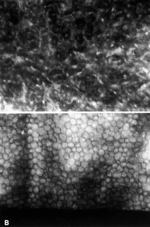
| Fig. 2. A. Specular microscopy of the corneal endothelium in ICE syndrome. Cell borders are obscured, resulting in loss of the normal endothelial mosaic. Note dark areas within endothelial cells. Brighter reflections are believed to be from cell borders. B. Specular microscopy of fellow eye showing normal endothelial mosaic. (Courtesy of Ira J. Udell, MD) |