Chapter 50
Heredity and Glaucoma
BRIAN P. BROOKS, JULIA E. RICHARDS, and PAUL R. LICHTER
Main Menu   Table Of Contents

Search

SIGNIFICANCE
NOMENCLATURE
OPEN-ANGLE GLAUCOMA
ANGLE-CLOSURE GLAUCOMA
INFANTILE GLAUCOMA
GLAUCOMA ASSOCIATED WITH OTHER GENETIC DISORDERS
THE FUTURE
REFERENCES

Genetics' contribution is not new to glaucoma. The hereditary nature of glaucoma is beautifully displayed in Julia Bell's 1931 Treasury of Human Inheritance, which presents 66 glaucoma pedigrees gleaned from the literature, much of it spanning the preceding decade, but beginning with a report by Benedict in 1842.1,2 Early discussions of glaucoma genetics were largely descriptive—following the disease through familial pedigrees or associating it with an inherited ocular or systemic phenotype—and included reports of both autosomal dominant and autosomal recessive inheritance.1–12

In the past decade, however, advances in molecular genetics have enabled the field to go beyond description. We can now identify mutations in the genetic code of patients that segregate with a disease phenotype such as elevated intraocular pressure, progressive optic nerve cupping, or anterior segment dysgenesis. This information may not only lead to a new method of classifying the glaucomas, but also to a unique approach to each glaucoma patient, based on his or her genetic makeup. The purpose of this chapter is to describe how heredity plays a role in various forms of glaucoma, withspecific emphasis on molecular advances. We also include a discussion of how genetic information presents novel challenges and opportunities in treating patients.

Back to Top
SIGNIFICANCE
Why study the genetics of glaucoma? First, glaucoma is an important public health problem. For example, progressive open angle glaucoma is estimated to affect about 2% of the white population and 4% of the African-American population over the age of 40.13–15 Any advance in understanding this disease will affect a great number of people. Second, identifying genes associated with glaucoma enables scientists and clinicians to understand its pathogenesis at the cellular and molecular level better. Model systems, such as transgenic mice, can be created using such information—providing a controlled milieu for studying such things as disease progression, interaction of genetic and environmental factors, and testing new therapies. Third, genetic information may allow clinicians to counsel, monitor and treat their patients more effectively. A patient with a mutation known to produce optic nerve cupping and visual field loss rapidly, for example, might be treated more aggressively—possibly even before the patient manifests advanced disease. Fourth, identification of patients who share an underlying molecular cause for their disease may allow us to tell whether they share any systemic disease or other risk factor not previously identified because it was not a characteristic found throughout the population of patients with glaucoma. Finally, identifying genes involved in disease pathogenesis may provide novel targets for therapy.
Back to Top
NOMENCLATURE
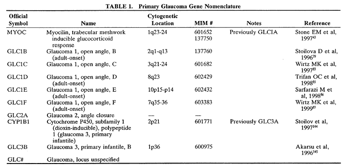
Glaucoma has traditionally been classified on criteria such as ocular anatomy (open- versus narrow-angle) or age of onset (infantile- versus juvenile- versus adult-onset). The Human Genome Organization (HUGO) Nomenclature Committee has adopted a similar strategy for naming gene loci associtd with glaucoma. Loci for open-angle glaucoma are designated, in general, by GLC1 (Table 1). To distinguish specific genes or loci, letters are added to the GLC1 in the order of their discovery (e.g., GLC1A, GLC1B). The designation GLC2 is reserved for angle-closure glaucoma and GLC3 for infantile glaucoma. Complicating this scheme is that two of these symbols, namely GLC1A and GLC3A, have been withdrawn and replaced by the names of the cloned glaucoma genes—MYOC and CYP1B1, respectively—present at these loci. Other inherited causes of glaucoma are classified under different names, especially in the case of secondary, developmental, and syndromic glaucomas in which the name is frequently based on the disease in which glaucoma may be one component. For example, a pedigree with autosomal dominant nanophthalmos resulting in angle-closure glaucoma or occludable angles maps to chromosome 11 and is designated by NNO116 rather than GLC2A. Areas of overlap between these designations may also exist. For example, one mutation in a gene may produce a dramatic phenotype early in life and be termed “juvenile open-angle glaucoma,” whereas a different mutation in the same gene may produce a clinical phenotype later in life and be termed “adult onset open-angle glaucoma.” Interestingly, in some cases the same mutation can cause juvenile onset of glaucoma in some individuals and adult onset of glaucoma in their relatives.


Although the HUGO classification scheme may be convenient and useful as new glaucoma genes are discovered, it must be stressed that it does not yet reflect substantial insights into the underlying biology. As we begin to understand how mutations in genes cause disease at the cellular and molecular levels, we may be able to create a new classification scheme based on the mechanism of disease that might or might not correlate with the genetic classification scheme. The substitution of the gene names MYOC and CYP1B1 for the locus names GLC1A and GLC3A reflects a change toward nomenclature based on meaningful biologic insights. However, much remains to be learned before further progress in the development of functionally meaningful molecular genetic classification systems will be possible.

Back to Top
OPEN-ANGLE GLAUCOMA
This section discusses the genetics of both juvenile-onset and adult-onset open-angle glaucoma with no discernable angle anomalies. These diseases, along with some forms of infantile glaucoma, may be caused by similar pathophysiologic processes of different timing and severity.

EVIDENCE FROM EPIDEMIOLOGY

Epidemiologic studies have confirmed the clinical observation that open-angle glaucoma is more likely to occur in people who have relatives with open-angle glaucoma. Familial forms of primary open-angle glaucoma have been estimated at anywhere from 5.6% to 47% of all primary open-angle glaucoma cases.17–23 The great variation in apparent frequency of familial glaucoma in these studies may in some cases reflect underlying genetic differences in the populations being studied but may also reflect methodologic differences, ascertainment bias, or variation in inclusion criteria and/or the parameters being used to define affected status. Additional inaccuracy in any attempt to gauge frequency of familial glaucoma cases arises because glaucoma frequently is present only later in life and may not have yet developed in some patients who will eventually be affected. Bias in the direction of overreporting may occur in patients who report family history when the relatives in fact have ocular hypertension or a completely different form of glaucoma. A recent study of very large families in Tasmania indicates that underreporting can occur even when the family history of glaucoma is strong, because individuals diagnosed with primary open-angle glaucoma may be unaware of the presence of glaucoma among their relatives.24 The Tasmania study points to a need to encourage affected individuals to communicate with their relatives about the existence of glaucoma in the family.

Twin studies also support the existence of a genetic component to open-angle glaucoma.25–27 One recent study that took age-related prevalence of glaucoma into account found 98.0% concordance among monozygotic twins age 55 or over.27 Twin studies have also provided evidence for genetic components to the optic cup diameter,28 optic disc diameter,29 cup:disc ratio,30 and anterior chamber depth.27

Finally, the idea of genetic components to glaucoma is supported by observations on differential prevalence of particular forms of glaucoma in particular racial or ethnic groups. Open-angle glaucoma is clearly found more often in African Americans than in whites.13 Open-angle glaucoma is less common than angle-closure glaucoma in Innuit and Asian populations.31–36

MYOC (GLC1A)

Genetics

Open-angle glaucoma caused by mutations at the MYOC gene (previously, the GLC1A locus) is generally inherited in an autosomal dominant fashion. Patients often have signs of elevated intraocular pressure, optic nerve cupping, and glaucomatous visual field loss within the first 3 decades of life.37 Sheffield and colleagues reported linkage of glaucoma to chromosome 1q38 and subsequent reports by this and other groups helped refine localization of the gene.39–42 After narrowing this locus further, Stone and partners reported use of a positional candidate gene approach to identify three distinct mutations in five of eight families with 1q-linked juvenile-onset glaucoma in a gene called the trabecular meshwork (TM)-inducible glucocorticoid response (TIGR) gene.43 Two of these mutations (Gly364Val and Gln368STOP) were found in 10 of 227 (4.4%) adult-onset open-angle glaucoma patients who had at least one living first-degree relative with a documented history of glaucoma. In addition, three of 103 (2.9%) unselected patients with open-angle glaucoma also harbored a Gln368STOP mutation, as did one of 380 people from the general population tested. Additional mutations, many of which cluster in the C-terminal domain, have since been reported in GLC1A families from around the world.37,44–51

Efforts to follow and understand the literature are helped by understanding the revisions that have taken place in the nomenclature of this gene. Before mutations were found in these families, the classification GLC1A was used to designate the first genetically-defined locus mapped for open-angle glaucoma. The gene that Stone and colleagues found mutated in these families, however, had already been identified as a candidate glaucoma gene based on a cellular pharmacology model and named TIGR (for TM-inducible glucocorticoid response protein) by Polansky and colleagues.52,53 Working independently, Kubota and colleagues cloned a gene for a cytoskeleton-associated protein from human retina that they named myocilin (MYOC) because of its homology to myosin and its localization to the cilium of the photoreceptor.54 However, MYOC was, unbeknown to them, in fact, the TIGR gene. In addition, during their studies of genes expressed by human ciliary body, Ortega and colleagues had deposited short stretches of the TIGR sequence into the Genbank sequence repository as anonymous expressed sequence tags.55 The official name of this gene and locus, as recognized by the HUGO Nomenclature Committee, is MYOC, but GLC1A, TIGR, and TIGR/MYOC are still frequently used in the literature.

Molecular Biology and Pathogenesis

The mechanism by which sequence changes in the myocilin protein causes glaucoma is unclear. Studying the normal functions of this protein, however, may help in understanding disease pathogenesis. The MYOC gene codes for the 504 amino acid, 55 kDa myocilin protein.53,54 Its sequence includes several domains (Fig. 1): (1) a hydrophobic amino-terminal domain with homology to nonmuscle myosin heavy chain, (2) a leucine zipper domain, possibly important in protein-protein interactions, and (3) a domain near the carboxy-terminus with homology to an olfactory epithelium-specific extracellular protein, olfactomedin, where many reported mutations cluster.53,54,56 Rozsa and colleagues have proposed the existence of an apparent charge-sensitive domain between the leucine zipper and the olfactomedin homology domain.47,49 Potential phosphorylation and glycosylation sites also exist, although Huang and colleagues did not detect glycosylated protein in human ocular tissues.57

Fig. 1. Schematic of the human myocilin protein. The amino terminus contains a signal peptide plus a leucine zipper, while the carboxy terminus contains an olfactomedin-like domain where many mutations are found. The leucine zipper sequence may be important in protein-protein interactions. Numbers represent amino acid position. NH2 marks the amino terminus. COOH marks the carboxy terminus. Additional points on the protein predicted from the sequence by Nguyen and colleagues are two glycosaminoglycan initiation sites (G), an N-glycosylation site (N) and a hyaluronic acid binding site (H).52,53 T indicates the position of the Glu323Lys mutation that causes a change in the translocational pause of TIGR protein biogenesis.143 P marks conserved sequences at which phosphorylation is predicted to occur and C marks a predicted C-terminal peroxisomal targeting sequence.47 The charge-sensitive domain was predicted by Shimizu and colleagues.49

Myocilin messenger RNA (mRNA) is normally expressed in various ocular and nonocular tissues, including retina (inner segment of photoreceptors, outer granular layer), iris, ciliary body, TM, heart, skeletal muscle, choroid plexus, selected brain regions, thymus, prostate, small intestine, colon, stomach, thyroid, trachea, and bone marrow.52,54,55,58–60 Less abundant expression is found in cornea, lung, pancreas, testis, ovary, spinal cord, lymph node, and adrenal gland. MYOC transcripts have also been found in pial septae that divide nerve fiber bundles, the perivascular connective tissue surrounding the central retinal vessels, and in the meninges surrounding the retrolaminar optic nerve.61

Myocilin protein is secreted into the medium of cultured human TM cells in glycosylated and nonglycosylated forms and is capable of forming oligomers.52,53,62 Specific, high-affinity binding to TM cells has been demonstrated. Ueda and colleagues have reported that the protein is associated with mitochondria, cytoplasmic filaments, trabecular beams, and the extracellular matrix in juxtacanalicular regions in TM tissues.63 Myocilin expression appears increased and more widespread in the TM of glaucomatous eyes than in nonglaucomatous eyes.64 These data, combined with the widespread expression of this gene, suggest that myocilin may be a general, stress-response protein. That its elevated expression occurs in a high fraction of glaucoma eyes whereas only a small fraction of individuals appear to carry the causative exon-3 mutations suggests that altered expression levels could be involved at least secondarily in cases of glaucoma that lack exon-3 mutations. Kubota and colleagues, focussing on myocilin's intimate localization with the cilium connecting the inner and outer segments of photoreceptors, suggested a structural role associated with the basal body.54

One key point about the MYOC gene is that it is inducible by various mechanisms, and its inducibility appears to be relatively specific to the TM cell.52,65,66 Because the gene is expressed in many different tissues but so far only seen to cause disease through mechanisms occurring in the TM, it is tempting to hypothesize that the inducibility might play some role in the tissue specificity of disease. MYOC was first identified as a candidate glaucoma gene induced by glucocorticoids in human TM cells over a 3-week time and dosage course designed to parallel the time dose-response course observed for elevation of intraocular pressure in response to glucocorticoids in some patients.67–70 Expression can also be induced by oxidative stress and induction can be somewhat blunted by concomitant administration of basic fibroblast growth factor, transforming growth factor-β, triiodothyronine, and nonsteroidal antiinflammatory drugs.65 Glucocorticoids, transforming growth factor-β, and mechanical stretch similarly induce expression in perfused anterior segment organ cultured TM. Kubota and colleagues, focusing on myocilin's intimate localization with the cilium connecting the inner and outer segments of photoreceptors, suggested a structural role associated with the basal body.54

MYOC induction findings suggest an interesting relationship between open-angle and glucocorticoid-induced glaucoma. Histologic studies of TM from patients with juvenile open-angle glaucoma show accumulation of a fine, basement membrane-like material concentrated in the inner cribriform and outer corneoscleral regions71—a pattern similar to that seen in steroid-induced glaucoma. A simple model would suggest that the mechanism of MYOC glaucoma could therefore be related to the accumulation of mutated, misfolded myocilin protein in the TM—perhaps binding to trabecular cells or extracellular proteins—that obstructs aqueous outflow. However, this seems unlikely because initial reports suggest that the mutant form of the protein may not get out of the cell.73 An alternative model proposed by Raymond and colleagues in that same study calls for heterodimers containing one normal and one mutant copy of the protein to play a role in the disease. If the mutant protein is, in itself, capable of eliciting a stress response in the TM, a positive feedback loop may be created such that myocilin accumulation promotes further myocilin induction and accumulation, resulting in the presence of excess normal myocilin protein in the extracellular matrix of the TM. Alternatively, the misfolded protein, if retained by trabecular cells, could eventually elicit a stress response that evokes a similar, positive-feedback mechanism. Polansky and colleagues, who originated the model of overexpression of normal myocilin in response to stress in general and stress responses to the presence of mutant myocilin protein specifically, have reported increased expression and abnormal subcellular distribution of Pro370Leu mutant myocilin protein when expressed in human TM cells.65 There remains the possibility that the overexpression model and the heterodimer model might represent two different aspects of one overall mechanism.

Morisette and colleagues note that patients homozygous for the Lys423Glu MYOC mutation do not develop glaucoma, although their relatives who carry one normal and one mutant copy of the gene are affected with relatively early onset glaucoma. This observation implies several things about pos-sible disease mechanisms, at least for the muta-tion described: 1) disease is not caused by loss ofmyocilin function, because it would be predictedthat homozygous patients would be more severelyaffected than heterozygotes; 2) a new, gain-of-function (e.g., obstructing aqueous outflow or induction of overexpression by misfolded protein) is the likely mechanism of pathogenesis; and 3) the presence of both a normal and a mutated protein may be necessary to produce a phenotype.62 This implication that the active presence of the mutant form of the protein in the presence of the normal protein leads to disease would be consistent with the dimerization model they have proposed,62,73 with Polansky's proposed model for induction of overexpression and export from the cell,65 or with a model in which the heterodimers bring about the induction, overexpression and export from the cell. This observation of normal phenotype in individuals homozygous for a MYOC mutation may be mutation specific, as Sarfarazi and colleagues have published a preliminary report of a large Turkish pedigree with one severely affected Gly326Arg homozygote.74

Clinical Correlation: Is Genetic Screening for Glaucoma Worthwhile?

Patients with MYOC glaucoma exhibit phenotypic variability.37,45,47,75,76 This statement is true both when comparing patients from different families, with different mutations and when comparing patients in the same family, with the same mutation. Such phenotypic differences may be due to modifying genetic or environmental factors, or the severity of a mutation's effect on myocilin expression or function. Although many patients with MYOC mutations develop glaucoma in childhood and early adult life, some mutations, such as Gln368STOP, are associated with a later age at diagnosis. Some patients with myocilin mutations appear nonpenetrant, living their adult lives without signs of glaucoma. Furthermore, Alward and associates37 noted that two unrelated patients with Tyr437His and Thr377Met mutations, respectively, had marked asymmetry between their eyes' glaucoma progression, suggesting environmental influences. Wiggs and colleagues found a difference in disease severity between monozygotic twins with MYOC glaucoma.50 Sorting out the genetic and nongenetic modifying factors for MYOC glaucoma may help in the understanding of disease pathophysiology of other forms of glaucoma and, perhaps, provide novel approaches to treatment.

Ongoing studies are working to figure out who should actually be screened for sequence changes in the MYOC gene. Certainly after a mutation has been identified in an affected individual, screening of their at-risk relatives has great potential value. The value of whole scale screening is less clear in situations in which negative findings provide little clinical insight, but some studies suggest that it may be possible to identify a subset of the glaucoma population with greater likelihood of MYOC mutations and thus a greater likelihood of a positive test result that would be clinically useful at our current level of understanding. Not all patients with juvenile-onset open-angle glaucoma have MYOC glaucoma and only about 4% of familial open-angle glaucoma harbor MYOC exon 3 mutations.43,50,77 The findings of Michels-Rautenstrauss and colleagues suggest that positive screening results are more likely in individuals with a family history of glaucoma.78 Shimuza and associates found that, of the 74 probands with open-angle glaucoma tested, none with a purely adult-onset pattern in the family had apparently-causative MYOC mutations.49 However, they did find that approximately one third of families with either purely juvenile-onset or mixed juvenile- or adult-onset glaucoma had MYOC mutations. Although larger population studies are needed, these data imply that MYOC mutation screening would be more fruitful in families where at least one individual had juvenile-onset open-angle glaucoma. Finally, even with an accurate genotype, at our current level of understanding, phenotypic variability may limit accuracy of prediction of disease course for an individual patient. After a mutation has been identified in one member of a family, that information offers a powerful mechanism for identifying other family members with elevated risk who merit monitoring.

GLC1B

In 1996, Stoilova and colleagues identified a locus for an autosomal dominant form of adult-onset primary open-angle glaucoma on 2cen-q13, designated GLC1B.79 These primarily British patients had low-to-moderate intraocular pressure (mostly teens and twenties), onset late in the fifth decade, and a good response to medical treatment. Two point and haplotype analyses narrowed the locus to an 8.5cM region flanked by markers D2S2161 and D2S2264. One patient, an 86-year-old man with the disease haplotype but no glaucoma, was considered a carrier. Analysis of eight other families with similar clinical characteristics showed no linkage to this region, emphasizing the genetic heterogeneity of adult-onset primary open-angle glaucoma. Those authors pointed out that many members of this pedigree had intraocular pressures within the normal range and that identification of the GLC1B gene may aid in our understanding of normal-tension glaucoma. The presence of both normal and elevated pressures in affected individuals raises some interesting questions about the role of elevated pressure in this disease. Faucher and colleagues have reported a large French-Canadian family in which glaucoma is linked to the GLC1B locus.80 Wiggs and colleagues have confirmed this locus as a glaucoma susceptibility locus in their molecular genetic study of sibling pairs affected with glaucoma.81 At present, the specific gene responsible for GLC1B glaucoma has not been isolated, but the physical region of the chromosome containing the gene has been isolated and mapped by research groups in Québec and Connecticut and nine candidate genes have so far been excluded as being the cause of the disease.80,82

GLC1C

Wirtz and associates reported on a family with adult-onset primary open-angle glaucoma inherited in an autosomal dominant fashion.83 The disease gene was mapped in twelve affected family members to 3q21-q24. Analysis of recombinant haplotypes suggests a total inclusion region of 11.1cM between D3S3637 and D3S1744. They suggest a membrane metalloendopeptidase that maps to this region, is widely expressed, and may be involved in the control of intraocular pressure, as a candidate gene. Their investigation of another candidate gene, a new type I procollagen C-proteinase enhancer gene, PCOLCE2, identified no mutations in an affected member of their GLC1C family.84 At present, the specific gene for GLC1C glaucoma has not been isolated.

GLC1D

Trifan and coworkers studied 20 members of a three-generation family with adult-onset primary open-angle glaucoma transmitted in an autosomal dominant fashion.85 Genetic mapping localized the disease haplotype in eight affected family members and three glaucoma suspects to 8q23. Haplotype transmission data identified two recombination events that placed the gene in a 6.3-cM region flanked by D8S1830 and D8S592. Although some phenotypic variability was observed, these patients tend to have onset of glaucoma in middle-age, with modest increases in intraocular pressure. The gene responsible for GLC1D glaucoma has not yet been identified, but an ongoing search for the gene includes investigation of candidate genes that encode heparan sulfate proteoglycan 2, angiopoietin 1, and an estrogen receptor-binding fragment associated protein.82 This array of candidate genes in part represents the type of diversity of candidate genes often encountered in positional candidate studies, but it also reflects diversity of possible biologic models that make sense relative to the known pathophysiology of the disease.

GLC1E

Safarazi and colleagues described a British family in 1998 with familial, normal-tension, open-angle glaucoma that localized to 10p15-p14.86 The mode of inheritance was autosomal dominant. Analysis of 42 meioses demonstrated agreement between genotype and prior clinical designation in 39 patients. The locus was further refined to a distance of 21 cM, flanked by D10S1729 and D10S1664O by analyzing critical recombination events in two affected patients. More than a half dozen candidate genes discussed include a serine protease inhibitor IT1H2 and alpha subunits of two different interleukin receptors, Il2RA and IL15RA, but continuing studies of the genetic inclusion interval plus information from the human genome sequence should provide a much more complete list of potential candidates for screening. This locus, like GLC1B, is particularly interesting because it is a potential gene for normal-tension glaucoma and may help to elucidate the pathogenesis of this disorder, if, in fact, it is different from open-angle with high intraocular pressure.

GLC1F

In 1999, Wirtz and coworkers analyzed DNA from 25 members of a four-generation pedigree with adult-onset open-angle glaucoma.87 These patients were diagnosed with glaucoma over a wide range of ages (25 to 69 years old) with initial intraocular pressures mostly in the upper twenties or low thirties. The disease was inherited as an autosomal dominant trait and mapped to 7q35-36 between markers D7S2442 and D7S483 with a multipoint lod score of 4.06, which constitutes highly significant evidence that this is the locus responsible for the disease. Recent work has narrowed the region believed to contain the gene to about 3 million base pairs that has been mapped and is being studied in an effort to identify the gene.88 It is interesting to note that a gene responsible for pigment dispersion syndrome maps to a region immediately adjacent to the GLC1F locus, raising interesting questions about the relationship between pigment dispersion and glaucoma.89

COMPLEX INHERITANCE

Mapping of the first six GLC1 loci took place in families in which the disease is inherited in a simple, autosomal dominant mendelian fashion. However, many complications suggest that finding additional glaucoma genes will call for considering more complex scenarios. In very late onset forms of the disease, there may be so few affected individuals alive at the same time that it is not possible to determine a mode of inheritance correctly. Some cases are nonpenetrant, that is, patients possess a mutation in a gene known to be associated with glaucoma, but they do not develop the disease. Other genes or environmental factors may modify the expression or presentation of glaucoma. Finally, development of open-angle glaucoma in some patients may depend on the additive or synergistic effects of more than one gene. Thus, it would appear that glaucoma may be genetically complex within the overall glaucoma population because there are many glaucoma genes, with the gene responsible for glaucoma in one family differing from the gene responsible for glaucoma in another family. However, some forms of glaucoma may even be genetically complex on an individual basis, with more than one gene contributing to the cause and progression of glaucoma in one person.

To deal with these two possible forms of genetic complexity in glaucoma, complex models of inheritance are currently being studied using robust sib-pair analysis, affected pedigree member studies, and two-trait locus linkage studies. These methods, unlike traditional two-point linkage analysis, do not require the assumption of a specific mode of inheritance. Wiggs and colleagues have used a two-stage genome scan involving both model-dependent (lod score) and model-independent affected relative- and sib-pair analyses to identify several glaucoma-susceptibile loci including mapped locations on chromosomes 2, 14, 17p, 17q, and 19.81 After more is understood about these genes, we may find that some are outright causes of glaucoma or that other are risk factors that contribute to a phenotype that can result only when the actions of more than one gene are involved. Additional studies (e.g., linkage studies in individual families) would assist in sorting out the causative role of these additional loci, but the large family structures needed for such con-firmations may not always be available for loci involving late onset disease, nonpenetrance, or multigene effects.

Mutations in myocilin and CYP1B1 have been found in a broad array of racial, ethnic, and geographic groups. Although myocilin mutations seem to be present in the African-American population,48 mutations in this gene do not seem to be responsible for the higher number of primary open-angle glaucoma cases in this population. Many other GLC1 loci have been identified in white families from Europe, Australia, the Middle East, and the United States. In many cases, the families have been relatively large and the mode of inheritance the kind of simple autosomal dominant inheritance that increases the chances of successfully mapping a locus. Even in cases in which information from multiple families has been pooled, there have been families of reasonable size and simplicity of inheritance with which to work. With so much concern for the problems of high prevalence in populations with African ancestry, it should be noted that the failure so far to map a simple autosomal dominant glaucoma locus in a family of African ancestry is not for lack of active study of these populations. Ongoing glaucoma research programs are studying DNA samples from Africa and the Caribbean in addition to samples from populations of African ancestry in the United States. Although much remains to be learned about genetic causes of glaucoma in populations, evaluation of pedigrees so far identified is raising questions about how complex glaucoma may turn out to be in individuals or populations of African ancestry. Thus, the lack of primary mapping breakthroughs in families and populations of African ancestry may be telling us something about the genetic complexity of their disease, but a solid answer awaits the finding of more loci and genes.

Similarly, glaucoma genetics studies are ongoing in Japan, Korea, Hong Kong, and China. Findings so far include identified myocilin mutations but no new loci mapped in an Asian population.77,90–94 Thus, although various myocilin mutations have been observed in open-angle glaucoma in Asian populations, much remains to be understood about the underlying genetics of the rest of the open-angle glaucoma in these populations.

Overall, our current expectation is that we are far from finished with finding new glaucoma loci in the populations currently under investigation and that yet more will be found as studies expand into new geographic areas and subpopulations. The genes involved in the most complex and late onset forms of the disease are going to take longer to find, and we expect that there will be many additional genes to identify that are not simple causes of glaucoma but that contribute to variability in the phenotype including differences in penetrance, age at onset of disease, levels of pressure elevation, and degree of optic nerve susceptibility to effects of pressure, excitotoxicity, and nitric oxide levels. Schwartz and Yoles have suggested that neuroprotective effects may result from the products of genes expressed by specific T cells.95 This offers hope for identification of genes that could be associated with beneficial effects in addition to the detrimental glaucoma genes so far being identified. Their work also puts forward the interesting possibility of eventual development of a glaucoma vaccine that will bring about the carefully orchestrated recruitment of the necessary T cells to the vicinity of the damaged optic nerve.

Back to Top
ANGLE-CLOSURE GLAUCOMA
Angle-closure glaucoma occurs when iris apposition against the TM prevents normal aqueous outflow, creating elevated intraocular pressure. The predominant trigger mechanism for this form of glaucoma is relative pupillary block, although subacute, chronic, and secondary forms also exist.

The influence of heredity on angle-closure glaucoma is less well established than for primary open-angle glaucoma or infantile glaucoma. Incidence of angle-closure glaucoma varies by ethnic group, suggesting a genetic influence. For example, angle-closure glaucoma is more common than open-angle glaucoma in certain Innuit populations.31–33 As is the case generally, risk for developing angle-closure glaucoma in this population is related to such biometric parameters as anterior chamber depth.96–98 Similarly, chronic angle-closure glaucoma appears to be significantly more common in the peoples of East and Southeast Asia that in European populations.34–36 Kellerman and Posner found that one quarter of relatives of patients with angle-closure glaucoma had narrow angles.17 Similarly, Spaeth found that 20% of relatives of people with an angle-closure attack had potentially occludable angles by gonioscopic criteria.99 Tomlinson and Leighton found that first degree relatives of patients with angle-closure glaucoma had smaller anterior chamber depths, corneal diameters and corneal heights, as well as thicker lenses, than a normal population.100 An important distinction here is that, while characteristics that predispose one to angle-closure likely have an inherited basis, angle-closure itself occurs only in a fraction of these patients. The causes of an angle-closure attack are likely multifactorial, resulting from a combination of anatomic—thus, potentially genetic—and environmental causes. Although a few families have been described, where multiple members have suffered angle-closure attacks, the family history is not very useful in predicting a future angle-closure attack.101

Although human leukocyte antigens (HLAs) showed no correlation in a study of 35 angle-closure glaucoma patients,102 Brooks and Gillies found a lower incidence of Rh negative blood-type patients in patients with chronic angle-closure compared to controls.103 The HUGO Nomenclature Committee has designated the GLC2 locus for angle-closure glaucoma, although no genes have been either mapped or identified to date.

NANOPHTHALMOS

Angle-closure glaucoma can occur as part of other ocular disorders. Nanophthalmos, an uncommon disorder of ocular development characterized by a small eye in the absence of systemic abnormalities, is associated with a high incidence of angle-closure glaucoma.104,105 These patients have short axial lengths, high hyperopia, and high lens:eye volume ratios. Several pedigrees with autosomal recessive106–109 and autosomal dominant inheritance110 have been described. More recently, Othman and colleagues isolated a locus for autosomal dominant nanophthalmos (NNO1) with high hyperopia and angle-closure glaucoma in a single, large pedigree.16 Linkage analysis localized the gene to a 14.7cM interval between D11S905 and D11S987 on chromosome 11. A preliminary confirming report of this locus describes linkage of nanophthalmos to the NNO1 locus in a large Brazilian kindred.111 This gene is distinct from the congenital microphthalmia locus on 14q32.112

Back to Top
INFANTILE GLAUCOMA
Infantile glaucoma is due to a primary defect in the aqueous filtration system of the eye, generally producing clinical changes such as buphthalmos, cloudy cornea, breaks in Descemet's membrane (Haab's striae), corneal enlargement, and optic nerve cupping usually within the first year of life. Children may experience tearing, photophobia, and increasing myopia. The term infantile glaucoma reflects the fact that it occurs in infants but may not be present at birth as implied by the alternative term congenital glaucoma that is sometimes used.

Infantile glaucoma may be associated with other syndromic conditions (see later for the example of nail-patella syndrome) or as an isolated, primary condition. Discussion here is limited to the latter. Angle abnormalities range from mild to severe—where few traditional structures can be identified—and classically include a putative membrane (Barkan's membrane) covering angle structures. These abnormalities tend to be bilateral, even if glaucoma is present unilaterally.113 Estimates of incidence range from 1:10,00 births in the West to 1:2500 births in the Middle East to 1:1250 births among the Slovakian Roms.114–116

Inheritance of infantile glaucoma can follow a number of patterns. Most cases are sporadic. For familial cases, early studies suggested primarily an autosomal recessive mode of inheritance, with variable penetrance.117,118 Authors studying populations with a high incidence of infantile glaucoma have found similar patterns, although with variable expressivity.115,119 Others have suggested a multifactorial or polygenic mode of inheritance. Both traditional pedigree analysis and more recent molecular advances have supported the notion of genetic heterogeneity.121–123

GLC3A

As previously mentioned, the designation GLC3 is reserved for loci linked to infantile glaucoma. In 1995, Sarfarazi and colleagues found that eleven out of seventeen infantile glaucoma families mapped to a locus on the short arm of chromosome 2.124 Haplotype and multipoint linkage analysis narrowed the locus to the 2p21 region, flanked by markers D2S1788/D2S1325 and D2S1356. In 1997, the same group found three different mutations in the cytochrome P450 1B1 gene (CYP1B1) in five families with infantile glaucoma.125 These mutations—two deletions and an insertion—were predicted to result in functional null alleles. Molecular analysis of other infantile glaucoma families of Turkish, Saudi, or gypsy descent revealed additional mutations, including both truncating and missense mutations.119,126–129 In some families that map to this locus, mutations have not been found in the coding regions of the gene but the possibility remains that causative changes may be found in promoter or control regions. A dominant modifier locus not linked to CYP1B1 may affect the penetrance of mutation expression in some Saudi families.128 This would suggest that testing of more than one gene may eventually be needed for optimal prediction of phenotype.

The mutations found thus far likely result in a loss of enzyme function. Membrane-bound cytochrome P450 enzymes, such as CYP1B1, share a conserved structure consisting of a transmembrane amino terminal, followed by a proline-rich “hinge” region.130 The latter permits flexibility between the membrane anchor and the cytoplasmic portion of the protein. Mutations in this region likely affect the overall conformation—and therefore the activity—of the protein.131,132 In addition, a conserved core sequence is found at the carboxy terminus of cytochrome P450 enzymes. Most reported missense mutations occur at conserved amino acid residues in the hinge region or the conserved core sequence and would be expected to affect vital enzymatic properties such as proper folding and heme-binding.123 Frameshift mutations are predicted to cause premature stops and delete the critical heme-binding region of the protein. This “loss of function” hypothesis is consistent with autosomal recessive inheritance, because heterozygotes are likely able to compensate for their mutant allele through production of normal enzyme off the normal allele.

How might loss of cytochrome P450 1B1 activity result in infantile glaucoma? The mechanism by which cytochrome P450 1B1 mutations cause disease remains unclear. CYP1B1 is expressed in the ciliary body, nonpigmented ciliary epithelium, iris, and TM of the eye.23,125 Although a general set of functions can be ascribed to the cytochrome P450 gene products, the target substrate for cytochrome P450 1B1 remains unknown. Cytochrome P450 proteins are heme-containing enzymes that participate in a number of oxidative reactions, inserting an atom of molecular oxygen into their substrates.133 They are involved in the metabolism of xenobiotics as well as the intermediary metabolisms of hormones and other small molecules. One possibility is that cytochrome P450 1B1 helps control the steady state levels of ligands that modulate cell growth, division, and morphogenesis.134 Sarfarazi and Stoilov have offered two alternative models that cannot currently be distinguished.123 One possibility is that the normal protein oxidizes its substrate to produce a metabolic product needed for correct ocular development. Alternatively, if interaction of the protein with its unknown substrate eliminates something, failure of mutant protein to properly metabolize the substrate could result in toxic accumulation of the substrate. Identification of the natural ligand for cytochrome P450 1B1 is likely to help distinguish between these two models. Additional information on the underlying basis of infantile glaucoma and the role of cytochrome P450 genes in development can be found in a pair of articles by Stoilov and Sarfarazi.123,135 If the natural ligand turns out to be an external substance it could raise important issues regarding the influence of environmental exposures during pregnancy.

GLC3B

During their initial mapping of infantile glaucoma loci, Sarfarazi and colleagues noted that several families did not map to the GLC3A locus located at 2p21.124 Four of these families mapped to a second locus, GLC3B, to 1p36.2-36.1. No recombination was found between markers D1S2834 and D1S402 in these patients. The specific gene responsible for GLC3B glaucoma has not been identified. Pairwise and multipoint linkage analysis found that four other families did not map to either the 2p21 or the 1p36 locus, suggesting the existence of at least one other gene for infantile glaucoma in this population.

Back to Top
GLAUCOMA ASSOCIATED WITH OTHER GENETIC DISORDERS
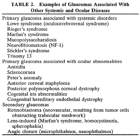
Glaucoma can be associated with various other genetic and congenital disorders (Table 2). The mechanism may be primary or secondary. A thorough discussion of these disorders is beyond the scope of this chapter. The reader is referred to other sections of this text dealing with these disorders.


NAIL-PATELLA SYNDROME

Because progressive, open-angle glaucoma is largely a silent disease until its later stages, it is possible that it remains unrecognized in various genetic diseases, especially those that are primary diagnosed and treated at a young age. An example of this scenario is nail-patella syndrome (NPS). NPS is inherited in an autosomal dominant fashion and has recently been recognized to include open-angle glaucoma in some families. Families with NPS have disorders of the nails (e.g., absence, splitting, ridging, triangular lunulae), hypoplastic or absent patellae, iliac horns, and long-bone abnormalities that can affect arm and leg motion.

In 1997, Lichter and colleagues reported on two families with NPS wherein glaucoma was found to cosegregate with NPS.136 Of particular interest is the open-angle glaucoma in these families that at first seemed to be phenotypically identical to primary open-angle glaucoma. The probands in the two families reported by the Lichter group show no discernible anterior segment abnormalities that accompany their elevated intraocular pressure, glaucomatous optic nerve cupping, and visual field loss. Most other affected members of these families have a similar glaucoma phenotype. However, in one of the families, infantile glaucoma is present in two NPS-affected children and one mother and daughter show iris hypoplasia in the form of a matted and architecturally bland iris surface.

Reports on this disorder go back across a span of more than a hundred years in the genetic and orthopedic literature without the connection to glaucoma having been made. This may not seem so surprising if you consider that the orthopedist performing surgery on an 8-year-old NPS child had no reason to ask about the ophthalmologic status of the child's grandmother. Similarly, the grandmother's ophthalmologist would have no reason to realize that the grandmother has NPS when the only visible signs of NPS in her family that have not been surgically and cosmetically corrected are present in her 8-year-old grandson. Now that the connection has been made, thumbnails and kneecaps should be points of concern to the observant ophthalmologist, and a query about NPS should be included when taking the medical and family histories during an ophthalmologic examination.

As suggested by the cosegregation of NPS and glaucoma phenotypes, glaucoma in these families apparently results from the same underlying genetic defect that causes their orthopedic anomalies. NPS results from mutations in a gene that encodes the LIM-homeodomain transcription factor LMX1B. Molecular genetic studies by Vollrath and and by Dreyer and their respective colleagues identified apparent loss-of-function mutations in LMX1B in cases of NPS, including the affected individuals from the families showing cosegregation of the two phenotypes.137–141 Identification of mutations in multiple families with both NPS and glaucoma allowed Vollrath and colleagues to rule out the model of a founder effect involving two tightly linked genes and determine that mutations in the LMX1B gene are responsible for both the ocular and orthopedic characteristics in the affected individuals.137

If only information from the ophthalmologic examination is used, glaucoma associated with NPS receives the same diagnosis as many patients without NPS—that of primary open-angle glaucoma. So what clinical difference is there between glaucoma classified as primary open-angle glaucoma and glaucoma that looks like primary open-angle glaucoma but is associated with NPS? The first item of clinical importance is that NPS is now a risk factor for glaucoma, and individuals with NPS can be referred for ophthalmologic evaluation that is warranted because of their increased risk.142 Education of NPS family members can alert them to the fact that their affected relatives have a greater risk of glaucoma than their unaffected relatives, which will optimistically lead to timely screening of additional individuals who have not had direct advice on the topic from their own physician. Except in cases of uncertainty about the NPS diagnosis and cases of prenatal testing, mutation screening may not even be needed to identify these individuals with increased risk.

Other glaucoma families may know that their relatives are at increased risk of glaucoma, but they cannot tell which relatives are the ones who should be undergoing more intensive monitoring. Because open-angle glaucoma is so genetically complex, a risk factor associated with any one glaucoma gene may be rare in the overall glaucoma population. Such rare risk factors will be even harder to recognize if they are not clinically significant items that come to the physician's attention during an examination. However, after a complete panel of glaucoma genes become available for use in genetic testing, individuals who have been grouped according to their glaucoma genes can then be evaluated for the presence of other shared characteristics. For instance, now that MYOC mutation screening can be done, it is possible to ask whether the individuals with the mutations have physical risk factors that would allow them to predict which of their relatives are at elevated risk of glaucoma without even doing any DNA testing on the other family members. The phenotype that could be used as a marker for increased risk may not be something that consistently manifests as a major health problem or anything else that gets recorded in a clinical chart. Although NPS can be severe and even fatal in some individuals, in some NPS families the disease phenotype is mild enough that some affected individuals do not even realize that they have a genetic “disease.” In the case of the MYOC gene, no pattern of major systemic disease has so far been identified as accompanying glaucoma in these families, even though MYOC is expressed in various important organs, including the heart. If we take a lesson from the NPS studies, we have to consider that there may still be other subtle characteristics associated with MYOC glaucoma that will not be identified until someone asks just the right question or until detailed enough physical characterization of whole families is carried out. These answers remain to be found.

Some of the most important insights are yet to come. Consider this hypothetical situation: if glaucoma due to an LMX1B mutation shares the clinical characteristics of glaucoma due to a MYOC mutation, does our ability to classify these individuals at the molecular genetic level say anything about functionally important relationship between them in either clinical or mechanistic terms? Given that LMX1B is a transcription factor, and regulation of expression of MYOC has been postulated to play a role in disease pathogenesis, it is tempting to hypothesize that these two genes are acting on the same pathophysiologic pathway leading to identical events downstream of the points at which these genes play their role. Although an obvious connection would be made if LMX1B turned out to play a role in MYOC transcription through interaction with the MYOC promoter, the connection could be much less direct and still have the same consequence: different genetic defects starting off the pathway at different initiation points but leading to a shared set of later events in the disease process. Clearly, this connection has not yet been made and there may be other reasons for the clinical similarities of genetically distinct glaucomas. Because the growing number of different glaucoma genes leaves us with the worrisome situation that the targeted cures we dream of may in fact have to target many different disease genes, this concept of multiple glaucoma genes “hitting” different points along a common glaucoma pathway offers some hope that we might not have to customize cures for every genetically distinct form of glaucoma. Instead, by careful elucidation of such a shared pathway it might be possible to target therapeutic interventions at steps farther along the pathway beyond points in the pathway at which the different glaucoma genes initiate problems. It remains to be seen whether the future will find us trying to find a new cure for each glaucoma gene or dealing with the perhaps simpler problem of finding a new cure for each “glaucoma pathway.” Either way, molecular genetic classification of glaucoma in each patient will be the key to first sorting out the answers and then later translating them into clinical practice. Before we ever get to any of those hoped-for new cures, it is a very real expectation that molecular genetic classification of glaucoma based on genetic testing of patient DNA will provide meaningful information that will assist physician's in making choices among traditional treatments, for at least some forms of glaucoma.

Back to Top
THE FUTURE
The past decade has witnessed great advances in the understanding of the genetic and molecular basis of glaucoma. However, there is still much to understand. We have in hand only a fraction of the genes likely to cause or contribute to causing glaucoma. We have yet to understand the many factors, genetic or environmental, that affect phenotypic variability in patients with the same gene mutation. The precise mechanisms by which myocilin or cytochrome P450 1B1 mutations lead to glaucoma have yet to be elucidated. The implications of genetic testing for these diseases are still being sorted out. Ongoing screening of affected populations should move these genes into the clinical testing arena as we gain enough information on the relationship of genotype to risk, genotype to phenotype, and genotype to treatment outcomes. The future also holds the promise of designing specific strategies, based on the molecular mechanism of disease, to slow or halt the progression of glaucoma.
Back to Top
REFERENCES

1. Bell J. On some structural anomalies of the eye and on the inheritance of glaucoma. In Pearson K (ed). The Treasury of Human Inheritance. Cambridge, UK: Cambridge University Press, 1931:425

2. Benedict TGG. Abhandlungen aus dem Gebiete der Augen-heilkunde. Breslau: L. Freund, 1842:123

3. Mooren A. Ophthalmologische Beobachtungen. Berlin: A. Hirschwald, 1867

4. von Graefe A. Beiträge zur Pathologie und Therapie der Glaucoms. Arch Ophthalmol 1869;15:108

5. Courthney RH, Hill E. Hereditary juvenile glaucoma simplex. JAMA 1931;97:1602

6. Stokes WH. Hereditary primary glaucoma. Arch Ophthalmol 1940;24:885

7. Allen TD, Ackerman WG. Hereditary glaucoma in a pedigree of three generations. Arch Ophthalmol 1942;27:139

8. Beiguelman B, Prado D. Recessive juvenile glaucoma.J Genet Hum 1963;12:53

9. Weatherill JR, Hart CT. Familial hypoplasia of the iris stroma associated with glaucoma. Br J Ophthalmol 1969;53:433

10. Jerndal T. Familial congenital glaucoma with dominant heredity. Acta Ophthalmol 1968;46:459

11. Jerndal T. Dominant goniodysgenesis with late congenital glaucoma: A re-examination of Berg's pedigree. Am J Ophthalmol 1972;74:28

12. Martin JP, Zorab EC. Familial glaucoma: In nine generations of a South Hampshire family. Br J Ophthalmol 1974;58:536

13. Tielsch JM, Sommer A, Katz J et al. Racial variations in the prevalence of primary open-angle glaucoma: The Baltimore Eye Survey. JAMA 1991;266:369

14. Tielsch JM, Katz J, Singh K et al. A population-based evaluation of glaucoma screening: The Baltimore Eye Survey. Am J Epidemiol 1991;134:1102

15. Klein BE, Klein R, Sponsel WE et al. Prevalence of glaucoma: The Beaver Dam Eye Study. Ophthalmology 1992;99:1499

16. Othman MI, Sullivan SA, Skuta GL et al. Autosomal dominant nanophthalmos (NNO1) with high hyperopia and angle-closure glaucoma maps to chromosome 11. Am J Hum Genet 1998;63:1411

17. Kellerman L, Posner A. The value of heredity in the detection and study of glaucoma. Am J Ophthalmol 1955;40:681

18. Becker B, Kolker AE, Roth FD. Glaucoma family study. Am J Ophthalmol 1960;50:557

19. Battacharjee H, Chowdury HK. A survey on heredity of open angle glaucoma. Ind J Ophthalmol 1987;35:138

20. Biro I. Notes on heredity of glaucoma. Ophthalmologica 1939;98:43

21. Francois J, Heintz-deBree. Personal research on the heredity of chronic simple (open-angle) glaucoma. Am J Ophthalmol 1966;62:1067

22. Tielsch JM, Katz J, Sommer A et al. Family history and risk of primary open angle glaucoma: The Baltimore Eye Survey. Arch Ophthalmol 1994;112:69

23. Probert LA. A survey of hereditary glaucoma. Can Med Assoc J 1951;66:563

24. McNaught AI, Allen JG, Healey DL et al. Accuracy and implications of a reported family history of glaucoma: Experience from the Glaucoma Inheritance Study in Tasmania. Arch Ophthalmol 2000;118:900

25. Goldschmidt E. The heredity of glaucoma. Acta Ophthalmol Suppl 1973;120:27

26. Teikari JM. Genetic factors in open-angle (simple and capsular) glaucoma: A population-based twin study. Acta Ophthalmol 1987;65:715

27. Gottfredsdottir MS, Sverisson T, Musch DC et al. Chronic open-angle glaucoma and associated ophthalmic findings in monozygotic twins and their spouses in Iceland. J Glaucoma 1999;8:134

28. Schwartz JT, Reuling FH, Feinleib M. Heritability study on size of the physiologic cup of the optic nerve head: A summary report. Acta Genet Med Gemellol 1976;25:181

29. Bengtsson B. The inheritance and development of cup and disc diameters. Acta Ophthalmol 1980;58:733

30. Knobloch WH, Leavenworth NM, Bouchard TJ et al. Eye findings in twins reared apart. Ophthalmic Paediatr Genet 1985;5:59

31. Drance SM. Angle closure glaucoma among Canadian Eskimos. Can J Ophthalmol 1973;8:252

32. Arkell SM, Lightman DA, Sommer A et al. The prevalence of glaucoma among Eskimos of northwest Alaska. Arch Ophthalmol 1987;105:482

33. Clemmesen V, Alsbirk PH. Primary angle-closure glaucoma (a.c.g.) in Greenland. Acta Ophthalmol 1971;49:47

34. Congdon NG, Quigley NG, Hung PR et al. Screening techniques for angle-closure glaucoma in rural Taiwan. Acta Ophthalmol Scand 1996;74:113

35. Congdon N, Wang F, Tielsch JM. Issues in the epidemiology and population-based screening of primary angle-closure glaucoma. Surv Ophthalmol 1992;36:411

36. Salmon JF. Predisposing factors for chronic angle-closure glaucoma. Prog Retin Eye Res 1999;18:121

37. Alward WL, Fingert JH, Coote MA et al. Clinical features associated with mutations in the chromosome 1 open-angle glaucoma gene (GLC1A). N Engl J Med 1998;338:1022

38. Sheffield VC, Stone EM, Alward L et al. Genetic linkage of familial open angle glaucoma to chromosome 1q21-q31. Nat Genet 1993;4:47

39. Richards JE, Lichter PR, Boehnke M et al. Mapping of a gene for autosomal dominant juvenile-onset open-angle glaucoma to chromosome Iq. Am J Hum Genet 1994;54:62

40. Wiggs JL, Haines JL, Paglinauan C et al. Genetic linkage of autosomal dominant juvenile glaucoma to 1q21-q31 in three affected pedigrees. Genomics 1994;21:299

41. Morissette J, Cote G, Anctil JL et al. A common gene for juvenile and adult-onset primary open-angle glaucomas confined on chromosome 1q. Am J Hum Genet 1995;56:1431

42. Lichter PR. Genetic clues to glaucoma's secrets: The L. Edward Jackson Memorial Lecture. Part 2. Am J Ophthalmol 1994;117:706

43. Stone EM, Fingert JH, Alward WLM et al. Identification of a gene that causes primary open angle glaucoma. Science 1997;275:668

44. Adam MF, Belmouden A, Binisti P et al. Recurrent mutations in a single exon encoding the evolutionarily conserved olfactomedin-homology domain of TIGR in familial open-angle glaucoma. Hum Mol Genet 1997;6:2091

45. Richards JE, Ritch R, Lichter PR et al. Novel trabecular meshwork inducible glucocorticoid response mutation in an eight-generation juvenile-onset primary open-angle glaucoma pedigree. Ophthalmology 1998;105:1698

46. Mansergh FC, Kenna PF, Ayuso C et al. Novel mutations in the TIGR gene in early and late onset open angle glaucoma. Hum Mutat 1998;11:244

47. Rozsa FW, Shimizu S, Lichter PR et al. GLC1A mutations point to regions of potential functional importance on the TIGR/MYOC protein. Mol Vis 1998;4:20

48. Fingert JH, Heon E, Liebmann JM et al. Analysis of myocilin mutations in 1703 glaucoma patients from five different populations. Hum Mol Genet 1999;8:899

49. Shimizu S, Lichter PR, Johnson AT et al. Age-dependent prevalence of mutations at the GLC1A locus in primary open-angle glaucoma. Am J Ophthalmol 2000;130:165

50. Wiggs JL, Del Bono EA, Schuman JS et al. Clinical features of five pedigrees genetically linked to the juvenile glaucoma locus on chromosome 1q21-q31. Ophthalmology 1995;102:1782

51. Stoilova D, Child A, Brice G et al. Novel TIGR/MYOC mutations in families with juvenile onset primary open angle glaucoma. J Med Genet 1998;35:989

52. Polansky JR, Fauss DJ, Chen P et al. Cellular pharmacology and molecular biology of the trabecular meshwork inducible glucocorticoid response gene product. Ophthalmologica 1997;211:126

53. Nguyen TD, Chen P, Huang WD et al. Gene structure and properties of TIGR, an olfactomedin-related glycoprotein cloned from glucocorticoid-induced trabecular meshwork cells. J Biol Chem 1998;273:6341

54. Kubota R, Noda S, Wang Y et al. A novel Myson-like pretin (Myocilin) expressed in the connecting cilium of the photoreceptor: Molecular cloning, tissue expression, and chromosomal mapping. Genomics 1997;41:360

55. Ortego J, Escribano J, Coca-Prados M. Cloning and characterization of subtracted cDNAs from a human ciliary body library encoding TIGR, a protein involved in juvenile open angle glaucoma with homology to myosin and olfactomedin. FEBS Lett 1997;413:349

56. Snyder DA, Rivers AM, Yokoe H et al. Olfactomedin: Purification, characterization, and localization of a novel olfactory glycoprotein. Biochemistry 1991;30:9143

57. Huang W, Jaroszewski J, Ortega J et al. Expression of the TIGR gene in the iris, ciliary body, and trabecular meshwork of the human eye. Ophthalmic Genet 2000;21:155

58. Tomarev SI, Tamm ER, Chang B. Characterization of the mouse Myoc/Tigr gene. Biochem Biophys Res Commun 1998;245:887

59. Fingert JH, Ying L, Swiderski RE et al. Characterization and comparison of the human and mouse GLC1A glaucoma genes. Genome Res 1998;8:377

60. Swiderski RE, Ying L, Swiderski RE et al. Expression pattern and in situ localization of the mouse homologue of the human MYOC (GLC1A) gene in adult brain. Brain Res Mol Brain Res 1999;68:64

61. Swiderski RE, Ross JL, Fingert JH et al. Localization of MYOC transcripts in human eye and optic nerve by in situ hybridization. Invest Ophthalmol 2000;41:3420

62. Morissette J, Clepet C, Moisan S et al. Homozygotes carrying an autosomal dominant TIGR mutation do not manifest glaucoma. Nat Genet 1998;19:319

63. Ueda J, Wentz-Hunter KK, Cheng EL et al. Ultrastructural localization of myocilin in human trabecular meshwork cells and tissues. J Histochem Cytochem 2000;48:1321

64. Lütjen-Drecoll E, May CA, Polansky JR et al. Localization of the stress protein aβ-crystallin and trabecular meshwork inducible glucocorticoid response protein in normal and glaucomatous trabecular meshwork. Invest Ophthalmol 1998;39:517

65. Polansky J, Fauss DJ, Zimmerman CC. Regulation of TIGR/MYOC gene expression in human trabecular meshwork cells. Eye 2000;14:503

66. Tamm ER, Russell P, Epstein DL et al. Modulation of myocilin/TIGR expression in human trabecular meshwork. Invest Ophthalmol 1999;40:2577

67. Polansky JR, Kurtz RM, Fauss DJ et al. HTM cell culture model for steroid effects on IOP: overview. In Krieglstein GK (ed). Glaucoma Update IV, Berlin: Springer-Verlag, 1991:20–29

68. Nguyen TD, Huang WD, Bloom E et al. Glucocorticoid effects on HTM cells: Molecular biology approaches. In Lutjen-Drecoll E (ed). Basic Aspects of Glaucoma Research III. Stuttgart: Schattauer, 1993:331–343

69. Armaly MF. Effect of corticosteroids on intraocular pressure and fluid dynamics. I: The effect of dexamethasone in the normal eye. Arch Ophthalmol 1963;70:482

70. Armaly MF. Effect of corticosteroids on intraocular pressure and fluid dynamics. II: The effect of dexamethasone in the glaucomatous eye. Arch Ophthalmol 1963;70:492

71. Furuyoshi N, Furuyoshi M, Futa R et al. Ultrastructural changes in the trabecular meshwork of juvenile glaucoma. Ophthalmologica 1997;211:140

72. Johnson D, Gottanka J, Flügel C et al. Ultrastructural changes in the trabecular meshwork of human eyes treated with corticosteroids. Arch Ophthalmol 1997;115:375

73. Raymond V, Rodrigue MA, Gobeil S et al. Heterodimerization between wild-type and mutant TIGR/myocilin polypeptides is critical for autosomal dominant open-angle glaucoma. Am J Hum Genet 2000;76:A2220

74. Sarfarazi M, Chaterji R, Kocak Midillioglu I et al. Identification of a severely affected homozygote TIGR/MYOC mutation in a large family with juvenile-onset primary open angle glaucoma. Invest Ophthalmol 2000;41:S822

75. Johnson AT, Drack AV, Kwitek AE et al. Clinical features and linkage analysis of a family with autosomal dominant juvenile glaucoma. Ophthalmology 1993;100:524

76. Johnson AT, Richards JE, Boehnke M et al. Clinical phenotype of juvenile-onset primary open-angle glaucoma linked to chromosome 1q. Ophthalmology 1996;103:808

77. Suzuki Y, Shirato S, Taniguchi F et al. Mutations in the TIGR gene in familial primary open-angle glaucoma in Japan letter. Am J Hum Genet 1997;61:1202

78. Michels-Rautenstrauss KG, Mardin CY, Budde WM et al. Juvenile open angle glaucoma: Fine mapping of the TIGR gene to 1q24.3- q25.2 and mutation analysis. Hum Genet 1998;102:103

79. Stoilova D, Child A, Trifan OC et al. Localization of a locus (GLC1B) for adult-onset primary open angle glaucoma to the 2cen-q13 region. Genomics 1996;36:142

80. Faucher M, Dubois S, Cote G et al. An integrated map of the GLC1B locus for primary open-angle glaucoma at chromosome 2. Invest Ophthalmol 2000;41:A4373

81. Wiggs JL, Allingham RR, Hossain A et al. Genome-wide scan for adult onset primary open angle glaucoma. Hum Mol Genet 2000;9:1109

82. Desai TD, Child A, Rezaie T et al. Searching for identification of mutations involved in GLC1B, GLC1D, and GLC1E linked glaucoma loci. Invest Ophthalmol 2000;41:S526

83. Wirtz MK, Child A, Rezaie T et al. Mapping a gene for adult-onset primary open-angle glaucoma to chromosome 3q. Am J Hum Genet 1997;60:296

84. Xu H, Acott TS, Wirtz MK. Identification and expression of a novel type I procollagen C-proteinase enhancer protein gene from the glaucoma candidate region on 3q21-24. Genomics 2000;66:264

85. Trifan OC, Traboulsi EI, Stoilova D et al. A third locus (GLC1D) for adult-onset primary open-angle glaucoma maps to the 8q23 region. Am J Ophthalmol 1998;126:17

86. Sarfarazi M, Child A, Stoilova D et al. Localization of the fourth locus (GLC1E) for adult-onset primary open-angle glaucoma to the 10p15-p14 region. Am J Hum Genet 1998;62:641

87. Wirtz MK, Samples JR, Rust K et al. GLC1F, a new primary open-angle glaucoma locus, maps to 7q35-q36. Arch Ophthalmol 1999;117:237

88. Sverrisson, TM, Rust K, Huynh L et al. Refining the GLC1F physical map and analysis of candidate genes. Invest Ophthalmol Vis Sci 2000;41:A4373

89. Andersen JS, Pralea AM, DelBono EA et al. A gene responsible for the pigment dispersion syndrome maps to chromosome 7q35-q36. Arch Ophthalmol 1997;115:384

90. Yokoyama A, Nao-i N, Date Y et al. Detection of a new TIGR gene mutation in a Japanese family with primary open angle glaucoma. Jpn J Ophthalmol 1999;43:85

91. Yoon SJ, Kim HS, Moon JI et al. Mutations of the TIGR/MYOC gene in primary open-angle glaucoma in Korea [letter]. Am J Hum Genet 1999;64:1775

92. Kee C, Ahn BH. TIGR gene in primary open-angle glaucoma and steroid-induced glaucoma. Korean J Ophthalmol 1997;11:75

93. Lam DS, Leung YF, Chua JK et al. Truncations in the TIGR gene in individuals with and without primary open-angle glaucoma. Invest Ophthalmol Vis Sci 2000;41:1386

94. Zhou Y, Ge J, Guo Y. To screen, clone and sequence TIGR gene mutation in Chinese patients with primary open-angle glaucoma. Chin J Ophthalmol 2000;36:416

95. Schwartz M, Yoles E. Self-destructive and self-protective processes in the damaged optic nerve: implications for glaucoma. Invest Ophthalmol Vis Sci 2000;41:349

96. Alsbirk PH. Anterior chamber depth and primary angle-closure glaucoma. II: A genetic study. Acta Ophthalmol 1975;53:436

97. Drance SM, Morgan RW, Bryett J et al. Anterior chamber depth and gonioscopic findings among the Eskimos and Indians in the Canadian Arctic. Can J Ophthalmol 1973;8:255

98. Lowe RF. Primary angle-closure glaucoma: Inheritance and environment. Br J Ophthalmol 1972;56:13

99. Spaeth GL. Gonioscopy: Uses old and new—The inheritance of occludable angles. Ophthalmology 1978;85:222

100. Tomlinson A, Leighton DA. Ocular dimensions in the heredity of angle-closure glaucoma. Br J Ophthalmol 1973;57:475

101. Lichter PR and Anderson DR. Discussions on Glaucoma. New York: Grune & Stratton, 1977

102. Gieser DK, Wilensky JT. HLA antigens and acute angle-closure glaucoma. Am J Ophthalmol 1979;88:232

103. Brooks AM, Gillies WE. Blood groups as genetic markers in glaucoma. Br J Ophthalmol 1988;72:270

104. Brockhurst RJ. Nanophthalmos with uveal effusion: A new clinical entity. Trans Am Ophthalmol Soc 1974;72:371

105. Kimbrough RL, Trempe CS, Brockhurst RJ et al. Angle-closure glaucoma in nanophthalmos. Am J Ophthalmol 1979;88:572

106. Cross HE, Yoder F. Familial nanophthalmos. Am J Ophthalmol 1976;81:300

107. Martorina M. Familial nanophthalmos. J Fr Ophthalmol 1988;11:357

108. Neelakantan A, Venkataramakrishnan P, Rao BS et al. Familial nanophthalmos: Management and complications. Ind J Ophthalmol 1994;42:139

109. Altintas AK, Acar MA, Yalvac IS et al. Autosomal recessive nanophthalmos. Acta Ophthalmol Scand 1997;75:325

110. Vingolo EM, Steindl K, Forte R et al. Autosomal dominant simple microphthalmos. J Med Genet 1994;31:721

111. Malta RF, Povoa CA, Castro EFS et al. Nanophthalmos (NNo1): Study of a Brazilian family. Invest Ophthalmol Vis Sci 2000;41:A4370

112. Bessant DA, Khaliq S, Hameed A et al. A locus for autosomal recessive congenital microphthalmia maps to chromosome 14q32. Am J Hum Genet 1998;62:1113

113. deLuise VP, Anderson DR. Primary infantile glaucoma (congenital glaucoma). Surv Ophthalmol 1983;28:1

114. Lehrfeld L, Reber J. Glaucoma at the Wills Eye Hospital (1925-1935). Arch Ophthalmol 1937;18:712

115. Gencik A. Epidemiology and genetics of primary congenital glaucoma in Slovakia. Description of a form of primary congenital glaucoma in gypsies with autosomal-recessive inheritance and complete penetrance. Dev Ophthalmol 1989;16:76

116. Jaafar MS. Care of infantile glaucoma patients. In Reinecke RD (ed). Ophthalmology Annual. New York: Raven Press, 1988:15

117. Westerlund E. On the heredity of congenital hydrophthalmus. Acta Ophthalmol 1943;21:330

118. Shaffer RN. Genetics and the congenital glaucomas. Am J Ophthalmol 1965;60:981

119. Bejjani BA, Lewis RA, Tomey KF et al. Mutations in CYP1B1, the gene for cytochrome P4501B1, are the predominant cause of primary congenital glaucoma in Saudi Arabia. Am J Hum Genet 1998;62:325

120. Merin S, Morin D. Heredity of congenital glaucoma. Br J Ophthalmol 1972;56:414

121. Demenais F, Elston RC, Bonaiti C et al. Congenital glaucoma: genetic models. Hum Genet 1979;46:305

122. Gencik A, Gencikova A, Gerinec A. Genetic heterogeneity of congenital glaucoma. Clin Genet 1980;17:241

123. Sarfarazi M, Stoilov I. Molecular genetics of primary congenital glaucoma. Eye 2000;14:422

124. Sarfarazi M, Akarsu AN, Hossain A et al. Assignment of a locus (GLC3A) for primary congenital glaucoma (buphthalmos) to 2p21 and evidence for genetic heterogeneity. Genomics 1995;30:171

125. Stoilov I, Akarsu AN, Sarfarazi M. Identification of three different truncating mutations in cytochrome P4501B1 (CYP1B1) as the principal cause of primary congenital glaucoma (buphthalmos) in families linked to the GLC3A locus on chromosome 2p21. Hum Mol Genet 1997;6:641

126. Stoilov I, Akarsi AN, Alonzie I et al. Sequence analysis and homology modeling suggest that primary congenital glaucoma on 2p21 results from mutations disrupting either the hinge region or the conserved core structures of cytochrome P4501B1. Am J Hum Genet 1998;62:573

127. Plasilova M, Stoilov I, Sarfarazi M et al. Identification of a single ancestral CYP1B1 mutation in Slovak Gypsies (Roms) affected with primary congenital glaucoma. J Med Genet 1999;36:290

128. Bejjani BA, Stockton DW, Lewis RA et al. Multiple CYP1B1 mutations and incomplete penetrance in an inbred population segregating primary congenital glaucoma suggest frequent de novo events and a dominant modifier locus Hum Mol Genet 2000;9:367

129. Kakiuchi T, Isashiki Y, Nakao K et al. A novel truncating mutation of cytochrome P4501B1 (CYP1B1) gene in primary infantile glaucoma. Am J Ophthalmol 1999;128:370

130. Graham-Lorence S, Peterson JA. P450s: Structural similarities and functional differences. FASEB J 1996;10:206

131. Chen CD, Kemper B. Different structural requirements at specific proline residue positions in the conserved proline-rich region of cytochrome P450 2C2. J Biol Chem 1996;271:28607

132. Yamazaki S, Sato K, Suhara K et al. Importance of the proline-rich region following signal-anchor sequence in the formation of correct conformation of microsomal cytochrome P-450s. J Biochem 1993;114:652

133. Nelson DR, Koymans L, Kamataki T et al. P450 superfamily: Update on new sequences, gene mapping, accession numbers and nomenclature. Pharmacogenetics 1996;6:1

134. Nebert DW. Proposed role of drug-metabolizing enzymes: Regulation of steady state levels of the ligands that effect growth, homeostasis, differentiation, and neuroendocrine functions. Mol Endocrinol 1991;5:1203

135. Stoilov I, Jannson I, Sarfarazi M et al. Role of cytochrome P450 in development. Drug Metab Drug Interact 2001;18:33

136. Lichter PR, Farley F, Stringham HM et al. Co-segregation of open-angle glaucoma and the nail-patella syndrome. Am J Ophthalmol 1997;124:506

137. Vollrath D, Jaramillo-Babb VL, Clough MV et al. Loss-of-function mutations in the LIM-homeodomain gene, LMX1B, in nail-patella. Hum Mol Genet 1998;7:1091

138. Chen H, Lun Y, Ovchinnikov D et al. Limb and kidney defects in Lmx1b mutant mice suggest an involvement of LMX1B in human nail patella syndrome. Nat Genet 1998;19:51

139. Dreyer SD, Morello R, German MS et al. LMX1B transactivation and expression in nail-patella syndrome. Hum Mol Genet 2000;9:1067

140. Dreyer SD Zhou G, Baldini A et al. Mutations in LMX1B cause abnormal skeletal patterning and renal dysplasia in nail patella syndrome. Nat Genet 1998;19:47

141. Clough MV, Hamlington JD, McIntosh I. Restricted distribution of loss-of-function mutations within the LMX1B genes of nail-patella syndrome patients. Hum Mutat 1999;14:459

142. Farley FA, Lichter PR, Downs CA et al. An orthopaedic scoring system for nail-patella syndrome and application to a kindred with variable expressivity and glaucoma.J Pediatr Orthop 1999;19:624

143. Zimmerman CC, Lingappa VR, Richards JE et al. A trabecular meshwork glucocorticoid response (TIGR) gene mutation affects translocational processing. Mol Vis 1999;5:19

144. Stoilov I., Akarsu AN, Sarfarazi M. Identification of three different truncating mutations in cytochrome P4501B1 (CYP1B1) as the principal cause of primary congenital glaucoma (buphthalmos) in families linked to the GLC3A locus on chromosome 2p21. Hum Mol Genet 1997;6:641

145. Akarsu AN, Turacli ME, Aktan SG, et al. A second locus (GLC3B) for primary congenital glaucoma (buphthalmos) maps to the 1p36 region. Hum Mol Genet 1996;5:1199

Back to Top