Chapter 28
Acute Retinal Necrosis and Progressive Outer Retinal Necrosis Syndromes
CAROLINE R. BAUMAL, JAY S. DUKER and DAVID FISCHER
Main Menu   Table Of Contents

Search

Acute Retinal Necrosis Syndrome
PROGRESSIVE OUTER RETINAL NECROSIS
REFERENCES

The herpesvirus family includes herpes simplex virus (HSV), varicella-zoster virus (VZV), cytomegalovirus (CMV), and Epstein-Barr virus (EBV). CMV produces a characteristic hemorrhagic necrotizing retinitis in immunocompromised patients. Acute retinal necrosis (ARN) and progressive outer retinal necrosis (PORN), which have been described more recently, are two distinct variants of necrotizing herpetic retinopathy.
Back to Top
Acute Retinal Necrosis Syndrome
Acute retinal necrosis is a clinical syndrome that has been associated with either HSV or VZV infection.1–5 Features of ARN syndrome include a vaso-occlusive angiitis of both the retinal and choroidal vessels, a necrotizing retinitis that preferentially involves the peripheral retina, and significant intraocular inflammation.6 Rhegmatogenous retinal detachment is a major late sequela.

ARN was originally described in Japan in 1971 by Urayama and coworkers, who studied six healthy patients with acute onset of a unilateral panuveitis and retinal arteritis that resulted in widespread peripheral, confluent retinal necrosis and eventual retinal detachment.7 This entity was first reported in English medical literature in 1977 by Willerson and associates, who described two patients with a bilateral necrotizing vaso-occlusive retinitis.8 The term acute retinal necrosis (ARN; or BARN, when bilateral), was coined by Young and Bird in 1978.9 Although this syndrome was initially described in healthy patients, it subsequently has been reported in individuals with compromised systemic immunity—most notably in those with human immunodeficiency virus (HIV) infection.

ARN can be a visually devastating disease with a poor visual prognosis, although advances in antiviral therapy and vitreoretinal techniques for retinal detachment have improved visual outcomes. Prompt diagnosis and treatment are necessary to limit retinal damage and preserve vision.

CLINICAL PRESENTATION

Acute retinal necrosis does not appear to have any racial predilection; however, it may be more common in males. Although ARN typically affects individuals between 20 and 50 years of age, cases have been reported in patients as young as 4 years and as old as 89 years of age.10–13

Initially, patients may complain of mild to moderate ocular or periorbital pain, often accompanied by a red eye. Early visual symptoms may or may not be present and can be insidious. When symptoms are present, these usually consist of floaters, blurred vision, or rarely, decreased peripheral vision. Acute central visual loss is an atypical presentation because the posterior pole usually is spared from the retinopathy until late in the course of the disease. Optic nerve involvement, retinal artery occlusion, and retinal detachment can affect central vision but usually are not manifest at presentation.7–10,14,15

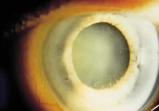
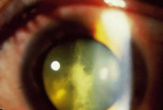
External examination may reveal diffuse episcleritis, scleritis, or orbital inflammatory disease. Mild to moderate anterior segment cellular reaction is common and either granulomatous or fine keratic precipitates typically are present 10,16,17(Fig. 1). A hypopyon typically does not develop.8 The intraocular pressure may be low, even in the absence of retinal detachment; however, elevated intraocular pressure secondary to anterior segment inflammation also may occur. Concurrent herpesvirus infection of the cornea with dendritic or stromal keratitis is not a typical feature of ARN syndrome in healthy individuals, although it has been described in patients with HIV infection.18,19 One case of simultaneous HSV type 1 keratitis and ARN was reported in a patient with Ramsay Hunt syndrome.20,21

Fig. 1. Typical anterior segment appearance during active ARN syndrome. Anterior segment cellular reaction with keratic precipitates and episcleral injection are noted.

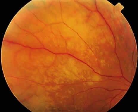
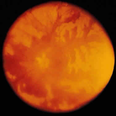
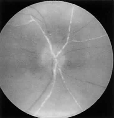
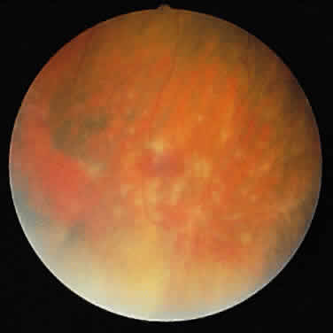
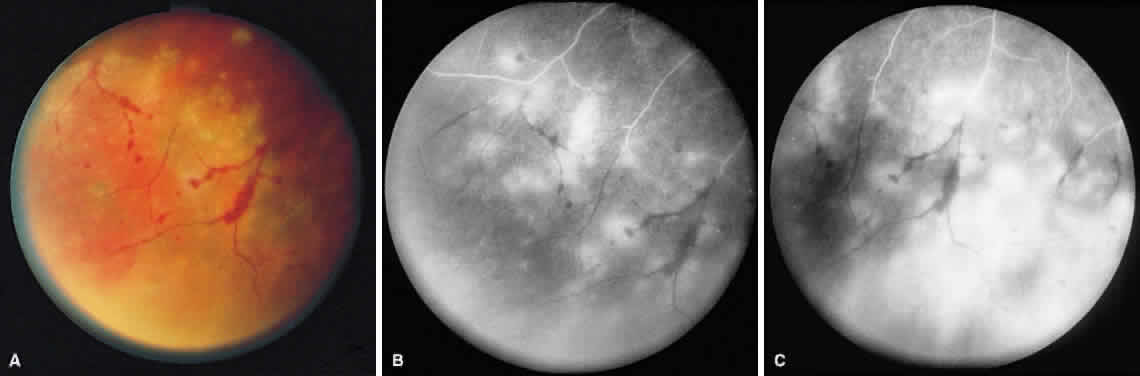
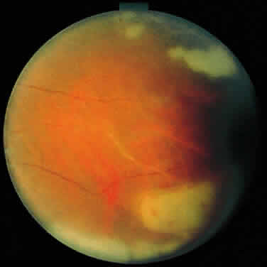
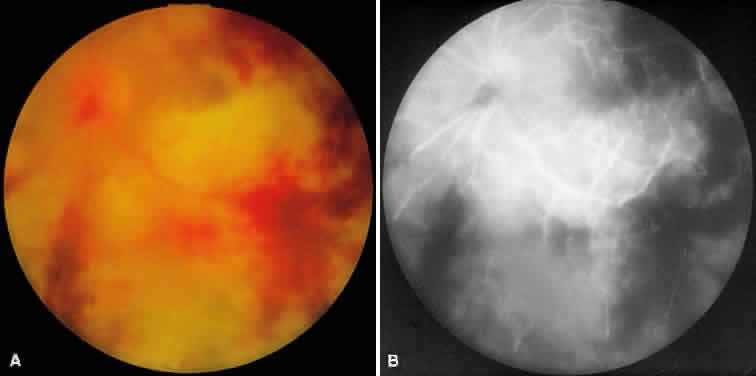
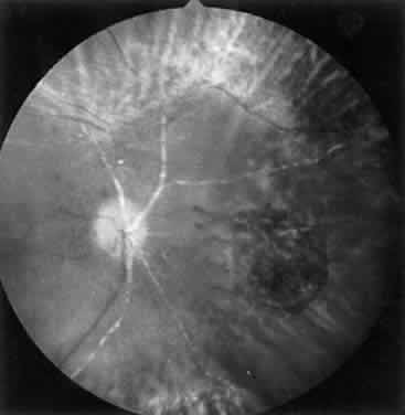
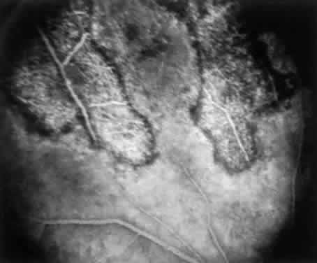
ARN is characterized by a retinal vasculitis affecting both the arteries and veins in the fundus, which is manifested by sheathing of the larger vessels (Fig. 2).7–10,16 Initially, patchy areas of peripheral retinal whitening (“thumbprint lesions”) representing full-thickness retinal necrosis are present or develop shortly after the vasculitis (Fig. 3). During a course that may span days or weeks, these patches coalesce into geographic areas (Fig. 4A). The entire peripheral retina (360 degrees) may be involved, or, more commonly, there are several noncontiguous patches of necrosis, each covering from a half to three clock hours (Figs. 5 and 6). The posterior segment lesions may not be detected without examination of the peripheral retina.

Fig. 2. Fundus photograph of the posterior pole in active ARN syndrome showing prominent arterial sheathing and mild blurring of the optic disc margin.

Fig. 3. In early ARN, multifocal white patches of retinal necrosis occur in the peripheral retina.

Fig. 4. A. With progression of ARN, the areas of retinal whitening begin to coalesce. Mild perivascular retinal hemorrhages are noted. B. Fluorescein angiography in the venous phase reveals retinal vascular nonperfusion in areas of active retinitis. C. In the late-phase angiogram, fluorescein hyperfluorescence is seen in areas of retinitis and around the retinal vasculature.

Fig. 5. The peripheral retinal whitening becomes confluent with vascular obliteration.

Fig. 6. Equator-plus photograph of a patient with 360° ARN syndrome.

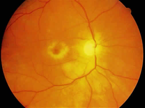
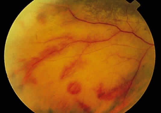
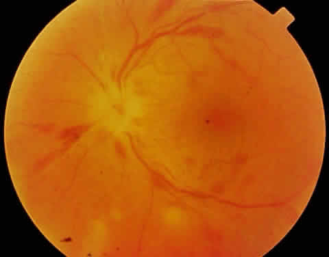
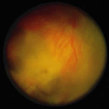
As the infection progresses, the leading edge of confluent retinal whitening advances toward the posterior pole (Fig. 7A). The retinitis may not progress posteriorly to the vascular arcades, sparing the macula and central vision. It is hypothesized that the retinal necrosis in ARN results from the combined effect of intracellular viral replication with subsequent cell death and vascular occlusion secondary to acute vasculitis. In some patients, the retinal vasculitic component may be much more prominent than the retinal necrosis.7 Optic disc swelling, either hyperemic or pallid, is a common feature of the ARN syndrome.1 Perivascular hemorrhages may be present (Fig. 8); however, widespread areas of retinal hemorrhage are atypical. Retinal vascular occlusion, often involving the arteries, can occur at any point during the clinical course. Without treatment, the inflammatory component of ARN typically burns out in 6 to 12 weeks, leaving behind a thin atrophic retina with associated pigmentary changes.10

Fig. 7. A. Active ARN with diffuse retinal necrosis progressing into the macula and areas of retinal hemorrhage. The hazy view is secondary to vitreous inflammation. B. Fluorescein angiogram in the venous phase of active ARN syndrome in this eye reveals peripheral vascular nonperfusion, as well as optic nerve and perivascular hyperfluorescence.

Fig. 8. Perivascular hemorrhage and optic nerve head swelling with small foci of retinal necrosis inferior to the optic nerve.

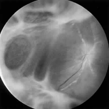
Along with anterior uveitis, a mild to moderate vitritis is present early in the course of ARN (Fig. 9). As the retina becomes necrotic, increasing amounts of vitreous cells and debris are seen. The vitritis may impair visualization of the posterior segment. Eventual opacification and organization of the vitreous can occur. Severe vitreous fibrosis with traction resembling proliferative vitreoretinopathy is not an uncommon late complication.1,8–12,14,15,17 Other features described in ARN include exudative retinal detachment, macular edema, glaucoma, and cataract.4

Fig. 9. Prominent vitreous reaction and a mild cataract are noted in ARN.

As the active retinal inflammatory process recedes, either spontaneously or with treatment, the retinal areas previously infected appear thinned and atrophic, with underlying retinal pigment epithelial alterations (Fig. 10). These pigmentary changes usually begin to develop around 4 weeks after the onset of the infection.22 Often a distinct scalloped pattern can be seen that clearly demarcates previously involved and spared retina (Fig. 11). The pigmentary alterations result from both retinal pigment epithelial cell death by direct infection and secondary inflammatory changes and hyperplasia induced by the adjacent retinal necrosis. Full-thickness retinal breaks are a prominent feature of the ARN syndrome, and often develop during the recovery stage of ARN. Retinal breaks may be large, irregular, and multiple and classically occur at the junction of normal and affected retina (Fig. 12)23; they can, however, occur elsewhere in the retina, with or without accompanying vitreous fibrosis and traction. Clarkson and coworkers noted traction-associated retinal tears in uninvolved retina.14 Rhegmatogenous retinal detachment is a major cause of visual loss and occurs in 50% to 75% of cases not treated with prophylactic laser photocoagulation.1,10,14 Retinal detachment caused by ARN usually develops several weeks after the onset of inflammation; however, retinal detachment has been reported as early as 6 days or as late as several months after the onset of symptoms.24 These detachments are often complicated because of the combined effect of vitreous organization and multifocal retinal breaks. Unusual late sequelae of ARN syndrome include neovascularization of the retina and optic disc with vitreous hemorrhage (Fig. 13).25,26 In contrast to other uveitic syndromes, chronic or relapsing episodes of intraocular inflammation are not common in ARN.27

Fig. 10. Healed phase of ARN syndrome in same eye as in Figure 2 after 4 weeks shows optic atrophy with retinal pigment epithelial changes.

Fig. 11. Venous phase of fluorescein angiogram demonstrating the healed stage of ARN syndrome. Classic retinal pigment epithelial alterations in a scalloped pattern clearly separate previously involved retina from spared retina.

Fig. 12. Complicated retinal detachment secondary to multiple, posterior retinal holes and PVR associated with ARN syndrome. Despite pars plana vitrectomy with silicon oil injection, the retina could not be reattached. This photograph was taken 6 weeks after Figure 10.

Fig. 13. Peripheral retinal neovascularization may occur on occasion as a sequela of ARN syndrome.

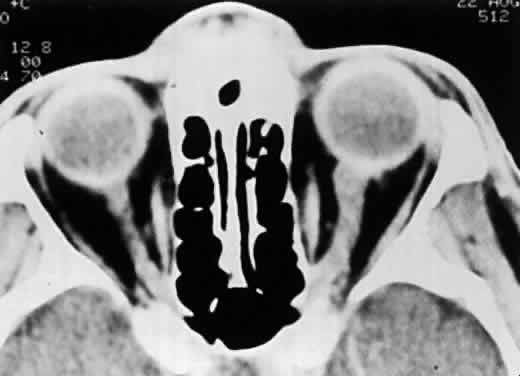
In the absence of direct macular involvement, arterial obstruction, or retinal detachment, decreased central vision that is inconsistent with the associated retinal findings is most likely secondary to optic nerve involvement.28 The presence of optic disc swelling with ARN was noted by early investigators.10 Histopathologic studies have confirmed the presence of intraneural inflammation and necrosis.1,29–31 Both direct inflammation of the optic nerve substance and a secondary ischemic component are believed to produce ARN optic neuropathy, paralleling the pattern of visual dysfunction in the retina. When optic nerve involvement occurs in ARN, an acquired dyschromatopsia, a relative afferent pupillary defect in disproportion to the amount of retinal necrosis, central or arcuate visual field defects, and an enlarged optic nerve sheath can be detected with neuroimaging28,31 (Fig. 14). It often is difficult to distinguish whether visual loss is caused by optic neuropathy or retinal disease in these severely affected patients. An accurate assessment of the incidence of ARN optic neuropathy has not been made. Clinically, optic nerve swelling occurs in a significant number of patients with ARN and may resolve rapidly with therapy.

Fig. 14. CT scan of a patient with unilateral ARN syndrome showing enlarged left optic nerve ipsilateral to the affected eye. (Sergott RC, Belmont JB, Savino PJ et al: Optic nerve involvement in the acute retinal necrosis syndrome. Arch Ophthalmol 103:116, 1985. Reprinted with permission from the American Medical Association)

Bilateral involvement in ARN eventually occurs in up to 65% of patients.10,23 There is no correlation between the severity of ARN in the first eye and severity of the disease in the fellow eye. Involvement of the second eye may not be apparent initially; there may be a delay of several weeks. Thirty-four years is the longest reported interval for involvement of the contralateral eye in ARN.32 Although this is an atypical scenario, it emphasizes the need for long-term ophthalmologic examinations in patients with ARN.

Initial reports indicated that the long-term visual prognosis in ARN syndrome was poor; 60% of eyes had a final visual acuity of 20/200 or worse.10,31Potential causes of visual loss in ARN syndrome include the following:

  • Media opacity
  • Retinal cell necrosis, infection, ischemia, retinal detachment, macular pucker
  • Optic neuropathy

A modified classification of the stages of ARN syndrome is presented in Table 1. However, not all cases of ARN have a fulminant course and result in a poor outcome. Mild cases of ARN with limited peripheral retinitis and good visual outcomes have been reported, and some cases resolve spontaneously without treatment.33 In a recent study of eyes with limited retinal involvement at the time of diagnosis, final visual acuity was 20/40 or better in 50% and 20/400 or better in 92% of eyes after treatment with acyclovir and prophylactic laser photocoagulation.34 The growing number of ARN cases with a good visual outcome is likely owing to increased physician awareness of this disorder, which leads to earlier diagnosis and aggressive therapy. The features of ARN may vary in severity depending on the specific etiologic agent and the immune status of the individual. For example, ARN associated with primary VZV infection (chickenpox) may have less severe features and a better visual prognosis.35


TABLE 28-1. Stages of Acute Retinal Necrosis


StageDescription
INecrotizing retinitis
 IaDiscrete areas of peripheral retinitis
 IbConfluent areas of peripheral retinitis, papillitis, macular edema
IIVitreous opacification or organization
IIIRegression of retinal necrosis; secondary pigmentation of the lesions with contraction and condensation of the vitreous base
IVRetinal detachment
 IVaAcute retinal tears or detachment with traction, or proliferative vitreoretinopathy
 IVbChronic retinal detachment
(After Carney MD, Peyman GA, Goldberg MF et al: Acute retinal necrosis. Retina 6:85, 1986)


Central nervous system abnormalities occasionally have been associated with ARN syndrome. Cerebrospinal fluid pleocytosis is well documented.10,28,36 One case of labyrinthine deafness in association with diffuse cerebral atrophy has occurred.37 True viral encephalitis usually is not concomitant with ARN syndrome; however, posterior segment inflammatory conditions that resemble ARN have been reported in association with florid viral encephalitis.38,39 A rapidly progressive, bilateral retinal necrosis caused by HSV has been described in two patients with concomitant HSV encephalitis and acquired immunodeficiency syndrome (AIDS).5 Because of the presence of central nervous system abnormalities as well as an acute ARN optic neuropathy, it has been suggested that ARN be reclassified as a “uveo-meningeal syndrome” instead of an isolated retinitis.28

Initial reports of ARN were in healthy patients with normal systemic immunity and no extraocular manifestations of herpetic infection. This continues to be true in most ARN cases. Most cases of ARN are believed to be secondary to reactivation of a latent herpetic infection, and often patients have a prior history of dermatomal VZV (herpes zoster) or primary VZV (chickenpox) infection, or perioral blisters, presumably caused by HSV infection. Reactivation of the viral genome from the trigeminal ganglion may result in the development of ARN after transneural spread. Trauma-induced reactivation of latent congenital HSV type 2 infection has been reported in three cases of ARN.13 It is uncommon for healthy individuals to develop ARN simultaneously with cutaneous VZV or HSV infection.40,41 Concurrent aphthous ulcers have been reported in two patients with ARN.42 Rare cases of ARN have been described in immunocompetent individuals (primarily adults) with primary VZV infection and in those who have had close contact with an individual with chickenpox but who did not experience skin lesions.35,43

ARN subsequently has been reported in immunocompromised patients with HIV infection, autoimmune disorders, cancer, and organ transplants.44–46 It has been recommended that ARN syndrome be defined by its ocular features rather than by the immune status of the affected individual.4 In patients with HIV infection and ARN, the CD4+ T lymphocyte count generally is greater than 60 cells/μl, although ARN is sufficiently infrequent in these patients to allow any conclusions to be drawn regarding the CD4+ count. HIV-infected patients are more likely to have simultaneous or recurrent VZV or HSV infections.45 Bilateral ARN after unilateral herpes zoster ophthalmicus has been reported as a presenting sign of AIDS.45 In a study of patients with VZV involving the ophthalmic division of the trigeminal nerve, 5 of 29 (17%) patients with HIV infection developed ARN within 9 months of follow-up, but there were no cases of ARN in immunocompetent individuals.47 ARN syndrome associated with HIV infection is typical in that it is rapidly progressive, peripheral in location, and responsive to acyclovir treatment. However, the retinitis often is more severe and bilateral, and often results in a worse visual outcome than in immunocompetent individuals.5,47

ETIOLOGY AND HISTOPATHOLOGY

Similarities between ARN syndrome and forms of viral retinitis such as neonatal HSV infections and CMV retinitis have been noted.8,9,16 Early studies either failed to detect rising serum antibody titers to common viruses or failed to culture virus from the vitreous of patients with ARN, and the viral theory remained speculative.17,48,49 In 1982, Culbertson and coworkers found viral particles consistent with a member of the herpes family of viruses in retinal tissue from an individual with ARN.1 Positive serum VZV titers and positive intraocular VZV antigen studies have been documented in some patients with ARN2,50–52; VZV also has been cultured from the vitreous during the active phase of ARN.2 Along with VZV, it is now clear that HSV types 1 and 2 also can produce ARN syndrome13,15,30,53; HSV has been cultured from the vitreous and HSV-DNA types 1 and 2 have been identified by polymerase chain reaction.3–5

Two members of the herpesvirus family, HSV and VZV, most commonly produce the clinical syndrome of ARN. Although CMV antigen was noted histopathologically in an enucleation specimen from a patient with bilateral ARN, subsequent studies have not supported an association between CMV and ARN syndrome.29 Under various clinical situations, HSV and VZV have been associated with a retinitis or choroiditis distinct from ARN and it is not clear what specific circumstances result in the ARN syndrome as opposed to other clinical ocular inflammatory syndromes. Most cases of cutaneous VZV and HSV are not associated with ARN syndrome; other factors must play a role in viral reactivation. An association between ARN and certain HLA haplotypes, such as the HLA-DQw7 antigen and the phenotype HLA-Bw62, DR4, has been postulated to account for the tendency of some individuals to develop ARN syndrome.54 It has been hypothesized that ARN may be a specific syndrome caused by a relatively recent mutation in the herpesvirus.42 There were few reports of similar cases before 1971 and most clinicians believe that ARN syndrome is a new disease.

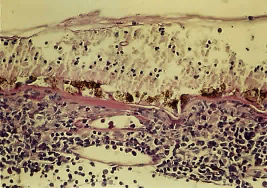
The histopathology of ARN shows profound necrosis of the retina, retinal arteritis, and viral inclusions in retinal cells.1 Examination of an eye with end-stage ARN secondary to VZV revealed diffuse full-thickness, necrotizing retinitis, replacement of sensory retinal structures by glial tissue, occlusive retinal arteritis, granulomatous choroiditis, optic neuritis with ischemic optic atrophy, and VZV in the choroid and the choriocapillaris55 (Fig. 15).

Fig. 15. Light microscopy of an eye with ARN reveals loss of the lamellar architecture of the retina. There is a mixed inflammatory cell infiltrate. A rare multinucleated giant cell is visible.

DIAGNOSTIC EVALUATION AND ANCILLARY TESTS

The diagnosis of ARN is based on a clinical examination and a characteristic funduscopic appearance. Although the signs of ARN may vary in severity, the American Uveitis Society proposed the following clinical criteria for diagnosis of ARN syndrome, regardless of an individual's systemic or immune status: a discrete peripheral necrotizing retinitis that progresses rapidly and circumferentially without antiviral therapy; prominent vitreous and anterior chamber inflammation; and an occlusive vasculitis with arteriolar involvement.6 Other features supporting the diagnosis of ARN include optic neuropathy or atrophy, scleritis, and ocular pain.

Diagnostic vitrectomy or retinal biopsy may be indicated in some atypical cases. However, evaluation of intraocular samples with various techniques, such as viral culture, serology, polymerase chain reaction, and histopathologic examination, do not always yield proof of a viral infection. Intraocular antibodies in the vitreous or aqueous have been measured to determine the cause of necrotizing retinitis.56 In one study, intraocular antibody production to VZV or HSV was detected in 57% of patients with typical ARN syndrome.57 The viral cause of ARN often is difficult to determine in an end-stage atrophic or detached retinal specimens, and may be more readily identified in ocular specimens obtained during acute stages of the disease.58

Systemic laboratory evaluations are suggested to help delineate the specific viral cause of ARN syndrome, to rule out possible associated immunodeficiency, and to guard against potential complications of therapy. Despite the fact that they may not be of benefit in the initial treatment of the patient, acute and convalescent serologic viral titers to HSV type 1, HSV type 2, VZV, and CMV may be obtained for diagnostic confirmation and epidemiologic data. Positive viral titers may prove particularly helpful in guiding therapy for an immunosuppressed patient who may harbor a subclinical, disseminated infection. A diagnostic increase or decrease in herpes viral antibody titers during serial sampling has been noted in only 40% of ARN cases because serum antibody levels may not reflect localized viral reactivation in the eye.59 Evaluation of the ratio of viral antibody levels in paired serum and intraocular fluid samples, which identified the etiologic agent of ARN syndrome in approximately 80% of cases, appears to be more efficacious.59 A complete blood cell count, as well as renal and liver function tests, should be monitored if acyclovir therapy will be used. Antibody testing for HIV should be obtained. If systemic corticosteroids will be used, exposure to tuberculosis should be ruled out with skin testing and chest roentgenography.

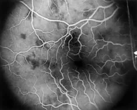
Intravenous fluorescein angiography may be helpful in delineating the extent of infection and elucidating the cause of central visual loss. In the early frames, choroidal perfusion defects may be seen; these defects are caused by areas of focal choroidal inflammatory cell accumulation and overlying retinal pigment epithelial damage (Fig. 16). Such choroidal perfusion defects may occur away from zones of active necrosis.42 Acute obstruction of the central retinal artery or any of its branches may be present. Peripheral views in the areas of active retinitis commonly show little or no intravascular fluorescein in the retinal arteries and veins. Often an abrupt “cut-off” of the intravascular fluorescein may be apparent at the edges of the retinal inflammation (see Fig. 4B and C). Areas of active retinitis show blockage of the underlying choroidal fluorescein pattern (see Fig. 7B). Recirculation phase views may reveal macular leakage, optic disc, and retinal vasculature staining.

Fig. 16. Fluorescein angiogram during early venous phase of active ARN syndrome showing choroidal blocking defects secondary to choroidal involvement.

Ultrasonography is invaluable for detecting retinal detachment when the degree of vitritis precludes examination of the posterior segment. In addition, ultrasound may be used to diagnose enlargement of the optic nerve sheath, which occurs with ARN optic neuropathy.28 Color Doppler imaging has identified hemodynamic compromise in the central retinal artery, suggesting that ischemia plays a role in the pathogenesis of ARN.60

The results of electroretinography have been reported for a limited number of patients. Bright-flash electroretinography tends either to be normal or to show reduced amplitudes, depending on the amount of retina affected.11,30 In a fulminant case of ARN caused by HSV type 1, the electroretinography was completely extinguished early in the disease course.4

Computed tomography scans often show optic nerve sheath enlargement on the side affected with ARN.28 Bilateral optic nerve sheath enlargement has been documented despite the presence of unilateral ARN.3 Magnetic resonance imaging (MRI) has shown concurrent lesions of the optic tracts, chiasm, and lateral geniculate body in a patient with ARN, suggesting that infection may spread through the axons of the ganglion cells.3

TREATMENT

Intravenous acyclovir is the current medical treatment of choice for active ARN. Acyclovir works by selectively inhibiting herpesvirus DNA polymerase. It has antiviral activity against HSV type 1, HSV type 2, and VZV. Most strains of CMV are resistant to acyclovir. The retinitis in ARN typically shows a rapid response to acyclovir therapy, in which progression of retinitis ceases in 3 to 5 days and eventual atrophy of the infected retina occurs. Untreated eyes tend to show regression of the necrotic lesions spontaneously over a period of 6 to 12 weeks. Acyclovir therapy speeds this regression and prevents new lesion formation. In unilateral cases of ARN, acyclovir reduces but does not completely eliminate the risk of fellow eye involvement.61 Initial treatment with a 10 day course of high dose intravenous acyclovir (10 mg/kg every 8 hours) is followed by oral acyclovir (800 mg taken 5 times a day) for up to 14 weeks.61 The time interval for therapy is based on the observation that ARN in the second eye most often occurs within 6 to 14 weeks of the initial symptoms in the first eye. ARN in healthy patients does not generally recur in the same eye after antiviral treatment.27 Long-term oral maintenance therapy may be required in immunosuppressed patients who develop recurrent lesions. Acyclovir has not conclusively been shown to decrease the incidence of subsequent retinal detachment.24 Ganciclovir and foscarnet are alternative intravenous agents that are effective against herpesviruses; however, their use is less favorable because of potential systemic toxicity. New antiviral agents such as famciclovir and valcyclovir are available, but there is no conclusive data regarding their efficacy for treatment of ARN. Intravitreal injections of ganciclovir or foscarnet may be considered to treat individuals limited by systemic drug toxicity.

Anticoagulation to treat the vascular obstructive component of the ARN syndrome has been recommended by some investigators.11,42 Experimental evidence supporting this form of therapy was presented by Ando and associates, who found hyperaggregation of platelets in six of seven patients with bilateral ARN.62 Aspirin and corticosteroids in combination were found to ameliorate this hypercoagulation state. Aspirin may be included in the therapeutic regimen of ARN if there are no systemic contraindications. The efficacy of anticoagulants such as oral warfarin and intravenous heparin to prevent vaso-occlusive phenomena remains unknown; however, a beneficial effect has not been conclusively demonstrated and potentially fatal systemic side effects may occur.

Systemic and topical corticosteroids are advocated to suppress the inflammatory component of ARN and to speed clearing of the vitreous reaction.24 Systemic corticosteroids used alone have not demonstrated any beneficial effect in the early stages of ARN, and because of their immunoinhibitory effects, they should not be administered in active cases of ARN without concurrent acyclovir antiviral therapy.7–10,14,22 Other antiviral medications such as vidarabine have been given to patients with ARN without a clear-cut beneficial effect.10 Cytotoxic agents were used in some early cases based on the similarity between ARN and Behçet's disease; however, it currently is known that ARN is a viral retinitis and that cytotoxic therapy should not be used.8,48 If a patient with ARN syndrome is medically immunosuppressed, the immunosuppression should be reversed unless systemically contraindicated. Topical steroids and cycloplegics are useful in treating associated anterior segment inflammation.

Culbertson and coworkers recommended that confluent laser photocoagulation be applied posterior to the active retinitis in all cases of ARN.14 This treatment creates a “new” ora serrata within the healthy retina in order to localize to the periphery any subsequent retinal detachment. Laser treatment does not stop the progression of retinitis, and therefore repeat treatments may be necessary. Because clear media is required to perform laser treatment successfully, the cases manifesting the greatest vitreous reaction are least likely to be suitable for laser photocoagulation. Sternberg and associates noted retinal detachment in 17% of eyes treated prophylactically with laser compared with 67% of untreated eyes.12 Han and colleagues reported on five eyes with ARN that received laser treatment in which the retina did not subsequently detach.63 Most clinicians currently perform prophylactic laser photocoagulation circumferentially for 360 degrees at the junction of necrotic and healthy retina at the time of diagnosis of ARN if visualization is adequate (Fig. 17).

Fig. 17. Prophylactic laser photocoagulation applied at the margin of healthy and affected retina may delimit a subsequent peripheral retinal detachment.

Of the first 52 cases of ARN reported, 72% progressed to retinal detachment and only 22% of those retinas were successfully reattached.10 In another study, a 50% incidence of ARN-associated retinal detachment was noted and 60% of these retinas were successfully reattached.14 Retinal detachment in eyes with a limited area of necrosis can be treated with scleral buckling, and a high posterior buckle is recommended to relieve vitreous traction on posterior breaks. In many cases, retinal detachments associated with ARN syndrome require pars plana vitrectomy for anatomic repair. This is owing to multiple factors, including media opacity, vitreous traction and fibrosis, and the nature, number, and posterior location of the retinal holes. Temporary or permanent retinal tamponade with long-acting gases or silicone oil often is necessary. Silicone oil tamponade has been shown to be efficacious and safe for repair of retinal detachments in necrotizing retinitis.64 Long-term complications of silicone oil include cataract, corneal opacification, hypotony, glaucoma, and silicone oil emulsification. Despite anatomical success, final visual acuity may be limited by optic atrophy secondary to optic nerve involvement, photoreceptor damage caused by detachment, or retinal cell death from infection. Prophylactic pars plana vitrectomy, scleral buckling, and intravitreal infusion of acyclovir has been reported to treat seven cases of ARN; although none of these patients subsequently developed retinal detachment, there was no control group available for comparison.11,15

Recently, increasing evidence has indicated that some of the acute loss of central vision in ARN is caused by an accompanying optic neuropathy.30 For this reason, Sergott and coworkers performed a modified optic nerve sheath decompression in selected patients with ARN who, after clinical examination and neuroimaging, were believed to have acute ARN optic neuropathy.31 Compared with a small group of concurrent patients who did not receive optic nerve surgery, those undergoing optic nerve sheath decompression were left with better central visual acuity. This work requires confirmation because it was based on an uncontrolled study.

Back to Top
PROGRESSIVE OUTER RETINAL NECROSIS
Progressive outer retinal necrosis syndrome is a visually devastating, acute necrotizing retinitis that occurs in immunocompromised patients. Forster and associates initially identified PORN as a distinct entity and described 2 cases.65 It has been documented to occur in patients with AIDS. PORN affects AIDS patients when their immune function is severely compromised and life expectancy is limited. VZV is the only infectious agent that has been associated with PORN. Ocular features include multifocal, deep retinal lesions that progress rapidly to involve the entire full-thickness retina. The visual prognosis is extremely poor despite antiviral therapy directed at VZV.

CLINICAL PRESENTATION

Although the exact incidence is unknown, PORN syndrome appears to be relatively uncommon. In a 9-year retrospective review of 1007 patients with symptomatic HIV infection, four cases of PORN were diagnosed.66 PORN occurs in the late stages of AIDS and long-term survival after diagnosis generally is less than 1 year. The CD4+ T-lymphocyte count in patients often is less than 50 cells/μl when PORN is diagnosed, with a median CD4+ count of 21 cells/μl in one study.67 Only one patient with PORN has been reported to have a CD4+ count greater than 100 cells/μl.67 PORN syndrome may coexist with other opportunistic retinal infections. Cases of PORN syndrome in one eye and CMV retinitis in the fellow eye have been confirmed by polymerase chain reaction identification of VZV and CMV-DNA from the vitreous.68,69

Although cutaneous VZV is reported in 20% of AIDS patients, previous or active cutaneous VZV infection is noted in up to 67% of patients with PORN.67,70 The infection may be in any dermatome and may be temporally remote from the onset of PORN syndrome. Cutaneous VZV involved the ophthalmic division of the trigeminal nerve in 27% of patients from one study.67 A history of chronic oral acyclovir use may account for the resistance and poor clinical response often observed during acyclovir therapy. PORN syndrome may be associated with an increased risk of developing VZV encephalitis in AIDS patients. Two patients with PORN syndrome and neurologic symptoms were reported to have radiographically demonstrable and histologically proven VZV encephalitis.68

Engstrom and associates evaluated 38 patients with PORN and all patients reported visual symptoms at presentation.67 A decrease or dimming of vision, constriction of visual fields, and floaters were frequently described, but pain was an uncommon feature. The visual symptoms may be disproportionate to the clinical findings early in the course of the disease. Asymptomatic disease often was noted in the contralateral eye in bilateral cases. Initial visual acuity ranged from 20/20 to loss of light perception, with a median acuity of 20/30.67

PORN can be unilateral or bilateral at onset and involvement of the contralateral eye eventually occurs in most patients. Bilateral involvement was noted in over 70% of patients at the time of their final examination.67 In contrast to ARN, there is minimal or no clinically evident inflammation in the anterior segment or vitreous with PORN. Fine white keratic precipitates have been observed in some eyes and posterior synechiae are rarely noted.67

PORN syndrome is a progressive retinal infection with a distinct early, middle, and late stage (Table 2). The early stage is characterized by multifocal, homogeneous, opacified deep retinal lesions (Fig. 18). These lesions lack a granular border, which is a feature of CMV retinal infection. Lesions measure from 50 μm to several thousand microns in diameter and can be located peripheral to the arcades or in the macula at presentation. Early macular lesions are noted in up to 65% of patients, often appearing as central cherry-red spots67 (Fig. 19). Multifocal lesions rapidly progress to confluence and to full-thickness retinal involvement, forming large yellow-white areas of retinal necrosis with minimal retinal hemorrhage (Fig. 20). In eyes with initial peripheral involvement, progression often extends into the macula, and the entire retina may be involved within days. Primary retinal vascular inflammation does not appear to occur, although retinal vasculopathy in the form of sheathing and occlusion may be noted only within or adjacent to areas of retinal necrosis.67,68 A characteristic finding is perivenular lucency within areas of opaque retina, which may represent early clearing of necrotic retina71 (Fig. 21). Optic nerve abnormalities such as edema and hyperemia may occur and an afferent pupillary defect may be present secondary to severe asymmetric retinal necrosis or unilateral optic neuropathy. In end-stage PORN, dense white, plaque-like scarring with a “cracked-mud” appearance or retinal atrophy with peripheral hole formation may develop. Optic atrophy and narrowing of retinal vasculature typically are present.


TABLE 28-2. Stages of Progressive Outer Retinal Necrosis


StageDescription
EarlyOuter retinal lesions: multifocal, opaque, homogeneous
 Minimal or absent viteous and anterior segment inflammation
 ± Macular involvement with cherry-red spot
 ± Papillitis
MiddleConfluence of multifocal lesions
 Full-thickness retinal whitening
 Extremely rapid progression to involve the entire retina over days
 Perivascular clearing of retinal opacification
LatePlaque-like retinal scarring (“cracked-mud”)
 Retinal atrophy with resolution of opacification
 Optic atrophy
 Narrowing of retinal vasculature
 Retinal detachment
Recurrence or progressionDevelopment of new lesions in either eye
 Lesion activity with retinal opacification at the border of an inactive lesion


Fig. 18. Early PORN is characterized by multifocal, homogeneous areas of deep retinal opacification without vitreous inflammation.

Fig. 19. A macular lesion in PORN produces a cherry red spot.

Fig. 20. Equator-plus photograph in PORN syndrome reveals confluence of the retinal lesions and progression into the macula. Vitreous inflammation is absent.

Fig. 21. Perivenular lucency is noted as the retina adjacent to the retinal veins appears to be uninvolved by necrosis.

Despite antiviral therapy, most patients experience involvement of the fellow eye and profound bilateral visual loss to loss of light perception within weeks.67,71,72 Visual loss results from either progressive retinitis involving the macula or retinal detachment. Forty-two of 63 eyes (67%) had no light perception at a median of 4 weeks after diagnosis of PORN.67 There are limited reports of retention of good vision in one eye after treatment for PORN.73–75

A successful clinical response to treatment includes decreased retinal opacification, cessation of confluence of multifocal lesions, and no further development of new lesions. Cessation of disease spread usually occurs after 3 days of therapy and decrease in opacification may require as many as 14 days.76 Long-term antiviral treatment is required to prevent recurrences of PORN syndrome. Disease progression or recurrence is characterized by the development of new lesions in either eye or lesion activity identified by retinal opacification at the border of previously inactive lesions. Johnston and associates hypothesized that reactivation at the border of a lesion was related to persistent virus in the retina.72 Engstrom and colleagues reported that new lesions in the contralateral unaffected eye were common and that border reactivation was uncommon in their studies; they speculated that recurrence may result from episodes of viremia rather than persistent virus in the retina.67

Despite treatment, rhegmatogenous retinal detachment has been reported in approximately 70% of eyes with PORN at a median of 4 weeks after diagnosis.67 In early studies, there was no difference in the proportion of retinal detachments between eyes grouped by extent of disease at diagnosis and disease activity after initial treatment.67 Tractional retinal detachments secondary to intraocular inflammation may occur in ARN but are not a feature of PORN syndrome.

ETIOLOGY AND HISTOPATHOLOGY

Varicella-zoster virus has been implicated as the causative agent of PORN. This is based on a history of previous or active cutaneous zoster infection in many patients and laboratory identification of VZV in some chorioretinal biopsy specimens.65,71,77 VZV also has been identified in some cases of ARN. While both PORN and ARN can be produced by VZV and have been described in individuals with HIV infection, they are clinically distinct syndromes. The reason for a differential presentation of VZV in PORN and ARN is not clear, although it may represent a differential host immunologic response to the same etiologic agent or may be caused by different strains of VZV. The degree of immunosuppression varies widely in AIDS, and with progressive immune dysfunction the clinical characteristics of VZV infection may change from the ARN syndrome to the hyperacute fulminant retinitis seen in AIDS patients with PORN. Patients who develop PORN have severely impaired systemic immunity, which is demonstrated by a very low CD4+ T-lymphocyte count that is usually less than 50 cells/μl. These patients may not be able to produce an inflammatory response, so VZV infection progresses without intraocular inflammation. In contrast, HIV-infected patients with higher CD4+ counts are able to respond with intraocular inflammation when exposed to VZV and may develop typical ARN.

Although PORN syndrome was defined in 1990, it may have been diagnosed previously as atypical ARN or CMV infection. It may be recognized with increasing frequency because of the improved survival rate of immunocompromised patients with AIDS. Forster initially identified intraretinal herpesvirus particles by electron microscopy and polymerase chain reaction in 2 patients with PORN.65 Margolis and associates provided additional evidence that PORN was a form of VZV retinopathy, and VZV has been identified in retina biopsy specimens with immunohistochemistry, electon microscopy, and polymerase chain reaction.71,75,78 Evaluation of eyes with PORN for HSV, CMV, Pneumocystis carinii, and other infectious agents has been negative.68

Histopathology varies with the stage of PORN. While early PORN appears to involve the outer retina clinically, the inner retina is involved on pathologic examination. Retinal biopsy of an eye with outer retina involvement revealed full thickness involvement in areas of retinitis with alteration and necrosis of cellular elements in all retinal layers.75 Sparse neutrophils were present in the outer retina, but not in the inner retina. Lymphoid cells were present primarily in the inner retina. VZV antigen was more heavily concentrated in the RPE and outer retina than in the inner retina in a specimen with early PORN. This may account for the clinical impression of outer retinal involvement.68 An established full-thickness lesion on clinical examination demonstrates complete necrosis of the retina with a variable chronic inflammatory response in the retina and choroid. The degree of choroidal inflammation does not appear to be related to the retinal inflammation.71 Intranuclear inclusions have been noted in choroidal cells.78 Necrosis and lymphocytic infiltration have been observed in the optic nerve.71 An eye enucleated with end-stage PORN and with absent light perception revealed advanced necrosis of the RPE and almost complete dissolution of the neurosensory retina with minimal chronic inflammatory cells in the choroid.77

DIAGNOSTIC EVALUATION AND ANCILLARY TESTS

The diagnosis of PORN is based on clinical appearance and a rapidly progressive course, and is supported by a history of marked systemic immunosuppression, HIV infection, prior VZV infection, or chronic acyclovir use. Chorioretinal biopsy to identify VZV may be indicated if the retinitis is atypical, although most specimens are obtained at the time of retinal detachment repair to confirm the diagnosis. VZV has not been demonstrated in all cases of PORN because necrotic tissue specimens may no longer contain viral particles and laboratory isolation of VZV may be difficult.67 VZV-DNA and VZV antigen have been identified in vitreous biopsy specimens, but VZV has not been cultured from the vitreous.69,71 Blood culture was positive for VZV in one patient with end-stage PORN.71

Fluorescein angiographic features vary depending on the stage of PORN, but demonstrate involvement of the choroid, RPE, and multiple layers of the retina. In the early stages, peripheral retinal microvascular alterations are noted within and extending beyond deep retinal lesions.79 Retinal leakage is present in large areas of retinal whitening. With progression of PORN, pruning of the retinal vasculature—as well as capillary loss, RPE destruction, and choiroidal leakage—occurs. Disease reactivation at the border of normal retina is noted by a prominent brush-fire pattern of fluorescein leakage involving the retina, RPE, and choroid.

TREATMENT

The optimal regimen for treatment of PORN and prevention of recurrences is unknown. PORN produces rapid destruction of the retina with severe and often bilateral visual loss, which can progress or recur despite antiviral therapy. Aggressive therapy of the progressive retinitis and any associated retinal detachments is vital to preserve vision. In early studies, an initial response to acyclovir therapy appeared to delay visual loss, but this was not associated with a better visual outcome at final examination.67 Acyclovir is the drug of choice for treatment of VZV infection at other sites in immunocompromised patients. However, a poor response has been observed with intravenous acyclovir therapy alone for PORN. Previous chronic oral acyclovir use in many of these patients may contribute to the development of VZV-acyclovir resistance. Decreased systemic immunity may preclude inhibition of VZV infection. In the murine model of herpes retinitis, animals depleted of CD4+ cells had minimal intra-ocular inflammation and poor clearance of the herpes virus.80 Spaide and associates noted that the CD8+ T lymphocyte was the predominant retinal cell infiltrate in PORN and hypothesized that a poor response to treatment may be related to a relative deficiency of CD4+ cells.75 Retinal ischemia secondary to retinopathy may prevent achievement of therapeutic drug levels in the retina after systemic administration. This factor may be overcome by increasing the systemic dose or by using local intra-ocular therapy. An improved therapeutic response has been noted by using an increased dose of systemic acyclovir (10 to 20 mg/kg every 8 hours).67,69

Other antivirals agents, such as ganciclovir and foscarnet, have activity against VZV and have been used to treat PORN. Acyclovir-resistant VZV may be sensitive to foscarnet because of its different mechanism of action. High-dose foscarnet (60 mg/kg every 8 hours) has been recommended by some authors for primary treatment.76 The combination of ganciclovir and foscarnet may have additive or synergistic effects.75 Visual acuity of 20/100 or better was retained in at least one eye in six patients for the remainder of their lives with a combination of antiviral therapy (systemic foscarnet and either acyclovir or ganciclovir) and vitreoretinal surgical repair of retinal detachment as required.75 High doses of acyclovir (10 to 20 mg/kg every 8 hours) or ganciclovir (5 mg/kg every 12 hours) combined with foscarnet (60 mg/kg every 8 hours) have been recommended for initial therapy of PORN.76 Combination antiviral therapy appears to be effective, although no clinical trials have compared the efficacy of a single antiviral agent with combination therapy.66,67,69 Initial treatment is continued until existing lesions are inactive and no new lesions appear. Other antiviral agents effective against VZV, including famciclovir, cidofovir, and sorivudine, have yet to be fully evaluated for treatment of PORN.74,81

Chronic antiviral therapy, similar to the management of other ocular opportunistic infections such as CMV and toxoplasmosis in AIDS patients, appears to be necessary to prevent recurrences of PORN. Potential mechanisms for disease recurrence after initial treatment may be related to the development of viral resistance, inadequate drug dose to suppress viral replication, or decreased patient immune function.76 The median length of time from stabilization of PORN to recurrence was 51 days, with a range of 14 to 90 days.72 In one study, recurrence was often associated with a discontinuation or reduction in the amount of antiviral medications or a switch from intravenous to oral acyclovir.72 The low bioavailability of oral acyclovir probably results in too low a drug level to prevent disease recurrence. Lifelong maintenance intravenous foscarnet therapy (90 to 120 mg/kg daily) or a combination of systemic antivirals has been recommended by some authors.76 This is in contrast to the treatment of ARN in immunocompetent patients, who usually respond well to antiviral therapy and do not require long-term maintenance therapy.

Intravitreal injections of ganciclovir or foscarnet provide immediate and local high drug concentrations and have been used with some success. Injection of intravitreal acyclovir (0.2 mg/0.1 ml) has been used with equivocal results.69 Disadvantages include the potential risk of intraocular infection and the need for multiple weekly intravitreal injections to maintain therapeutic drug levels. The sustained-release ganciclovir intraocular implant was used to treat one patient with PORN, although simultaneous intravenous foscarnet was required for control.75 A combination of intravitreal and intravenous antivirals is an alternative in patients limited by systemic drug toxicity.

Systemic corticosteroids are not used to treat PORN because of the paucity of intraocular inflammation and the risk of potentiating systemic infections in immunocompromised patients. Anticoagulation is not recommended in PORN because occlusion vasculopathy is not a feature.

Treatment of retinal detachment in PORN syndrome is similar to that used in other infectious necrotizing retinopathies, such as CMV or ARN syndrome, that produce multiple atrophic holes in necrotic retina. Retinal detachment developed in 93% of eyes with PORN that received prophylactic laser photocoagulation around areas of necrotic retina.67 Contraction of detached retina may produce traction on areas in which previous laser photocoagulation has been performed and lead to extension of the retinal detachment.75 An unusual case of spontaneous regression of PORN has been described after prophylactic laser photocoagulation was used to surround the posterior pole.82 The ultimate repair of a retinal detachment in PORN usually requires pars plana vitrectomy and silicone oil tamponade in an effort to preserve some vision. Despite anatomical repair, vision may be severely impaired. Vision was described as absent light perception in 5 of 12 eyes (42%) after successful retinal reattachment surgery.67

DIFFERENTIAL DIAGNOSIS

The differential diagnoses of the ARN and PORN syndromes are listed in Table 3. ARN and PORN syndromes are distinguished by an individual's immune status and the intraocular inflammatory response to the virus. The ocular features of ARN include a peripheral retinitis, periarteritis, and vitritis. PORN is distinguished from ARN by initial outer retinal whitening, early involvement of the posterior pole, and the absence of intraocular inflammation and vasculitis. Only patients who are markedly immunosuppressed, such as those with AIDS, develop PORN. ARN has been described in individuals with relatively intact systemic immunity, regardless of their HIV status.


TABLE 28-3. Major Differential Diagnoses of the ARN and PORN Syndromes


FeaturesARN SyndromePORN SyndromeCytomegalovirusDiffuse Toxoplasmosis
Typical location of retinal lesionsPeripheral retinaPeripheral retina and/or maculaAnywhereAnywhere
Initial ophthalmoscopic appearanceGray-white, opaque; one or more areas of full-thickness retinal necrosis; discrete bordersMultifocal lesions; deep retinal opacification; no granularity; perivenular clearning of opacificationWhite; granular; “crumbled cheese”White
Hemorrhagic component+ +-+ + + ++ /-
Vitreous reaction+ + + ++ /-++ + + + *
Arteritis+ + + +-+ ++ + +
ProgressionRapid circumferential spread around retinal periphery and geographic areas simultaneouslyExtremely rapid progression to involve the entire retinaAdvancing edge with “burned out” areasSpread to geographic areas simultaneously
Late retinal detachment50%–75%>70%29%Uncommon
Systemic immunityTypically normal†ImpairedImpairedNormal or impaired

*Especially directly overlying lesions.
†ARN syndrome in immunocompromised hosts has been reported.


The other herpesviruses should be considered among the differential diagnoses. Although some early reports suggested that CMV may produce ARN syndrome, this has not been demonstrated in most studies.29 CMV retinitis occurs in immunosuppressed patients. CMV lesions are often granular and hemorrhagic and feature a slower clinical course than either ARN or PORN. The marked vitritis described in ARN is not a usual feature of CMV retinitis. PORN is distinguished from CMV retinitis by the deep, multifocal, retinal opacification; a relative absence of hemorrhage; and a very rapid clinical progression. The differentiation of CMV retinitis is therapeutically relevant because CMV does not usually respond well to acyclovir, although other antivirals, such as ganciclovir, effectively control CMV infection. Late retinal detachment also has been reported in CMV retinitis. Tiedeman studied 10 patients with a diffuse, unilateral, or bilateral panuveitis associated with choroiditis that, evidence suggests, was secondary to EBV.83 In contrast to ARN and PORN, large areas of confluent retinal necrosis did not occur.

Other types of posterior uveitis may be confused with ARN or PORN syndromes. Behçet's disease shares many similarities with ARN, including a diffuse uveitis, prominent retinal arteritis, and patches of retinal whitening.8,9 The presence or absence of associated systemic findings, such as aphthous and genital ulcers, rash, and arthritis should differentiate the two entities. Uveitis and vascular sheathing may occur with ocular sarcoidosis; however, arteritis and broad patches of retinal whitening are atypical. Again, systemic evaluation is invaluable in separating sarcoidosis from ARN. Ocular toxoplasmosis presenting as a focal retinitis with overlying vitritis may be confused with ARN. Serum or ocular toxoplasma titers or polymerase chain reaction may be helpful for differentiation, but in some instances, especially in the case of an immunocompromised patient, careful observation with or without a therapeutic trial may be indicated.

Large cell lymphoma (formerly called reticulum cell sarcoma) with vitreous cells and white retinal lesions may masquerade as ARN. The course of large cell lymphoma tends to be more chronic and lacks the acute inflammatory signs of ARN.10 Adult T-cell lymphoma associated with human T-lymphotropic virus type I retroviral infection may present as severe uniocular intraocular inflammation, retinitis, and vasculitis simulating ARN syndrome.84 In children, the diffuse form of retinoblastoma may be confused with ARN. In young patients with diffuse uveitis, pars planitis or toxocariasis also should be considered.10,42 Acute ophthalmic artery obstruction, ocular ischemic syndrome, and commotio retinae (Berlin's edema) may demonstrate diffuse retinal whitening and should be considered among the differential diagnoses.10,42

Back to Top
REFERENCES

1. Culbertson WW, Blumenkranz MS, Haines H et al: The acute retinal necrosis syndrome. Part 2: Histopathology and etiology. Ophthalmology 89:1317, 1982

2. Culbertson WW, Blumenkranz MS, Pepose JS et al: Varicella zoster virus is a cause of the acute retinal necrosis syndrome. Ophthalmology 93:559, 1986

3. Lewis ML, Culbertson WW, Post JD et al: Herpes simplex virus type 1: A cause of the acute retinal necrosis syndrome. Ophthalmology 96:875, 1989

4. Duker JS, Nielsen JC, Eagle RC Jr et al: Rapidly progressive acute retinal necrosis secondary to herpes simplex virus type 1. Ophthalmology 97:1638, 1990

5. Cunningham ET Jr, Short GA, Irvine AR et al: Acquired immunodeficiency syndrome-associated herpes simplex virus retinitis: Clinical description and use of a polymerase chain reaction-based assay as a diagnostic tool. Arch Ophthalmol 114:834, 1996

6. Holland GN, Executive Committee of the American Uveitis Society: Standard diagnostic criteria for the acute retinal necrosis syndrome. Am J Ophthalmol 117:663, 1994

7. Urayama A, Yamada N, Sasaki T et al: Unilateral acute uveitis with periarteritis and detachment. Jpn J Clin Ophthalmol 25:607, 1971

8. Willerson D Jr, Aaberg TM, Reeser FH: Necrotizing vaso-occlusive retinitis. Am J Ophthalmol 84:209, 1977

9. Young NJ, Bird AC: Bilateral acute retinal necrosis. Br J Ophthalmol 62:581, 1978

10. Fisher JP, Lewis ML, Blumenkranz M et al: The acute retinal necrosis syndrome. Part 1: Clinical manifestations. Ophthalmology 89:1309, 1982

11. Carney MD, Peyman GA, Goldberg MF et al: Acute retinal necrosis. Retina 6:85, 1986 [published erratum appears in Retina 1986 Summer-Fall;6:168]

12. Sternberg P Jr, Han DP, Yeo JH et al: Photocoagulation to prevent retinal detachment in acute retinal necrosis. Ophthalmology 95:1389, 1988

13. Thompson WS, Culbertson WW, Smiddy WE et al: Acute retinal necrosis caused by reactivation of herpes simplex virus type 2. Am J Ophthalmol 118:205, 1994

14. Clarkson JG, Blumenkranz MS, Culbertson WW et al: Retinal detachment following the acute retinal necrosis syndrome. Ophthalmology 91:1665, 1984

15. Peyman GA, Goldberg MF, Uninsky E et al: Vitrectomy and intravitreal antiviral drug therapy in acute retinal necrosis syndrome: Report of two cases. Arch of Ophthalmol 102:1618, 1984

16. Kometani J, Asayama T: A case of specific uveitis occurring acutely in the right eye. Folia Ophthalmol Jpn 29:1397, 1978

17. Saari KM, Boke W, Manthey KF et al: Bilateral acute retinal necrosis. Am J Ophthalmol 93:403, 1982

18. Chambers RB, Derick RJ, Davidorf FH et al: Varicella-zoster retinitis in human immunodeficiency virus infection: Case report. Arch Ophthalmol 107:960, 1989

19. Litoff D, Catalano RJ: Herpes zoster optic neuritis in human immunodeficiency virus infection. Arch Ophthalmol 108:782, 1990

20. Sado K, Kimura T, Hotta Y et al: Acute retinal necrosis syndrome associated with herpes simplex keratitis. Retina 14:260, 1994

21. Solomon MJ: Unilateral acute retinal necrosis syndrome with herpes simplex keratitis. Retina 15:359, 1995

22. Okinami S, Tsukahara I: Acute severe uveitis with retinal vasculitis and retinal detachment. Ophthalmologica 179:276, 1979

23. Culbertson WW, Clarkson JG, Blumenkranz M, Lewis ML: Acute retinal necrosis. Am J Ophthalmol 96:683, 1983

24. Blumenkranz MS, Culbertson WW, Clarkson JG et al: Treatment of the acute retinal necrosis syndrome with intravenous acyclovir. Ophthalmology 93:296, 1986

25. Hayreh MMS, Kreiger AE, Straatsma BR et al: Acute retinal necrosis. Invest Ophthalmol Vis Sci 19(Suppl):48, 1980

26. Wang CL, Kaplan HJ, Waldrep JC, Pulliam M: Retinal neovascularization associated with acute retinal necrosis. Retina 3:249, 1983

27. Altamirano D, Rochat C, Claeys M et al: Acute retinal necrosis: A result of immune dysfunction? Report of a case with subacute evolution and relapses in a patient with impaired cellular immunity. Ophthalmologica 208:49, 1994

28. Sergott RC, Belmont JB, Savino PJ et al: Optic nerve involvement in the acute retinal necrosis syndrome. Arch Ophthalmol 103:1160, 1985

29. Rungger-Brandle E, Roux L, Leuenberger PM: Bilateral acute retinal necrosis (BARN): Identification of the presumed infectious agent. Ophthalmology 91:1648, 1984

30. Matsuo T, Date S, Tsuji T et al: Immune complex containing herpesvirus antigen in a patient with acute retinal necrosis. Am J Ophthalmol 101:368, 1986

31. Sergott RC, Anand R, Belmont JB et al: Acute retinal necrosis neuropathy: Clinical profile and surgical therapy. Arch Ophthalmol 107:692, 1989

32. Falcone PM, Brockhurst RJ: Delayed onset of bilateral acute retinal necrosis syndrome: A 34-year interval. Ann Ophthalmol 25:373, 1993

33. Matsuo T, Nakayama T, Koyama T et al: A proposed mild type of acute retinal necrosis syndrome. Am J Ophthalmol 1988; 105:579

34. Crapotta JA, Freeman WR, Feldman RM et al: Visual outcome in acute retinal necrosis. Retina 13:208, 1993

35. Culbertson WW, Brod RD, Flynn Jr HW et al: Chickenpox-associated acute retinal necrosis syndrome. Ophthalmology 98:1641, 1991

36. Sternberg P Jr, Knox DL, Finkelstein D et al: Acute retinal necrosis syndrome. Retina 2:145, 1982

37. Severin M, Neubauer H: Bilateral acute retinal necrosis. Ophthalmologica 182:199, 1981

38. Minckler DS, McLean EB, Shaw CM et al: Herpes hominis encephalitis and retinitis. Arch Ophthalmol 94:89, 1976

39. Bloom JN, Katz JI, Kaufman HE: Herpes simplex retinitis and encephalitis in an adult. Arch Ophthalmol 95:1798, 1977

40. Gorman BD, Nadel AJ, Coles RS: Acute retinal necrosis. Ophthalmology 89:809, 1982

41. Ludwig IH, Zegarra H, Zakov ZN: The acute retinal necrosis syndrome: Possible herpes simplex retinitis. Ophthalmology 91:1659, 1984

42. Gass JDM: Stereoscopic Atlas of Macular Disease, p 490. Vol 2. St. Louis: CV Mosby, 1987

43. Matsuo T, Koyama M, Matsuo N: Acute retinal necrosis as a novel complication of chickenpox in adults. Br J Ophthalmol 74:443, 1990

44. Friberg TR, Jost BF: Acute retinal necrosis in an immunosuppressed patient. Am J Ophthalmol 98:515, 1984

45. Chess J, Marcus DM: Zoster-related bilateral acute retinal necrosis syndrome as presenting sign in AIDS. Ann Ophthalmol 20:431, 1988

46. Murray PI: Wegener's granulomatosis, pituitary adenoma and BARN. Int Ophthalmol 18:361, 1994

47. Sellitti TP, Huang AJ, Schiffman J et al: Association of herpes zoster ophthalmicus with acquired immunodeficiency syndrome and acute retinal necrosis. Am J Ophthalmol 116:297, 1993

48. Price FW Jr, Schlaegel TF Jr: Bilateral acute retinal necrosis. Am J Ophthalmol 89:419, 1980

49. Hayasaka S, Asano T, Yabata K et al: Acute retinal necrosis. Br J Ophthalmol 67:455, 1983

50. Pepose JS, Stewart JA, Grose C et al: ARN: Serologic studies and further characterization of the virus. Invest Ophthalmol Vis Sci 25 (Suppl):12, 1986

51. Soushi S, Ozawa H, Matsuhashi M et al: Demonstration of varicella-zoster virus antigens in the vitreous aspirates of patients with acute retinal necrosis syndrome. Ophthalmology 95:1394, 1988

52. Sarkies N, Gregor Z, Forsey T et al: Antibodies to herpes simplex virus type I in intraocular fluids of patients with acute retinal necrosis. Br J Ophthalmol 70:81, 1986

53. Margolis T, Irvine AR, Hoyt WF et al: Acute retinal necrosis syndrome presenting with papillitis and arcuate neuroretinitis. Ophthalmology 95:937, 1988

54. Holland GN, Cornell PJ, Park MS et al: An association between acute retinal necrosis syndrome and HLA-DQw7 and phenotype Bw62,DR4. Am J Ophthalmol. 108:370, 1989

55. Rummelt V, Wenkel H, Rummelt C et al: Detection of varicella zoster virus DNA and viral antigen in the late stage of bilateral acute retinal necrosis syndrome. Arch Ophthalmol 110:1132, 1992

56. Davis JL, Feuer W, Culbertson WW et al: Interpretation of intraocular and serum antibody levels in necrotizing retinitis. Retina 15:233, 1995

57. de Boer JH, Luyendijk L, Rothova A et al: Detection of intraocular antibody production to herpesviruses in acute retinal necrosis syndrome. Am J Ophthalmol 117:201, 1994

58. Freeman WR, Thomas EL, Rao NA et al: Demonstration of herpes group virus in acute retinal necrosis syndrome. Am J Ophthalmol 102:701, 1986

59. Pepose JS, Flowers B, Stewart JA et al: Herpesvirus antibody levels in the etiologic diagnosis of the acute retinal necrosis syndrome. Am J Ophthalmol 113:248, 1992

60. Regillo CD, Sergott RC, Ho AC et al: Hemodynamic alterations in the acute retinal necrosis syndrome. Ophthalmology 100:1171, 1993

61. Palay DA, Sternberg PJ, Davis J et al: Decrease in the risk of bilateral acute retinal necrosis by acyclovir therapy. Am J Ophthalmology 112:250, 1991

62. Ando F, Kato M, Goto S et al: Platelet function in bilateral acute retinal necrosis. Am J Ophthalmol 96:27, 1983

63. Han DP, Lewis H, Williams GA et al: Laser photocoagulation in the acute retinal necrosis syndrome. Arch Ophthalmol 105:1051, 1987

64. Davis JL, Serfass MS, Lai MY et al: Silicone oil in repair of retinal detachments caused by necrotizing retinitis in HIV infection. Arch Ophthalmol 113:1401, 1995

65. Forster DJ, Dugel PU, Frangieh GT et al: Rapidly progressive outer retinal necrosis in the acquired immunodeficiency sydrome. Am J Ophthalmol 110:341, 1990

66. Jabs DA: Ocular manifestations of HIV infection. Trans Am Ophthalmol Soc 93:623, 1995

67. Engstrom RE Jr, Holland GN, Margolis TP et al: The progressive outer retinal necrosis syndrome: A variant of necrotizing herpetic retinopathy in patients with AIDS. Ophthalmology 101:1488, 1994

68. Kuppermann BD, Quiceno JI, Wiley C et al: Clinical and histopathologic study of varicella zoster virus retinitis in patients with the acquired immunodeficiency syndrome. Am J Ophthalmol 118:589, 1994

69. Pavesio CE, Mitchell SM, Barton K et al: Progressive outer retinal necrosis (PORN) in AIDS patients: A different appearance of varicella-zoster retinitis. Eye 9:271, 1995

70. Jansen DJ, Saylin AJ, Cannon L et al: Neurological and neuropsychological manifestations of AIDS. Ann Neurol 26:592, 1989

71. Margolis TP, Lowder CY, Holland GN et al: Varicella-zoster virus retinitis in patients with the acquired imunodeficiency syndrome. Am J Ophthalmol 112:119, 1991

72. Johnston WH, Holland GN, Engstron REJ Jr et al: Recurrence of presumed varicella-zoster virus retinopathy in patients with the acquired imunodeficiency syndrome. Am J Ophthalmol 116:42, 1993

73. Morley MG, Duker JS, Zacks C: Successful treatment of rapidly progressive outer retinal necrosis in the acquired immunodeficiency syndrome. Am J Ophthalmol 117:264, 1994

74. Pinnolis MK, Foxworthy D, Kemp B: Treatment of progressive outer retinal necrosis with sorivudine. Am J Ophthalmol 119:516, 1995

75. Spaide RF, Martin DF, Teich SA et al: Successful treatment of progressive outer retinal necrosis syndrome. Retina 16:479, 1996

76. Holland GN: The progressive outer retinal necrosis syndrome. Int Ophthalmol 18:163, 1994

77. Friedman SM, Margo CE, Connelly BL: Varicella-zoster virus retinitis as the initial manifestation of the acquired imunodeficiency syndrome. Am J Ophthalmol: 117:536, 1994

78. Greven CM, Ford J, Stanton C et al: Progressive outer retinal necrosis secondary to varicella zoster virus in acquired immune deficiency syndrome. Retina 15:14, 1995

79. Walton RC, Byrnes GA, Chan CC et al: Fluorescein angiography in the progressive outer retinal necrosis syndrome. Retina 16:393, 1996

80. Azumi A, Cousins SW, Kanter MY et al: Modulation of murine herpes simplex virus type 1 retinitis in the uninoculated eye by CD4+ T lymphocytes. Invest Ophthalmol Vis Sci 35:54, 1994

81. Flores-Aguilar M, Huang JS, Wiley CA et al: Long-acting therapy of viral retinitis with (S)-1-(3-hydroxy-2-phosphonylmethoxypropyl) cytosine. J Infect Dis 169:642, 1994

82. Cooper HM, Beer PM: Spontaneous regression and successful laser prophylaxis in progressive outer retinal necrosis syndrome. Am J Ophthalmol 121:723, 1996

83. Tiedeman JS: Epstein-Barr viral antibodies in multifocal choroiditis and panuveitis. Am J Ophthalmol 103:659, 1987

84. Kohno T, Uchida H, Inomata H et al: Ocular manifestations of adult T-cell leukemia/lymphoma: A clinicopathologic study. Ophthalmology 100:1794, 1993

Back to Top