|
|
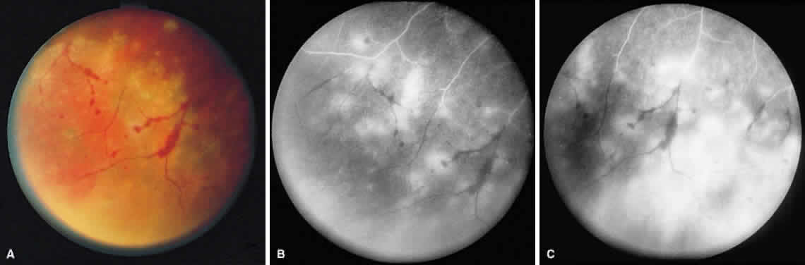
| Fig. 4. A. With progression of ARN, the areas of retinal whitening begin to coalesce. Mild perivascular retinal hemorrhages are noted. B. Fluorescein angiography in the venous phase reveals retinal vascular nonperfusion in areas of active retinitis. C. In the late-phase angiogram, fluorescein hyperfluorescence is seen in areas of retinitis and around the retinal vasculature. |