Chapter 25
Gyrate Atrophy
STEVE A. ARSHINOFF, KEVIN LEUNG and YI NING J. STRUBE
Main Menu   Table Of Contents

Search

HISTORY
DIAGNOSIS
THE METABOLISM OF GYRATE ATROPHY
GENETICS OF GYRATE ATROPHY
TREATMENT OF GYRATE ATROPHY
REFERENCES

HISTORY
Gyrate atrophy (GA) (MIM 258870) of the choroid and retina is a rare autosomal recessive disorder characterized by progressive, metabolic, retinal, and choroidal degeneration due to deficiency of the pyridoxal phosphate (PLP)–dependent, nuclear-encoded, mitochondrial matrix enzyme ornithine delta(δ)-aminotransferase (OAT; L-ornithine:2-oxoacid aminotransferase; EC 2.6.1.13), which has been mapped to chromosome 10q26.1,2 GA was initially described as an atypical type of retinitis pigmentosa by Jacobsohn in 18883 and by Cutler in 1895.4 The disease was given its current name by Fuchs in 1896.5 The extreme rarity of this disorder is evident from the fact that when Usher6 reviewed all previously known cases in 1935 and correctly concluded that the inheritance pattern was autosomal recessive, he could find reports of only 26 cases. When Kurstjens7 did his extensive review in 1965 and characterized many of the typical ophthalmic findings, he could find only 44 cases in the world literature. A careful review at the present time still reveals just over 150 biochemically documented cases, about one-third of which are from Finland and only seven (less than 5%) of which have been responsive to therapy with vitamin B6 dietary supplementation.8

Simell and Takki's discovery of associated hyperornithinemia, the primary biochemical manifestation of OAT deficiency, in 1973 caused an explosion of interest in this disease, making it definable metabolically as well as ophthalmologically.9 Since then, GA has been transformed from an obscure curiosity to the prototype of potentially treatable metabolic retinal degenerations. It was also the first primary retinal degeneration to be recognized as an inborn error of metabolism, as defined by Garrod in 1908.10 Readers interested in metabolic details of GA beyond this chapter are referred to the chapter by Valle and Simell in the eighth edition of The Metabolic and Molecular Bases of Inherited Disease.8

Back to Top
DIAGNOSIS
The diagnosis of GA can easily be made with certainty if the five most prominent features can be recalled.11

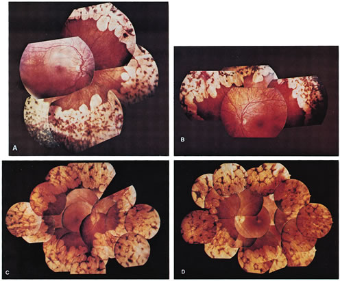
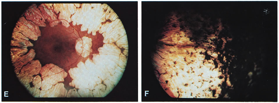
  1. Typical gyrate retinal and choroidal lesions (Fig. 1): The appearance of confluent arcuate equatorial full-thickness lesions of the choroid and retina, sparing some of the larger choroidal vessels and separated from one another by thin margins of pigment, is characteristic of GA. The lesions extend without interruption to the ora serrata, where a dense aggregation of pigment can be seen, but they invade the circummacular vascular arcade only in advanced cases. Peripapillary gyrate lesions are common, but foveal lesions are rare until late in the course of the disease.
  2. Early cataract: Posterior subcapsular lens changes usually begin in the late teens, and fully developed posterior subcapsular cataracts with diffuse cortical opacities are almost invariably present by age 30.
  3. High myopia with marked astigmatism: Myopia of –6 to –10 diopters (D) or more, accompanied by 2 D or more of astigmatism is present in about 90% of cases so far reported. The occasional case may be only mildly myopic and astigmatic.
  4. Hyperornithinemia: Extreme hyperornithinemia is universal in this disease. Its absence necessitates a search for another diagnosis. All body fluids measured to date (whole blood, plasma, cerebrospinal fluid, aqueous humor, and urine) have been found to contain 10 to 20 times the normal levels of ornithine.9,11
  5. Autosomal recessive inheritance pattern: To date, all patients with the oculometabolic disease have family trees and laboratory genetic findings consistent with autosomal recessive inheritance. It is possible to diagnose heterozygotes and to confirm the inheritance pattern in a family with a single affected member (see below).12

Fig. 1. Representative fundus photographs of patients with gyrate atrophy (GA). A and B. The right and left fundi, respectively, of a 9-year-old white female patient with relatively early lesions. There is total sparing of the retina within the circummacular vascular arcade and a lack of pigment between the gyrate lesions (corrected visual acuity, 20/30 RE, 20/25 LE). C and D. The appearance of the right and left fundi, respectively, of a 16-year-old white male patient. The lesions extend considerably farther posteriorly than in the younger patient in A and B. Also, in the left eye, there is increased pigmentation between the lesions, as well as a foveal lesion and peripapillary lesions (corrected visual acuity, 20/100 RE, 20/80 LE). E. The posterior pole in the right eye of a 31-year-old white female patient with advanced GA. The gyrate lesions extend within the circummacular vascular arcade; a large peripapillary lesion is present. F. The far periphery of the eye shown in E. There is extensive accumulation of pigment, typical of advanced GA.

OPHTHALMIC FINDINGS

A detailed ophthalmic examination of a patient with GA usually reveals the following findings:

  Visual acuity: Acuity in most cases is normal with correction, up until about age 10. There appears to be a gradual decline in visual acuity thereafter, unrelated to the development of cataracts. The slowly progressive irreversible loss of vision from retinal degeneration often leads to blindness, usually by the fourth or fifth decade of life.13 The appearance of a large peripapillary gyrate lesion is often accompanied by a decline in central vision. Rarely, a patient may develop a foveal gyrate lesion during the early stages of the disease, but these lesions are often present in advanced cases. Occasionally, acuity is inexplicably decreased in one eye, when compared with its fellow, in the same patient. Variation in the severity of visual loss among patients from different families is considerable and may be due to the tremendous variety in the specific mutations in the GA gene found in different families (see metabolic discussion below; e.g., pyridoxine-responsive mutations tend to be less severe).
  Pupils: Pupillary appearance and reactivity to light and near are generally normal. Patients with marked asymmetry in acuity, visual fields, and other parameters may have an afferent pupillary defect in the worse eye.
  Motility: Extraocular muscle balance is usually normal, except for a moderate exophoria not unlike that seen in other myopes. Patients with significantly worse vision in one eye may have manifest exotropia.
  Stereopsis: Stereopsis is normal as long as the visual acuity remains symmetric. A decrease in stereopsis may be an early sign of developing exotropia and decline in the visual acuity of one eye.
  Color vision: Decreased color discrimination occurs when there is a drop in visual acuity on the basis of retinal lesions, or with the development of cataracts. Otherwise, there are no remarkable changes in color vision, as the central color sensitive cones appear to be preserved until late in the disease.14
  Visual fields: Visual fields coincide fairly well with the remaining area of healthy-appearing retina (i.e., about 15 to 50 degrees), with an enlarged blind spot corresponding to the peripapillary lesion, if present. Using fundus photoperimetry, Enoch and associates15 found that the limit of the functional visual field typically conforms to the observed gyrate lesions with a sharp drop-off in visual function at the border of the lesions. However, the functional visual field may occasionally be considerably smaller than the central area of the retina that appears to be healthy on ophthalmoscopy, and this is often associated with a large peripapillary lesion.11
  Lens: Posterior subcapsular cataracts develop in the late teens or early twenties and progress to include extensive cortical changes primarily involving opacification of the lens along the suture lines, extending toward the equator of the lens.16 Anterior subcapsular fibrous plaques have been reported.17
  Ciliary body: Abnormally short and scant ciliary processes have been reported from cycloscopic examination of these patients.18
  Vitreous: The vitreous is typically syneretic and frequently contains clusters of cloudy fibrils. Recurrent vitreous hemorrhages during adolescence have been noted in a few patients.19–21
  Fundus: Gyrate appearance is typical (see Diagnosis section). Takki and associates have described the presence of crystals in the demarcation lines of the peripheral gyrate lesions in some patients.9,12,22 Choroidal large vessels may be seen where the retina and pigment epithelium have atrophied. At the macula, a granular appearance may be observed before atrophic changes become evident. A zone of pigmentary change may separate normal and atrophic areas. These areas may represent abnormally functioning retina and pigment epithelium prior to the stage of visible atrophy.22,23 Bakker and colleagues24 described subretinal neovascularization and foveal disciform hemorrhages in a case of GA. Marano and colleagues25,26 described GA associated with low-activity subfoveal choroidal neovascularization (CNV) development, possibly a result of the high myopia present.
  Intraocular pressure: Alteration in intraocular pressure has not been described in the literature as an associated finding in GA. Since 1977, however, the author (SAA) has been following a patient who developed typical pigmentary glaucoma 5 years after bilateral intracapsular cataract extraction, with dense bands of presumed retinal pigment epithelial (RPE) pigment in the trabecular meshwork bilaterally. This patient has been successfully managed with filtration surgery and topical medications bilaterally. Subsequent patients have had their cataracts managed with extracapsular cataract extraction or phacoemulsification and have not developed glaucoma, possibly because of preservation of the posterior capsule as a barrier to pigment migration (Steve A. Arshinoff, unpublished data, 1977–2003).
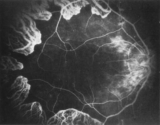
  Fluorescein angiography: There is mild leakage of fluorescein at the margin of the healthy-appearing retina, where it abuts the gyrate lesions (Fig. 2). The earliest detectable changes include pigment epithelial transmission defects, which eventually progress into patches of atrophy with drop-out of the choriocapillaris in the involved area. The edges of such patches stain by leakage, indicating functioning choriocapillaris in the surrounding tissue.27 The ophthalmoscopically normal areas of retina appear normal on fluorescein angiography.

Fig. 2. A fluorescein angiogram shows slight leakage from the margins of the choriocapillaris neighboring the atrophic areas. The posterior pole appears surprisingly normal.

  Dark adaptation: The dark adaptation curve is usually monophasic, delayed, and elevated above normal by about 3 log units.
  Electroretinography: The full-field electroretinogram (ERG) has been shown to be reduced in the early stages of GA, and no normal ERG recordings have been reported.28,29 Both rod- and cone-mediated a- and b-wave amplitudes are usually severely reduced (less than 10 μV) or even isoelectric, indicating that rod and cone systems are affected jointly, although occasionally a patient's ERG may be reduced by only 50% to 75% (i.e., recordable at 100 μV to 300 μV). Direct current ERG findings indicate that the c wave can be recorded only in the very early stages of the disease. The disappearance of the c wave prior to the a and b waves in GA suggests that the ocular disease is primarily RPE in origin.30 Some patients have a delayed implicit time on 30 Hz flicker testing. Computer averaging with narrow-band filtering has made it possible to detect ERG responses under 1 μV, allowing objective monitoring of the progress of even severely affected subjects.31
  Electro-oculography: The ratio of the light peak to the dark trough is severely reduced, parallel to the reduction in the ERG.
  Visual evoked response: The visual evoked response appears to vary as a direct function of visual acuity.
  Conjunctival pathology: Three conjunctival biopsies of patients with GA have shown changes in the epithelial cells and in the stromal fibroblasts of the conjunctiva. The presence of osmiophilic particles (hypothesized to be lipidic or fatty acid drops), hypertrophy of the Golgi apparatus with rupture of intracellular membranes, and accumulation of lysosomes have been detected on electron microscopy.32
  Whole-globe pathologic examinations: A postmortem histopathologic examination of whole globes from a 98-year-old woman with a well-documented (confirmed with fibroblast cultures and hyperornithinemia), atypically mild case of vitamin B6–responsive GA, who had retained 20/50 vision up until death, was reported by Wilson and associates.33 Distinctive, circumscribed patches of RPE atrophy were observed only at the periphery. The outer retina, RPE, choriocapillaris, and most of the choroidal vessels were absent from these patches. Where the RPE terminated, the photoreceptor cells directly abutted Bruch's membrane. On electron microscopy, the mitochondria in the corneal endothelium, nonpigmented ciliary epithelium, smooth muscle of the iris, and ciliary body were enlarged and had disrupted cristae and an electronlucent matrix. Similar but less severe mitochondrial abnormalities were found in the photoreceptors. No mitochondrial abnormalities were found in the RPE.

SYSTEMIC CLINICAL AND PATHOLOGIC FINDINGS

Although ocular involvement is the most clinically significant manifestation of GA, other organs may be affected. On systemic examination, ornithine levels about 10 to 20 times normal values (fasting morning plasma ornithine = 400 to 1400 μM [normal = 40 to 120 μM, mean = 60 to 80 μM]) were observed.8 Hyperornithinemia is an essential component of GA and was the finding that proved GA to be an inborn error of metabolism, with the eye as the most apparent target organ of the metabolic derangement. McCulloch and Marliss34 demonstrated that ornithine was being released from many organ systems and suggested multisystem involvement. The following organs and tissues have so far been found to be affected in GA:

  Brain: Takki22 first reported that abnormal electroencephalographic (EEG) recordings and borderline low intellectual function were common in GA. McCulloch and coworkers11,34 confirmed these findings and stated that they do not necessarily coexist in the same patient. Kaiser-Kupfer and associates35 also reported EEG abnormalities in GA. Using brain magnetic resonance imaging (MRI), Valtonen and colleagues36 found degenerative lesions in the white matter of 50% of 23 GA patients studied and premature atrophic changes in 70%, with a striking increase in the number of Virchow spaces. In agreement with the above-mentioned studies,11,22,32,35 EEG recordings revealed abnormal slow background activity, focal lesions, or high-amplitude B rhythms (greater than 50 mV) in 58% of 33 GA patients tested. There was no correlation between EEG and MRI results or the age or sex of the patients. The MRI and EEG results did not differ between untreated and creatine (Cr)-supplemented GA patients. The authors concluded that early degenerative and atrophic brain changes and abnormal EEG findings are additional clinical features of GA. Nanto-Salonen and colleagues37 analyzed the proton magnetic resonance (MR) spectra of the basal ganglia in 20 GA patients. They found reduced brain Cr stores in GA, which was partially corrected by low-dose Cr supplementation and an arginine-restricted diet. They concluded that these results support the hypothesis that hyperornithinemia in GA results in chronic Cr depletion and decreased phospho-Cr (PCr) stores in the retina, central nervous system, and muscle, and may contribute to the observed pathology in those organs.
  Peripheral nervous system: Peltola and colleagues38 used neurography, quantitative sensory threshold testing, and evoked potential testing to evaluate peripheral nervous system involvement in 40 patients with GA. All the patients tested were homozygotes or compound heterozygotes for one single OAT mutation (L402P). Fifty-three percent of the patients had at least two abnormal findings on neurography, prolonged F-latency being the earliest detectable change characteristic of sensorimotor axonal distal neuropathy. None of the patients studied had disabling neuropathy, 40% of the patients had mild and asymptomatic neuropathy, and 10% had symptomatic peripheral neuropathy. The neurography findings correlated with the severity of GA fundus changes, and patients with signs of neuropathy had the most severe ophthalmologic GA stage; Cr supplements had no effect on neuropathy prevalence.
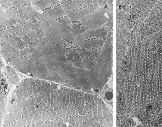
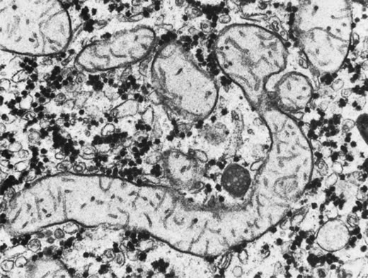
  Skeletal muscle: McCulloch and Marliss34 were the first to report atrophy of, and the presence of tubular aggregates in, type 2 skeletal muscle fibers of one patient with GA (Fig. 3), a finding later confirmed by various other investigators.35,38–40 All reported abnormalities have been confined to type 2 fibers, type 1 fibers being normal in appearance and number in all examined biopsy specimens reported to date. Similar histologic muscle changes were noted in the iris dilator muscle of one patient.41 Tubular aggregates appear to be more common in GA than in any other disease, and aside from patients with GA, they are rarely observed in female patients. They have been noted in male patients in periodic paralysis, hyperthyroidism, porphyria cutanea tarda, acromegaly, myasthenia gravis, polymyositis, myotonic dystrophy, muscular viral infections, denervations, and drug and alcohol abuse, and in some apparently normal persons. The reason that female patients with these disorders do not have tubular aggregates is unknown. Tubular aggregates are thought to represent a detoxifying mechanism or a response to injury and may perhaps be a result of muscle degeneration, eventually disappearing with the affected fiber. Consistent with this hypothesis is the finding by Sipila and colleagues39 of nonspecifically abnormal electromyograms in 16 of 17 deltoid muscle recordings in GA patients, indicative of a mild to moderate skeletal muscle myopathy in GA. The progression of the myopathy is slower than that of the chorioretinopathy, but muscle is nevertheless fairly severely affected by the underlying metabolic defect. Approximately 10% of GA patients demonstrate mild proximal muscle weakness.8 Kaiser-Kupfer and co-workers,35 who found tubular aggregates in skeletal muscle biopsies of three of four patients, grew the muscle cells of these patients in tissue culture. The addition of 20 mmol/L ornithine to the culture media was lethal to GA muscle cells within 48 to 72 hours, but had no effect on cultured normal muscle cells. In addition to type 2 muscle fiber atrophy and tubular aggregates, Valtonen and colleagues43 found computed tomography and MRI changes in the thigh muscles of seven GA patients, although relaxation time measurements gave little additional muscle metabolism insights. Heinanen and colleagues,44 in conjunction with studying brain Cr,36,37 studied the creatine phosphate (CrP) levels in skeletal muscle of GA patients. Abnormal 31P-MR spectra were found in resting calf muscle of GA patients, with decreased ratios of PCr/adenosine triphosphate (ATP) and PCr/Pi, proposed to reflect a decreased CrP concentration in calf muscle of GA patients. However, these decreases did not reflect the clinical stage of the disease, degree of muscle destruction, or ornithine concentrations. The researchers proposed that disturbed cellular energy production resulting from intracellular ornithine-induced inhibition of Cr synthesis, and decreased PCr muscle stores may partially explain the pathogenesis of GA. Daily oral supplementation with physiological levels of Cr (1.5–2 g/day) for 8 to 15 years led to almost normalized PCr/P and PCr/ATP ratios in the calf muscle of GA patients, likely by increasing intracellular PCr.45

Fig. 3. A. A muscle fiber of a patient with gyrate atrophy shows a large inclusion. B. On high power, the inclusion is composed of tubular aggregates.

  Liver: Liver biopsies in patients with GA have demonstrated bizarre, elongated, and segmented mitochondria—an abnormality perhaps similar to that seen in the hyperornithinemia, hyperammonemia, and homocitrullinuria (HHH) syndrome (see below; Fig. 4). These changes are thought to be a direct result of hyperornithinemia, since similar changes have been produced in rats maintained on diets high enough in ornithine content to produce a constant moderate hyperornithinemia.46 Fibroblasts from GA patients, as well as normal fibroblasts exposed to high levels of ornithine in the growth media, exhibit similar mitochondrial changes in tissue culture.47 Similar mitochondrial changes have also been observed in skeletal muscle of GA patients.41

Fig. 4. An elongated mitochondrion in a liver cell from a patient with gyrate atrophy. (Courtesy of M.J. Phillips, MD.)

  Hair: Ten patients with GA have been noted to have peculiar, fine, straight, occasionally sparse scalp hair with areas of alopecia. Scanning electron microscopy revealed that the medullary portions of sampled hairs contained loosely packed macrofibrils. The intervening spaces appeared to be packed with a structureless, electronlucent compact substance that was insoluble in both water and common laboratory solvents. The exact nature of this substance and the relationship of this finding to the oculometabolic disease remain obscure.35
  Other: Francois48 reported an association of Alder's anomaly (the presence of numerous azurophilic granules in the cytoplasm of neutrophils, which stain dark violet by Pappenheim's technique) with GA in one patient. However, Alder's anomaly has generally been associated with Hurler syndrome and has not been reported in association with GA by other investigators. In a study performed at the U.S. National Eye Institute, and therefore not expected to deal with a homogenous genetic population (as one might find with Finnish GA patients), an increased occurrence of thyroid disease in patients with GA compared with control patients was found by Whitcup and colleagues.49 Seven of 34 patients with GA had thyroid disease, resulting in an estimated odds ratio for thyroid disease in GA patients of 12.7 that of normal controls. Similar but less pronounced results were found in retinitis pigmentosa patients, with an estimated odds ratio of 6.2 compared with normal controls.

ANIMAL DATA

Hepatic mitochondrial changes similar to those seen in patients with GA have been produced in normal rats maintained on high-ornithine diets.50 A case of an adult male cat with retinal degeneration, extreme hyperornithinemia, and absence of OAT—an apparent replica of human GA—was reported by Valle and associates.51 Attempts to breed the cat were unsuccessful, and it later died. Histopathologic examination of the cat's eyes demonstrated extensive neuroretinal and RPE damage and cell loss. There was a decrease in the number of small choroidal vessels, but not the large ones. In cats with other retinal degenerations, such loss of RPE and choriocapillaris is uncommon. This “GA cat” did not have cataracts.

Kuwabara and associates52 demonstrated selective destruction of RPE cells in rats and monkeys after intravitreal injections of ornithine. Secondary destruction of overlying photoreceptors and underlying choroid developed later. Based on these data, they suggested that the RPE may be the primary target organ in GA. A more recent attempt by Daune-Anglard and colleagues53 to mimic the damage of GA with induced ornithine toxicity in mice and chickens using 5-fluoromethylornithine (5FMOrn) administered orally for 53 days showed no ocular pathologic changes in these animals despite a tenfold increase in tissue ornithine. However, 10% to 20% of tissue OAT is refractory to inactivation by 5FMOrn, so these results only show that mice and chickens will not develop GA as a result of sustained hyperornithinemia due to incomplete blockage of OAT for this period of time.

Subsequently, Wang and colleagues54 successfully created a mouse model of GA that closely mimics GA in human patients. Using targeted disruption of the murine OAT gene, they produced OAT-deficient mice that exhibit chronic hyperornithinemia, ten to 15 times normal. These OAT-deficient mice developed slowly progressive retinal degeneration over the first 12 months of life. The RPE cells were the initial site of damage. Using this mouse model of GA, Wang and colleagues55 further evaluated the effect of long-term reduction in ornithine on prevention of retinal degeneration. OAT-deficient mice fed an arginine-restricted diet for 12 months had significantly reduced plasma ornithine levels. Importantly, retinal degeneration, as measured by ERG and retinal histologic and ultrastructural studies, was prevented. The researchers concluded that ornithine accumulation, and not OAT activity in the retina and RPE, is a necessary factor in the pathophysiology of retinal degeneration in GA.

DIFFERENTIAL DIAGNOSIS

Ocular

Although cases of GA have been mislabeled as many other things, usually “atypical retinitis pigmentosa,” and patients referred as having GA often turn out to be suffering from some other disorder, the diagnosis is actually quite easy to make if the five main features (see previous Diagnosis section) can be recalled (Fig. 5). Confusion in diagnosis is primarily due to the rarity of the disease. It is extremely unlikely that any primary care ophthalmologist would have the opportunity to make this diagnosis more than once in a career, since the incidence of GA appears to be less than one in 1,000,000 everywhere except in Finland, where it occurs in about one in 50,000 individuals.8

Fig. 5. The five main features in the diagnosis of gyrate atrophy.

Almost invariably, the main ophthalmic features of high myopic astigmatism, posterior subcapsular cataracts, and typical retinal appearance are obvious to the ophthalmologist. One can be quite certain that if the patient does not have the typical fundus picture, as well as extreme hyperornithinemia, GA is not the correct diagnosis. The autosomal recessive inheritance pattern can usually be quickly ascertained with a brief family history asking about affected relatives and parental consanguinity (see Fig. 5), even before confirmatory laboratory biochemical and genetic testing is undertaken.

Non-GA patients with extremely high myopia (usually more than –15 D) may occasionally have posterior polar, peripapillary, or peripheral clusters of round, full-thickness, chorioretinal atrophic lesions, often causing significant reduction in visual acuity.56 This, however, is not the pattern of the lesions seen in GA.

An entity of “central gyrate atrophy” has been described,57 but this is probably equivalent to end-stage serpiginous choroidopathy rather than a separate inherited chorioretinopathy.58,59

Large areas of paving stone degeneration may superficially resemble GA, albeit on a much smaller scale. Paving stone degeneration is usually found in the inferior quadrants peripherally, whereas GA is not segmental and involves all 360 degrees of the fundus.60 Occasional reports of atypical GA appear in the literature.61–64 Review of these cases usually reveals either that the retina did not quite have the typical appearance of GA, myopia and cataracts were not present, and/or hyperornithinemia was absent. It is interesting to speculate as to why other disorders have retinal lesions similar in appearance to GA and whether or not some final common pathway to chorioretinal destruction exists, especially in cases where some similar systemic involvement may coexist, as in muscular dystrophy. However, labeling such cases with similar lesions as “atypical gyrate atrophy” can be misleading, especially to the majority of practitioners who have never encountered GA, and may lead to corruption of research efforts and reports by the inclusion of different etiologic disorders in supposedly homogeneous study groups.65

Metabolic

Hyperornithinemia is not unique to GA.11,46 It was reported in two other conditions. In the late 1960s, Kekomaki and colleagues66 and Bickel and associates67 described two siblings who presented with failure to thrive, prolonged neonatal jaundice, atypical hepatic cirrhosis, renal tubular dysfunction, and mental retardation. Hyperornithinemia (about three times normal), renal glycosuria, generalized aminoaciduria, and mild hyperammonemia were present. Hepatic OAT levels were about one-sixth those of normal patients. Garnica and coworkers68 later confirmed these findings. Kekomaki and coworkers66 studied the enzyme kinetics of the residual OAT in the hepatocytes of one patient and found them to be normal, suggesting that the problem in this disorder is one of decreased enzyme synthesis or excessive degradation. Both siblings, at ages 15 and 9, were severely retarded, had normal ocular examinations, normal plasma ornithine, normal liver function tests, generalized aminoaciduria, elevated serum creatinine, and hypertension. The etiology of this syndrome remains unknown.8

The second disorder with associated hyperornithinemia is HHH syndrome, a rare autosomal recessive disorder that is about one-third as common as GA. HHH patients exhibit hyperornithinemia about one-half as elevated as that of GA patients, but there is considerable overlap. In the late 1960s to early 1970s, Shih and colleagues69,70 and later (1975) Gatfield and colleagues71 described patients presenting in infancy with feeding difficulty, mental retardation, and seizures, in which the biochemical triad of hyperornithinemia, hyperammonemia, and homocitrullinuria was observed. Abnormal hepatic mitochondria, not unlike those seen in GA, have been identified in this disease. The biochemical defect was hypothesized by Fell and coworkers72 to reside in defective transport of ornithine across the inner mitochondrial membrane into the mitochondrial matrix, and the biochemical and clinical evidence supports this hypothesis.8,73–82 The ornithine/citrulline transporter was identified and confirmed as the defect in the HHH syndrome.83–85 The gene has been labeled ORNT1 and mapped to 13q14.86 HHH patients may voluntarily restrict their protein intake to avoid having symptoms. The symptoms are thought to be due to hyperammonemia as they are similar to other hyperammonemia syndromes, and can be dramatically ameliorated with ornithine HC1, 0.5 to 1.0 mmol/kg/day dietary supplementation.78 The additional dietary ornithine is thought to elevate cytosolic ornithine levels, thereby driving it into the mitochondrial matrix, where it acts as the limiting substrate in the elimination of ammonia via the urea cycle. HHH syndrome, like GA, is inherited as an autosomal recessive disorder. Some patients do not respond to ornithine or arginine supplementation, suggesting heterogeneity in the mutation of the affected protein. Ocular examination has been normal in all patients examined.8

In neither of the above two metabolic syndromes have ocular abnormalities been noted. The senior author (SAA) repeatedly examined a currently 26-year-old patient and a teenage patient, both with HHH syndrome, as they underwent ornithine supplementation treatment over many years. Neither patient manifested any retinal or ERG changes (Steve A. Arshinoff, unpublished data, York Finch Eye Associates, Toronto, Ontario, Canada, 1985–2003). GA retinal lesions have not been observed in an infant. Hayasaka and coworkers19 noted that a 2-year-old patient with biochemical GA did not develop fundus lesions until age 4, and Stoppoloni and colleagues87 reported on a 13-year-old patient who had early fundus lesions. The senior author has followed a GA patient, who first presented at age 7 with well-defined peripheral GA lesions involving 360 degrees of her retina.

Back to Top
THE METABOLISM OF GYRATE ATROPHY

ALTERED PLASMA AMINO ACID PROFILE

Ornithine is a dibasic amino acid that is not normally found in proteins (Figs. 6 and 7). However, it plays a pivotal role in the urea cycle (Fig. 8). The initial discovery by Simell and Takki9 of extreme elevations of ornithine levels in the plasma, urine, cerebrospinal fluid, and aqueous humor of patients with GA encouraged others to search for secondary alterations in amino acids. McCulloch and coworkers11 and Arshinoff and coworkers46 reported the presence of hypolysinemia, hypoglutamic acidemia, and hypoglutaminemia, which was confirmed by Valle and coworkers,88 who also found the presence of hypoammonemia. The mechanism of the hypolysinemia, hypoglutamic acidemia, hypoglutaminemia, and hypoammonemia has been proposed to be excessive excretion secondary to renal tubular readsorption competitive blockade by hyperornithinuria in the renal proximal tubular filtrate.46,88,89

Fig. 6. Whole-blood amino acid levels in gyrate atrophy. The level of ornithine is ten times normal. There are decreases in the levels of lysine, glutamate, and glutamine. A. Basic. (ORN, ornithine; LYS, lysine; HIS, histidine; ARG, arginine) B. Acidic and neutral. (TAU, taurine; THR, threonine; SER, serine; GLU, glutamate; ASN, asparagine; GLN, glutamine; PRO, proline; CIT, citrulline; GLY, glycine; ALA, alanine; ABA, α-amino-n-butyrate; VAL, valine; ILE, isoleucine; LEU, leucine; TYR, tyrosine; PHE, phenylalanine)

Fig. 7. Chemical formula of the dibasic amino acid ornithine.

Fig. 8. The metabolic pathways involving ornithine include polyamine synthesis, the urea cycle, ornithine catabolism, and creatine synthesis. A defect in the mitochondrial matrix enzyme ornithine aminotransferase (OAT) is the biochemical basis of gyrate atrophy.

PATHOPHYSIOLOGY OF THE RETINAL DEGENERATION

The biochemical mechanism of the observed pathology in an enzyme-deficiency state may be due to a deficiency of the product of the reaction or subsequent dependent reactions, or accumulation of excessive substrate, which, in turn, may either be toxic or may drive another reaction to produce a toxic product. Elucidating which of these two conditions is present in any given disorder is often a very complex process owing to the myriad interdependent reactions involved in the metabolism of different tissues. The following hypotheses for the GA mechanism have been proposed.

Hyperornithinemia Hypothesis

A simple and attractive hypothesis regarding the pathophysiology behind the ocular degeneration in GA is that the high ornithine levels in themselves are toxic (Fig. 9). In GA, high intramitochondrial concentrations of ornithine presumably occur and must somehow contribute to the etiology of the disease.90 Any attempt to explain the pathophysiology of the observed retinal degeneration in GA must also take into account the fact that other syndromes with hyperornithinemia (notably HHH, which has high cytosolic ornithine but low mitochondrial levels), do not manifest ocular symptoms; the reduced involvement of other organ systems in GA and the slowly progressive nature of GA must also be explained.69,72 The hyperornithinemia, particularly elevated intramitochondrial ornithine, hypothesis is strongly supported by the mouse model studies discussed earlier, but the issue is often more complex than it would initially seem, and it is discussed in more detail later. Even if it is determined that the retinal and RPE degeneration is due to elevated intramitochondrial ornithine, the other theories discussed below may account for other observations in GA, so all may be correct and contribute to the observed GA pathology to varying degrees.

Fig. 9. Proposed hypotheses regarding the pathophysiology of gyrate atrophy. (1, hyperornithinemia hypothesis; 2, phosphocreatine deficiency hypothesis; 3, Δ1-pyrroline-5-carboxylate/proline deficiency hypothesis; 4, decarboxylation product excess (putrescine and other polyamines) hypothesis; OAT, ornithine aminotransferase; –, negative inhibition; ↑ , increase; ↓ , decrease)

Phosphocreatine Deficiency Hypothesis

Sipila and colleagues39,91,92 drew attention to decreased plasma, tissue, and urinary levels of Cr and creatinine in GA (see Fig. 9) and suggested that because of the importance of PCr in the energy metabolism of skeletal muscle and RPE and the decreased production of Cr in GA (due to competitive inhibition of intramitochondrial arginine-glycine amidinotransferase by increased ornithine levels), this metabolic alteration may be the biochemical etiology of the observed chorioretinal and/or muscular pathology. This hypothesis is given credence by the investigations of neural and muscular pathology reported earlier, except that the pathology was not normalized in patients treated with supplemental Cr. If a PCr deficiency is the etiology of the retinal degeneration in GA, HHH patients may be spared if arginine-glycine amidinotransferase is located in the mitochondrial matrix, since it is has been demonstrated that the defect in HHH is the inability of ornithine to cross into the mitochondrial matrix and that intramitochondrial ornithine levels are low. HHH patients, therefore, would not have inhibition of PCr synthesis.

Δ1-Pyrroline-5-Carboxylate/Proline Deficiency Hypothesis

A third hypothesis was proposed by Valle and Simell8 in which the OAT deficiency leads to a decreased level of Δ1-pyrroline-5-carboxylate (P5C), an intermediate product between ornithine and proline (see Fig. 9). P5C is normally synthesized by OAT and P5C synthase. In GA patients, the OAT pathway is defective. In addition, physiologic concentrations of ornithine have an inhibitory effect on P5C synthase.8 Thus, the two normal pathways that form P5C are either defective or shut down in GA. Ultimately, the decreased levels of P5C may cause the ocular pathology because of decreased proline synthesis93,94 or the disruption of the regulatory roles of P5C.8 If a P5C deficiency is the etiology of the retinal degeneration in GA, HHH patients are spared because they can rely on their intact OAT pathway to synthesize P5C.95

Ueda and colleagues96 proposed that the combination of ornithine accumulation and an increased sensitivity to ornithine due to OAT deficiency, together with abnormal proline metabolism, causes specific RPE degeneration. They created an in vitro model of GA using human RPE cell lines treated with 5-fluoromethylornithine, a specific, irreversible OAT inhibitor. They found that ornithine, only in the presence of OAT inactivation, caused time- and dose-dependent inhibition of DNA synthesis, accompanied by morphologic changes and cell death. Proline prevented the cytotoxicity of ornithine in this system. Following these results, the same researchers, using primary cultured bovine RPE cells, found that ornithine caused cytotoxicity specifically in the epithelioid but not fusiform phenotype of bovine RPE cells.97 These results suggest abnormal proline metabolism may contribute to the pathophysiology of GA.

Hypothesis of Excess Decarboxylation Product (Polyamine)

Jaeger and associates65 suggested that an excess of the products of ornithine decarboxylase, which may result from hyperornithinemia, is toxic to the brain, liver, and kidneys (see Fig. 9). The decarboxylase pathway had not been considered by others to have a high likelihood of significance in GA because of its relative inactivity when compared with the other pathways that utilize ornithine.46,98 This pathway has not been investigated to any great extent in GA, and any definitive statement concerning its relative importance must await further study. Kennaway and associates measured polyamine levels in four patients, but the results were inconclusive with respect to any effect of hyperornithinemia on levels of these decarboxylation products. Sulochana and colleagues99 measured blood and urine polyamines in seven patients with phenotypic GA and found elevated levels of putrescine (six out of seven cases) and spermine (three out of seven cases), as well as decreased levels of cadaverine, a metabolite of lysine, in all seven patients. They concluded that measuring urinary polyamines is more sensitive diagnostically, and correlates better clinically, than measuring ornithine or OAT alone. Not all of these patients fulfilled the diagnostic criteria for GA (see earlier), and it may be that the polyamine findings of the Sulochana study represent a final common pathway to retinal degeneration and were therefore judged by the authors to be more sensitive in predicting retinal damage than OAT activity.

Back to Top
GENETICS OF GYRATE ATROPHY

THE MOLECULAR GENETICS OF ORNITHINE AMINOTRANSFERASE

Soon after the discovery of the association between hyperornithinemia and GA, OAT deficiency was hypothesized by many investigators to be the underlying enzymatic defect (see Fig. 8).9,11,42 Takki22 was the first to report negligible OAT activity in cultured fibroblasts in this disease. This finding was confirmed by many others in cultured fibroblasts, phytohemagglutinin-transformed lymphocytes, skeletal muscle, and liver.21,35,100–111 Enzymatic heterogeneity was first demonstrated by the fact that a minority of patients' fibroblasts showed marked increases in OAT activity (from negligible to about one-third normal) when large amounts of PLP (vitamin B6) were added to the assay medium.98 Wirtz and co-workers112 demonstrated the enzymatic heterogeneity between B6-responsive and -nonresponsive patients using complementation analysis on GA patients.

More recently, DNA sequencing of GA patients has revealed a multitude of primary defects, due to over 60 different mutations in the OAT gene.8 In the majority of cases, the basic genetic defect resides in production of abnormal enzyme with an amino acid substitution at the DNA level.113 However, Inana and associates,114,115 Hotta and colleagues,116 and Shull and Pitot117 characterized a single patient whose heterozygous-defective OAT alleles resulted in a complete absence of OAT mRNA expression, further expanding the genetic heterogeneity observed in GA. This patient's defect, unlike all the others to date, results in a failure to produce enzyme protein. Other data published to date on the kinetics of residual enzyme activity and on immunologic quantification of enzyme protein in GA patients, heterozygotes, and normal controls, demonstrated production of normal amounts of kinetically abnormal enzyme protein.110,111,118

The human OAT gene has now been well characterized at both the DNA and mRNA levels, and extensive studies by various groups have successfully cloned the gene defect responsible for GA and localized it to chromosome 10q26.113,119,120 It is interesting to note that several nonfunctional OAT-like sequences exist on chromosome Xp11.2-p11.3 and map to the same region as two other retinal degenerative diseases: X-linked retinitis pigmentosa and Norrie's disease.121 The human OAT gene is 21 kilobases in length (genomic DNA), with 11 exons and a promoter region. The gene transcribes a 2073-base pair mRNA, which is translated into a protein precursor consisting of 439 amino acids (48, 534 daltons) (Fig. 10).119,120 The precursor is then translocated into mitochondria, where it is processed to yield a mature OAT monomer consisting of 407 amino acids (45, 136 daltons).114,123 The human holoenzyme was found to be a 256-kilodalton homohexamer with one molecule of cofactor PLP bound to each subunit.124 DNA regulatory sequences are present at the 5' end of the gene, including a 22–base pair element that resembles an estrogen-responsive element. DNA sequencing of the OAT gene from GA patients has revealed that the overwhelming majority of them express normal quantities of normal-sized OAT mRNA. Among the mutations reported are 34 missense mutations, ten nonsense mutations, ten microdeletions, one microinsertion, and three errors resulting in splicing defects, giving us an idea of the relative frequencies of different genetic defects in GA patients. The more than 60 mutations identified thus far in the OAT gene8 and the wide range of residual OAT activity in GA patients serve to emphasize the genetic and biochemical heterogeneity in this disease. It would appear that GA confers a genetic disadvantage on the patient as evidenced by the relative lack of propagation of these mutations (i.e., identical mutations recur infrequently in different family studies).8,125 Localization of the gene has confirmed the autosomal recessive inheritance pattern of GA.1,2,107

Fig. 10. The molecular genetics of ornithine aminotransferase (OAT). The OAT gene is located on chromosome 10q26 and is transcribed into a 2073 base pair (bp) mRNA. Cytosolic polysomes translate the mRNA into a 439 amino acid (aa) proenzyme precursor. A mitochondrial localization signal targets the proenzyme for translocation into the mitochondria, where it is subsequently cleaved into a mature enzyme monomer. The functional OAT enzyme is believed to be a homohexamer, each monomer of which is associated with a molecule of B6.

THE ENZYME

OAT was first characterized in the rat, but is similar in all mammals studied to date except where noted below. OAT is a PLP-dependent, mitochondrial matrix enzyme that is manufactured from the cell's nuclear, rather than mitochondrial, DNA (see Fig. 8). It is initially synthesized by cytoplasmic polysomes and is subsequently processed into the mature enzyme following transport into the mitochondria. It catalyzes the reaction


ornithine + α-ketoglutarate glutamate + glutamic γ-semialdehyde

with strong directional preference to the right under laboratory conditions. The standard free energy change of the reaction in rat liver is about –2500 calories at 25°C.126 The molecular weight of OAT was initially determined for rat liver and kidney and was found to be approximately 170,000 daltons, consisting of four subunits of 43,000 daltons each, with one molecule of PLP bound to each subunit,127 and was then found to be identical in different rat tissues.128 Recent work has shown that the mature human OAT monomer consists of 407 amino acids and weighs 45,136 daltons.114,129 X-ray diffraction studies have suggested that the human enzyme is a 256-kilodalton hexameric enzyme consisting of three dimers as the basic packing unit.130 Tyr-55 and Arg-180 have been found to be important sites for positioning ornithine within the active site of OAT for specific transamination at the δ position; both these residues have been found mutated in GA patients, thus affecting binding affinity of OAT.131 There is a PLP-binding residue at Lys-292 of each processed protein monomer.120 Crystal structure studies have revealed eight residues important for PLP positioning in OAT: Gly-142, Bal-143, Phe-177, Asp-263, Ile-265, Gln-266, Ser-321, and Thr-322.132 Ramesh and colleagues133 proposed that B6-responsive GA patients have a mutation at or near the B6 binding site that hinders but does not prevent B6 binding (see Fig. 10). In agreement with this hypothesis, four mutations have been found to cause impaired binding of PLP to OAT. Two mutations, V332M133,134 and A226V,135 have been shown to be B6 responsive by in vitro expression studies; and two other mutations, T181M136 and E318K,137 have been found in B6-responsive GA patients. As expected, these patients have less severe, slower progressing GA than do nonresponsive patients.

As might be expected from the regulatory sites found on the OAT gene, there is a marked increase in OAT activity in response to estrogen and thyroid hormone administration in nonocular tissues other than skeletal muscle.138–140 Enzyme activity has been found to be high in RPE, retina, liver, kidney, small intestine, and skeletal muscle. Various investigators have shown both OAT activity and concentration to be greatest in human, bovine, and chick ocular tissues relative to other tissues in the body138,141–144; specifically, tissues of neuroepithelial origin (e.g., neuroretina, RPE, ciliary body, epithelium of iris) have been demonstrated in rats and cats.145–147 In humans, normal OAT activity is three times higher in the retina than in the brain and is 80% that of the liver.148 These data do not fully explain why the eye is the most prominent target organ in GA. In analyzing total RNA from humans and rhesus monkeys, Bernstein and Wong149 found that regional expression of OAT mRNA does not directly correlate with the pattern of gene expression in GA. In contrast to previous findings that found OAT mRNA expressed most in RPE, Bernstein and Wong found the highest levels of OAT mRNA in the neuroretina—specifically the fovea, with the midperipheral retina next—whereas the RPE/choroid expressed the lowest level of OAT mRNA. These findings suggest that factors other than simple OAT gene expression are involved in the pathophysiology of GA.

CLINICAL GENETICS

More than 150 cases of GA have been reported, with the greatest concentration of patients in Finland (about 70 cases), but the disease does not appear to be confined to any one geographic or racial group. GA has been reported in patients around the world, including the following ethnic origins: Finnish, French, English, Welsh, Scottish, Swedish, Lebanese, Spanish, Portuguese, Italian, Mexican, German, Japanese, Nicaraguan, Jewish, Brazilian, African American, West African, Algerian, Hungarian, Turkish, and Indian.8,131,132,150–153 The inheritance pattern is autosomal recessive, as are most other classic inborn errors of metabolism. Heterozygotes demonstrate roughly one-half the level of OAT activity that normals exhibit, yet they do not express the disease phenotypically, further supporting the autosomal recessive inheritance pattern.

The gene frequency of GA in Finland is about 1:220, with an estimated frequency of heterozygosity of 1:110 and an incidence of GA about 1:50,000.8 The gene frequency in the general North American population would seem to be approximately 1:2000, with a resultant incidence of heterozygosity of about 1:1000 and the incidence of GA about 1:4,000,000. Our estimate is based on the number of reported cases and consideration of the expectation that some cases may go unreported. Although simple mass screening methods for hyperornithinemia have been described, mass screening for these disorders has not been adopted.154 It is interesting that newborn urine amino acid screening programs have failed to detect any cases of GA, suggesting that ornithine does not rise rapidly postnatally in GA patients.

DETECTION OF HETEROZYGOSITY

Obligate heterozygotes have not been found to have any ocular abnormalities, and consequently proof of heterozygosity must rely on biochemical or genetic testing.

Prior to the elucidation of the biochemical basis of GA, an ornithine tolerance test was devised by Takki and Simell12 (Fig. 11) in which an ornithine load of 100 mg/kg body weight is administered orally after an overnight fast. Plasma ornithine levels are then measured at 30-minute intervals for 3 to 4 hours, and a graph of ornithine levels versus time is drawn and compared with normal controls and homozygous GA patients. Some investigators have suggested that adequate segregation of controls from heterozygotes is not always achieved with this test.40

Fig. 11. The ornithine tolerance test, indicating plasma ornithine levels after an oral loading dose of 100 mg/kg l-ornithine. Note the clear segregation of homozygous patients. The heterozygous parents and controls show less complete segregation from each other. (Modified from Takki D, Simell O: Genetic aspects in gyrate atrophy of the choroid and retina with hyperornithinemia. Br J Ophthalmol 58:907, 1974.)

Biochemical assays of OAT activity in cultured fibroblasts and phytohemagglutinin-stimulated lymphocytes yield levels for heterozygotes intermediate between homozygotes and normal controls, with adequate segregation of each group.104,105,107,155 More recently, two rapid, direct methods of OAT activity measurement have been described: a reverse-phase high-performance liquid chromatography assay based on the separation of P5C156 and a more sensitive microradioisotopic assay that directly measures [S14C] glutamate.157 Both methods reliably recognize homozygotes but since the measured values of healthy controls and heterozygotes overlap, are not reliable for detecting heterozygotes. An enzyme assay utilizing hair roots to detect heterozygosity for GA has also been described.158

Molecular detection of the genetic defect in patients or potential carriers via a rapid protocol involving polymerase chain reactions, denaturing gradient gel electrophoresis, and direct sequencing has been described by Mashima and colleagues,159 who detected subtle point mutations in genomic DNA with a gene detection accuracy rate of 95.5% (21 of 22 mutant alleles tested). Kaufman and colleagues160 also developed exon scanning, a quick method in which even point mutations may be detected in the gene. These new detection and sequencing methods are capable of rapidly elucidating the genetic defect in a given patient or carrier, compared with previous technology.159 These tests, however, are too cumbersome for mass GA screening; assays of ornithinemia or ornithinuria are still the most cost-effective.

PRENATAL DIAGNOSIS

To date, we are unaware of any family that has presented itself for prenatal diagnosis of GA. Nevertheless, O'Donnell and coworkers161,162 have proposed two methods by which such diagnosis could be achieved. Kennaway and coworkers104 and Shih and Shulman70 have confirmed the presence of measurable OAT levels in cells from the amniotic fluid of normal humans. Rapid, sensitive, and direct enzyme assays allow for analysis of villus biopsy cell lysates156,157and instigation of treatment as early as possible. Using the microradioisotopic assay described above, Roschinger and colleagues157 detected OAT expression in native and cultured chorionic villi, possibly allowing detection of GA during the third trimester of pregnancy. Prenatal diagnosis has been attempted in a pregnant HHH patient to determine whether the fetus' genotype could be detected with accuracy. The fetus was diagnosed as normal with confirmation by the measurement of ornithine levels in the baby postnatally.163 To our knowledge, however, no cases have yet been described in which there was a positive diagnosis of GA or HHH using prenatal diagnostic methods.

Back to Top
TREATMENT OF GYRATE ATROPHY
The proposed treatments for GA all attempt to correct one or more of the metabolic alterations present, based on the assumption that the particular abnormality being addressed is the one that is the primary etiology for the chorioretinal degeneration. In actuality, it has yet to be conclusively demonstrated what the toxic metabolic alteration is in humans, and of course this presents great difficulties both in the planning and assessment of any treatment program. Hyperornithinemia, Cr deficiency, lysine deficiency, and P5C/proline deficiency have all been the targets of therapeutic programs. We are unaware of any attempts to prevent overproduction of polyamines through the decarboxylation pathway in GA.

CORRECTION OF HYPERORNITHINEMIA

It is apparent that the primary metabolic consequence of OAT deficiency is accumulation of excess ornithine, and intramitochondrial accumulation appears to be the factor that is toxic to the retina in the mouse model (above). The products of OAT (glutamic acid and proline) can be derived from other sources; thus, the observed alteration in their levels is not nearly as great, and probably not as significant in the production of disease, as the change in ornithine levels. The remainder of the observed biochemical alterations in GA is a result of hyperornithinemia and is not due to the enzyme deficiency directly. It therefore seems logical that, unlike other strategies for treatment, the normalization of ornithine levels as a primary therapeutic objective should secondarily normalize all the other metabolic parameters and thereby correct the etiologically significant parameter, regardless of which one or ones it eventually proves to be. Three routes to accomplish this, either singly or together, have been attempted.

Administration of Supplemental Pyridoxine (Vitamin B6)

The administration of pharmacologic doses of pyridoxine in a disorder caused by decreased activity of a B6-dependent enzyme is an established procedure.164 Among diseases in which this therapy has been used successfully are cystathioninuria, B6-dependent convulsions of infancy, and one type of homocystinuria. Berson and coworkers,165 Shih and colleagues,166 and Wirtz and associates112 demonstrated marked increases of in vitro OAT activity in GA fibroblasts, isolated from a B6-responsive patient, in response to increasing B6 levels in the assay medium. A corresponding 50% fall in plasma ornithine levels was noted in the GA patient who received an oral dosage of 300 ng/day of pyridoxine and from whom the fibroblast culture was derived. Since these findings, it has become standard practice to test all new GA patients for B6 responsiveness, both in vitro and in vivo.

Of the approximately 70 Finnish GA patients reported to date, none have been B6 responsive.167 Of the remaining patients worldwide, who are currently being studied in the United States, Canada, Japan, Great Britain, Italy, Germany, the Netherlands, and Israel, seven have been responsive to B6 and the rest have not.11,18,98,101,108,165,168–173 It would seem that in vivo responsiveness usually, but not always, correlates well with in vitro responsiveness. The required oral dose of pyridoxine has not been established and varies with each patient's specific defect, since individual pyridoxine sensitivity varies widely due to different amino acid substitutions in the enzyme protein.40,168,174 Treatment protocols have indicated doses ranging from 15 to 750 mg/day. The advantage of B6 supplementation is that it is an easy treatment, and consequently patient compliance is good.95 The fall in serum ornithine by 50% in B6 responders is accompanied by normalization of serum lysine and a rise in P5C. One study reported improvement in the ERGs of two patients after they were placed on B6 supplementation. In addition, Weleber and coworkers175 reported that B6-responsive patients typically have a milder course of disease compared with B6-nonresponsive patients in terms of visual function, as well as less extensive lesions. This was clearly evident in the 98-year-old patient's eyes, which were obtained for pathologic examination (see Ophthalmic Findings section).

Arginine-Reduced Diet

Arginine is not an essential dietary amino acid in the normal human adult. In eukaryotic organisms, including humans, it can be generated from ornithine in the urea cycle, and ornithine in turn can be derived from glutamic acid, through OAT.176 In the absence of functioning OAT, arginine becomes an essential amino acid (EAA) and the major, if not the only, source of de novo ornithine.108,176 Diets may therefore be designed that, by controlling arginine intake, can be titrated to achieve a desired plasma ornithine level.177,178 These diets require that the patient drastically reduce consumption of normal proteins, since arginine is a normal constituent of all natural proteins, and substitute the balance of the required daily essential dietary protein with artificially flavored solutions of EAAs containing all essential amino acids except arginine.90 (One protocol called for 0.5 g protein/kg/day with 0.3 to 0.5 g EAA/kg/day.179) Compliance with such severe dietary restriction has been a problem with some patients, whereas others manage surprisingly well.180,181

Ambiguous results were obtained in early studies, which initially led several groups to conclude that normo-ornithinemia achieved through dietary therapy (arginine restriction) is inadequate to halt the progression of the chorioretinal degeneration.182,183 However, long-term follow-up studies suggest that with good compliance, progression of ocular symptoms in the disease can be slowed significantly. The diets have been available for about 25 years, and improvements in visual acuity and fields, dark adaptation, ERG, and color vision have been reported.184,185 Perhaps the most compelling evidence yet presented in favor of dietary arginine restriction therapy has been put forth by Kaiser-Kupfer and associates,179 who studied six pairs of siblings with genetically identical GA over a 5- to 7-year period. Retinal degeneration progressed in all of the GA patients on the diet, but the younger of the sibling pairs displayed less ocular involvement than the older siblings did at the same age, which was explained by the fact that the older siblings began the diet at an older age than their younger siblings. Long-term follow-up of the two youngest sibling pairs further supports the contention that the early introduction, ideally before any detectable retinal lesions, of an arginine-restricted diet can slow the progression of retinal degeneration.186 After 16 to 17 years of maintaining excellent ornithine levels, the youngest siblings had minimal retinal lesions, with the phenotype of retinal changes resembling early retinitis pigmentosa more than GA. However, loss of retinal function as measured by ERG amplitude and visual field sensitivity still occurred, which might have been prevented by even earlier institution of the arginine-restricted diet. This and other positive clinical results, supported by the mouse model data, have led to the use of arginine-restricted diet therapy as a standard of GA treatment.90,177–179,184–185 In our own series, one B6-unresponsive patient, who began the diet at age 7 and has followed it diligently for 25 years, has suffered far less progression than similar patients had developed by ages 10 years less (Steve A. Arshinoff, personal observation). Longer-term follow-up continues to evaluate the benefits of this diet.

The major reported risk in arginine-restriction therapy is that if the arginine restriction is carried to excess, both arginine and ornithine levels may become depleted, with resultant impairment of urea cycle function and consequent hyperammonemia.108,177 In one hospitalized patient, McInnes and coworkers intentionally induced hyperammonemia by excessive restriction of arginine intake in order to assess this theoretically possible complication of dietary treatment.178 The patient developed nausea and lethargy and responded quickly to intravenous administration of arginine. Our experience to date has indicated that in the day-to-day management of these patients, the risk of hyperammonemia is very low, provided that arginine restriction is not permitted to reduce serum ornithine levels in plasma below normal, which is usually about 0.2 mmol/L. Nevertheless, patients must be cautioned against excessive arginine restriction and monitored frequently while on this diet. In practice, the diet is unpalatable enough that the problem is invariably inadequate, as opposed to overzealous, compliance.

The advantage of the arginine-restricted diet over other treatment regimens is that the patient can be titrated to a predetermined target level for ornithine. Once this level is achieved, not only are the patient's ornithine levels reasonably normal, but lysine, glutamic acid, glutamine, and ammonia levels are also normalized.98,177 Presumably, levels of Cr, P5C, proline, and any other metabolites are also normalized, but these specific points have yet to be confirmed. The diet, which may also be combined with B6 administration in B6 responders, making the diet far less restrictive for these patients, is unlike other treatments in that it has the theoretic advantage of “biochemical normalization” of the patient, which hopefully should correct the basic cause of the chorioretinal degeneration.181

Administration of α-Aminoisobutyric Acid

α-Aminoisobutyric acid acts to accelerate the lowering of plasma ornithine by facilitating urinary excretion, but only at a relatively high plasma ornithine concentration. The effectiveness of α-aminoisobutyric acid decreases as the plasma ornithine concentrations decrease, so it is probably of little use in the long-term reduction of ornithine levels in GA patients. No real attempts have been made to administer it therapeutically to patients with GA.90

CORRECTION OF CREATINE DEFICIENCY

To test their hypothesis that decreased levels of Cr in GA may impair energy metabolism and thereby may be etiologic in the observed retinochoroidal atrophy and muscular abnormalities in GA, Sipila and associates91,92,167 supplemented the diets of 13 GA patients with 1.5 g of Cr/day (normal adult daily requirement = 2 g) for 5 years. At the end of this period, significant improvements in skeletal muscle morphology (notably, the type 2 muscle fiber atrophy and tubular aggregates) were noted, and interruption of treatment resulted in the return of the muscular atrophy. Enlargement of the retinal lesions and further deterioration of visual function were observed in all patients while on Cr supplementation. Vannas-Sulonen and colleagues187 suggested three possible explanations for the failure of Cr supplementation to improve ocular symptoms: (1) inadequate amounts of Cr supplementation given in the study; (2) inability of the Cr to penetrate the blood–eye barrier; and (3) changes in the eye and muscle being caused by entirely different mechanisms. Ornithine levels were unaffected by this treatment. Recently, Heinanen and colleagues45 reported the correction of abnormal 31P spectra in calf muscle of GA patients by long-term, oral Cr supplementation. In light of the recently demonstrated neural Cr deficiency in GA patients associated with central nervous system and peripheral neuropathy (above), further understanding of the dose of Cr required to correct the deficiency and the clinical results of such treatment over the long term is needed.

CORRECTION OF LYSINE DEFICIENCY

Hypolysinemia is an invariable finding in GA and is probably due to renal tubular readsorption blockade of dibasic amino acids secondary to competitive inhibition by the excessive ornithine load. Hodes and coworkers,169 Giordano and associates,170 and Yatziv and colleagues173 reported on the administration of exogenous oral lysine (4 to 5 g/day) for prolonged periods of time. Increasing blood lysine concentration has therefore been studied as a means of blocking ornithine and arginine reabsorption in the kidney, thus increasing renal ornithine loss and decreasing plasma ornithine. The first two studies reported correction of the hypolysinemia with increased lysinuria. Giordano and associates and Hodes and coworkers reported a 20% to 40% reduction in hyperornithinemia, whereas Yatziv and coworkers found no significant effect on hyperornithinemia. Peltola and colleagues188 found that oral lysine at 10 g/day reduced plasma ornithine concentrations by 30% to 39% within a week in three adult patients. Elpeleg and Korman189 found oral lysine at 10 to 15 g/day effective in reducing plasma ornithine concentrations by 21% to 31% within two days in three teenage patients, but no further reduction was noted during the 40 to 55 days of continued therapy. They concluded that oral lysine may be a useful adjunct to a low-arginine diet. None of these studies attempted to document any changes in the ophthalmologic course of these patients while under treatment, and hypolysinemia is not currently considered to be important in the genesis of the clinical findings.

CORRECTION OF Δ1-PYRROLINE-5-CARBOXYLATE/PROLINE DEFICIENCY

Based on the assumption that depletion of intramitochondrial P5C as a consequence of elevated intramitochondrial ornithine causes generalized hypoprolinemia, which may be etiologic in the retinal degeneration of GA, Hayasaka and coworkers190 attempted to correct the hypoprolinemia (see Δ1-Pyrroline-5-Carboxylate/Proline Deficiency Hypothesis section). They conducted a clinical trial of proline supplementation in four GA patients and concluded that proline supplementation may minimize the progression of chorioretinal atrophy. However, since plasma proline levels are normal in GA patients and the hypothesized pathophysiology involves decreased intramitochondrial proline and an inability of the normal plasma proline levels in GA patients to correct the intracellular proline depletion in the retina, the efficacy of oral proline supplementation has been questioned by other investigators.8

FUTURE TREATMENT POSSIBILITIES

Enzyme Replacement

Enzyme replacement therapy for patients with inborn errors of metabolism is an attractive concept. However, multiple unresolved problems, including difficulties isolating and purifying sufficient enzyme, lack of delivery systems that target the tissues and subcellular compartments involved, and the potential for immunologic reactions against the therapeutic enzyme, are all barriers that make enzyme replacement therapy in GA impractical at present.90

Gene Therapy

The potential of gene therapy for diseases involving inherited defects of metabolism was recognized by Inana and colleagues123 when they first cloned the OAT complementary DNA (cDNA). Rivero and colleagues191 described the successful transfer and expression of functional human OAT (hOAT) gene into mice embryonic fibroblasts using a retrovirus MoMuLV vector. Enzymatic assays showed a four- to tenfold increase in OAT activity following transfection.

Similarly, mouse fibroblasts (NIH3T3) transfected with a plasmid vector showed a 49% to 95% increase in hOAT activity.192,193 The increase in activity following transfection indicates that the necessary mitochondrial matrix localization signals were present in the cDNA transcript and that functional enzyme was produced. (The ability to express active OAT in mammalian cells using an expression clone of OAT cDNA opens up the possibility of replacement gene therapy using a retrovirus- or plasmid-based delivery system to transfect a functional OAT gene into GA patients).192

Difficulties with dietary compliance and variable results with arginine restriction have prompted interest in somatic gene therapy as an alternative or adjunct treatment for GA. Initial intraocular gene therapy studies used adenovirus-mediated delivery of OAT into primary cultures of human RPE cells.194 Although the RPE could tolerate a greater than 150-fold increase in OAT-specific activity, vector-induced toxicity and excess induced OAT activity, which causes mitochondrial disruption, limited this approach.

Another approach to the metabolic treatment of GA requires removal of toxic ornithine, and not OAT replacement, by creating a “metabolic sink.”195 The tissue selected for gene replacement should be easy to access and manipulate and have adequate metabolic activity. Keratinocytes are the ideal target because of the ease of obtaining, culturing, modifying, and returning skin cells; ease of access in vivo; and high metabolic capacity.196 Such an “ornithine sink” to clear ornithine from the circulation by expressing OAT in cultured epidermal keratinocytes has shown success in metabolizing ornithine in tissue culture, and potentially could maintain plasma ornithine within normal limits via skin grafting of patches onto GA patients. Sullivan and colleagues195 used adenovirus vector to transfer OAT gene into human keratinocytes to quantify ornithine metabolism in intact cells. Overexpression of OAT resulted in a greater than normal rate of ornithine catabolism with GA patient–transducted keratinocytes. In preparation for a phase I clinical study investigating the effectiveness of ex vivo transduction of keratinocytes from GA patients, an efficient retroviral vector capable of high OAT enzyme activity in GA patient keratinocytes was developed.197 The vector used must result in stable OAT expression in keratinocytes to achieve clinical effectiveness, and must not contain genes that result in an immune response. This new technology poses a number of unresolved problems, but may offer potential in GA.

ASSESSMENT OF TREATMENT

One of the remaining problems in GA is the assessment of the proposed treatment regimens. This is a disease in which the patient almost invariably presents with moderately decreased visual acuity; 40 degrees or less of central retina remaining, with total absence of peripheral retina and choroid; nonrecordable ERGs; and monophasic dark adaptation curves. No treatment can be expected to restore vision to areas where only sclera remains. Consequently, each patient must be meticulously examined to determine parameters in which follow-up might demonstrate change, and these parameters must then be carefully followed. Furthermore, the natural history of this disease is only slowly becoming well documented, and the rarity of GA and its clinical heterogeneity with respect to the development and progression of symptoms makes evaluation of any treatment difficult.198 Takki and Milton199 studied the largest available series, but much remains to be learned about the manner of progression of the lesions, the underlying causes of decreased central acuity when the posterior pole appears to be normal, and other parameters. Because GA is very slowly progressive, it is hard to quantitate the efficacy of any therapeutic approach in halting or slowing the gradual decline of visual function seen in these patients, but it becomes critically important when planning a trial of a new therapy, such as transplantation of transduced GA keratinocyte patches. Consequently, follow-up protocols have been created for this purpose.200 Current evidence, however, already substantiates some efficacy of the ornithine-reducing strategies of arginine-restriction therapy and B6 supplementation (in the responsive patient) in delaying progression of this disease.179 GA has proved to be an extremely complicated problem to solve. As we slowly progress in our understanding of its metabolism, organ predilection, and method of organ damage, we are developing a model that should prove useful in adding to our understanding of many ocular diseases by elucidating critical biochemical pathways and their relative importance in different ocular tissues.

Back to Top
REFERENCES

1. Barrett DJ, Bronwyn-Bateman J, Sparkes RS, et al: Chromosomal localization of human ornithine aminotransferase gene sequences to 10q26 and Xpl 1.2. Invest Ophthalmol Vis Sci 28:1037, 1987

2. O'Donnell JJ, Vannas-Sulonen K, Shows TB, et al: Gyrate atrophy of the choroid and retina: Assignment of the ornithine aminotransferase structural gene to human chromosome 10 and mouse chromosome 7. Am J Hum Genet 43:922, 1988

3. Jacobsohn E: Ein fall von retinitis pigmentosa atypica. Klin Monatsbl Augenheilkd 26:202, 1888

4. Cutler C: Three unusual cases of retinochoroidal degeneration. Arch Ophthalmol 24:334, 1895

5. Fuchs E: Ueber zwei der retinitis pigmentosa verwandte krankheiten (retinitis punctata albescens und atrophia gyrata choroideae et retinae). Arch Augenheilkd 32:111, 1896

6. Usher CH: Choroideremia (the Bowman lecture: On a few hereditary affections). Trans Ophthalmol Soc UK 55:164, 1935

7. Kurstjens JH: Choroideremia and gyrate atrophy of the choroid and retina. Doc Ophthalmol 19:1, 1965

8. Valle D, Simell O: The hyperornithinemias. In Scriver CR, Beaudet AL, Sly WS, et al (eds): The Metabolic and Molecular Bases of Inherited Disease., pp 1857–1895. 8th ed. New York, McGraw-Hill, 2001

9. Simell O, Takki K: Raised plasma ornithine and gyrate atrophy of the choroid and retina. Lancet 1:1031, 1973

10. Garrod AE: Inborn Errors of Metabolism. 2nd ed. London, London H. Frowde, 1923

11. McCulloch JC, Arshinoff SA, Marliss EB, et al: Hyperornithinemia and gyrate atrophy of the choroid and retina. Ophthalmology 85:918, 1978

12. Takki K, Simell O: Genetic aspects in gyrate atrophy of the choroid and retina with hyperornithinemia. Br J Ophthalmol 58:907, 1974

13. Poll-The BT, Billette de Villemeur T, Abitbol M, et al: Metabolic pigmentary retinopathies: Diagnosis and therapeutic attempts. Eur J Pediatr 151:2, 1992

14. Mantyjarvi M, Tuppurainen : Colour vision in gyrate atrophy. Vis Res 38:3409, 1998

15. Enoch JM, O'Donnell J, Williams RA, et al: Retinal boundaries and visual function in gyrate atrophy. Arch Ophthalmol 102:1314, 1984

16. Kaiser-Kupfer M, Kuwabara T, Uga S, et al: Cataract in gyrate atrophy: Clinical and morphologic studies. Invest Ophthalmol Vis Sci 24:432, 1983

17. Steel D, Wood CM, Richardson J, et al: Anterior subcapsular plaque cataract in hyperornithinemia gyrate atrophy: A case report. Br J Ophthalmol 76:762, 1992

18. Hayasaka S, Saito T, Nakajima H, et al: Gyrate atrophy with hyperornithinemia: Different types of responsiveness to vitamin B6. Br J Ophthalmol 65:478, 1981

19. Hayasaka S, Shiono T, Mizuno K, et al: Gyrate atrophy of the choroid and retina: 15 Japanese patients. Br J Ophthalmol 70:612, 1986

20. Takahashi O, Hayasaka S, Kiyosawa M, et al: Gyrate atrophy of the choroid and retina complicated by vitreous hemorrhage. Jpn J Ophthalmol 29:170, 1985

21. Kaiser-Kupfer MI, Valle D, Del Valle LA: A specific enzyme defect in gyrate atrophy. Am J Ophthalmol 85:200, 1978

22. Takki K: Gyrate atrophy of the choroid and retina associated with hyperornithinemia. Br J Ophthalmol 58:3, 1974

23. Potter MJ, Berson EL: Diagnosis and treatment of gyrate atrophy. Int Ophthalmol Clin 33:229, 1993

24. Bakker HD, Abeling NGGM, van Schooneveld MJ, et al: A far advanced case of gyrate atrophy in a 12-year-old girl. J Inherit Metab Dis 14:379, 1991

25. Marano F, Deutman AF, Pinckers AJ, et al: Gyrate atrophy and choroidal neovascularization. Arch Ophthalmol 114:1295, 1996

26. Marano F, Deutman AF, Leys A, et al: Hereditary retinal dystrophies and choroidal neovascularization. Graefes Arch Clin Exp Ophthalmol 238:760, 2000

27. Vannas-Sulonen K: Progression of gyrate atrophy of the choroid and retina: A long-term follow-up by fluorescein angiography. Acta Ophthalmol 65:101, 1987

28. Kaiser-Kupfer MI, Ludvig IH, de Monasterio FM, et al: Gyrate atrophy of the choroid and retina: Early findings. Ophthalmology 92:395, 1985

29. Kaiser-Kupfer MI, Valle DL: Clinical, biochemical, and therapeutic aspects of gyrate atrophy. Prog Retinal Res 6:179, 1987

30. Raitta C, Carlson S, Vannas-Sulonen K: Gyrate atrophy of the choroid and retina: ERG of the neural retina and pigment epithelium. Br J Ophthalmol 74:363, 1990

31. Andreasson SOL, Sandberg MA, Berson EL: Narrow-band filtering for monitoring low-amplitude cone electro-retinograms in retinitis pigmentosa. Am J Ophthalmol 105:500, 1988

32. Francois J, Victoria-Troncoso V, Vannas-Sulonen K, Bohijn W: Conjunctival biopsies in gyrate atrophy of the choroid and retina. Ophthalmologica 189:165, 1984

33. Wilson DJ, Weleber RG, Green WR: Ocular clinicopathologic study of gyrate atrophy. Am J Ophthalmol 111:24, 1995

34. McCulloch C, Marliss EB: Gyrate atrophy of the choroid and retina with hyperornithinemia. Am J Ophthalmol 80:1047, 1975

35. Kaiser-Kupfer MI, Kuwabara T, Askanas V, et al: Systemic manifestations of gyrate atrophy of the choroid and retina. Ophthalmology 88:302, 1981

36. Valtonen M, Nanto-Salonen K, Jaaskelainen S, et al: Central nervous system involvement in gyrate atrophy of the choroid and retina with hyperornithinaemia. J Inherit Metab Dis 22:855, 1999

37. Nanto-Salonen K, Komu M, Lundbom N, et al: Reduced brain creatine in gyrate atrophy of the choroid and retina with hyperornithinemia. Neurology 53:303, 1999

38. Peltola KE, Jaaskelainen S, Heinonen OJ, et al: Peripheral nervous system in gyrate atrophy of the choroid and retina with hyperornithinemia. Neurology 59:735, 2002

39. Sipila I, Simell O, Rapola J, et al: Gyrate atrophy of the choroid and retina with hyperornithinemia: Tubular aggregates and type 2 fiber atrophy in muscle. Neurology 29:996, 1979

40. Kennaway N, Weleber RG, Buist NRM: Gyrate atrophy of the choroid and retina with hyperornithinemia: Biochemical and histologic studies and response to vitamin B6. Am J Hum Genet 32:529, 1980

41. Shapira Y, Yatziv S, Merin S, et al: Myopathy in hyperornithinemic gyrate atrophy of choroid and retina. Isr J Med Sci 17:271, 1981

42. Vannas-Sulonen K, Vannas A, O'Donnell JJ, et al: Pathology of iridectomy specimens in gyrate atrophy of the retina and choroid. Acta Ophthalmol 61:9, 1983

43. Valtonen M, Nanto-Salonen K, Heinanen K, et al: Skeletal muscle of patients with gyrate atrophy of the choroid and retina and hyperornithinaemia in ultralow-field magnetic resonance imaging and computed tomography. J Inherit Metab Dis 19:729, 1996

44. Heinanen K, Nanto-Salonen K, Komu M, et al: Muscle creatine phosphate in gyrate atrophy of the choroid and retina with hyperornithinaemia—clues to pathogenesis. Eur J Clin Invest 29:426, 1999

45. Heinanen K, Nanto-Salonen K, Komu M, et al: Creatine corrects muscle 31P spectrum in gyrate atrophy with hyperornithaemia. Eur J Clin Invest 29:1060, 1999

46. Arshinoff SA, McCulloch HC, Matuk Y, et al: Amino acid metabolism and liver ultrastructure in hyperornithinemia with gyrate atrophy of the choroid and retina. Metabolism 28:979, 1979

47. O'Donnell JJ, Wood I, Hopkins SR: Mitochondrial abnormalities in cultured fibroblasts from a gyrate atrophy patient. Invest Ophthalmol Vis Sci Annual Supplement , p 79, 1981

48. Francois J: Metabolic tapetoretinal degenerations. Surv Ophthalmol 26:293, 1982

49. Whitcup SM, Iwata F, Podgor MJ, et al: Association of thyroid disease with retinitis pigmentosa and gyrate atrophy. Am J Ophthalmol 122:903, 1996

50. Daily R, Matuk Y: Experimentally produced hyperornithinemia. Am J Ophthalmol 82:646, 1976

51. Valle DL, Boison AP, Jezyk P, et al: Gyrate atrophy of the choroid and retina in a cat. Invest Ophthalmol Vis Sci 20:251, 1981

52. Kuwabara T, Ishikawa Y, Kaiser-Kupfer M: Experimental model of gyrate atrophy in animals. Ophthalmology 88:331, 1981

53. Daune-Anglard G, Bonaventure N, Seiler N: Some biochemical and pathophysiological aspects of long-term elevation of brain ornithine concentrations. Pharmacol Toxicol 73:29, 1993

54. Wang T, Milam AH, Steel G, Valle D: A mouse model of gyrate atrophy of the choroid and retina. J Clin Invest 97:2753, 1996

55. Wang T, Steel G, Milam AH, Valle D: Correction of ornithine accumulation prevents retinal degeneration in a mouse model of gyrate atrophy of the choroids and retina. Proc Natl Acad Sci U S A 97:1224, 2000

56. Gass JDM: Stereoscopic Atlas of Macular Disease, pp 86–87. 2nd ed. St. Louis, CV Mosby, 1977

57. Franceschetti A, Francois J, Babel J: Les Heredo-Degenerescences Chorioretiennes, p 627. Vol 2. Paris, Masson & Cie, 1963

58. Krill AE, Archer D: Classification of the choroidal atrophies. Am J Ophthalmol 72:562, 1971

59. Krill AE: Hereditary Retinal and Choroidal Diseases, p 976. Vol 2. Hagerstown, MD, Harper & Row, 1977

60. Tasman W, Shields JA: Disorders of the Peripheral Fundus, pp 176–179. Hagerstown, MD, Harper & Row, 1980

61. Hayasaka S, Mizuno K, Yabata K, et al: Atypical gyrate atrophy of the choroid and retina associated with imino-glycinuria. Arch Ophthalmol 100:423, 1982

62. Mishima H, Hirata H, Ono H, et al: A Fukuyama type of congenital muscular dystrophy associated with atypical gyrate atrophy of the choroid and retina. Acta Ophthalmol (Copenh) 63:155, 1985

63. Bargum R: Differential diagnosis of normoornithinemic gyrate atrophy of the choroid and retina. Acta Ophthalmol (Copenh) 64:369, 1986

64. Kellner U, Weleber G, Kennaway NG, et al: Gyrate atrophy-like phenotype with normal plasma ornithine. Retina 17:403, 1997

65. Jaeger W, Kettler JV, Lutz P, et al: Differential diagnosis of gyrate atrophy of the choroid and retina. (Gyrate atrophy of the choroid and retina with and without hyperornithinemia.) Metab Pediatr Ophthalmol 3:189, 1979

66. Kekomaki MP, Raiha NCR, Bickel H: Ornithine ketoacid aminotransferase in human liver with reference to patients with hyperornithinemia and familial protein intolerance. Clin Chim Acta 23:203, 1968

67. Bickel H, Feist D, Muller H, et al: Ornithinemia: Another disorder of amino acid metabolism associated with brain damage. Ger Med Mon 14:101, 1969

68. Garnica AD, Rennert OM, Chaw WY: Ornithine ketoacid transaminase deficiency associated with hyperammo-nemia, ornithinemia. Pediatr Res 10:365, 1976

69. Shih VE, Efron ML, Moser HW: Hyperornithinemia, hyperammonemia, and homocitrullinuria: A new disorder of amino acid metabolism associated with myoclonic seizures and mental retardation. Am J Dis Child 117:83, 1969

70. Shih VE, Shulman JD: Ornithine ketoacid transaminase activity in human skin and amniotic fluid cell culture. Clin Chim Acta 27:73, 1970

71. Gatfield PD, Taller E, Wolfe DM, et al: Hyperornithinemia, hyperammonemia, and homocitrullinuria associated with decreased carbamyl synthetase 1 activity. Pediatr Res 9:488, 1975

72. Fell V, Pollitt RJ, Sampson GA, et al: Ornithinemia, hyperammonemia, and homocitrullinuria. Am J Dis Child 127:752, 1974

73. Hommes FA, Ho CK, Roesel RA, et al: Decreased transport of ornithine across the inner mitochondrial membrane as a cause of hyperornithinemia. J Inherit Metab Dis 5:41, 1982

74. Hommes FA, Roesel RA: Studies on a case of HHH syndrome (hyperammonemia, hyperornithinemia, homocitrullinuria). Neuropediatrics 17:48, 1986

75. Gjessing LR, Lunde HA, Undrum T, et al: A new patient with hyperornithinaemia, hyperammonaemia, and homocitrullinuria treated early with low protein diet. J Inherit Metab Dis 9:186, 1986

76. Rodes M, Ribes A, Pineda M, et al: A new family affected by the syndrome of hyperornithinaemia, hyperammonaemia, and homocitrullinuria. J Inherit Metab Dis 10:73, 1987

77. Inoue I, Koura M, Saheki T, et al: Abnormality of citrulline synthesis in liver mitochondria from patients with hyperornithinaemia, hyperammonaemia, and homocitrullinuria. J Inherit Metab Dis 10:277, 1987

78. Gordon BA, Gatfield DP, Haust MD: The hyperornithinemia, hyperammonemia, homocitrullinuria syndrome: An ornithine transport defect remediable with ornithine supplements. Clin Invest Med 10:329, 1987

79. Kirsch SE, McInnes RR: Control of hyperammonemia in the 3H syndrome by ornithine administration [abstract]. Pediatr Res 20:267A, 1986

80. Koike R, Fujimori K, Yuasa T, et al: Hyperornithinemia, hyperammonemia, and homocitrullinuria: Case report and biochemical study. Neurology 37:1813, 1987

81. Dionisi Vici C, Bachmann C, Gambarara M, et al: Hyperornithinemia-hyperammonemia-homocitrullinuria syndrome: Low creatine excretion and effect of citrulline, arginine, or ornithine supplement. Pediatr Res 22:364, 1987

82. Nakajima M, Ishii S, Mito T, et al: Clinical, biochemical and ultrastructural study on the pathogenesis of hyperornithinemia-hyperammonemia-homocitrullinuria syndrome. Brain Dev 10:181, 1988

83. Inoue I, Saheki T, Kayanuma K, et al: Biochemical analysis of decreased ornithine transport activity in the liver mitochondria from patients with hyperornithinemia, hyperammonemia and homocitrullinuria. Biochim Biophys Acta 964:90, 1988

84. Bradford NM, McGivan JD: Evidence for the existence of an ornithine/citrulline antiporter in rat liver mitochondria. FEBS Lett 113:294, 1980

85. Indiveri C, Tonazzi A, Palmieri F: Identification and purification of the ornithine/citrulline carrier from rat liver mitochondria. Eur J. Biochem 207: 449, 1992

86. Camacho JA, Obie C, Biery B, et al: Hyperornithinemia-hyperammonemia-homocitrullinemia (HHH) syndrome is caused by mutations in a gene encoding a mitochondrial ornithine transporter. Nat Genet 22:151, 1999

87. Stoppoloni G, Prisco F, Santinelli R, Tolone C: Hyperornithinemia and gyrate atrophy of the choroid and retina: Report of a case. Helv Paediatr Acta 33:429, 1978

88. Valle D, Walser M, Bruisilow S, et al: Gyrate atrophy of the choroid and retina: Amino acid metabolism and correction of hyperornithinemia with an arginine deficient diet. J Clin Invest 65:371, 1980

89. Inana G, Hotta Y, Inouye L, et al: Single point mutation and amino acid change in ornithine aminotransferase from a gyrate atrophy patient. Invest Ophthalmol Vis Sci 29:14, 1988

90. Weleber RG, Kennaway NG, Buist NRM: Gyrate atrophy of the choroid and retina: Approaches to therapy. Int Ophthalmol 4:23, 1981

91. Sipila I, Simell O, Arjomaa P: Gyrate atrophy of the choroid and retina with hyperornithinemia: Deficient formation of guanidoacetic acid from arginine. J Clin Invest 66:684, 1980

92. Sipila I: Inhibition of arginine glycine amidinotransferase by ornithine: A possible mechanism for the muscular and chorioretinal atrophies in gyrate atrophy of the choroid and retina with hyperornithinemia. Biochim Biophys Acta 613:79, 1980

93. Saito T, Omura K, Hayasaka S, et al: Hyperornithinemia with gyrate atrophy of the choroid and retina: A disturbance in de novo formation of proline. Tohoku J Exp Med 135:395, 1981

94. Hayasaka S, Shiono T, Mizuno K, et al: Hyperornithinemia, gyrate atrophy, and ornithine ketoacid transaminase. Adv Exp Med Biol 153:353, 1982

95. Valle D, Kaiser-Kupfer M: Gyrate atrophy of the choroid and retina. Prog Clin Biol Res 82:123, 1982

96. Ueda M, Masu Y, Ando A: Prevention of ornithine cytotoxicity by proline in human retinal pigment epithelial cells. Invest Ophthalmol Vis Sci 39:820, 1998

97. Ando A, Ueda M, Uyama M, et al: Heterogeneity in ornithine cytotoxicity of bovine retinal pigment epithelial cells in primary culture. Exp Eye Res 70:89, 2000

98. Valle D, Walser M, Bruisilow S, et al: Gyrate atrophy of the choroid and retina: Biochemical considerations and experience with an arginine-restricted diet. Ophthalmology 88:325, 1981

99. Sulochana KN, Ramakrishnan S, Mahesh L, et al: Possible role of polyamines in gyrate atrophy. Indian J Ophthalmol 48:37, 2000

100. Sengers RCA, Trijbels JMF, Brussart JH, et al: Gyrate atrophy of the choroid and retina and ornithine ketoacid aminotransferase deficiency [abstract]. Pediatr Res 10:894A, 1976

101. Trijbles JMF, Sengers RCA, Bakkeren JAJM, et al: L-ornithine ketoacid transaminase deficiency in cultured fibroblasts of a patient with hyperornithinemia and gyrate atrophy of the choroid and retina. Clin Chim Acta 79:371, 1977

102. Deutman AF, Sengers RCA, Trijbels JMF: Gyrate atrophy of the choroid and retina with reticular pigmentary dystrophy and ornithine ketoacid transaminase deficiency. Int Ophthalmol 1:49, 1978

103. Kennaway NG, Weleber RG, Buist NRM: Deficiency of ornithine ketoacid aminotransferase in patients with gyrate atrophy of the choroid and retina [abstract]. Am J Hum Genet 29:61A, 1977

104. Kennaway NG, Weleber RG, Buist NR: Gyrate atrophy of the choroid and retina: Deficient activity of ornithine ketoacid aminotransferase in cultured skin fibroblasts. N Engl J Med 297:1180, 1977

105. Valle D, Kaiser-Kupfer MI, Del Valle LA: Gyrate atrophy of the choroid and retina: Deficiency of ornithine aminotransferase in transformed lymphocytes. Proc Natl Acad Sci U S A 74:5159, 1977

106. O'Donnell JJ, Sandman RP, Martin SR: Deficient L-ornithine-2-oxoacid aminotransferase activity in cultured skin fibroblasts from a patient with gyrate atrophy of the retina. Biochem Biophys Res Commun 79:396, 1977

107. Shih VE, Berson EL, Mandell R, et al: Ornithine ketoacid transaminase deficiency in gyrate atrophy of the choroid and retina. Am J Hum Genet 30:174, 1978

108. McInnes RR, Bell L, Arshinoff SA, et al: Hyperornithinemia and gyrate atrophy of the retina: Improvement in vision during treatment with a low arginine diet. Lancet 1:513, 1981

109. O'Donnell JJ, Sandman RP, Martin SR: Gyrate atrophy of the retina: Inborn error of L-ornithine-2-oxoacid aminotransferase. Science 200:200, 1978

110. Vannas-Sulonen K, O'Donnell JJ, Sipila I: Gyrate atrophy of the retina: Kinetic mutation of liver ornithine aminotransferase. Invest Ophthalmol Vis Sci Annual Supplement , p 210, 1981

111. Sipila I, Simell O, O'Donnell JJ: Gyrate atrophy of the choroid and retina with hyperornithinemia: Characterization of mutant liver L-ornithine:2-oxoacid aminotransferase kinetics. J Clin Invest 67:1805, 1981

112. Wirtz MK, Kennaway NG, Weleber RG: Heterogeneity and complementation analysis of fibroblasts from vitamin B6 responsive and non-responsive patients with gyrate atrophy of the choroid and retina. J Inherit Metab Dis 8:71, 1985

113. Ramesh V, Shaffer MM, Allaire JM, et al: Investigation of gyrate atrophy using a cDNA clone for human ornithine aminotransferase. DNA 5:493, 1986

114. Inana G, Hotta Y, Zintz C, et al: Molecular basis of ornithine aminotransferase defect in gyrate atrophy. Prog Clin Biol Res 362:191, 1991

115. Inana G, Hotta Y, Zintz C, et al: Expression defect of ornithine aminotransferase gene in gyrate atrophy. Invest Ophthalmol Vis Sci 29:1001, 1988

116. Hotta Y, Kennaway NG, Weleber RG, et al: Inheritance of ornithine aminotransferase gene, mRNA, and enzyme defect in a family with gyrate atrophy of the choroid and retina. Am J Hum Genet 44:353, 1989

117. Shull JD, Pitot HC: The ornithine aminotransferase gene in gyrate atrophy of the retina: Analysis of expression and gross structure of this gene in cultured fibroblasts. In Vitro Cell Dev Biol 25:971, 1989

118. O'Donnell JJ, Sandman R, Stein P, et al: Gyrate atrophy of the retina: Ornithine aminotransferase protein is present. Invest Ophthalmol Vis Sci Annual Supplement, p 227, 1982

119. Inana G, Totsuka S, Redmond M, et al: Molecular cloning of human ornithine aminotransferase mRNA. Proc Natl Acad Sci U S A 83:1203, 1986

120. Mitchell GA, Looney JE, Brody LC, et al: Human ornithine-δ-aminotransferase: cDNA cloning and analysis of the structural gene. J Biol Chem 263:14288, 1988

121. Ramesh V, Gusella JF, Shih VE: Molecular pathology of gyrate atrophy of the choroid and retina due to ornithine aminotransferase deficiency. Mol Biol Med 8:81, 1991

122. Mitchell GA, Looney JE, Brody LC, et al: Human ornithine-δ-aminotransferase: cDNA cloning and analysis of the structural gene. J Biol Chem 263:14288, 1988

123. Inana G, Totsuka S, Zintz C, et al: Molecular genetics of gyrate atrophy. Prog Clin Biol Res 247:163, 1987

124. Markovic-Housley Z, Kania M, Lustig A, et al: Quaternary structure of ornithine aminotransferase in solution and preliminary crystallographic data. Eur J Biochem 162:345, 1987.

125. Inana G, Hotta Y, Zintz C, et al: Molecular genetics of ornithine aminotransferase defect in gyrate atrophy. Prog Clin Biol Res 314:99, 1989

126. Strecker HJ: Purification and properties of rat liver ornithine δ-transaminase. J Biol Chem 240:1225, 1965

127. Kalita CC, Kerman JD, Strecker HJ: Preparation and properties of ornithine-oxo-acid aminotransferase of rat kidney: Comparison with the liver enzyme. Biochim Biophys Acta 429:780, 1976

128. McGiven JD, Bradford NM, Baevis AD: Factors influencing the activity of ornithine aminotransferase in isolated rat liver mitochondria. Biochem J 162:147, 1977

129. Inana G, Totsuka S, Zintz C, et al: Molecular genetics of gyrate atrophy. Prog Clin Biol Res 247:163, 1987

130. Shen BW, Ramesh V, Mueller R, et al: Crystallization and preliminary X-ray diffraction studies of recombinant human ornithine aminotransferase. J Mol Biol 243:128, 1994

131. Shah SA, Shen BW, Brunger AT: Human ornithine aminotransferase complexed with L-canaline and gabaculine: Structural basis for substrate recognition. Structure 5:1067, 1997

132. Shen BW, Hennig M, Hohenester E, et al: Crystal structure of human recombinant ornithine aminotransferase. J Mol Biol 277:81, 1998

133. Ramesh V, McClatchey AI, Ramesh N, et al: Molecular basis for ornithine aminotransferase deficiency in B-6-responsive and -nonresponsive forms of gyrate atrophy. Proc Natl Acad Sci U S A 85:3777, 1988

134. Dougherty KM, Swanson DA, Brody LC, et al: Expression and processing of human ornithine-δ-aminotransferase in Saccharomyces cerevisiae. Hum Mol Genet 2:1835, 1993

135. Michaud J, Thompson GN, Brody LC, et al: Pyridoxine-responsive gyrate atrophy of the choroids and retina: Clinical and biochemical correlates of the mutation A226V. Am J Hum Genet 56:616, 1995

136. Mashima Y, Shiono T, Tamai M, Inana G: Heterogeneity and uniqueness of ornithine aminotransferase mutations found in Japanese gyrate atrophy patients. Curr Eye Res 15:792, 1996

137. Mashima Y, Weleber RG, Kennaway NG, et al: Genotype-phenotype correlation of a pyridoxine-responsive form of gyrate atrophy. Ophthalmic Genet 20:219, 1999

138. Herzfeld A, Knox WE: The properties, developmental formation, and estrogen induction of ornithine aminotransferase in rat tissues. J Biol Chem 243:3327, 1968

139. Drake MV, O'Donnell JJ, Sipila I, et al: Ornithine aminotransferase in eye and skeletal muscle: Hormonal regulation. Invest Ophthalmol Vis Sci Annual Supplement, p 211, 1981

140. Fagan RJ, Sheffield WP, Rozen R: Regulation of ornithine aminotransferase in retinoblastomas. J Biol Chem 264:20513, 1989

141. Baich A, Ratzlaff K: Ornithine aminotransferase in chick embryo tissues. Invest Ophthalmol Vis Sci 19:411, 1980

142. Hayasaka S, Shiono T, Takaku Y, et al: Ornithine ketoacid aminotransferase in the bovine eye. Invest Ophthalmol Vis Sci 19:1457, 1980

143. Shiono T, Hayasaka S, Mizuno K: Presence of ornithine ketoacid aminotransferase in human ocular tissues. Graefes Arch Clin Exp Ophthalmol 218:34, 1982

144. Mito T, Shiono T, Ishiguru SI, et al: Immunocytochemical localization of ornithine aminotransferase in human ocular tissues. Arch Ophthalmol 107:1372, 1989

145. Takahashi O, Ishiguro SI, Mito T, et al: Immunocytochemical localization of ornithine aminotransferase in rat ocular tissues. Invest Ophthalmol Vis Sci 28:1617, 1987

146. Hotta Y, Kato T: Ornithine aminotransferase distribution in ocular tissues and retinas of cat and mouse. Invest Ophthalmol Vis Sci 30:1173, 1989

147. Kasahara M, Matsuzawa T, Kokubo M, et al: Immunohistochemical localization of ornithine aminotransferase in normal rat tissues by fab'-horseradish peroxidase conjugates. J Histochem Cytochem 34:1385, 1986

148. Rao GN, Cotlier E: Ornithine delta-aminotransferase activity in retina and other tissues. Neurochem Res 9:555, 1984

149. Bernstein SL, Wong P: Regional expression of disease-related genes in human and monkey retina. Mol Vis 4:24, 1998

150. Brody LC, Mitchell GA, Obie C, et al: Ornithine 6-aminotransferase mutations in gyrate atrophy: allelic heterogeneity and functional consequences. J Biol Chem 267:3302, 1992

151. Hasanoglu A, Biberoglu G, Tumer L: Gyrate atrophy of the choroid and retina. Turk J Pediatr 38:253, 1996

152. Christopher R, Babu SVS, Shetty KT: Hyperornithinaemia associated with gyrate atrophy of the choroid and retina: Two cases from India. Ann Clin Biochem 36:529, 1999

153. Arvind S, Nirmala L., Christopher R: Hyperornithinemia associated with gyrate atrophy of the choroid and retina in a child with myopia. Indian Pediatr 38:914, 2001

154. Fujimura Y, Matsuzawa T, Kawamura M, et al: Mass screening of urea cycle diseases: A new mass screening method of hyperornithinemia by using two coupling enzymes. Tohoku J Exp Med 141:257, 1983

155. Shih VE, Mandell R, Berson EL: Pyridoxine effects on ornithine ketoacid transaminase activity in fibroblasts from carriers of two forms of gyrate atrophy of the choroid and retina. Am J Hum Genet 43:929, 1988

156. Heinanen K, Nanto-Salonen K, Leino L, et al: Gyrate atrophy of the choroids and retina: Lymphocyte ornithine-δ-aminotransferase activity in different mutations and carriers. Pediatr Res 44:381, 1998

157. Roschinger W, Endres W, Shin YS: Characteristics of L-ornithine: 2-oxoacid aminotransferase and potential prenatal diagnosis of gyrate atrophy of the choroids and retina by first trimester chorionic villus sampling. Clinica Chimica Acta 296:91, 2000

158. Janssen AJM, Plakke T, Trijbels FJM, et al: L-ornithine ketoacid-transaminase assay in hair roots of homozygote and heterozygotes for gyrate atrophy. Clin Chim Acta 113:213, 1981

159. Mashima Y, Shiono T, Inana G: Rapid and efficient molecular analysis of gyrate atrophy using denaturing gradient gel electrophoresis. Invest Ophthalmol Vis Sci 35:1065, 1994

160. Kaufman DL, Ramesh V, McClatchey AI, et al: Detection of point mutations associated with genetic diseases by an exon scanning technique. Genomics 8:656, 1990

161. O'Donnell JJ, Sipila I, Vannas A, et al: Two methods of prenatal diagnosis of gyrate atrophy of the retina. Invest Ophthalmol Vis Sci Annual Supplement p 120, 1979

162. O'Donnell JJ, Sipila I, Vannas A, et al: Gyrate atrophy of the choroid and retina: Two methods of prenatal diagnosis. Int Ophthalmol 4:33, 1981

163. Cadefaux B, Bonnefont JP, Rabier D, et al: Potential for the prenatal diagnosis of hyperornithinemia, hyperammonemia, and homocitrullinuria syndrome. Am J Med Genet 32:264, 1989

164. Mudd SH: Pyridoxine responsive genetic disease. Fed Proc 30:970, 1970

165. Berson EL, Schmidt SY, Shih VE: Ocular and biochemical abnormalities in gyrate atrophy of the choroid and retina. Ophthalmology 85:1018, 1978

166. Shih VE, Berson EL, Mandel R, et al: Ornithine ketoacid transaminase deficiency in gyrate atrophy of the choroid and retina. Am J Hum Genet 30:174, 1978

167. Sipila I, Rapola J, Simell O, et al: Supplementary creatine as treatment for gyrate atrophy of the choroid and retina. N Engl J Med 304:867, 1981

168. Weleber RG, Kennaway NG: Clinical trial of vitamin B6 for gyrate atrophy of the choroid and retina. Ophthalmology 88:316, 1981

169. Hodes DT, Oberholzer VG, Mushin AS, et al: Hyperornithinemia with gyrate atrophy of the choroid and retina in two siblings. J R Soc Med 73:588, 1980.

170. Giordano C, De Santo NG, Pluvio M, et al: Lysine treatment in hyperornithinemia. Nephron 22:97, 1978

171. Rinaldi E, Stoppoloni GP, Savastano S, et al: Gyrate atrophy of the choroid associated with hyperornithinemia: Report of the first case in Italy. J Pediatr Ophthalmol Strabismus 16:133, 1979

172. Behrens-Baumann W, Konig U, Schroder K, et al: Biochemical and therapeutic studies in a case of atrophia gyrata. Graefes Arch Clin Exp Ophthalmol 218:21, 1982

173. Yatziv S, Statter M, Merin S: Metabolic studies in two families with hyperornithinemia and gyrate atrophy of the choroid and retina. J Lab Clin Med 93:749, 1970

174. Weleber RG, Kennaway NG, Buist NRM: Vitamin B6 in the management of gyrate atrophy of the choroid and retina. Lancet 2:1213, 1978

175. Weleber RG, Wirth MK, Kennaway NG: Gyrate atrophy of the choroid and retina: Clinical and biochemical heterogeneity and response to vitamin B6. Birth Defects 18:219, 1982

176. Ross G, Dunn D, Jones ME: Ornithine synthesis from glutamate in rat intestinal mucosa homogenates: Evidence for the reduction of glutamate to γ-glutamyl semialdehyde. Biochem Biophys Res Commun 85:140, 1978

177. Valle D, Walser M, Bruisilow SW, et al: Gyrate atrophy of the choroid and retina: Amino acid metabolism and correction of hyperornithinemia with an arginine deficient diet. J Clin Invest 65:371, 1980

178. Bell L, McInnes RR, Arshinoff SA, et al: Dietary treatment of hyperornithinemia in gyrate atrophy. J Am Diet Assoc 79:139, 1981

179. Kaiser-Kupfer MI, Caruso RC, Valle D: Gyrate atrophy of the choroid and retina: Long-term reduction of ornithine slows retinal degeneration. Arch Ophthalmol 109:1539, 1991

180. Berson EL, Shih VE, Sullivan PL: Ocular findings in patients with gyrate atrophy on pyridoxine and low protein, low arginine diets. Ophthalmology 88:311, 1981

181. Shih VE, Berson EL, Gargiulo M: Reduction of hyperornithinemia with a low protein, low arginine diet and pyridoxine in patients with a deficiency of ornithine ketoacid transaminase (OKT) activity in gyrate atrophy of the choroid and retina. Clin Chim Acta 113:243, 1981

182. Vannas-Sulonen K, Simell O, Sipila I: Gyrate atrophy of the choroid and retina: The ocular disease progresses in juvenile patients despite normal or near normal plasma ornithine concentration. Ophthalmology 94:1428, 1987

183. Berson EL, Hanson AH, Rosner B, Shih VE: A two year trial of low protein, low arginine diets or vitamin B6 for patients with gyrate atrophy. Birth Defects 18:209, 1982

184. Kaiser-Kupfer MI, de Monasterio F, Valle D, et al: Visual results of a long term trial of a low arginine diet in gyrate atrophy of the choroid and retina. Ophthalmology 88:307, 1981

185. Kaiser-Kupfer MI, de Monasterio F, Valle D, et al: Gyrate atrophy of the choroid and retina: Improved visual function following reduction of plasma ornithine by diet. Science 210:1128, 1980

186. Kaiser-Kupfer MI, Caruso R, Valle D: Gyrate atrophy of the choroid and retina: Further experience with long-term reduction of ornithine levels in children. Arch Ophthalmol 120:146, 2002

187. Vannas-Sulonen K, Sipila I, Vannas A, et al: Gyrate atrophy of the choroid and retina: A five-year follow-up of creatine supplementation. Ophthalmology 92:1719, 1985

188. Peltola K, Heinonen OJ, Nanto-Salonen K, et al: Oral lysine feeding in gyrate atrophy with hyperornithinaemia—a pilot study. J Inherit Metab Dis 23:305, 2000

189. Elpeleg ON, Korman SH: Sustained oral lysine supplementation in ornithine 6-aminotransferase deficiency. J Inherit Metab Dis 24:423, 2001

190. Hayasaka S, Saito T, Nakjima H, et al: Clinical trials of vitamin B6 and proline supplementation for gyrate atrophy of the choroid and retina. Br J Ophthalmol 69:283, 1985

191. Rivero JL, Lacorazza HD, Kozhich A, et al: Retrovirus-mediated gene transfer and expression of human ornithine (6) aminotransferase into embryonic fibroblasts. Hum Gene Ther 5:701, 1994

192. Hotta Y, Inana G: Gene transfer and expression of human ornithine aminotransferase. Invest Ophthalmol Vis Sci 30:1024, 1989

193. Hotta Y, Inana G: Expression of human ornithine aminotransferase (oat) in OAT-deficient Chinese hamster ovary cells and fibroblasts of gyrate atrophy patient. Jpn J Ophthalmol 36:28, 1992

194. Sullivan DM, Chung DC, Anglade E, et al: Adenovirus-mediated gene transfer of ornithine aminotransferase in cultured human retinal pigment epithelium. Invest Ophthalmol Vis Sci 37:766, 1996

195. Sullivan DM, Jensen TG, Taichman LB, et al: Ornithine-δ-aminotransferase expression and ornithine metabolism in cultured epidermal keratinocytes: Toward metabolic sink therapy for gyrate atrophy. Gene Ther 4:1036, 1997

196. Spirito F, Meneguzzi G, Danos O, et al: Cutaneous gene transfer and therapy: The present and the future. J Gene Med 3:21, 2001

197. Jensen TG, Sullivan DM, Morgan RA, et al: Retrovirus-mediated gene transfer or ornithine-δ-aminotransferase into keratinocytes from gyrate atrophy patients. Human Gene Therapy 8:2125, 1997

198. Kaiser-Kupfer MI, Valle D, Bron AJ: Clinical and biochemical heterogeneity in gyrate atrophy. Am J Ophthalmol 89:219, 1980

199. Takki KK, Milton RC: The natural history of gyrate atrophy of the choroid and retina. Ophthalmology 88:292, 1981

200. Caruso RC, Nussenblatt RB, Csaky KG, et al. Assessment of visual function in patients with gyrate atrophy who are considered candidates for gene replacement. Arch Ophthal. 119:667, 2001

Back to Top