|
|
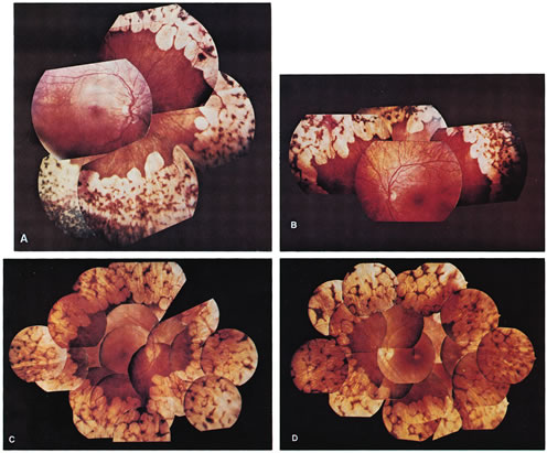
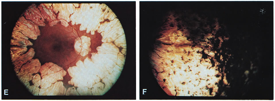
| Fig. 1. Representative fundus photographs of patients with gyrate atrophy (GA). A and B. The right and left fundi, respectively, of a 9-year-old white female patient with relatively early lesions. There is total sparing of the retina within the circummacular vascular arcade and a lack of pigment between the gyrate lesions (corrected visual acuity, 20/30 RE, 20/25 LE). C and D. The appearance of the right and left fundi, respectively, of a 16-year-old white male patient. The lesions extend considerably farther posteriorly than in the younger patient in A and B. Also, in the left eye, there is increased pigmentation between the lesions, as well as a foveal lesion and peripapillary lesions (corrected visual acuity, 20/100 RE, 20/80 LE). E. The posterior pole in the right eye of a 31-year-old white female patient with advanced GA. The gyrate lesions extend within the circummacular vascular arcade; a large peripapillary lesion is present. F. The far periphery of the eye shown in E. There is extensive accumulation of pigment, typical of advanced GA. |