Chapter 73
Cataract: Clinical Types
NATHAN G. CONGDON, MARGARET A. CHANG, PAUL BOTELHO, WALTER J. STARK and MANUEL B. DATILES III
Main Menu   Table Of Contents

Search

AGE-RELATED CATARACTS
NUCLEAR CATARACTS
CORTICAL CATARACTS
POSTERIOR SUBCAPSULAR CATARACT
MIXED CATARACTS
OTHER, LESS-FREQUENT AGE-RELATED ANATOMIC CATARACT TYPES
OTHER CLINICAL CATARACT TYPES
TRAUMATIC CATARACT
CATARACT ASSOCIATED WITH PRIMARY OCULAR DISEASES
CATARACT ASSOCIATED WITH SYSTEMIC DISEASES
CATARACT ASSOCIATED WITH METABOLIC DISORDERS
CATARACT ASSOCIATED WITH RENAL DISEASE
CATARACT ASSOCIATED WITH CUTANEOUS DISEASE
CATARACT ASSOCIATED WITH CONNECTIVE TISSUE AND SKELETAL DISORDERS
CATARACT ASSOCIATED WITH CENTRAL NERVOUS SYSTEM DISEASE
CATARACTS CAUSED BY ENVIRONMENTAL EXPOSURE: RADIATION AND PHARMACEUTICALS
CATARACTS CAUSED BY ELECTRIC SHOCK
OTHER RELATED CONDITIONS OF THE LENS
REFERENCES

Of the various clinical types of cataracts known today, the majority are age-related cataracts. It has been estimated that more than 75% of persons older than 75 years have lens opacities.1 Therefore, the first part of this chapter addresses the various types of age-related cataracts, and the second part is concerned with the less-common types of cataracts. Table 1 serves as a classification of the various types of cataracts and as a general outline of this chapter.

TABLE 1. Classification of Cataracts

  1. By anatomic location
    1. Nuclear
    2. Cortical
    3. Posterior subcapsular
    4. Mixed
    5. Other
      1. Capsular (polar)
      2. Anterior subcapsular
      3. Lens epithelial decompensation
      4. Retrodots
      5. Advanced
  2. By etiology
    1. Age-related
    2. Congenital and juvenile
      1. Part of systemic syndrome (see below)
      2. Isolated
      3. AD
      4. AR
      5. X-linked
    3. Traumatic
    4. Associated with primary ocular diseases: uveitis, glaucoma (glaukomflecken), retinal detachment, retinal degeneration (retinitis pigmentosa, gyrate atrophy), sclerocornea, microphthalmos, intraocular tumor (various), Norrie's disease, persistent hyperplastic primary vitreous, retinopathy of prematurity, aniridia, Peters' anomaly, high myopia, retinal anoxia, anterior segment necrosis
    5. Associated with systemic disease:
      1. Metabolic disorders: diabetes mellitus, galactosemia, hypoparathyroidism/hypocalcemia; Lowe's syndrome; Wilson's, Fabry's, and Refsum's diseases; homocystinuria
      2. Renal disease: Alport's disease (Lowe's syndrome)
      3. Cutaneous disease: congenital ectodermal dysplasia; Werner's and Rothmund-Thomson's syndromes; atopic dermatitis
      4. Connective tissue/skeletal disorders: myotonic dystrophy, WMS, and Conradi's, Stickler's, and Marfan's syndromes
      5. Central nervous system: Marinesco-Sjögren's syndrome, bilateral acoustic neuroma (neurofibromatosis type 2)
      6. Down's syndrome (Trisomy 21)
    6. Caused by environmental exposure
      1. Ionizing radiation: x-ray, ultraviolet rays, infrared rays, microwaves
      2. Pharmaceuticals: steroids, naphthalene, triparanol, lovastatin, ouabain, ergot, chlorpromazine, thallium (acetate and sulfate), dinitrophenol, dimethyl sulfoxide, psoralens, miotics, paradichlorobenzene, sodium selenite
      3. Electric
      4. Infectious: rubella, other TORCH infections
      5. Postsurgical: glaucoma, parsplana vitrectomy
    7. Other
      1. Ectopic lentis
      2. Lenticonus and lentiglobus

A cataract is commonly defined as any opacity in the ocular lens, and a wide variety of descriptive terms have been used by clinicians to describe these opacities. In this chapter, we mention and attempt to clarify some of the more commonly used descriptive terms, but for the most part we describe the various types of cataracts in terms of their anatomic location.

Back to Top
AGE-RELATED CATARACTS
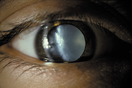
During the Middle Ages, Arabian physicians called the diffuse white appearance (Fig. 1) of total cataracts nuzul-el-ma, which means “a flowing down of water” or “blue water.” The Carthaginian monk Constantius Africanus, of the Monte Cassino Monastery, translated the ancient Arabic manuscripts into medieval Latin and applied the word cataracta, meaning “to rush down, as a waterfall.”2 In modern times, the development of devices that allow the lens to be examined at high magnification (e.g., the slit-lamp biomicroscope) has helped reveal many early changes that can occur in the human lens. Patients' increasing visual demands, as well as advances in the field of cataract surgery, have resulted in earlier removal of cataracts, and this has made the waterfall-like appearance of advanced cataracts a rarity.

Fig. 1. Bilateral mature cataract with white opacities visible to the naked eye. Cataract is the most common cause of treatable blindness in the world.

The lens maintains its clarity because of its architectural structure and composition. The lens fibers are laid down in specific arrays and are composed mainly of crystallin proteins. When biochemical or physicochemical changes develop that disrupt the architecture or result in the formation of molecules larger than 1,000 nm (10,000 Å), the affected area loses its transparency and opacity results.3

However, the resulting opacity must be located at or near the visual axis for it to affect vision and become clinically important. Very often peripheral cataracts develop without affecting the visual acuity of the patient, and are therefore considered clinically benign. These opacities may exist for many years, even decades, without necessitating any treatment other than optical correction.

Age-related cataracts appear in late adulthood and may cause a painless progressive loss of vision over the course of several years. Cataract is the leading cause of low vision in the United States4 and of blindness in the world.5 To date, studies on possible medical treatments for cataracts have not proved them to be safe and effective in humans.6

When the patient's visual function becomes compromised by the cataract, the only effective treatment available today is surgical extraction and replacement of the cataractous lens with an intraocular lens. Various studies have suggested that cataract surgery is one of the most cost-effective surgical interventions.7,8 The techniques used for the surgical extraction of cataracts are constantly being refined and improved. In the last decade, the frequency of cataract extraction has increased to more than one million per year in the United States alone, and it has become the most commonly performed surgical procedure for Medicare beneficiaries.9 An estimated 20.5 million Americans 40 years and older (17.2%) have cataract, and approximately 6.1 million Americans (5.1%) have undergone cataract surgery.1

In recent attempts to develop a more uniform classification, clinical researchers have used the anatomic locations of lens opacities as a means of classifying cataracts based on photographic standards. The current consensus divides age-related cataracts into three major pure anatomic types (cortical, nuclear, and posterior subcapsular) and mixed types (combinations of these three). Classification schemes such as the Lens Opacities Classification System (LOCS) II10 and III,11 the Oxford Cataract Classification System,12 and the systems used in the Beaver Dam Eye Study,13 at Johns Hopkins,14 and in the Age Related Eye Diseases Study15 use photographic standards to further subdivide each major type into grades. These grades are based either on density and color (in the case of nuclear cataract) or the anatomic area affected by opacity (in the cases of cortical cataract and posterior subcapsular cataract (PSC)). One can directly grade the patient's lens as seen with the slit-lamp (clinical grading), or evaluate photographs of the lens being studied (photographic grading). In addition, an automated grading system that uses densitometry was recently developed.16,17

Back to Top
NUCLEAR CATARACTS
Nuclear cataracts are the most common subtype in most European-derived populations.18 Studies have documented a gradual increase in optical density of the nucleus with increasing age in normal adults with 20/20 vision (Fig. 2).19

Fig. 2. Densitometry of the lens in various age groups, showing a gradual increase in total density related to normal aging.

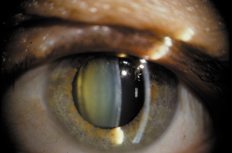
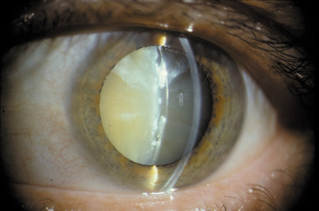
Nuclear cataracts tend to progress slowly. The refractive index of the lens changes as the nucleus progressively hardens, which usually results in increasing myopia.20,21 In some patients this is accompanied by optical distortion, especially of distant images, while near vision remains unaffected. A nuclear cataract is best seen with the narrow-beam direct illumination employed by the slit-lamp, which reveals the color and generalized haze or opalescence of the nucleus. In the early stages, the two halves (cotyledons) of the embryonic nucleus remain visible (Fig. 3). Later the entire nucleus appears as a homogeneous mass in contrast to the cortex (Fig. 4). Retroillumination may show the “oil droplet” effect (Fig. 5). Sometimes one may notice crystals in the lens nucleus (known as a Christmas tree cataract; Fig. 6A and B).

Fig. 3. Early nuclear cataract. Note the “cotyledons” in the nucleus.

Fig. 4. Advanced nuclear cataract. Note the homogeneous nuclear opacity.

Fig. 5. “Oil droplet” appearance of a pure nuclear cataract seen on retroillumination examination.

Fig. 6. Slit-lamp appearance of a Christmas tree cataract, showing crystals in the nucleus.

Nuclear cataracts are associated with physiochemical changes in the lens structural proteins (α-, β-, and γ-crystallins). These proteins undergo oxidation, nonenzymatic glycosylation, proteolysis, deamidation, phosphorylation, and carbamylation, which lead to the aggregation and formation of high-molecular-weight proteins. It was recently proposed22 that α-crystallin may play a role as a molecular chaperon in preventing the aggregation of protein. These high-molecular-weight (>1,000 nm) protein aggregates interfere with light transmission and cause light scattering in nuclear cataracts. Chemical modification of the nuclear lens protein also leads to yellowing, followed by browning and, in advanced stages, blackening.23 Whether these color changes are related to the cataract or are independent of it is still being debated and investigated. Recent studies have also suggested that phase separation inhibitors (PSIs) may play a role in maintaining a clear nucleus, and that the loss of these PSIs may lead to nuclear cataract formation.24

Back to Top
CORTICAL CATARACTS
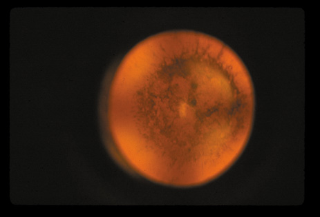
The cortical cataract is often the most common subtype of opacity seen in African-derived populations.18 The cortical layer is approximately 2 mm thick anterior and posterior to the nucleus in adults, and its metabolically active new cortical fibers are continuously laid down by the bow region cells. The cortical layer is less compact than the nucleus and is therefore more prone to becoming overhydrated as the result of an electrolyte imbalance. This may eventually lead to disruption of the lens cortical fibers, as demonstrated in diabetes25 (Fig. 7) and galactosemia.26 It has therefore been proposed that this type of cataract may be partly caused by osmotic stress. Early changes may include signs such as vacuoles, water clefts, and lamellar separation (Fig. 8). These changes may come and go over time, but eventually they may lead to damage and irreversible opacification of some fibers. Recent studies have also suggested that an abnormality of the maturation process of the lens fibers may lead to subsequent poor development of the primary, secondary, and tertiary sutures, resulting in the formation of cortical cataracts.27

Fig. 7. True diabetic cataract with marked swelling of the cortex. This type of cataract is usually of acute onset and typically occurs in type I diabetes.

Fig. 8. Early cortical changes. Vacuoles, water clefts, and lamellar separation.

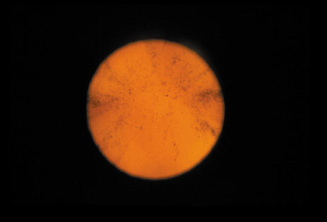
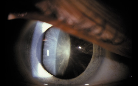
Cortical opacities have been clinically observed to develop earliest in the inferior half of the lens, especially the lower nasal quadrant.28 Epidemiologic29 and laboratory studies30 have suggested that cortical cataracts may be caused by ultraviolet rays from sunlight. The supraorbital margins may block the ultraviolet rays from falling over the upper part of the lens, thus making cortical cataract less frequent in the upper quadrants. Eventually these opacities also develop in the periphery in other quadrants, resulting in a circular array of spokes and peripheral cuneiform opacities (Fig. 9). Bands of central cortical fibers may become prominent and opacify centrally (Fig. 10). However, most cortical cataracts remain in the periphery for many years, even decades, before the central axis of the lens becomes involved, causing loss of vision late in the development of the cataract.

Fig. 9. Moderate cortical changes. Wedge-shaped (cuneiform) or spoke-like (wheel) peripheral changes are seen. These changes may be extensive but may not affect Snellen visual acuity since they occur in the periphery.

Fig. 10. Central cortical opacities. This type of cortical opacity may causes early diplopia and glare disability.

It has been observed that some individuals may have cortical opacities covering the entire anterior cortical and posterior cortical area (Fig. 11), and yet have 20/40 or better Snellen visual acuity under standard testing conditions. However, these patients may have severe disability glare such that under simulated bright lights their visual acuity may decrease to 20/80 or worse.31 They may also have decreased contrast sensitivity. These individuals tend to do well indoors but have difficulty driving during bright, sunny days, and at night because of oncoming headlights. Treatment in these cases must be decided on an individual basis, and surgery may be indicated when the expected benefits outweigh the surgical risks.

Fig. 11. Advanced cortical cataract. Although this type of cataract may be compatible with a Snellen visual acuity of 20/40 or better, it may give rise to severe glare disability.

This type of cataract is best seen with retroillumination, which gives an enhanced picture of the cortical spokes and vacuoles by the shadows they cast as the light is reflected back by the fundus. Direct illumination helps clarify the level of the opacities (see discussion in the Posterior Subcapsular Cataract section below).

Back to Top
POSTERIOR SUBCAPSULAR CATARACT
The PSC is the least prevalent subtype in most population-based studies.18 These cataracts often occur in combination with nuclear or cortical cataracts in the later stages. They are easily noticed on retroillumination because they are usually located centrally, and may interfere with funduscopy (Fig. 12). In early stages, patients usually complain of subjective symptoms such as glare disability32 and difficulty focusing on near objects. This is because when the pupil constricts during accommodation, the light entering the eye becomes concentrated centrally, where the PSC is also located. This causes light scattering and interferes with the ability of the eye to focus an image on the macula. In addition, these opacities lie at or near the nodal point of the eye, further interfering with focusing of the image on the macula.

Fig. 12. PSC. Note the central location, which gives rise to severe glare disability.

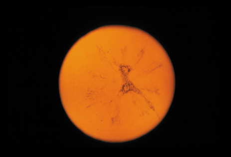
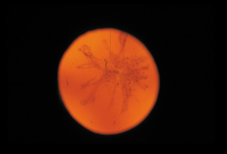
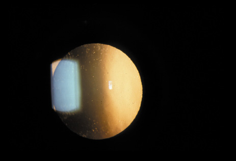
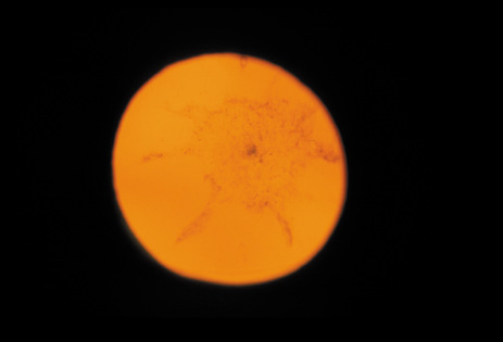
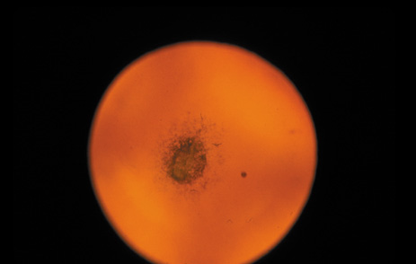
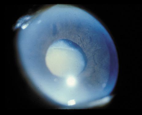
One can examine this type of cataract with direct illumination, using the narrow and broad beams of the slit-lamp to show the characteristic granular inner surface immediately in front of the posterior capsule (Fig. 13). The problem with this technique, however, is that patients may not tolerate any prolonged direct illumination because of the glare. Retroillumination is therefore more useful for revealing the outline of the opacity, since it is usually seen as an “island” in the center of the posterior capsule, which is further highlighted by the shadow cast by the opacities.33 However, in the early stages of this type of cataract, the dust-like particles that might be noticeable in the central posterior subcapsular area with direct illumination disappear or are difficult to see with retroillumination (Fig. 14). Eventually this “dusting” becomes dense enough to cast a shadow and thus appear on retroillumination. The smooth orange background of the fundus helps to highlight the rough, irregular pseudopodia-like edges of the central opacity. In advanced stages, the PSC may become a thick, calcified plaque (Fig. 15). During surgery, excessively vigorous scraping or vacuuming of the calcified opacity can lead to rupture of the posterior capsule. Usually, small remnants that are left behind after surgery are reabsorbed and do not interfere with vision; otherwise, they are easily treated with a neodymium : yttrium (Nd:YAG) aluminum garnet laser. Pathologic evidence suggests that most PSCs result from the migration of bow region cells into the potential space (along with accumulated cellular debris) between the posterior capsule and the cortex.34–36

Fig. 13. Direct slit-lamp illumination of a PSC, showing an irregular granular surface in front of the posterior capsule.

Fig. 14. A PSC, showing vacuoles and dust-like material in the potential space between the posterior cortex and the capsule.

Fig. 15. Example of an Oxford camera retroillumination image of a PSC, and the automated method used to measure the area of cataract.

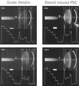
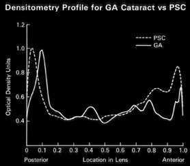
PSC may also result from irradiation or steroid ingestion, or it may be associated with diabetes, high myopia,37–41 retinal degeneration (e.g., retinitis pigmentosa),42,43 and gyrate atrophy.44,45 In some cases, the PSC eventually may be pushed to the cortex as new fibers are laid down and the offending agent is no longer present (Fig. 16A and B).

Fig. 16. A: Scheimpflug slit image of two types of PSCs (steroid-induced and gyrate atrophy-related) showing the positions of the opacities. B: Densitometry profiles showing the position of the gyrate atrophy-related PSC in comparison with the steroid-induced PSC. These suggest that the PSC migrates anteriorly as the newly laid down fibers push the PSC deeper into the cortex.45

Back to Top
MIXED CATARACTS
Sometimes more than one of the above-described varieties of cataract will occur together in a lens (Fig. 17). In general, a cataract will start as a pure type but eventually become mixed as the other lens regions become involved in the degenerative process.

Fig. 17. Slit-lamp photographs of a mixed cataract, showing opacities in the cortex, nucleus, and posterior subcapsular areas.

Back to Top
OTHER, LESS-FREQUENT AGE-RELATED ANATOMIC CATARACT TYPES

CAPSULAR (INCLUDING POLAR) CATARACTS

The lens capsule may develop localized opacities in age-related cataracts. However, these opacities are more often associated with persistent pupillary membranes or epicapsular stars, and they may also occur in uveitis in association with posterior synechiae or secondary to injury caused by drugs, radiation, or trauma. Capsular thickening may also occur in heat (glassblowers') cataract, and localized thickening may occur in both Lowe's syndrome and Miller's syndrome.46

Localized central capsular cataracts (polar cataracts) can occur in the anterior and posterior capsules and are usually congenital, although they may also occur secondary to trauma. Polar cataracts (Fig. 18) are usually dense, localized, and nonprogressive. Because they are stable, many patients may be able to tolerate them and may retain good or adequate vision with conservative treatment (e.g., dilation of the pupil, wearing sunglasses on bright days, and optical correction).

Fig. 18. Posterior polar cataract. This type of cataract is usually congenital, but may also be acquired.

ANTERIOR SUBCAPSULAR CATARACTS

Anterior subcapsular cataracts (Fig. 19) result from the aberrant differentiation and growth of lens epithelial cells to form fibrotic plaques.47–49 Immunolabeling studies of anterior subcapsular cataracts have revealed cytoskeletal and extracellular matrix proteins that are not normally expressed by the lens.50,51Transforming growth factor beta (TGF-β) has been shown to induce anterior subcapsular cataracts in animal models.47,48,52,53 Anterior subcapsular cataracts have been reported following ocular trauma and in association with atopic dermatitis.48,54,55 Pure anterior subcapsular cataracts may be more common in the Korean population.50

Fig. 19. Anterior subcapsular cataract. Note the blurred image of a PSC behind the anterior subcapsular cataract.

LENS EPITHELIAL DECOMPENSATION

In some cases the entire anterior lens epithelium is observed to be edematous with resulting generalized haze (Fig. 20A). This is usually followed within a short period of time (a few months to 1 year) by the development of cortical cataracts and PSCs that mature quickly and cause severe, rapid loss of vision (Fig. 20B).

Fig. 20. A: Slit-lamp photograph of lens epithelial decompensation. Note the early nuclear opacity and cloudy lens epithelium. B: Four months later, the same lens is completely opacified.

RETRODOTS

Retrodots are round, translucent opacities that usually occur in the deep cortex or perinuclear region. It has been proposed that they contain calcium oxalate, probably from ascorbic acid.46 In general, they do not seem to affect vision until a mixed cataract appears (nuclear or cortical), and patients may have these retrodots for years and still retain good vision (Fig. 21).

Fig. 21. Retrodots seen on retroillumination examination as refractile bodies. These retrodots are usually seen in nuclear cataracts, are located in the perinuclear or deep cortical areas, and usually do not affect vision.

ADVANCED CATARACTS

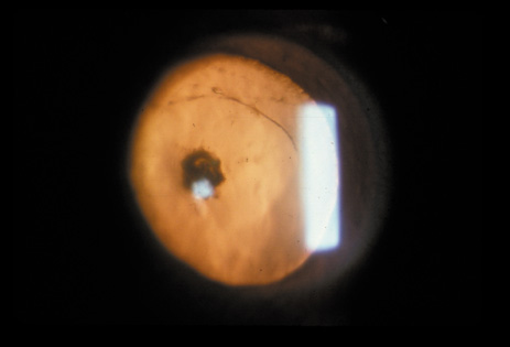
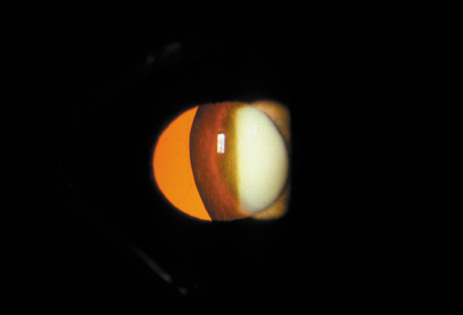
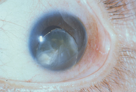
Advanced cataracts are usually mixed cataracts that reach the mature stage. A cataract is termed “mature” if the cortex and nucleus are so opaque that one can no longer see the red reflex; at this stage the lens looks white (hence the appellation “waterfall” or “cataract”)2 (see Fig. 1). In even more advanced cases, the white cortex becomes so liquefied that one can see the outline of the brown nucleus floating free inside the lens, which usually settles down with gravity when the patient rests for a while. This is called a morgagnian cataract (Fig. 22). If the lens is noticeably swollen, the cataract is called an intumescent cataract. If the lens appears silvery white and desiccated, with some leakage of the cortical fluid, it is called a hypermature cataract (Fig. 23). As mentioned above, these advanced stages are now rarely seen in the United States but are still fairly common in less-developed countries where surgical treatment is not readily available.

Fig. 22. Morgagnian cataract. This is a mature cataract that is also intumescent (swollen), being filled with liquefied cortex. A free-floating nucleus can be seen at the bottom of the lens.

Fig. 23. Trauma-induced hypermature cataract, showing changes in the capsule. The hole seen at the 9 o'clock position was caused by penetration with a foreign body that perforated both the iris and the lens.

Surgery on these advanced cases must be done carefully because the lens capsule and zonules can easily break during manipulation. When carefully performed, however, both intra- and extracapsular extraction techniques have successfully achieved complete restoration of sight for patients with advanced cataracts.

Before we leave the topic of age-related cataracts, we should point out that the mortality rates of patients who have cataract or have undergone cataract surgery may be elevated compared to matched controls.56 This has been interpreted to indicate that the presence of an advanced cataract may reflect a generalized state of decreased health in the patient. In addition, recent studies have also suggested that body mass is indirectly related to the incidence of cataracts. Again, this may indicate that a person's state of health is an important factor in cataract formation.57–59

Back to Top
OTHER CLINICAL CATARACT TYPES

CONGENITAL AND JUVENILE CATARACTS

By definition, congenital cataracts are detected at birth, whereas juvenile cataracts develop during the first 12 years of life. Both range from mild and benign to advanced and sight-threatening. Congenital cataracts are an important cause of blindness in children.60 Approximately one-third of congenital and juvenile cataracts are inherited. A wide degree of variation is seen in the morphologic characteristics of these cataracts. In the following paragraphs we present a morphologic classification of congenital cataracts.

I. Total or Complete Cataracts

Total or complete cataracts are completely opaque or hazy at birth. Most of these are associated with systemic disorders or abnormalities such as galactosemia, rubella, and Lowe's syndrome. They may also be hereditary (autosomal-dominant (AD) or autosomal-recessive (AR)) (Fig. 24).

Fig. 24. Total congenital cataract.

II. Partial or Incomplete Cataracts

A. ANTERIOR AND POSTERIOR POLAR CATARACTS

(see Fig. 18). These cataracts involve the lens capsule in the anterior or posterior pole of the lens. They are sometimes associated with a localized anatomic abnormality in the region. For example, posterior polar cataracts commonly occur in cases of posterior lenticonus. They may cause more visual symptoms because they are closer to the nodal point of the eye; however, they are usually stable, and patients may do well with conservative measures. Anterior polar cataracts may be associated with absence of the lens capsule. Anterior and posterior polar cataracts are associated with abnormalities in the gene that encodes the major intrinsic protein of the lens fiber (chromosome 12q3), which is inherited as an AD trait. Posterior polar cataract may also be associated with AD defects in α-2 crystallin (chromosome 21q22.3) and γ-D crystallin (chromosome 2q33-q35).

B. ZONULAR CATARACT

In this type of cataract, only a region or zone of the lens is opaque. They may be stationary, but may also progress. There are various subtypes, as described below:

1. Lamellar. This is the most common type of congenital cataract. Such cases are usually bilateral and symmetric, and the density of opacification may vary considerably (Fig. 25A and B). Less opaque lamellar cataracts may be compatible with good vision and minimal medical intervention (e.g., optical correction and therapeutic mydriasis). These cataracts may be inherited as an AD trait through defects in γ-C and γ-D crystallin (chromosome 2q33-q35), but in some cases they may be attributed to a transient intrauterine toxic agent that affects only the layer of cells that develop at the time of fetal exposure.

Fig. 25. A. Direct illumination photograph of a lamellar cataract, showing a central opacity. B. Retroillumination photograph of a lamellar cataract, showing the red reflex from the fundus visible through the cataract. Patients with such cataracts do well with conservative treatment (e.g., dilation and optical correction) and can achieve 20/40 vision or better.

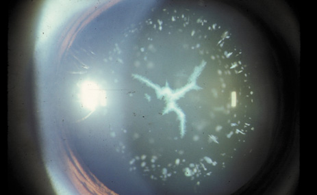
2. Stellate. These cataracts affect the region of the sutures. They may be Y-shaped if the cataract occurs in the intrauterine stage of development, since the sutures have this configuration during this period. Anterior sutural cataracts are Y-shaped, and posterior sutural cataracts are shaped like an inverted Y. Sutural cataracts that develop later on have a more stellate shape (Fig. 26), in keeping with the shape of the sutures after birth. Stellate cataracts can be inherited in an AD pattern through mutations in the β-B1 crystallin (chromosome 22q11.2-q12.1) and the major intrinsic protein of the lens fiber (chromosome 12q3).

Fig. 26. Sutural or stellate cataract. Note the blue-green dot opacities in the cortex (Courtesy of Ernest Kuehl.)

3. Nuclear (Fig. 27). These cataracts are usually bilateral and involve the fetal or embryonal nucleus. They may be inherited as an AD, AR, or X-linked trait. The known loci of genetic mutation include α-1 crystallin (chromosome 21q22.3), γ-C crystallin (chromosome 2q33-q35), and γ-D crystallin (chromosome 2q33-q35), all of which are AD.

Fig. 27. Nuclear congential cataract.

4. Coronary (Fig. 28). These cataracts are radial, club-shaped discrete opacities that are located in the cortex. They are called “coronary” because they resemble the top of a crown. Because of their peripheral location, they do not decrease visual acuity. Coronary cataracts are dominantly inherited and have been described in cases of Down's syndrome, glutathione peroxidase deficiency (chromosome Xq28), and myotonic dystrophy.

Fig. 28. Coronary cataract.

5. Cerulean. These cataracts consist of small, discrete opacities that have a distinct bluish hue (see Fig. 29). They are located in the cortex, are nonprogressive, and do not cause visual symptoms. They may be present together with other congenital cataracts. Others are dominantly inherited through mutations in γ-D crystallin (chromosome 2q33-q35).

Fig. 29. Nuclear, cerulean, and stellate cataracts.

C. MEMBRANOUS CATARACTS

These cataracts are thin but dense and contain fibrous tissue. They may occur when lens proteins are reabsorbed (e.g., traumatized lens; see following section), such that the anterior and posterior lens capsules fuse and produce a dense membrane. They are also associated with defects in type II collagen (chromosome 12q13.11-q13.2).

Back to Top
TRAUMATIC CATARACT
Cataracts can occur secondary to trauma to the lens.61 The morphologic characteristics differ between cataracts caused by blunt trauma and cataracts secondary to penetrating trauma. Cataracts secondary to blunt trauma often have a rosette-shaped appearance (Fig. 30) or are of the PSC variety. In cataracts secondary to penetrating trauma, the size of the opening in the lens capsule determines the morphology of the cataract. When the opening is large, the whole lens may become cataractous. When the opening is small, it may sometimes seal by itself and leave behind an opacity that is localized to the site of penetration.

Fig. 30. Traumatic cataract. Note the stellate opacity in the nucleus, which was seen on retroillumination examination after a blunt trauma.

Back to Top
CATARACT ASSOCIATED WITH PRIMARY OCULAR DISEASES
Cataracts can occur secondary to a large number of intraocular diseases. Intraocular inflammation is the most common cause of this type of cataract. These cataracts are typically of the PSC variety. PSCs seen in retinitis pigmentosa (Fig. 31), gyrate atrophy (Fig. 32), and Usher's syndrome (Fig. 33) show finger-like projections. In persistent hyperplastic primary vitreous, the PSC is often associated with abnormal blood vessels from the hyaloid system that arborize from the posterior pole of the lens. Cataracts seen in retinal anoxia and anterior segment necrosis are thought to occur due to interference with the nutrient supplies of the lens. This leads to decreased anabolism, increased catabolism and acidity, and necrosis. The cataracts in these conditions are also of the PSC type. An acute increase in intraocular pressure can cause focal necrosis of the subcapsular epithelium and localized, fleck-like opacities (glaukomflecken). These opacities are initially located immediately under the capsule, but when new fibers are laid down they slowly become buried in the lens. Their presence indicates that the patient has experienced an acute increase in intraocular pressure. The premature occurrence of PSC and possibly nuclear-type cataracts has been noted in eyes with high myopia (Fig. 34).37–41

Fig. 31. PSC in a 25-year-old man with retinitis pigmentosa. (Courtesy of Muriel Kaiser Kupfer, M.D.)

Fig. 32. PSC in a patient with gyrate atrophy of the choroid and retina. (Courtesy of Muriel Kaiser Kupfer, M.D.).

Fig. 33. PSC in a patient with Usher's syndrome (retinitis pigmentosa plus deafness). (Courtesy of Muriel Kaiser Kupfer, M.D.).

Fig. 34. PSC in a patient with high myopia.

In patients with aniridia (Fig. 35),62 Peters' anomaly, and neurofibromatosis type II (Fig. 36),63 both PSCs and cortical cataracts64 may occur.

Fig. 35. Congenital cataract in a patient with aniridia. (Courtesy of Muriel Kaiser Kupfer, M.D.).

Fig. 36. PSC in a patient with bilateral acoustic neuroma (neurofibromatosis type 2). (Courtesy of Muriel Kaiser Kupfer, M.D.).

Back to Top
CATARACT ASSOCIATED WITH SYSTEMIC DISEASES
The systemic conditions associated with cataract formation can be grouped into five broad categories (see Table 1):
  1. Metabolic disorders
  2. Skin disease
  3. Connective tissue/skeletal disorders
  4. Renal disease
  5. Central nervous system disorders
Back to Top
CATARACT ASSOCIATED WITH METABOLIC DISORDERS

DIABETES MELLITUS

Diabetes mellitus is a significant risk factor for cataract formation. It has been reported that cataracts reach visual significance 10 years earlier in the presence of diabetes than they would otherwise.65,66

In diabetes, hyperglycemia leads to the diffusion of increased amounts of glucose into the lens, which is then converted into the sugar alcohol sorbitol by the enzyme aldose reductase.25,26,67,68

Sorbitol does not readily pass through the cell membranes, and is therefore trapped inside the lens, causing an osmotic imbalance. This results in the influx of water into the lens to balance the osmotic pressure, which causes the lens fibers to swell and eventually rupture. This cataract type has been demonstrated in experimental animals and has been completely prevented in animal models with the use of various potent aldose reductase inhibitors.68–70 However, aldose reductase enzyme levels in human lenses have been found to be lower than in animal models of diabetic cataract, and so far the few aldose reductase inhibitors that have been tested (in a limited way) have not been shown to have much effect on human diabetic cataracts.

Another possible mechanism of cataract formation in patients with diabetes is overaction of the proteolytic enzyme calpain 2. This intracellular cysteine protease, in the setting of elevated lens calcium 2+ levels, was found to be cataractogenic in several diabetic animal models.71,72,73 Calpain 2 has been identified in human lenses, and was reported by one study to be the major calpain expressed in epithelial cells of human lenses with cortical cataracts.74,75

Other potential causes of increased cataract formation in diabetes include elevated lens advanced glycated end-products found in human subjects compared to controls,65 and oxyradical-induced lens DNA changes with apoptosis in a diabetic animal model (with a protective effect from pyruvate).76

Transient changes in refraction, which may be either a myopic or a hyperopic shift, may accompany this swelling in some diabetic patients. Diabetics also exhibit a decreased amplitude of accommodation.

Two types of cataracts result from diabetes. The first one is the true diabetic (snowflake) cataract, which is quite rare. It is usually bilateral, occurs rapidly, and is usually related to very high, uncontrolled levels of serum glucose in young type II diabetics. The word “snowflake” is used to describe these cataracts because in the early stages one notices multiple white anterior and posterior subcapsular and cortical opacities, as well as vacuoles (Fig. 37). Later, as more lens fibers become involved, water clefts appear (Fig. 38) in the cortex, and the lens becomes swollen and opaque. This closely mimics what occurs in experimental animals, as mentioned above. The second, more common type is the age-related diabetic cataract, which occurs in type I or II diabetics. It is very similar to other age-related cataracts, and may present as a cortical, posterior subcapsular (Fig. 39), or (less frequently) nuclear cataract. These cataracts have an earlier onset than other age-related cataracts, and patients with such cataracts undergo cataract surgery sooner. These cataracts may result from osmotic causes and increased glycosylation of lens proteins, in addition to the other mechanisms that lead to age-related cataract (e.g., oxidation and phase separation).

Fig. 37. True diabetic cataract. Early snowflake and spoke cortical opacities in type I diabetes. (Courtesy of David Cogan, M.D.).

Fig. 38. Early cortical swelling in a patient with a true diabetic cataract.

Fig. 39. PSC in a patient with type II diabetes.

GALACTOSEMIA

Galactosemia, which refers to a group of AR inborn errors of galactose metabolism, is a well-recognized cause of cataract in infants.77 Classic galactosemia is due to a deficiency in galactose-1-phosphate uridyltransferase (GALT) activity. Affected patients may develop significant neurologic abnormalities, including mental and motor retardation, dyspraxia, and hypergonadotropic hypogonadism, in addition to cataract formation. By contrast, numerous case series of galactokinase deficiency, a less common cause of galactosemia, have reported isolated cataract formation.78–80 As in diabetic cataract formation, galactosemic cataracts occur as a result of osmotic swelling of lens fibers secondary to a metabolic abnormality: as serum galactose accumulates, it enters the lens and is converted to the sugar alcohol galactitol by the enzyme aldose reductase. Because galactitol does not pass readily through cell membranes, it accumulates in the lens, resulting in the entry of water to correct the osmotic imbalance. This leads to swelling and rupture of lens fibers and cataract formation. As in diabetic animal experiments, galactosemic animal experiments have shown that these cataracts can be prevented completely with the use of potent aldose reductase inhibitors.81 Reversal of these cataracts is known to occur if galactose is removed from the diet.

Reduced activity of GALT has been proposed as a risk factor for the development of idiopathic cataract formation. Several studies have shown an increased rate of isolated cataract formation in heterozygotes for classic galactosemia, as well as homozygotes with the less severe Duarte or Los Angeles variant.82–87

Although galactokinase enzyme mutations are known to cause congenital cataracts, recent studies suggest that the Osaka variant of galactokinase is associated with modestly increased rates of formation of age-related cataracts.80 Given that galactokinase thus appears to be associated with both congenital and age-related cataracts, future research investigating other loci associated with congenital cataracts may likewise shed light on the pathogenesis of age-related cataracts.

HYPOPARATHYROIDISM/HYPOCALCEMIA

Cataracts secondary to hypocalcemia show fine, discrete white dots in the perinuclear zone. Patients with hypoparathyroidism/hypocalcemia also have skeletal abnormalities, such as frontal bossing and increased osteoporotic changes, as well as a serum calcium-to-phosphorus ratio that is well below normal

LOWE'S (OCULOCEREBRORENAL) SYNDROME

Lowe's syndrome is characterized by congenital glaucoma and cataract. Close relatives of patients with this disorder show punctate lenticular opacities suggestive of a carrier state (Fig. 40).88

Fig. 40. Cortical dot opacities in a carrier of Lowe's disease (oculocerebrorenal syndrome). (Courtesy of Muriel Kaiser Kupfer, M.D.).

WILSON'S DISEASE

Wilson's disease, which is characterized by increased serum copper levels and the deposition of copper in basement membranes throughout the body, causes a kind of lenticular opacity called sunflower cataract. This is characterized by a fine, powdery deposition of copper just underneath the lens capsule, which gives it a brilliant yellow, brown, or red sheen. The deposition is more concentrated in the pupillary area and is disc-like centrally. Extending from this disc-like area are radiating petal-like deposits (hence, the sunflower appearance). These cataracts do not interfere with vision and may resolve with systemic therapy for the increased copper levels.89

FABRY'S DISEASE

Fabry's disease occurs secondary to an abnormality in lipid metabolism, and is characterized by feathery opacities radiating in the posterior subcapsular regions.90 These cataracts are mild and seldom cause significant visual symptoms

HOMOCYSTINURIA

In 90% of patients with homocystinuria, the zonules show abnormalities leading to a subluxation of the lens that is often in the inferonasal direction.91 Occasionally, congenital cataract is associated with this condition.92 Patients with homocystinuria have thrombotic tendencies and must be monitored carefully when they are under general anesthesia

REFSUM'S DISEASE

Refsum's disease, whic is a condition of abnormal lipid metabolism, is often associated with PSCs.93 Patients with this disease have an atypical pigmentary degeneration of the retina.

Back to Top
CATARACT ASSOCIATED WITH RENAL DISEASE
Patients with Lowe's or Alport's syndrome are known to have cataracts. Ocular manifestations (most commonly anterior lenticonus) occur in approximately 15% of patients with Alport's syndrome.94 Often there is a localized posterior polar opacity associated with the conic area. The opacity or sometimes the severe irregular astigmatism induced by the lenticonus can cause disabling visual symptoms, which often necessitate cataract extraction.
Back to Top
CATARACT ASSOCIATED WITH CUTANEOUS DISEASE
That lenticular changes should act in consort with cutaneous changes in some cutaneous disorders is not surprising given their common ectodermal origin. Lenticular changes are common in Rothmund's syndrome, atopic dermatitis, and Werner's syndrome, and may be seen in incomplete forms of congenital ectodermal dysplasia.92 The features of these cataracts are outlined along with their respective clinical conditions as follows:

Atopic dermatitis: Cataracts occur in 8% to 10% of cases, are bilateral, develop in the third to fifth decade of life, and usually are in the form of a dense anterior or posterior subcapsular plaque with radiating opacities into the cortex. Eventually complete opacification of the lens is seen.

Rothmund's syndrome: Cataracts are of the zonular type and typically develop between 2 and 4 years of age. They are bilateral, occur mainly in girls, develop as punctate opacities, and evolve rapidly.

Werner's syndrome: Cataracts commence with the formation of striae that are located in the posterior cortical and subcapsular areas, and have a metallic sheen. They develop during the second or third decades of life, rapidly become intumescent, and progress to total opacification of the lens.

Congenital ectodermal dysplasia: The complete forms of this disorder are not usually associated with cataract. In incomplete forms of the disease, however, congenital cataract may be seen along with other ocular disorders, such as Rieger's anomaly and vitreous fibrosis.

Back to Top
CATARACT ASSOCIATED WITH CONNECTIVE TISSUE AND SKELETAL DISORDERS
Myotonic dystrophy and Stickler's, Conradi's, and Marfan's syndromes are all associated with cataract. The features of these cataracts are outlined along with their respective clinical conditions as follows:

Conradi's syndrome: Cataracts are associated with two of the three genetic types of this syndrome. In the lethal, AR type, the cataracts are congenital, dense, and symmetric. A milder, asymmetric, acquired cataract is the only ocular finding in the X-linked dominant type. No cataracts are associated with the AD type.95

Myotonic dystrophy: Cataracts are an essential part of the constellation of signs seen in this condition. They usually commence in the second or third decade of life and show a diffuse layer of dust-like, multicolored, punctate, and flaky opacities that may be located in the perinuclear zone or the posterior subcapsular area. They may remain stable for many years before progressing. The posterior subcapsular opacity may enlarge and increase in density, and an anterior subcapsular opacity may develop. The sutures may also opacify, causing a stellate opacity. Finally, the lens becomes diffusely opaque. Myotonic dystrophy is a well recognized cause of presenile cataract (Fig. 41), and should be suspected in patients with systemic features of the condition and the typical cataracts described above. These patients may also have associated ocular disorders, such as pigmentary changes in the retina, ocular hypotension,96 ptosis, and abnormal pupillary responses.

Fig. 41. Cortical cataract and PSC in a patient with myotonic dystrophy.

Marfan's syndrome: Marfan's syndrome is an AD disorder that affects the cardiovascular, musculoskeletal, and ocular systems. The causative defective gene located on the long arm of chromosome 15 codes for fibrillin 1, the main constitutive protein of the elastic tissue.97,98In one study, up to 70% of patients with Marfan's syndrome were found to have ectopic lentis.99 Dislocation of the lens (Fig. 42) has been positively correlated with increased axial length, which is a very common ocular abnormality in this syndrome. Localized opacities of the lens may also be present. The defective fibrillin 1 protein may be more susceptible to degradation by matrix metalloproteases, resulting in zonular instability.100,101 It has been proposed that the superotemporal subluxation may be due to UV-B light that is preferentially focused in the inferonasal quadrant, leading to decreased fibrillin expression in this location.102

Fig. 42. Subluxation of the lens in a patient with Marfan's syndrome. Note that the edge of the lens is seen in the pupillary space. (From Jaeger EA: Cataract types. In: Tasman W, Jaeger EA (eds). Atlas of Clinical Ophthalmology. Philadelphia: Lippincott-Raven, 1995.)

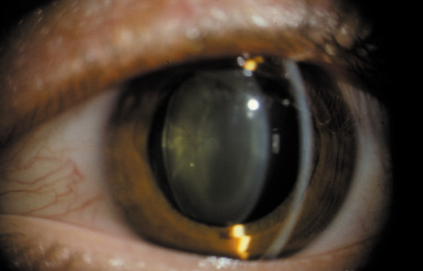
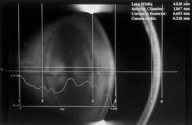
Weill-Marchesani syndrome (WMS): WMS is a systemic connective tissue disorder characterized by short stature, brachydactyly, joint stiffness, and characteristic ocular findings. These include microspherophakia (Fig. 43A and B), ectopic lentis, cataract formation, severe myopia, and acute or chronic glaucoma.103–105 Both AD and AR modes of inheritance have been reported in familial cases of WMS.106,107 A review of all published case reports of WMS found clinical homogeneity and genetic heterogeneity in WMS.105 Of note, AR cases were more commonly associated with microsherophakia (94% AR, 74% AD, Fischer 0.007) while ectopic lentis was present in a greater percentage of AD cases (84% AD, 64% AR, Fischer 0.016). Wirtz et al108 reported linkage of the AD form of WMS to the fibrillin-1 gene located on chromosome 15q21.1. A frame fibrillin-1 gene deletion in AD WMS was also identified.109 Underscoring the genetic heterogeneity of WMS was the recent identification of ADAMTS10 mutation in AR WMS. Three distinct mutations involving the ADAMTS10 gene on chromosome 19p13.3-p13.2 were identified in two consanguineous families and one sporadic case of WMS.110 ADAMTS10 is a member of the extracellular matrix protease family, and is believed to be anchored to the extracellular matrix through interactions with aggregan or other matrix components.111,112 A close interaction between fibrillin-1 and ADAMTS10 has been suggested as the explanation for the similarities in phenotypic expression found among genetic heterogeneous cases of WMS.110

Fig. 43. A: Slit photograph of a spherophakic lens. B: Scheimpflug photograph of a spherophakic lens in a 35-year-old patient, showing the anteroposterior dimension of the lens (lens width = 4.8 mm).

Stickler syndrome: Stickler syndrome, an AD disorder of connective tissue proteins, has multiple manifestations that affect the ophthalmic, orofacial, auditory, and articular systems. Micrognathia, depressed nasal bridge, and flat midface are the most commonly found orofacial features, and they tend to become less evident with age. Midline clefting may be of variable severity, ranging from a cleft soft palate to the Pierre-Robin sequence. Deafness may be present secondary to ossicle defects or sensorineural deficits. Joint hyperextensibility with later degenerative osteoarthropathy is also common.113,114 The most prominent ophthalmic manifestations are abnormalities of vitreous formation and architecture, and congenitally high myopia, which predispose to giant retinal tear formation and retinal detachment. Patients with Stickler syndrome may be more at risk of early-onset nuclear sclerosis and posterior subcapsular opacification.113 Congenital cortical cataracts may be present. These tend to be nonprogressive, and may have a curved113 or “wedge and fleck”114 distribution.

Back to Top
CATARACT ASSOCIATED WITH CENTRAL NERVOUS SYSTEM DISEASE
Cataracts have been described in conditions associated with anomalies of the brain, such as Little's disease, microencephaly, and Marinesco-Sjogren's syndrome. The characteristic features of such anomalies are congenital spinocerebellar ataxia, oligophrenia, and occasionally congenital cataract.92 Neurofibromatosis type 2 (bilateral acoustic neuroma) is associated with cortical cataract and PSC (see Fig. 36).63,64
Back to Top
CATARACTS CAUSED BY ENVIRONMENTAL EXPOSURE: RADIATION AND PHARMACEUTICALS

CATARACTS DUE TO IONIZING RADIATION

Continuous exposure to many of the wavelengths in the electromagnetic spectrum (e.g., x-rays, ultraviolet rays, microwaves, and infrared rays) can cause cataracts.115,116 An increased rate of cataract formation has been reported in astronauts and airline pilots.117–119 Often a latent period of 9 to 12 months or more exists between exposure to ionizing radiation and the onset of cataract. These cataracts are PSCs (Fig. 44) and classically show multiple vacuoles, as well as an evenly feathery appearance and weblike fringes. Ionizing radiation affects the germinative equatorial epithelium. The damaged cells form an abnormal plaque, which first appears at the posterior pole and progressively shows enlargement.

Fig. 44. Radiation-induced cataract in a 16-year-old girl.

CATARACTS SECONDARY TO PHARMACEUTICAL AGENTS

A large number of medications are known to induce cataracts (see Table 1). The following is a list of the more commonly used pharmaceuticals, along with the characteristics of their associated cataracts:

Corticosteroids: Both topically applied and systemically administered corticosteroids can cause cataract.120,121 The factors involved in the development of typical PSCs include 1) inhibition of the sodium-potassium-ATPase pump mechanism, which increases the permeability of the lens to cations;122 and 2) conformational changes in specific amino groups of the lens crystallins, which lead to the development of disulfide bonds and protein aggregation.123

Other possible mechanisms include a decreased expression of cadherin (a family of cell–cell adhesion molecules that control the calcium-dependent cell adhesion of lens proteins that are necessary to prevent cataract formation),124,125 binding of corticosteroids to lens proteins forming lysine-ketosteroid adducts that cause aggregation of lens crystallin proteins,126 or corticosteroid-induced oxidative stress caused by accelerated gluconeogenesis, with reduced levels of glutathione sulphate attributed to the possible inhibition of glucose-6-phosphate dehydrogenase.127–130

A genetic predisposition to the cataractogenic effect of corticosteroids has been proposed to explain the apparently exaggerated effect seen in some persons as opposed to others. These cataracts commence at the posterior pole and progress to form PSCs with dense central opacity and a feathery fringe (Fig. 45).

Fig. 45. Steroid-induced cataract. Note the dense central opacity and thinner periphery with pseudopodia-like fringes.

Hydrocarbons and substituted hydrocarbons: Naphthalene, dinitrophenol, p-dichlorobenzene, and other orally ingested compounds belonging to this group have been reported to cause cataracts (however, vapors from p-dichlorobenzene have not been found to be cataractogenic).131 Dinitrophenol, which was previously prescribed for obesity, is notorious for causing cataracts because it has a directly toxic effect. Bilateral cataracts develop after only a few months and are characterized by fine, grayish opacities in the anterior cortex, with a lusterless appearance of the anterior capsule. Golden granular opacities with a polychromatic specular reflection also appear in the posterior cortex. These opacities rapidly progress to a mature cataract. Most of the studies on naphthalene-induced cataracts were based on experiments in rat and rabbit models, and reports of naphthalene-induced cataracts in humans are anecdotal. Naphthalene-induced cataracts have been studied as a possible model for human age-related cortical cataracts, and aldose reductase inhibitors have been shown to prevent these cataracts in animal models.132,133

Miotics: Chronic use of all long-acting cholinesterase inhibitors (e.g., echothiophate iodide [phospholine iodide] and demecarium bromide) can produce anterior subcapsular vacuoles. Continued use of these strong miotics may cause posterior subcapsular and nuclear changes.131 Pilocarpine is also associated with cataract formation, but the changes are less marked and require more time to develop than those induced by the stronger miotics.134 The mechanism of cataract formation remains unclear.

Phenothiazines: This refers to a group of antipsychotic drugs, of which chlorpromazine is the most widely used. Fine, granular deposits are seen in the palpebral portion of the cornea and conjunctiva. In the lens, a dust-like granular pattern may develop in the pupillary area beneath the anterior capsule.133 These pigmented deposits may progress to a stellate pattern following the suture lines, but usually they are not visually significant. Development of these opacities appears to be dose related. Although it has been thought that these opacities do not lead to formation of an ordinary cataract, it has been proposed that these drugs may accelerate any predisposition to lens opacification from solar radiation because of their ability to form photosensitive products. Recent studies have also shown that phenothiazine use is a risk factor that results in the need for cataract surgery.135,136

Psoralen-ultraviolet A (PUVA) therapy: Psoralen is used to treat psoriasis and vitiligo, usually in conjunction with long-wave (320 to 400 nm) ultraviolet radiation.137,138 The notion of a possible association between PUVA and cataracts is based on experimental animal studies in which excessively large doses of psoralen (methoxsalen) were administered. A number of studies on humans have shown that cataract development is a rare occurrence in association with PUVA therapy. It is also possible that concomitant ultraviolet exposure may have an additive cataractogenic effect.

Statins: Statins have proved to be beneficial in a number of cardiovascular diseases, and their usage is increasing throughout the world. There has been concern that statins may induce cataract. Animal studies have shown an increased risk of lens opacity with large doses of statins, and a dose-dependent increase in cataract formation has been found in dogs.139–141 Thus far, no strong evidence has linked statin usage to increased lens opacity in humans.142–147 A large case-control study using data from the United Kingdom's General Practice Research Database found no significant risk of cataract with statin use, but did find an increased risk of cataract with concomitant use of simvastatin and erythromycin.148 This finding was not reproduced in a second, larger study using the same database.149 Long-term studies of statin use in humans will be necessary to determine whether chronic use may lead to a cumulative risk of lens opacity.

Back to Top
CATARACTS CAUSED BY ELECTRIC SHOCK
Clinical cases of lenticular opacification following shock from a high-tension conductor,150 lightning,151 and even electric shock therapy have been reported. The exact pathogenesis of these cataracts is unknown, but it is thought that direct coagulation of proteins and osmotic changes following damage to the subcapsular epithelium are responsible. These cataracts are typically anterior and posterior subcapsular, and have a coarse, fern-like appearance. They impair vision significantly because of the dispersive properties of the coarse opacities, and often necessitate surgical removal.
Back to Top
OTHER RELATED CONDITIONS OF THE LENS

DISLOCATION OF THE LENS (ECTOPIA LENTIS)

In ectopia lentis, the lens may be dislocated completely (luxated) into the anterior (Fig. 46), posterior, or vitreous chambers. It may be congenital, developmental, or acquired. Before Daviel performed the first cataract extraction in 1748, the popular method for treating cataract was “couching,” or the deliberate posterior dislocation of the lens into the vitreous chamber in order to clear the pupil and restore vision. This cleared the pupillary axis to enable the patient to see again, and long-term results were variable.152

Fig. 46. Ectopia lentis. Lens in the anterior chamber.

The lens may also be partially displaced (subluxated) in patients who have Marfan's syndrome or have been subjected to trauma (Fig. 47). Such patients may complain of decreased or fluctuating vision and monocular diplopia. The findings include iridodonesis (tremulous iris) and irregular astigmatism.

Fig. 47. Subluxation of the lens.

LENTICONUS AND LENTIGLOBUS

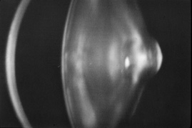
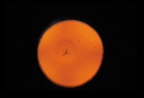
Lenticonus is a cone-shaped deformation of the anterior or posterior lens surface. Posterior lenticonus (Figs. 48 and 49) is more common than anterior lenticonus, and is usually unilateral and polar. This deformation results in a marked distortion of images and subsequent poor vision. Posterior lenticonus may be associated with extremely thin posterior lens capsules, and polishing of the posterior capsule should be avoided during cataract surgery.

Fig. 48. Scheimpflug image of the posterior lenticonus, showing outpouching at the posterior pole.

Fig. 49. Retroillumination photograph of the same lens as in Fig. 48, showing an “oil droplet” appearance and a central opacity.

Lentiglobus is a spheric deformation of the lens surface. Posterior lentiglobus is more common than anterior lentiglobus, and is often associated with posterior polar cataracts.

Back to Top
REFERENCES

1. Congdon N, Vingerling JR, Klein BEK, et al: The prevalence of cataract and pseudophakia/aphakia among adults in the United States. Arch Ophthalmol 122:487–494, 2004

2. Duke-Elder S: System of ophthalmology. Vol. 11. St. Louis: CV Mosby, 1969:63

3. Benedek GB, Chylack LT Jr, Libondi T, et al: Quantitative detection of the molecular changes associated with early cataractogenesis in the living human lens using quasielastic light scattering. Curr Eye Res 6:1421, 1987

4. Congdon N, O'Colmain B, Klaver CC, et al: Causes and prevalence of visual impairment among adults in the United States. Arch Ophthalmol 122:477–485, 2004

5. Thylefors B, Negrel AD, Pararajasegaram R, Dadzie KY: Global data on blindness. Bull World Health Org 73:115–121, 1995

6. Congdon NG: Prevention strategies for age related cataract: present limitations and future possibilities. Br J Ophthalmol 85:516–520, 2001

7. Baltussen R, Sylla M, Mariotti SP: Cost-effectiveness analysis of cataract surgery: a global and regional analysis. Bull World Health Org 82:338–345, 2004

8. Busbee BG, Brown GC, Brown MM: Cost-effectiveness of ocular interventions. Curr Opin Ophthalmol 14:132–138, 2003

9. Steinberg EP, Javitt JC, Sharkey PD, et al: The content and cost of cataract surgery. Arch Ophthalmol 111:1041, 1993

10. Chylack LT Jr, Leske MC, McCarthy D, et al: Lens opacities classification system II (LOCS II). Arch Ophthalmol 107:991, 1989

11. Chylack LT Jr, Wolfe JK, Singer DM, et al: Lens opacities classification system III. Arch Ophthalmol 111:831, 1993

12. Sparrow JM, Bron AJ, Brown NAP, et al: Oxford clinical cataract classification and grading system. Int Ophthalmol 9:207, 1986

13. Klein BEK, Klein R, Linton KLP, et al: Assessment of cataracts from photographs in the Beaver Dam Eye Study. Ophthalmology 97:1428, 1990

14. Taylor HR, West SK: The clinical grading of lens opacities. Aust N Z J Ophthalmol 17:81, 1989

15. Age-Related Eye Disease Study Research Group: The age-related eye disease study (AREDS) system for classifying cataracts from photographs: AREDS report no. 4. Am J Ophthalmol 131:167–175, 2001

16. Robman LD, McCarty CA, Garrett SK, et al: Comparison of clinical and digital assessment of nuclear optical density. Ophthalmic Res 31:119–126, 1999

17. Garrett SK, Robman LD, McCarty CA, et al: Reproducibility of automatic standard digital analysis of lens opacities. Aust N Z J Ophthalmol 26(Suppl 1):S29–S31, 1998

18. Congdon NG, Taylor HR: Age-related cataract. In: Johnson GJ, Minassian DC, West S, Weale R, (eds.): The Epidemiology of Eye Disease. London, UK: Arnold Publishers, 2003:105–119

19. Kashima K, Trus B, Unser M, et al: Aging studies on normal lens using the Scheimpflug slit lamp camera. Invest Ophthalmol Vis Sci 34:263, 1993

20. Panchapakesan J, Rochtchina E, Mitchell P: Myopic refractive shift caused by incident cataract: the Blue Mountains Eye Study. Ophthal Epidemiol 10:241–247, 2003

21. Lee KE, Klein BE, Klein R, Wong TY: Changes in refraction over 10 years in an adult population: the Beaver Dam Eye study. Invest Ophthalmol Vis Sci 43:2566–2567, 2002

22. Horwitz J: Proctor lecture: The function of alpha-crystallin. Invest Ophthalmol Vis Sci 34:10, 1993

23. Van Heyningen R: What happens to the human lens in cataract? In: Spivey B, Henkind P, Lichter P, et al (eds). American Academy of Ophthalmology: Selected Readings in Ophthalmology Companion Source Manual. Vol. 2. San Francisco: American Academy of Ophthalmology, 1976:112

24. Clark JI, Livesey JC, Steele JE: Phase separation inhibitors and lens transparency. Optom Vis Sci 70:873, 1993

25. Kinoshita JH, Kador P, Datiles M: Aldose reductase in diabetic cataract. JAMA 246:259, 1981

26. Kinoshita JH: Mechanisms initiating cataract formation: Proctor lecture. Invest Ophthalmol 13:713, 1974

27. Kuszak JR: The development of lens sutures. Prog Retinal Eye Res 14:567, 1995

28. Schein O, West S, Mundy B, et al: Cortical lenticular opacification: Distribution and location in a longitudinal study. Invest Ophthalmol Vis Sci 35:363, 1994

29. West SK, Duncan DD, Munoz B, et al: Sunlight exposure and risk of lens opacities in a population-based study: The Salisbury Eye Evaluation Project. JAMA 280:714–718, 1998

30. Zigman S, Paxhia T, McDaniel T, et al: Effect of chronic near-ultraviolet radiation on the gray squirrel lens in vivo. Invest Ophthalmol Vis Sci 32:1723–1732, 1991

31. Lasa S, Datiles M, Podgor M, Magno B: Contrast and glare sensitivity: Association with type and severity of the cataract. Ophthalmology 99:1045, 1992

32. Lasa S, Podgor M, Datiles M, et al: Glare sensitivity in early cataracts. Br J Ophthalmol 77:489, 1993

33. Datiles MB: Clinical evaluation of cataracts. In: Tasman W, Jaeger EA (eds). Duane's Clinical Ophthalmology. Vol. 1. Philadelphia: JB Lippincott, 1993:6

34. Greiner JV, Chylack LT Jr: Posterior subcapsular cataracts: histopathologic study of steroid-associated cataracts. Arch Ophthalmol 97:135–144, 1979

35. Harding CV, Chylack LT Jr, Susan SR, et al: Elemental and ultrastructural analysis of specific human lens opacities. Invest Ophthalmol Vis Sci 23:1–13, 1982

36. Chylack LT Jr: Mechanisms of senile cataract formation. Ophthalmology 91:596–602, 1984

37. Wong TY, Klein BE, Klein R, Tomany SC, Lee KE: Refractive errors and incident cataracts: the Beaver Dam Eye Study. Invest Ophthalmol Vis Sci 42:1449–1454, 2001

38. Lim R, Mitchell P, Cumming RG: Refractive associations with cataract: the Blue Mountains Eye Study. Invest Ophthalmol Vis Sci 40:3021–3026, 1999

39. Younan C, Mitchell P, Cumming RG, et al: Myopia and incident cataract and cataract surgery: the Blue Mountains Eye Study. Invest Ophthalmol Vis Sci 43:3625–3632, 2002

40. Wong TY, Foster PJ, Johnson GJ, Seah SK: Refractive errors, axial ocular dimensions, and age-related cataracts: the Tanjong Pagar survey. Invest Ophthalmol Vis Sci 44:1479–1485, 2003

41. Chang MA, Congdon NG, Munoz B, et al: The association between myopia and various subtypes of lens opacity: SEE Project. Ophthalmology (in press)ppyy

42. Fishman GA, Anderson RJ, Lourenco P: Prevalence of posterior subcapsular lens opacities in patients with retinitis pigmentosa. Br J Ophthalmol 69:263, 1985

43. Fagerholm PP, Philipson BT: Cataract in retinitis pigmentosa: an analysis of cataract surgery results and pathological lens changes. Acta Ophthalmol (Copenh) 63:50, 1985

44. Kaiser-Kupfer M, Kuwabara T, Uga S, et al: Cataract in gyrate atrophy: clinical and morphologic studies. Invest Ophthalmol Vis Sci 24:432, 1983

45. Kashima K, Datiles M, Trus B, et al: Localization of lens opacities in gyrate atrophy: Image analysis study with Scheimpflug camera. Folia Ophthalmol Jpn 40:986, 1989

46. Vrensen GF, Willekens B, De Jong PT, et al: Heterogeneity in ultrastructure and elemental composition of perinuclear lens retrodots. Invest Ophthalmol Vis Sci 35:199, 1994

47. Lovicu FJ, Schulz MW, Hales AM, et al: TGFbeta induces morphological and molecular changes similar to human anterior subcapsular cataract. Br J Ophthalmol 86:220–226, 2002

48. Wunderlich K, Pech M, Eberle AN, et al: Expression of connective tissue growth factor mRNA in plaques of human anterior subcapsular cataracts and membranes of posterior capsule opacification. Curr Eye Res 21:627–636, 2000

49. Lovicu FJ, Steven P, Saika S, McAvoy JW: Aberrant lens fiber differentiation in anterior subcapsular cataract formation: a process dependent on reduced levels of Pax6. Invest Ophthalmol Vis Sci 45:1946–1953, 2004

50. Joo CK, Lee EH, Kim JC, et al: Degeneration and transdifferentiation of human lens epithelial cells in nuclear and anterior polar cataracts. J Cataract Refract Surg 25:652–658, 1999

51. Marcantonio JM, Syam PP, Liu CS, Duncan G: Epithelial transdifferentiation and cataract in the human lens. Exp Eye Res 77:339–346, 2003

52. Srinivasan Y, Lovicu FJ, Overbeek PA: Lens-specific expression of transforming growth factor beta 1 in transgenic mice causes anterior subcapsular cataracts. J Clin Invest 101:625–634, 1998

53. Hales AM, Chamberlain CG, McAvoy JW: Susceptibility to TGF-beta2-induced cataract increases with aging in the rat. Invest Ophthalmol Vis Sci 41:3544–3551, 2000

54. Beethan WP: Atopic cataract. Arch Ophthalmol 24:21, 1940

55. Sasaki K, Kojima M, Nakaizumi H, et al: Early lens changes seen in patients with atopic dermatitis applying image analysis processing of Scheimpflug and specular microscopic images. Ophthalmologica 212:88–94, 1998

56. Street DA, Javitt JC: National five-year mortality after inpatient cataract extraction. Am J Ophthalmol 113:263, 1992

57. Glynn RJ, Christen WG, Manson JE, et al: Body mass index: an independent predictor of cataract. Arch Ophthalmol 113:1131, 1995

58. Tavani A, Negri E, La Vecchia C: Selected diseases and risk of cataract in women: a case-control study from northern Italy. Ann Epidemiol 5:234, 1995

59. Leske MC, Chylack LT Jr, Wu SY: Lens opacities case-control study: risk factors for cataract. Arch Ophthalmol 109:244, 1991

60. Robinson GC, Jan JE, Kinnis C: Congenital ocular blindness in children, 1945–1984. Am J Dis Child 141:1321, 1987

61. Wong TY, Klein BE, Klein R, Tomany SC: Relation of ocular trauma to cortical, nuclear, and posterior subcapsular cataracts: the Beaver Dam Eye Study. Br J Ophthalmol 86:152–155, 2002

62. Nelson LB, Spaeth GL, Nowinski TS, et al: Aniridia: a review. Surv Ophthalmol 28:621, 1984

63. Kaiser-Kupfer MI, Freidlin V, Datiles MB, et al: The association of posterior capsular lens opacities with bilateral acoustic neuromas in patients with neurofibromatosis type 2. Arch Ophthalmol 107:541, 1989

64. Bouzas EA, Freidlin V, Parry DM, et al: Lens opacities in neurofibromatosis 2: Further significant correlations. Br J Ophthalmol 77:354, 1993

65. Ederer F, Hiller R, Taylor HR: Senile lens changes and diabetes in two population studies. Am J Ophthalmol 91:381–395, 1981

66. Pokupec R, Kalauz M, Turk N, Turk Z: Advanced glycation endproducts in human diabetic and non-diabetic cataractous lenses. Graefe's Arch Clin Exp Ophthalmol 241:378–384, 2003

67. Kinoshita JH, Datiles MB, Kador PF, Robison WG Jr: Aldose reductase and diabetic eye complications. In: Rifkin H, Porte D (eds). Ellenberg and Rifkin's Diabetes Mellitus: Theory and Practice. 4th ed. New York: Elsevier, 1990:264

68. Fukushi S, Merola LO, Kinoshita JH: Altering the course of cataracts in diabetic rats. Invest Ophthalmol Vis Sci 19:313, 1980

69. Datiles M, Fukui H: Cataract prevention in diabetic Octodon degus with Pfizer's sorbinil. Curr Eye Res 8:233, 1989

70. Wyman M, Sato S, Akayi Y, et al: The dog as a model for ocular manifestation of high concentration of blood sugars. J Am Vet Med Assoc 193:1153, 1988

71. Biswas S, Harris F, Singh J, Phoenix D: Role of calpains in diabetes mellitus-induced cataractogenesis: A mini review. Mol Cell Biochem 261:151–159, 2004

72. Sakamoto-Mizutani K, Fukiage C, Tamada Y, et al: Contribution of ubiquitous calpains to cataractogenesis in the spontaneous diabetic WBN/Kob rat. Exp Eye Res 75:611–617, 2002

73. Thampi P, Hassan A, Smith JB, Abraham EC: Enhanced C-terminal truncation of alpha A- and alpha B-crystallins, in diabetic lenses. Invest Ophthalmol Vis Sci 43:3265–3272, 2002

74. Azuma M, Fukiage C, David LL, Shearer TR: Activation of calpain in lens: A review and proposed mechanism. Exp Eye Res 64:529–538, 1997

75. Andersson M, Sjostrand J, Andersson AK, et al: Calpains in lens epithelium from patients with cataract. Exp Eye Res 59:359–364, 1994

76. Hedge KR, Varma SD: Morphogenetic and apoptotic changes in diabetic cataract: Prevention by pyruvate. Mol Cell Biochem 262:233–237, 2004

77. Beutler E, Matsumono F, Kuhl W, et al: Galactokinase deficiency as a cause of cataracts. N Engl J Med 288:1203, 1973

78. Holton JB, Walter JH, Tyfield LA: Galactosemia. In: Scriver CR, Beaudet AL, Sly WS, Valle D (eds). The Metabolic and Molecular Bases of Inherited Disease. 8th ed. New York: McGraw-Hill, 2001:1553–1583

79. Bosch AM, Bakker HD, Van Gennip AH, et al: Clinical features of galactokinase deficiency: A review of the literature. J Inherit Metab Disord 25:629–634, 2002

80. Okano Y, Asada M, Fujimoto A, et al: A genetic factor for age-related cataract: identification and characterization of a novel galactokinase variant, “Osaka,” in Asians. Am J Hum Genet 68:1036–1042, 2001

81. Datiles M, Fukui H, Kuwabara T, Kinoshita JH: Galactose cataract prevention with sorbinil, an aldose reductase inhibitor: a light microscopic study. Invest Ophthalmol Vis Sci 22:174, 1982

82. Skalka HW, Prchal JT: Presenile cataract formation and decreased activity of galactosemic enzymes. Arch Ophthalmol 98:269–273, 1980

83. Stevens RE, Datiles MB, Srivastava SK, et al: Idiopathic presenile cataract formation and galactosemia. Br J Ophthalmol 73:48–51, 1989

84. Karas N, Gobec L, Pfeifer V, et al: Mutations in galactose-1-phosphate uridyltransferase gene in patients with idiopathic presenile cataract. J Inherit Metab Dis 26:699–704, 2003

85. Lukac-Bajalo J: Idiopathic presenile cataracts and galactosemia. Adv Clin Path 1(suppl 1):L5, 1997

86. Podskarbi T, Kohlmetz T, Gathof BS, et al: Molecular characterization of Duarte-1 and Duarte-2 variants of galactose-1-phosphate uridyltransferase J Inherit Metab Dis 19:638–644, 1996

87. Simonelli F, Nesti A, Picardi A, et al: Cataract formation in patients with lactose and galactose disorders. Dev Ophthalmol 17:82–86, 1989

88. Gardner RJ, Brown N: Lowe's syndrome: identification of carriers by lens examination. J Med Genet 13:449, 1976

89. Hedges TR Jr, Hedges TR 3rd: Wilson's disease (hepatolenticular degeneration). In: Gold DH, Weingeist TA (eds). The Eye in Systemic Disease. Philadelphia: JB Lippincott, 1990:390

90. Sher NA, Letson RD, Desnick RJ: The ocular manifestations in Fabry's disease. Arch Ophthalmol 97:671, 1979

91. Ramsay RC: Homocystinuria. In: Gold DH, Weingeist TA (eds). The Eye in Systemic Disease. Philadelphia: JB Lippincott, 1990:319

92. Morris DA: Cataracts and systemic disease. In: Duane TD, Jaeger EA (eds). Duane's Clinical Ophthalmology. Vol. 5. Philadelphia: JB Lippincott, 1993:8– 13

93. Claridge KG, Gibberd FB, Sidey MC: Refsum disease: the presentation and ophthalmic aspects of Refsum disease in a series of 23 patients. Eye 6:371, 1992

94. Grondalski SJ, Bennett GR: Alport's syndrome: review and case report. Optom Vis Sci 66:396, 1989

95. Mintz-Hittner H, Kretzer FL: Conradi's syndrome. In: Gold DH, Weingeist TA (eds). The Eye in Systemic Disease. Philadelphia: JB Lippincott,

96. Raitta C, Karli P: Ocular findings in myotonic dystrophy. Ann Ophthalmol 14:647, 1982

97. Cross HE, Jensen AD: Ocular manifestations in the Marfan syndrome and homocystinuria. Am J Ophthalmol 75:405–420, 1973

98. Hutchinson S, Furger A, Halliday D, et al: Allelic variation in normal human FBN1 expression in a family with Marfan syndrome: a potential modifier of phenotype? Hum Mol Genet 12:2269–2276, 2003

99. Hamod A, Moodie D, Clark B, Traboulsi EI: Presenting signs and clinical diagnosis in individuals referred to rule out Marfan syndrome. Ophthalmic Genet 24:35–39, 2003

100. Sachdev NH, Di Girolamo N, McCluskey PJ, et al: Lens dislocation in Marfan syndrome: Potential role of matrix metalloproteinases in fibrillin degradation. Arch Ophthalmol 120:833–835, 2002

101. Ashworth JL, Murphy G, Rock MJ, et al: Fibrillin degradation by matrix metalloproteinases: implications for connective tissue remodelling. Biochem J 340:171–181, 1999

102. Sachdev N, Wakefield D, Coroneo MT: Lens dislocation in Marfan syndrome and UV-B light exposure. Arch Ophthalmol 121:585–585, 2003

103. Weill G: Ectopie du cristallin et malformations generales. Ann Ocul 169:21–44, 1932

104. Marchesani O: Brachydaktylie und angeborene kugellines als systemerkrankung. Klin Mibl Augenheilkd 103:392–406, 1939

105. Faivre L, Dollfus H, Lyonnet S, et al: Clinical homogeneity and genetic heterogeneity in Weill-Marchesani syndrome. Am J Med Genet 123A:204–207, 2003

106. Veloes A, Hermia JP, Galand A, et al: Glaucoma-lens ectopia microsherophakia-stiffness-shortness (GEM-SS) syndrome: A dominant disease with manifestations of Weill-Marchesani syndromes. Am J Med Genet 44:48–51, 1992

107. Kloepfer HW, Rosenthal JW: Possible genetic carriers in the spherophakia-brachymorphia syndrome. Am J Hum Genet 7:398–425, 1955

108. Wirtz MK, Samples JR, Kramer PL, et al: Weill-Marchesani syndrome–possible linkage of the autosomal dominant form to 15q21.1. Am J Med Genet 65:68–75, 1996

109. Faivre L, Gorlin RJ, Wirtz MK, et al: In frame fibrillin-1 gene deletion in autosomal dominant Weill-Marchesani syndrome. J Med Genet 40:34–36, 2003

110. Dagoneau N, Benoist-Lasselin C, Huber C, et al: ADAMTS10 mutations in autosomal recessive Weill-Marchesani syndrome. Am J Hum Genet 75:801–806, 2004

111. Tang BL: ADAMTS: a novel family of extracellular matrix proteases. Int J Biochem Cell Biol 33:33–44, 2001

112. Cal S, Obaya AJ, Llamazares M, et al: Cloning, expression analysis and structural characterization of seven novel human ADAMTS, a family of metalloproteinases with disintegrin and thrombospondin-1 domains. Gene 283:49–62, 2002

113. Snead MP, Yates JRW: Clinical and molecular genetics of Stickler syndrome. J Med Genet 36:353–359, 1999

114. Seery CM, Pruett RC, Liberfarb RM, Cohen BZ: Distinctive cataract in the Stickler syndrome. Am J Ophthalmol 110:143–148, 1990

115. Lipman RM, Tripathi BJ, Tripathi RC: Cataracts induced by microwave and ionizing radiation. Surv Ophthalmol 33:200, 1988

116. Lydahl E: Infrared radiation and cataract. Acta Ophthalmol 166(suppl):1, 198

117. Cucinotta FA, Manuel FK, Jones J, et al: Space radiation and cataracts in astronauts. Radiat Res 156:460–466, 2001

118. Lett JT, Lee AC, Cox AB: Risks of radiation cataracts from interplanetary space missions. Acta Astronaut 32:739–748, 1994

119. Nicholas JS, Butler GC, Lackland DT, et al: Health among commercial airline pilots. Aviat Space Environ Med 72:821, 2001

120. Urban RC Jr, Cotlier E: Corticosteroid-induced cataracts. Surv Ophthalmol 31:102, 1986

121. Limaye SR, Pillai S, Tina LU: Relationship of steroid dose to degree of posterior subcapsular cataracts in nephrotic syndrome. Ann Ophthalmol 20:225, 1988

122. Mayman CI, Miller D, Mayman CI: In vitro production of steroid cataract in bovine lens. Part II: Measurement of sodium-potassium adenosine triphosphatase activity. Acta Ophthalmol 57:1107–1116, 1979

123. Lou MF, Dickerson JE Jr, Garadi R: The role of protein-thiol mixed disulfides in cataractogenesis. Exp Eye Res 50:819–826, 1990

124. Lyu J, Kim JA, Chung SK, et al: The alteration of cadherin in dexamethasone-induced cataract organ-cultured rat lens. IOVS 44:2034–2040, 2003

125. More MI, Kirsch FP, Rathjein FG: Targeted ablation of NrCAM or ankyrin-B results in disorganized lens fibers leading to cataract formation. J Cell Biol 154:187–196, 2001

126. Bucala R, Gallati M, Manabe S, et al: A glucocorticoid-lens protein adducts in experimentally induced steroid cataracts. Exp Eye Res 40:853–863, 1985

127. Hamamichi S, Kosano H, Nakai S, et al: Involvement of hepatic glucocorticoid receptor-mediated functions in steroid-induced cataract formation. Exp Eye Res 77:575–580, 2003

128. Kosano H, Nishigori H: Steroid induced cataract: other than in whole animal system, in lens culture system, androgens, estrogens, and progestins as well as glucocorticoids produce loss of transparency of the lens. Dev Ophthalmol 35:161–168, 2002

129. Reddy VN: Glutathione and its function in the lens–an overview. Exp Eye Res 50:771–778, 1990

130. Spector A: Oxidative stress-induced cataract: mechanism of action. FASEB J 9:1173–1182, 1995

131. Grant WM: Toxicology of the Eye. Springfield: Charles C. Thomas, 1974: 411–710

132. Xu GT, Zigler JS Jr, Lou MF: The possible mechanism of naphthalene cataract in rat and its prevention by aldose reductase inhibitor (ALO1576). Exp Eye Res 54:63, 1992

133. Tao RV, Takahashi Y, Kador PF: Effect of aldose reductase inhibitors on naphthalene cataract. Invest Ophthalmol Vis Sci 32:1630, 1991

134. Levene RZ: Uniocular miotic therapy. Trans Am Acad Ophthalmol Otolaryngol 79:OP376, 1975

135. Isaac NE, Walker AM, Jick H, Gorman M: Exposure to phenothiazine drugs and risk of cataract. Arch Ophthalmol 109:256, 1991

136. West SK, Valmadrid CT: Epidemiology of risk factors for age-related cataract. Surv Ophthalmol 39:323, 1995

137. Boukes RJ, van Balen AT, Bruynzeel DP: A retrospective study of ocular findings in patients treated with PUVA. Doc Ophthalmol 59:11, 1985

138. See JA, Weller P: Ocular complications of PUVA therapy. Australas J Dermatol 34:1, 1993

139. Geron RJ, MacDonald JS, Alberts AW, et al: On the etiology of subcapsular lenticular opacities produced in dogs receiving HMG-CoA reductase inhibitors. Exp Eye Res 50:65–78, 1990

140. Hartman HA, Myers LA, Evans M, et al: The safety evaluation of fluvastatin, an HMG-CoA reductase inhibitor, in beagle dogs and rhesus monkeys. Fundam Appl Toxicol 29:48–62, 1996

141. Rao PV, Robison WG Jr, Bettelheim F, et al: Role of small GTP-binding proteins in lovastatin-induced cataracts. Invest Ophthalmol Vis Sci 38:2313–2321, 1997

142. Boccuzzi SJ, Bocanegra TS, Walker JF, et al: No lens changes caused by simvastatin results from a prospective drug safety study. Lens Eye Toxic Res 7:643–650, 1990

143. Lundh BL, Nilsson SE: Lens changes in matched normals and hyperlipidemic patients treated with simvastatin for 2 years. Acta Ophthalmol (Copenh) 68:658–660, 1990

144. Schmidt J, Schmitt C, Hockwin O, et al: Ocular drug safety and HMG-CoA-reductase inhibitors. Ophthalmic Res 26:352–360, 1994

145. Harris ML, Bron AJ, Brown NA, et al: Absence of effect of simvastatin on the progression of lens opacities in a randomised placebo controlled study. Oxford Cholesterol Study Group. Br J Ophthalmol 79:996–1002, 1995

146. Pedersen TR, Berg K, Cook TJ, et al: Safety and tolerability of cholesterol lowering with simvastatin during 5 years in the Scandinavian Simvastatin Survival Study. Arch Intern Med 156:2085–2092, 1996

147. Qian W, Soderberg PG, Chen E, et al: 3 year simvastatin treatment and lens nuclear back scattering. Br J Ophthalmol 84:512–516, 2000

148. Schlienger RG, Haefeli WE, Jick H, Meier CR: Risk of cataract in patients treated with statins. Arch Intern Med 161:2021–206, 2001

149. Smeeth L, Hubbard R, Fletcher AE: Cataract and the use of statins: a case-control study. QJM 96:337–343, 2003

150. Boozalis GT, Purdue GF, Hunt JL, McCulley JP: Ocular changes from electrical burn injuries: a literature review and report of cases. J Burn Care Rehabil 12:458, 1991

151. Noel LP, Clarke WN, Addison D: Ocular complications of lightning. J Pediatr Ophthalmol Strabismus 17:245, 1980

152. Duke-Elder S: System of ophthalmology. Vol. 11. St. Louis: CV Mosby, 1969:248

153. Jaeger EA: Cataract types. In: Tasman W, Jaeger EA (eds). Atlas of Clinical Ophthalmology. Philadelphia: Lippincott-Raven, 1995:6B

Back to Top