|
|
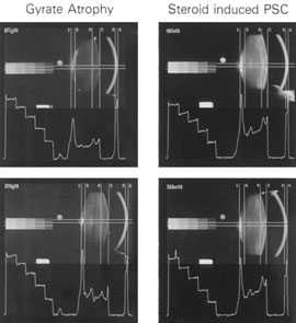
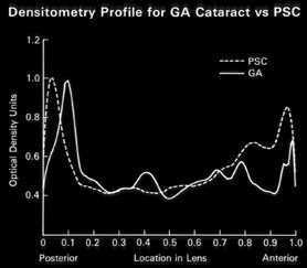
| Fig. 16. A: Scheimpflug slit image of two types of PSCs (steroid-induced and gyrate atrophy-related) showing the positions of the opacities. B: Densitometry profiles showing the position of the gyrate atrophy-related PSC in comparison with the steroid-induced PSC. These suggest that the PSC migrates anteriorly as the newly laid down fibers push the PSC deeper into the cortex.45 |