Chapter 10
Amblyopia
MARK J. GREENWALD and MARSHALL M. PARKS
Main Menu   Table Of Contents

Search

CAUSES
AMBLYOPIC VISION
MOTOR ASPECTS
ELECTROPHYSIOLOGY AND IMAGING
EXPERIMENTAL NEUROBIOLOGY
REFERENCES

Amblyopia is an acquired defect in monocular vision caused by abnormal visual experience early in life.* It is usually unilateral but may be bilateral. Amblyopia itself produces no change in the appearance of ocular structures, but it nearly always develops in association with some other condition, which is evident on physical examination and which is responsible for abnormal visual experience.


*The word amblyopia (derived from Greek roots meaning “blunt sight”) is occasionally still applied to other disorders in which partial loss of vision is congenital or acquired through processes unrelated to visual experience. Throughout this chapter, amblyopia is used to denote only the condition as defined in the text. Other authors sometimes indicate this restricted sense with the terms functional amblyopia and amblyopia ex anopsia.

The visual system is sensitive to the effects of abnormal visual experience only during a limited time in infancy and childhood when it is immature and plastic. For humans, this period extends roughly from birth through the end of the first decade.1,2 Vulnerability is greatest during the first few months of life and decreases gradually thereafter, with apparently considerable variation from person to person in the degree of sensitivity at a particular age (especially beyond 5 years). The age range within which amblyopia can be induced corresponds roughly to the span of time over which visual function normally develops to full maturity, but the resulting level of vision, particularly in unilateral cases, is often far below the normal level for the age of onset.3,4

Abnormal early visual experience can affect monocular vision through either or both of two amblyopiogenic mechanisms5: In the first, lack of exposure to the sharply focused images necessary for normal development disturbs and limits the maturation of form vision. In the second, marked disparity in the quality or directionality of inputs from the two eyes prevents binocular fusion and results in abnormal competitive binocular interaction, which leads to active interference with, or exclusion of, one eye's input to higher visual centers that persists during monocular viewing. Either mechanism can contribute to unilateral or bilateral amblyopia: The two eyes may be affected simultaneously by the first or sequentially by the second. Clinical and laboratory observations that have helped to elucidate the physiologic and anatomic bases for these mechanisms are reviewed in later sections of this chapter.

Although amblyopia may result from abnormal binocular interaction, it is not in essence a disorder of binocular vision. Amblyopia thus contrasts with monofixation syndrome, which is primarily a defect in binocularity and does not necessarily involve disturbance of monocular function. Monofixation and amblyopia often coexist, however, and may be acquired as a result of the same abnormal visual experience, possibly through a common mechanism. Amblyopia differs more fundamentally from strabismic suppression and anomalous correspondence, which are also frequently associated consequences of abnormal visual experience (although unlike amblyopia, they often result from intermittent strabismus). These are not defects but are adaptations of binocular vision. They can benefit the patient by eliminating diplopia without undermining the capacity for normal visual function. Amblyopia has no value for the patient (diplopia may occur even when one eye is severely amblyopic), and the amblyopic patient can never revert to a state of normal vision quickly. Those binocular mechanisms that underlie suppression and anomalous correspondence are thus probably quite distinct from those responsible for amblyopia. When the vision of an amblyopic eye is tested during binocular viewing, suppression or monofixation may contribute to its poor performance,6,7 but this effect vanishes during monocular testing, which reveals the extent of the strictly amblyopic visual deficit.

Back to Top
CAUSES
Many ocular disorders in infancy or childhood may be responsible for the abnormal visual experience that causes amblyopia. Traditionally, clinicians have classified persons with amblyopia into several categories on the basis of apparent etiology (strabismus, anisometropia, isoametropia, visual deprivation due to adnexal abnormality or media opacity, and so on). Only recently, however, has evidence from laboratory investigations begun to justify the assumption that different pathophysiologic disturbances underlie the occurrence of amblyopia in different clinical settings. Some of these findings are reviewed in later sections of this chapter. Distinctions tend to blur in practice because of the frequent coincidence of multiple causal disorders in a single patient. There also seems to be considerable heterogeneity even in cases with a “pure” etiologic basis, possibly attributable to variation in the proportionate contributions of the different amblyopiogenic mechanisms to visual loss, or to varying age of onset or duration of the condition.

With an overall prevalence of 2% to 4%, amblyopia is the most frequent cause of visual impairment in children and young adults in our population.8 A large fraction of cases results from strabismus with constant unilateral fixation, which leads to amblyopia in the deviating eye. Amblyopia generally does not develop if fixation alternates, providing each eye with similar access to higher visual centers, or if strabismic deviation is intermittent (as a result of fusional vergence or incomitance), so that there are periods of normal binocular interaction that preserve the integrity of the visual system. Chronic rejection of disparate input from the deviating eye by the binocular visual centers seems to be the principal factor responsible for strabismic amblyopia, but blurring of its foveal image due to inappropriate accommodation (determined by the distance from the viewer of the fixating eye's object of regard) also may contribute.

Amblyopia is considerably more common in persons who are esotropic than in those who are exotropic, and it can often be determined that an older amblyopic, exotropic patient actually developed the condition while esotropic at an earlier age. This observation probably reflects that most young persons with exodeviation are only intermittently tropic. The occasionally encountered child with constant exotropia is as likely to develop amblyopia as an esotropic one of the same age.

Uncorrected refractive error is the other common cause of abnormal visual experience that leads to amblyopia. Amblyopic visual loss becomes evident when optical correction is provided. Unilateral blur resulting from anisometropia may be the sole source of amblyopia (acting through either or both possible amblyopiogenic mechanisms), or it may interact with strabismus by determining a constant preference for fixation with the less ametropic eye. Modest degrees (i.e., + 1 to + 3) of unilateral excess hyperopia may cause mild to moderate amblyopia, especially when there is significant hyperopia in the less ametropic eye as well.9,10 When the more ametropic eye is mildly myopic, with a far point that approximates normal near viewing distance, amblyopia generally does not develop. Bilateral myopic shift during late childhood or adolescence may account for the occasional finding of amblyopia in the emmetropic eye of an adult with unilateral myopia, which during early childhood was more hyperopic than its fellow. Some published observations suggest that amblyopia may itself be a cause of unilateral excess hyperopia.11,12

Unilateral high hyperopia or myopia (greater than about + 6 diopters) can cause severe amblyopia, but in some patients (possibly those with relatively late age of onset) the disturbance of vision is surprisingly mild. When visual loss is profound there is often evidence of malformation or degenerative change in the ametropic eye that contributes directly to functional impairment or adds an additional amblyopiogenic factor.

Severe symmetric refractive error (isoametropia) may cause bilateral amblyopia of mild to moderate degree. Hyperopia in excess of about + 6 is usually involved; myopia, even when extreme, rarely causes bilateral amblyopia because the sharply focused images of objects held close to the eyes support normal visual development. Bilateral amblyopia is less likely to develop in the high hyperopic patient when accommodative esotropia is present. In such cases, the accommodation that causes excessive convergence has the beneficial effect of bringing the fixating eye's retinal image into sharp focus, whereas the nonesotropic, highly hyperopic person may not be accommodating sufficiently to have a clear view with either eye. When apparently severe bilateral amblyopia is encountered in a child with symmetrical refractive status, especially if nystagmus is present, the possibility of occult underlying retinal dystrophy (often associated with large refractive errors) should be considered.

A person who did not receive optical correction for severe astigmatic refractive error in childhood sometimes shows persistent impairment of corrected vision that is confined to the more ametropic meridians. This phenomenon is known as meridional amblyopia. It can be unilateral or bilateral. The effect on conventionally measured acuity is generally small.

Ptosis or tumors of the eyelids that result inconstant obstruction of the visual axis may cause amblyopia. If no other barrier to binocular fusion is present, infants and children with adnexal abnormalities, even rather severe ones, usually adopt compensatory gaze and head postures that prevent occlusion of the involved eye and preserve vision intact. When strabismus is also present, however, there is less incentive to keep the deviating eye visually active, and amblyopia is likely to develop because of preference for fixation with the uninvolved or less involved eye, especially with visual axis obstruction in the primary gaze position. Anisometropia, particularly astigmatism, which may result from distortion of the involved eye's cornea resulting from compression by an adnexal mass, is often a contributing factor as well.

Obstruction of vision by media opacities may cause serious damage to the immature visual system. Total cataracts, dense axial (nuclear or polar) opacities more than about 3 mm in diameter, and other comparable disturbances of media clarity can produce bilateral or unilateral amblyopia on the basis of form vision deprivation.13 The diffuse retinal illumination that results from media opacification seems also to be a particularly potent initiator of amblyopiogenic binocular interaction in unilateral cases, often leading to profound visual loss.14 Bilateral amblyopia from congenital cataracts removed later than a few months of age may be severe and permanent, but bilateral opacities acquired after the fixation reflex becomes well developed at age 3 to 4 months generally do not cause profound amblyopia. Unilaterally acquired cataracts may produce severe amblyopia as late as age 6 to 8 years. Milder forms of cataract (e.g., lamellar opacities that transmit light centrally, small discrete polar opacities) usually do not cause amblyopia when bilateral.13 A lamellar cataract that is present in only one eye, either primarily or following removal of the lens from the other eye, may produce significant amblyopia; small unilateral polar opacities generally do not result in amblyopia.

Congenital or early acquired abnormalities of the retina or optic nerve can interfere with visual development in addition to disturbing vision directly.15 When the primary disorder is not correctable or self-limited, it may be impossible to determine the precise extent of the amblyopic contribution to overall reduction of acuity.

Iatrogenic interference with vision in childhood (therapeutic occlusion or cycloplegia) has the potential to induce amblyopia. When such measures have been employed in the treatment of unilateral amblyopia, the initially amblyopic eye usually seems to achieve normal vision in the process, but occasionally bilateral amblyopia may result.16 Severe amblyopia has been reported after as little as 1 week of unilateral patching in children under 2 years of age after minor eyelid surgery.17

Back to Top
AMBLYOPIC VISION
The visual defect in amblyopia is complex and distinctive. Patients with unilateral amblyopia find it easy to recognize a qualitative difference between the vision in their normal and amblyopic eyes even when acuity is rendered equal by optical blur or diffusion. However, precise characterization of amblyopic vision remains difficult for clinicians and even laboratory researchers.18 It is still not generally possible to determine definitely whether amblyopia is present based solely on results of vision testing.

Amblyopia primarily affects spatial or form vision, although abnormalities of the light sense may also be found in at least some cases. Subsequent discussion considers several specific aspects of amblyopic vision, with emphasis on points that either are important in clinical practice or seem to shed light on the pathophysiology or anatomic localization of the underlying visual system disturbance.

VISUAL ACUITY

Amblyopic patients are deficient in their ability to resolve closely spaced contours and recognize the patterns they form. Visual acuity measured with conventional tests, which rely on both these functions, is always subnormal. Clinical diagnosis of amblyopia is in fact usually based on the observation of reduced acuity in association with a history of abnormal visual experience and a lack of any other abnormality on examination that can account for the acuity deficit.

Measurement of visual acuity is a major concern for the clinician in relation to the amblyopic child because, although reliable assessment of acuity may be quite difficult with young patients, amblyopia is most effectively and efficiently treated in childhood. Children old enough to identify block letters are usually tested with standard letter optotypes, either projected, wall mounted, or computer generated. Today's sophisticated youngster, exposed to educational television programs from infancy, is sometimes able to name letters even before age 3 years. For the less verbal child, testing must be modified to permit the use of manual pointing responses.

Currently, the nonverbal Snellen equivalents most widely used in North America are the tum-bling E test and the HOTV test (a simplificationof the older Sheridan-Gardiner test).19 The former relies on a child's ability to indicate with fingers the direction of the legs of a letter E that is rotated to point up, down, left or right; the latter involves matching each test letter to one of the four letters H, O, T, and V printed on a card that can be held in the child's hands. For most 3-year-olds and nearly all developmentally normal 4-year-olds, fairly reliable visual acuity measurements can be obtained with either of these tests after a minute or two ofinstruction (and confirmation of the patient's competence in responding) using large demonstration letters. Some children do better with one test than the other, both should therefore be available to the clinician. An advantage of the HOTV test is that there is no need to discriminate left/right mirror image figures, which may be difficult for the young patient. This problem can be eliminated with the E test by giving credit for correct identification of horizontal orientation to either a right or left response.

Various acuity tests have been devised that substitute pictures for letters. The most widely used test of this type in North America remains the set of cards developed by Allen, each of which displays a figure composed of dark and light elements having dimensions that correspond to those of a 30-foot(9 meter) Snellen letter. The examiner presents the cards in random order at progressively greater distances as the child identifies each pictured object by name (any consistently used word or phrase is acceptable) until recognition is no longer possible. The greatest distance at which correct responses are obtained is recorded in feet as the Snellen numerator over denominator 30.

Some small children who will not point to identify letters respond well to testing with pictures, but the shy child may actually be more difficult to test in this way because of the requirement for verbal responses. (It is sometimes helpful to have the child whisper answers to a parent.) Lack of familiarity with the pictured objects or inability to recognize the stylized images may be a problem for some children; testing should begin with a review of the cards up close, and any that are not readily identified should be eliminated (three or four remaining different pictures are sufficient for testing). Cluesderived from the overall shape of the Allen cardimages may undermine the accuracy of acuity measured. In general, this form of testing should be the last resort after failure with letter testing at each examination. On the occasion when a switch is made from Allen pictures to letters (or any significant change in the method of acuity assessment is made), results from both tests should be recorded to ensure comparability of measurements over time.

A newer approach to pictographic vision testing with significant advantages employs the set of 4 Lea symbols (Fig. 1). These simple forms are easy for most toddlers to deal with, they are more similar in configuration to Snellen letters than the Allen pictures and have been carefully calibrated and assessed for reliability.20 Several testing devices using Lea symbols are now available (Precision Vision, Lasalle, IL). Matching to shapes on a handheld card, as in the HOTV test, is an option.

Fig. 1. The four Lea symbols.

Whether letters or pictures are used, considerable patience, effort, and experience on the part of the examiner are required to ensure reliable measurement of the amblyopic child's visual acuity. It must always be kept in mind that a child's short attention span and lack of familiarity with a recognition task may be critical factors in acuity testing. Even when vision in the two eyes is equal, some young patients do better with the second eye tested as a result of practice provided by testing the first eye, or worse, because of fatigue and waning interest. Several brief sessions, beginning sometimes with the right eye and sometimes with the left, may need to be conducted before acuity assessment can be considered complete. It is worthwhile to record observations relating to the child's general behavior during testing as well as the numeric result. Because normal preschool children often test no better than 20/30 to 20/40 (although actual acuity may exceed this level), mild amblyopia may escape detection in children at an early age unless differences in the speed and confidence with which the patient responds are noted.

When amblyopia is present, its severity may be overestimated if the child is reluctant to identify distinguishable but distorted figures seen by the amblyopic eye. Even adults with amblyopia often begin to have difficulty and make errors in letter identification two lines or more above the actual visual acuity level. Testing should continue until incorrect responses are obtained for most letters on a given level; the level immediately above this is taken as the eye's acuity. A good deal of prodding is sometimes necessary to ensure continuation of responses to the point at which recognition becomes impossible. In approaching this limit it may be helpful at times briefly to back up to larger, “easier” letters to recover momentum.

The acuity tester must also be constantly alert to peeking and memorization or guessing on the part of the child. The eye not being tested should be occluded by a broad opaque object or an adhesive patch, not the fingers of a hand, and movement that allows glimpses around the occluder must be prevented. It is best to observe the child constantly during acuity measurement. This is facilitated by placing a mirror on the wall behind the patient, in which letters displayed at the opposite end of the room can be viewed without the need for head turning. If the acuity of an amblyopic eye abruptly “improves” to equality with the opposite better eye in the course of amblyopia treatment, peeking must always be considered as a possible explanation.

To avoid memorization of letter sequences on a standard chart (readily accomplished by many children), an eye with known or suspected amblyopia should be tested before the child has an opportunity to view the chart with the better eye. Charts on which a small number of different figures are repeated in random order (such as those designed for E and HOTV matching) make memorization more difficult; their use may thus be advantageous even for older patients who know the entire alphabet.

Some poker-faced children can mislead even an experienced examiner by guessing. When four different figures are used, the probability of guessing correctly at three out of four characters on a given acuity level is greater than 0.05; with a single character the probability is of course 0.25, or higher, if the patient remembers the chart from previous testing sessions. It is therefore important to avoid reliance on very short sequences of letters. This is especially a concern at low acuity levels (20/100 or less), for which there are usually four or fewer letters (and frequently one or two) on standard charts. Strategies that enable the examiner to minimize the effect of guessing by increasing the number of responses required include presentation of figures on cards that can be shuffled (such as the Allen pictures or the 150-foot letters that are provided for demonstration of HOTV matching) at increasing distances, repeat testing with the child standing half the usual distance from the chart (doubling each Snellen denominator), and confirmation of distance measurement with a near test that involves similar figures and presentation. Newer devices such as the B-VAT vision tester that employ a computer to generate endless random sequences of characters provide an excellent way to avoid the pitfalls created by memorization and guessing.

The use of preferential looking devices, visual evoked potential (VEP) recording, and observation of fixation behavior to assess amblyopic visual loss in infants and children too young or uncooperative for any form of conventional acuity testing is discussed in later sections of this chapter. Each of these approaches can provide valuable information to the clinician when used appropriately. None should be considered an adequate substitute for actual measurement of acuity when it can be performed reliably using the techniques already described, however great the effort required.

SPATIAL INTERACTIONS

It is well established that many persons with amblyopia have increased difficulty identifying test let-ters when they are presented in a linear or two-dimensional array rather than as isolated characters. A similar effect can be produced by placing interactive bars around a single letter (Fig. 2). This observation, sometimes described as the “crowding phenomenon” or “separation difficulty,” is an example of the effect of contour interaction on visual acuity. It can be demonstrated in normal and organically diseased as well as in amblyopic eyes when figures near the limit of resolution are surrounded by other closely spaced forms. In the normal fovea, contour interaction occurs when forms are separated by a distance of 1 to 3 minutes of arc (0.2 to 0.6 times the overall size of a 20-ft [6-m] Snellen letter); in the normal periphery, its extent is much greater. In the amblyopic fovea, contour interaction typically extends over an increased distance, to a degree that is roughly proportional to the reduction in acuity.21

Fig. 2. Interactive bars surrounding a Snellen test letter. Contour interaction is maximal with separation by a distance 0.3 to 0.5 times the total width or height of the letter.

Other conditions that decrease visual acuity may produce similar extension of contour interaction.22 What appears to be unique to amblyopia is the magnitude of the effect on measured acuity that may result from this phenomenon. Occasionally, an amblyopic eye that can resolve a 20/20 letter in isolation drops to as low as 20/100 in the presence of maximal contour interaction. Such a large discrepancy sometimes develops in the course of treatment as acuity measured with isolated letters improves more rapidly than interactive acuity. Thus, amblyopia cannot be considered cured until interactive acuity has become normal.

Several difficulties are encountered in attempting to quantify the effect of contour interaction with linear or multiline presentation of characters on traditional Snellen charts. Preschool children tend to be confused by a collection of letters and may not read them in the appropriate left to right, top to bottom order, or may be reluctant to respond at all. When using a wall-mounted chart, the examiner can help by physically pointing to each letter in turn, but this approach does not work well with projected charts because they are used in a darkened room. Spacing between characters on most charts is not in uniform proportion to their size from line to line, and with projected charts, separation between the letters and the edge of the light rectangle that surrounds them varies considerably. Contour interaction can be produced by this edge even when only a single letter is projected.

Given these concerns, the examiner should not hesitate to rely on isolated letter presentation for assessment of visual acuity in appropriate circumstances. In particular, when the goal of acuity measurement is initial detection of amblyopia or documentation of an interval change during the early stages of treatment there is no real disadvantage to the use of isolated letters. When concern about the effect of contour interaction increases in the late stages of treatment, effort should be made to obtain responses with multicharacter presentation. An alternative that should also be considered is the use of single letters surrounded by bars (drawn on cards for hand-held presentation, or computer-generated by a device such as the B-VAT vision tester). This approach permits measurement of interactive acuity with considerably greater ease and precision than is possible with standard charts.

Contour interaction is related to two other features of visual processing, spatial summation and lateral inhibition. Spatial summation refers to the reduction in the brightness required for detection of a small spot of light as its area is increased. Lateral inhibition is observed when the threshold for detection of a small test light is increased by illumination of the surrounding retina. Like contour interaction, spatial summation and lateral inhibition occur over very short distances in the normal fovea (about3-min arc for spatial summation and 10-min arc for lateral inhibition), and greater distances in the normal periphery. Spatial summation is responsible for the increased visibility of larger test spots in perimetry. Lateral inhibition can be demonstrated with a familiar optical illusion called the Hermann grid, which consists of black squares separated by white stripes (Fig. 3). The intersections of the stripes appear darker than the stripes themselves because the greater area of white surrounding points in the intersections produces greater lateral inhibition. The effect is less near the point of fixation because the smaller foveal zones of inhibition fall entirely within the width of a stripe. Spatial summation and lateral inhibition have both been found to extend over much greater distances than normal in the amblyopic fovea.23,24

Fig. 3. A. The Hermann grid illusion. B. The unshaded area of each circle represents the inhibitory zone for the point at the center of the circle. Near fixation the inhibitory zones are small and fit within the boundaries of the white stripes. Points in the intersections and points in the stripes outside the intersections receive the same amount of inhibition. Away from fixation, the larger inhibitory zones extend into the black regions, which do not contribute to lateral inhibition. Points in the intersections receive more inhibition than points outside and therefore appear darker.

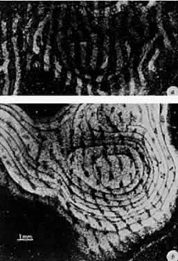
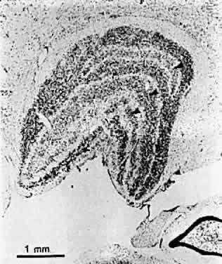
A probable explanation for contour interaction, spatial summation, and lateral inhibition is provided by data from electrophysiologic experiments inanimals.25 Studies employing microelectrode recordings from individual cells have shown that each responds to light within a small area of the field of vision known as its receptive field. This area is subdivided into zones within which light either excites or inhibits activity in the cell. In retinal and lateral geniculate neurons, the inhibitory zone forms an annulus around a central excitatory zone, or the reverse (Fig. 4). Cortical neurons generally respond best to oblong stimuli with a particular orientation, but they too have receptive fields of limited size divided into excitatory and inhibitory zones. The dimensions of excitatory zones appear to determine the extent of spatial summation at a particular location in the visual field; the dimensions of inhibitory zones determine the extent of lateral inhibition. The increased distance over which spatial interactions occur in the amblyopic fovea suggests that neurons serving foveal vision have enlarged receptive fields. The observed abnormalities could occur at any level from the retina to the visual cortex.

Fig. 4. An on-center receptive field. A. A spot of light directed into the center causes vigorous discharges. B. Illumination of the surround by a ring of light inhibits background discharge and causes responses when the light is turned off. C. Light covering the entire receptive field has a relatively small effect when turned on and off because antagonistic actions in the center and surround almost cancel each other. Illumination, indicated by the dark line, lasts 0.5 seconds. (Kuffler SW. The single-cell approach in the visual system and the study of receptive fields. Invest Ophthalmol Vis Sci 1973;12:794)

VISIBILITY OF GRATINGS

Patterns of alternating light and dark stripes known as gratings have been used extensively to analyze form vision in recent years. Gratings simplify acuity measurement (in a psychophysical sense) by substituting a task of resolution or detection alone for the combination of resolution and recognition involved in testing with Snellen letters. Their application to the study of amblyopia has led to a number of interesting and useful findings.

A grating pattern is characterized by the orientation of its stripes, its spatial frequency (the number of light/dark pairs or cycles per degree of visual angle), and the contrast between its light and dark elements (Fig. 5). A square-wave grating consists of uniform light and dark stripes with sharp edges. A sinusoidal grating has a sine wave luminance profile with gradual transitions from light to dark. Sinusoidal gratings are particularly important and widely used in vision research because any complex luminance profile can be resolved into sine wave components of different frequency by the mathematical technique of Fourier analysis.26

Fig. 5. Examples of gratings: sinusoidal (A-C) and square (D).A and B differ in spatial frequency, B and C in contrast. Luminance is diagrammed to the right of the figure. (Arden GB. The importance of measuring contrast sensitivity in cases of visual disturbance. Br J Ophthalmol 1978;62:198.)

The highest spatial frequency at which the stripes in a grating can be resolved with maximum contrast is called the cut-off frequency or grating acuity. For the normal human eye, this is 30 to 40 cycles per degree (for both square-wave and sinusoidal gratings), comparable with the minimum separation of elements required for recognition of conventional Snellen letters. In most disorders that affect visual resolution, grating acuity and Snellen acuity are reduced to a similar extent.

In amblyopia, grating acuity is generally reduced, but often by considerably less than Snellen acuity. Significant discrepancy between grating and Snellen acuity occurs much more frequently in strabismic than in anisometropic amblyopia.27,28 Some strabismic amblyopic patients actually have normal grating acuities. Although they may experience no difficulty detecting the presence of a grating pattern near the cut-off frequency, these patients report that they observe marked distortion of the stripes.29

The spatial distortion that seems to account for this difference between grating and recognition acuity (along with the related phenomenon of spatial uncertainty, a lack of precision in spatial localization) has long been noted by clinicians to be characteristic of amblyopic vision.30 It interferes with the performance of various visual tasks, including bisection of a line segment and counting a numberof closely spaced objects (such as the letters on aSnellen line). Laboratory investigators have developed refined and quantitative approaches to assess spatial distortion and have found it to be a feature primarily of strabismic amblyopia.31

Distortion of visual space also appears to under-lie the effect of amblyopia on ability to detect off-set between contiguous line segments, known as Vernier acuity. This function too is much more severely affected in strabismic than in anisometropic amblyopia.32,33 The physiologic basis for spatial distortion and its consequences in strabismic amblyopia remains a matter of controversy, but recent evidence suggests that processing beyond the level of the primary visual cortex is involved.34,35

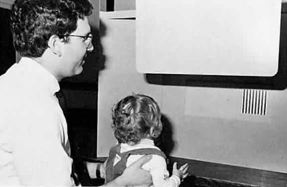
These considerations imply that measurement of grating acuity in amblyopic patients (and other tests based on detection rather than recognition, such as pointing to or picking up a minute object), is likely to underestimate the degree of visual loss, especially when strabismus is present. In some situations, grating acuity testing may nevertheless be of value to the clinician. It is well established that grating acuity can be measured reliably in very young patients using devices based on the technique of preferential looking.36,37 Infants naturally prefer to look at stripes rather than blank surfaces. In preferential looking tests, a trained examiner determines the patient's ability to see high contrast gratings of various spatial frequencies by noting the consistency with which the infant looks toward the pattern in a controlled setting (Fig. 6). At least one such test, the Teller acuity card system (Vistech Consultants, Dayton, OH), has now proved suitable for use in clinical practice, although the testing process remains somewhat cumbersome and time consuming (about 15 minutes per patient under optimal conditions).

Fig. 6. Use of Teller acuity cards to measure grating acuity by preferential looking assessment in an infant.

Teller cards or an alternative preferential looking device provide a reliable means for detecting amblyopia, estimating its severity, and monitoring the progress of treatment in the patient with anisometropia or media opacity who is too young for any form of recognition testing. For patients with strabismus, preferential looking assessment that indicates subnormal grating acuity confirms the presence of amblyopia, but it cannot be ruled out on the basis of normal responses. Because observation of fixation preference is available to the clinician as a rapid and highly sensitive means of detecting strabismic amblyopia, preferential looking tests are not usually helpful with such patients.

Gratings of different orientation are applicable to the assessment of meridional variation in acuity. Reduction in grating acuity that is limited to the more ametropic meridians is seen in patients with optically corrected astigmatism and meridional amblyopia (Fig. 7).38 Some patients with strabismic amblyopia (esotropic or exotropic) who do not have significant astigmatism show greater reduction of acuity for vertical gratings than for horizontal gratings. This is thought to reflect the greater impact of horizontal image displacement in the deviating eye on cortical neurons selective for vertical orientation.39

Fig. 7. The orientation differences in grating acuity for two optically corrected astigmias. A. Acuity is reduced for vertical gratings in an eye with a refractive error of + 4.50 -5.00 × 180.B. Acuity is reduced for horizontal gratings in a person withrefractive errors of -2.00 -4.25 × 180 right eye, -1.00 -4.50 × 180 left eye. The closed and open circles represent the right and left eyes, respectively. The drawing of an eye adjacent to each curve illustrates the focus of vertical and horizontal lines in the absence of optic correction and accommodation. Grating acuity for diagonal orientations is normally less than for either vertical or horizontal. (Mitchell DE, Freeman RD, Millodot M et al. Meridional amblyopia: Evidence for modification of the human visual system by early visual experience. Vis Res 1973;13:535.)

The visual system can respond to sinusoidal gratings over a broad range of spatial frequencies below the acuity cutoff. At different frequencies, varying amounts of contrast are required for detection of the grating pattern. Plotting contrast sensitivity (the reciprocal of threshold contrast) against spatial frequency yields a curve called the contrast sensitivity function. Normally, contrast sensitivity peaks at about three cycles per degree and falls off gradually at lower frequencies.26 Low frequency contrast sensitivity is a measure of ability to detect gradual transitions from light to dark. This is an important aspect of form vision (conveying information about the shape and position of large objects) that seems to be handled differently by the visual system from input concerning fine detail that is carried by higher spatial frequencies.40

Investigation of contrast sensitivity functions in amblyopia has revealed that some affected persons have reduced sensitivity only at high spatial frequencies, whereas others show reduction at all frequencies (Fig. 8).41 No consistent relationship has been established between contrast sensitivity profile and etiologic category or any other clinically significant feature of amblyopia, however. Although several devices that permit contrast sensitivitytesting in the clinical setting are now available,there is no generally agreed on indication for theirapplication to the care of amblyopic patients at present.

Fig. 8. A. Contrast sensitivity functions for the normal (open circles) and amblyopic (closed circles) eyes of a patient with strabismic amblyopia. The difference between the eyes is plotted in the upper part of the figure and is significant only at high spatial frequencies. B. Contrast sensitivity functions for a different strabismic amblyopic patient. In this case, the eyes differ significantly at all spatial frequencies. The grating acuity of the amblyopic eyes is the same in the two cases. (Hess RF, Howell ER. The threshold contrast sensitivity function in strabismic amblyopia: Evidence for a two-type classification. Vis Res 1977;1049, 1051.)

VISUAL FIELD

Amblyopia affects primarily foveal vision, but demonstration of a central scotoma by conventional means is difficult because of the difficulty that many amblyopic patients have in maintaining central fixation and the subtlety of the defect itself. Wald and Burian measured absolute light sensitivity in dark-adapted amblyopic patients and found no difference from normal across the visual field.42 In the dark-adapted state, however, both normal and amblyopic eyes show a relative reduction in light sensitivity in the rod-free fovea. When the visual field of an amblyopic eye is plotted in the light-adapted state, a relative central scotoma can be demonstrated if very small test targets are employed.23 If larger test spots are used, increased spatial summation of the amblyopic fovea eliminates the scotoma (Fig. 9). Recent observations made with automated perimetry suggest that there is slight reduction in light sensitivity of amblyopic eyes compared with their fellow normal eyes that extends well into the peripheral field even when acuity reduction is moderate.43

Fig. 9. The visual field of an amblyopic eye, obtained by static perimetry with test spots of different areas. (Flynn JT. Spatial summation in amblyopia. Arch Ophthalmol 78:470, 1967. Copyright 1967, American Medical Association.)

Severe amblyopia, such as that produced by a unilateral congenital cataract, may profoundly degrade peripheral vision, and in some cases depression of the visual field all the way to its nasal termination can be demonstrated. A region that seems consistently to be spared, however, is the temporal monocular crescent.44 This part of an amblyopic eye's projection to higher visual centers is not subject to competition from the input of the other eye, and thus can be affected only by the amblyopiogenic mechanism that is strictly dependent on form vision deprivation. Because the far periphery is normally exposed to relatively degraded images, it may be resistant to this effect as well. The monocular temporal field is functionally important for mobility and personal safety. Its preservation implies that the total extent of the binocular field of vision is normal even in a severely amblyopic patient, unless the amblyopic eye is esodeviated or obstructed by a dense media opacity.45

EFFECTS OF CHANGING LIGHT LEVELS

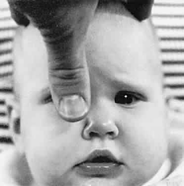
As already mentioned, the visual field of the amblyopic eye appears normal when plotted with very dim lights following dark adaptation. The difference between normal and amblyopic eyes with respect to other visual functions also tends to decrease when the light level is diminished. Von Noorden andBurian46 found that an amblyopic eye may show little or no reduction in acuity when viewing through neutral density filters that are dark enough to decrease the normal eye's acuity to the amblyopic eye's level. In contrast, performance of organically diseased eyes typically deteriorates more than normal with lowering of illumination (Fig. 10). Recent observations indicate that this characteristic behavior at reduced light levels is found only in strabismic amblyopic patients.47

Fig. 10. Visual acuity with different levels of illumination. The curve for normal eyes represents theaverage of measurements taken from 10 eyes. The curves for strabismic amblyopia and organic disease represent individual cases. (Von Noorden GK, Burian HM. Visual acuity in normal and amblyopicpatients under reduced illumination. II. The visual acuity at various levels of illumination. Arch Ophthal-mol 62:396, 1959. Copyright 1959, American Medical Association.)

Whether the process of dark adaptation is abnormal in amblyopia remains a matter of dispute. Wald and Burian observed normal dark adaptation in their classic study of amblyopic vision, but they tested this function in an extrafoveal area.42 Flynn found both a decreased rate of dark adaptation and residual threshold evaluation in some patients with amblyopia using a large central test field.48

These findings do not imply abnormality of the rods and cones in amblyopic eyes, for dark adaptation is a complex process involving functional changes in neurons as well as regeneration of visual pigments. Adjustment to varying light levels does occur primarily at lower levels of visual processing, however, and the effects of illumination on amblyopic vision may in fact have a subcortical origin.

COLOR VISION

In contrast to organic disease of the retina or optic nerve, amblyopia typically causes no major disturbance of color vision. Wald and Burian demonstrated normal spectral sensitivity in severely amblyopic patients in both the photopic and the scotopic states.42 Abnormalities have, however, been found in the increment threshold spectral sensitivity, which differs from the absolute sensitivity measured by Wald and Burian in that test lights are presented against a brightly illuminated white background.49 Like other aspects of amblyopic vision already considered, spectral sensitivity seems to be most disturbed at relatively high levels of overall illumination.

Mild to moderate amblyopia does not affect performance on clinical tests of color vision, but when acuity is severely reduced (20/200 or less) errors are common. The mistakes made by amblyopic patients appear random and are probably secondary to marked impairment of form vision rather than to a specific defect in color discrimination.

Back to Top
MOTOR ASPECTS
Amblyopia is frequently associated with characteristic abnormalities of ocular motor function. These may be a direct result of the sensory defect in amblyopic vision, independently induced consequences of the abnormal visual experience responsible for amblyopia, or manifestations of the underlying disorder (e.g., strabismus) that gain expression because of the amblyopic visual defect. Like the sensory features of amblyopia, associated motor abnormalities provide insight into the nature of the condition, and have a number of significant implications for the clinician who must diagnose and treat the problem.

MONOCULAR FIXATION

Visual experience early in life appears to be critical to development of the motor centers that direct and maintain fixation, just as it is critical to the development of the sensory visual system. An infant deprived of normal visual input from both eyes throughout the first 2 to 4 months of life is likely to develop the abnormal conjugate oscillatory eye movements of nystagmus in addition to bilateral amblyopia. This usually persists indefinitely even after subsequent elimination of the responsible obstacle to vision, and may itself ultimately become a limiting factor for visual development.

Nystagmus in bilaterally vision-deprived infants tends to be horizontal with a symmetric pendular quality in primary gaze and becomes jerky in lateral gaze, with faster movement in the direction of gaze. Vertical or cyclorotary components are sometimes seen, however. There may be a neutral or null zone (characterized by pendular oscillations of minimal amplitude) in a position of gaze other than primary, with a fast phase directed away from this position in primary gaze. Nystagmus due to delayed treatment of a correctable disorder such as cataract is indistinguishable from that caused by disease or maldevelopment directly involving visual system neurons. In fact, nystagmus associated with sensory disorders in general cannot be differentiated reliably from that due to primary motor system disturbance on the basis of eye movement characteristics alone.

Especially in strabismic patients, bilateral nystagmus of primarily motor origin may coexist with unilateral amblyopia. Typically, the nystagmus is of the latent or manifest latent variety, with a fast phase that reverses with alternation of fixation, consistently beating toward the side of the fixating eye. Amplitude of these oscillations often increases when the amblyopic eye fixates and may contribute to the measured reduction in its acuity.

Quantification of the purely sensory component of visual loss in an amblyopic eye can be difficult or impossible for the clinician when latent nystagmus is present. Sometimes the excess of eye movement during monocular viewing is attributable to exclusion of visual input from the nonamblyopic eye. It can then be reduced or eliminated during acuity testing by using a plus lens to blur the nonamblyopic eye to an acuity level below that of the amblyopic eye, by holding an occluder at a distance of several inches from the nonamblyopic eye to avoid obstructing its peripheral field (watching carefully to ensure that its visual axis is obstructed), or using a vectographic, red-green, or other viewing system that excludes only the test letters from the nonamblyopic eye. Nystagmus amplitude may occasionally be diminished by allowing the fixating eye to view in adduction. Sometimes, however, the act of fixation with the amblyopic eye is itself the principal determinant of the motor abnormality, and the clinician can do no more than establish a lower limit for amblyopic visual loss.

Severe unilateral amblyopia from any cause is usually associated with unsteady fixation by the amblyopic eye. The abnormal eye movements are typically coarse and less regular in direction, amplitude, and velocity than nystagmus seen with bilateral visual deprivation. In the mature patient, unsteadiness of monocular fixation may help to distinguish visual loss that occurred early in life from disorders of later onset. In the preverbal child, unilateral, unsteady fixation is a reliable indicator of severe amblyopia, implying visual acuity poorer than20/200. The quality of fixation tends to improve along with acuity in the course of successful treatment.

Eccentric fixation is a frequent finding in amblyopic eyes, especially in patients with strabismus. The eccentrically fixating eye directs a nonfoveolar retinal locus toward the object of regard when viewing monocularly. Grossly eccentric fixation, obvious from displacement of the corneal reflection during monocular viewing of a light, is seen only infrequently, in association with reduction in acuity to less than 20/200. Obvious unsteadiness of fixation in such eyes is nearly always present.

Less marked and relatively steady eccentricity of fixation (a few degrees or less of visual angle) is common and can be detected by the clinician with various tests. Visuscopic devices (no longer available as separate instruments, but simulated by some direct ophthalmoscopes that provide a selection of viewing apertures) project directly onto the retina an image of a target or a small object that can be fixated by the patient (Fig. 11). By comparing the position of this image with that of the foveolar light reflex, the examiner can estimate the degree of eccentric fixation.

Fig. 11. The Visuscope. (Von Noorden GK, Maumanee AE. Atlas of Strabismus. 2nd ed. St Louis: CV Mosby, 1973:81.)

The entoptic phenomenon of Haidinger brushes, seen best when a blue field is viewed through a rotating polarizing filter (available as an accessory with most synoptophores), allows the patient to identify subjectively the visual direction of the foveola. This corresponds to the apparent axis of rotation of a wispy spinning image that resembles a bowtie, butterfly, or airplane propeller shifting position with each fixation movement of the eye. Angular separation of this point from the fixation point can be determined with the aid of a superimposed figure (traditionally a frontal view of an airplanein relation to which the propeller is located whenthe nose is visually fixated). Both visuscopy andHaidinger brush assessment of eccentric fixation require a patient who is fully cooperative, and even then it may be difficult to obtain reliable information.

When sensitive tests are used, eccentric fixation is detectable in a large proportion of strabismic amblyopic patients. Those with esotropia typically fixate on the nasal side of the foveola whereas those who are exotropic fixate on the temporal side, but exceptions to this rule are common and some eccentric fixators do not have strabismus at all. Eccentric fixation may result in underestimation or overestimation of the angle of strabismic deviation on cover testing, and in the occasional patient with equal angles of eccentricity and strabismus, cover testing may fail altogether to detect the presence of a tropia.

The acuity of an eccentrically fixating eye is never better than the normal acuity of the retinal locus used for fixation, but it may be considerably poorer.50 Severe amblyopic reduction in acuity can occur in the absence of eccentric fixation (Fig. 12). Eccentric fixation thus cannot be considered the primary cause of reduced acuity in amblyopia, nor does it account fully for any of the other features of amblyopic vision described in the previous section. Like unsteady fixation, eccentric fixation tends toward normalization with improvement in acuity as amblyopia responds to treatment.

Fig. 12. The relationship between visual acuity and locus of fixation in ten eyes with strabismic amblyopia (closed circles). The open symbols represent the normal acuity of the peripheral retina (average of data from three eyes). Acuity was determined with single Landolt C figures. (Hess RF. On the relationship between strabismic amblyopia and eccentric fixation. Br J Ophthalmol 1977;61:767.)

Although it has been the subject of intense study and speculation, the origin of eccentric fixation remains obscure. It has been claimed that it is caused by anomalous retinal correspondence, but because, in many cases, the angle of eccentricity is quite different from the angle of anomaly this explanation is difficult to accept.51 Another theory holds that the locusof eccentric fixation has the best visual acuity of any point in the retina of the amblyopic eye. Careful mapping of acuity across the visual field, however, demonstrates that in some cases the foveola retains better vision than the eccentric fixation locus (Fig. 13).52 Several other hypotheses have been advanced to explain this curious phenomenon, but none can be considered fully satisfactory at present.

Fig. 13. Visual acuity at different retinal loci for the normal (dashed line) and amblyopic (solid line)eyes of an eccentrically fixating strabismic amblyopic patient. The arrow indicates the eccentric fixationlocus of the amblyopic eye. Acuity is better at the foveola than at the eccentric fixation locus. (KirschenDG, Flom MC. Visual acuity at different retinal loci of eccentrically fixating functional amblyopes. AmJ Optometry Physiol Opt 1978;55:144. Copyright 1982, American Academy of Optometry.)

BINOCULAR FIXATION PATTERN

Most amblyopic eyes with better than 20/200 acuity have fixation that is at least grossly central and steady. Observation of central steady fixation thus in no way rules out amblyopic visual loss. In such cases, when acuity cannot be measured directly, it is frequently possible to determine whether amblyopia is present by documenting a significant unilateral fixation preference. If the patient consistently objects strongly to occlusion of one eye but readily accepts occlusion of the other, it can be assumed that the latter eye is amblyopic if there is no other apparent cause for reduced acuity and a history of abnormal visual experience can be established.

Symmetric resistance to occlusion of either eye is common in infants and small children. It reveals nothing about vision and tends to thwart efforts to assess the quality of fixation. The examiner can minimize apprehensiveness and resistance that is unrelated to visual impairment by attending to certain details of technique in fixation assessment. It is helpful to use a thumb as an occluder (Fig. 14),casually lowering it in front of one eye and then the other (taking care to block the visual axis), with the fingers of the hand resting lightly on the head, with the patient seated on a parent's lap. Attention is directed toward a near or distant object as the examiner soothes the child with a running commentary about some feature of the fixation target. Ocular pursuit as the object moves or saccadic movement as attention is redirected from one objectto another confirms that active fixation is occur-ring.

Fig. 14. The thumb occluder.

Ideally, the fixation object itself should be colorful, attractive, and recognizable by the patient, and when activated should be in constant motion or should flash light and make music or other appealing sounds. To ensure that interest can be maintained, a variety of objects should be available. For distance, an array of separately illuminated animated toys that can be individually activated by remote switches, or an animated cartoon displayed by a continuous loop projector or video monitor are excellent devices. For near, rubber animals that squeak, spinning toys that reveal hidden objects or emit sparks of light, and electronic “badges” that light and play tunes work well. Incorporating a lightsource into a small toy for near fixation facilitates simultaneous assessment of the corneal light reflexes. Sometimes the examiner's face moving from side to side is the best object for holding an infant's attention.

For strabismic patients, spontaneous alternation of fixation (even if one eye is used most of the time) implies equal or nearly equal vision in the two eyes. In the nonstrabismic patient, if optical misalignmentinduced by placing a 20-diopter prism base down on one side results in a series of up and down refixation movements, equality of vision can likewise be inferred. Consistent cross fixation, with use of the right eye for viewing to the left and the reverse, has similar significance. (Asymmetric bilateral limitation of abduction in cross fixating patients with large angle esotropia may prevent alternation in primary gaze.) In the nonalternating strabismic patient, near equality of vision may be assumed present if, when the preferred eye is occluded, the consistently deviating eye promptly picks up fixation and retains it for at least a few seconds (or through a blink) after the occluder is removed. Alternating fixation is a reassuring finding even in strabismic patients whose acuity can be directly measured, especially when cooperation for conventional testing is borderline or the performance of both eyes is subnormal.

When there is absence of alternation and refusal to maintain fixation with the deviating eye, it is generally assumed that amblyopia is present. In most cases, this assumption is correct. It is not uncommon, however, for patients with strabismus that appears strictly unilateral to have vision that is equal or nearly equal in the two eyes.53 This seems to be especially likely in cases of small angle deviation. Consistent unilaterality of intermittent strabismic deviation is found frequently in conjunction with normal acuity and should not be regarded as an indication for treatment unless there is also another reason to suspect amblyopia.

In general, estimation of the degree of amblyopic visual loss on the basis of the strength of fixation preference is unreliable and should be avoided, although in a patient undergoing treatment comparison of behavior from one examination to the next may provide an indication that improvement is occurring. The strabismic patient who continues to fixate habitually with one eye through an extended course of therapy for amblyopia creates a dilemma for the clinician, who must consider the possibility that vision has in fact reached an optimal level despite the lack of alternation. Determined and repeated efforts to measure acuity directly should be made with every such child over the age of 2 years. In the case of an infant with persistent unilateral fixation, evaluation with preferential looking can at least place a lower limit on the residual amblyopic deficit.

ALIGNMENT

Strabismus may be a consequence as well as a cause of abnormal visual experience. It is common for an eye with a dense unilateral cataract to develop either esodeviation or exodeviation in childhood; severe, uncorrected anisometropia or ptosis may have a similar effect. A characteristic finding in strabismus that is due to unilateral visual deprivation is marked variability in the angle of deviation, but in many cases the motility disturbance is indistinguishable in appearance from primary esotropia or exotropia.

Development of secondary strabismus implies the presence of a significant obstacle to visual development, and once established it can potentiate the amblyopiogenic effect of the original underlying disorder. Its onset should thus increase the clinician's level of concern in patients who have been thought to have low potential for amblyopia (e.g., minor lens opacities; moderate ptosis) and may signal a need for more intensive treatment. If therapeutic occlusion is under way when the strabismic deviation appears, however, the possibility that loss of alignment is an effect of the treatment must also be considered.

Oscillations that resemble nystagmus, but are confined to one eye, sometimes develop in patients with unilateral visual loss from any cause that occurs early in life. These movements tend to be slow and irregular and may simply represent an extreme form of variability of strabismic deviation. They must be distinguished from the monocular oscillations sometimes seen in association with spasmus nutans (which are fine and rapid and usually do not have a significant effect on visual acuity) or with intracranial lesions in the region of the optic chiasm (in which case there may be asymmetric or unilateral reduction in acuity).54

PUPILLARY LIGHT REACTION

Most amblyopic eyes, including those with severely reduced visual acuity, show no clinically detectable abnormality of the pupil. Pupillographic studies have shown, however, that subtle alterations of the papillary light reaction do occur in amblyopia.55,56 With careful observation, these can be detected in 10% or more of all amblyopic patients by means of the “swinging flashlight” test for relative afferent pupillary defect.57,58 That amblyopia may affect the pupil suggests (but does not require) the occurrence of a physiologic abnormality at the retinal level in at least some cases. Although the presence of an afferent defect does not imply that amblyopia will be unresponsive to treatment, evidence suggests that occlusion may be less effective in such cases.58

The absence of an afferent pupillary defect in an eye with poor vision is often interpreted as evidence that optic nerve function is intact and may be claimed to support the diagnosis of amblyopia if the eye appears otherwise normal on examination. It must be recognized, however, that visual acuity can be significantly reduced in one eye as a result of optic nerve hypoplasia without any associated clinically detectable abnormality of the pupil, and that compression of the optic chiasm by an intracranial mass (especially craniopharyngioma or optic glioma in childhood) may produce unilateral or markedly asymmetric reduction in acuity without causing an afferent defect. In both of these situations subtle disturbances of optic disc appearance sometimes escape detection in the course of ophthalmoscopy, especially when the patient is uncooperative. If what seems to be amblyopia fails to respond to treatment, careful reexamination of the discs should be performed, and neuroradiologic evaluation with computed tomography (CT) or magnetic resonance imaging (MRI) should be considered.

ACCOMMODATION

The function of accommodation is to bring retinal images into sharp focus, a state with which the amblyopic eye often lacks experience and which it may have difficulty recognizing. It is therefore not surprising that amblyopia impairs the ability to control accommodation, typically resulting in a subnormal response.59,60 Moderate hyperopia that would not be expected to affect the vision of a normal eye, which will accommodate to sharpen its retinal image, may cause significant optic blur in association with amblyopia.

Baseline acuity should be determined for an amblyopic eye with full hyperopic correction under cycloplegia. Even if there is initially no difference between corrected and uncorrected acuity, glasses should be considered for substantial degrees of hyperopia to eliminate a possible obstacle to successful treatment.

Back to Top
ELECTROPHYSIOLOGY AND IMAGING
Characteristic abnormalities of the VEP are seen in amblyopia. The VEP is an electrical signal recorded from the occipital region of the scalp in response to visual stimulation. Its stimulus may be a diffuse flash of light or a pattern, usually consisting of light and dark checks, that either flashes briefly (producing a transient response) or reverses periodically while maintaining a constant mean luminance (to produce a steady-state response). VEP responses are very sensitive to variation in the specific parameters of the stimulus and recording technique; results from different laboratories may vary significantly. General agreement confirms that the VEP response to pattern stimuli of appropriate dimensions is reduced in amplitude and has a normal or slightly prolonged latency (the time interval between stimulus and response).

Clinically, VEP testing may be useful in distinguishing amblyopia from optic nerve disease such as retrobulbar neuritis, which markedly prolongs VEP latency, or from malingering, in which the VEP is normal. Amblyopic VEP responses are not sufficiently distinctive, however, to differentiate amblyopia reliably from other conditions with which it may be confused, including optic nerve hypoplasia and optic blur.

Investigators have demonstrated that VEP testing can provide a means of detecting amblyopic visual loss in patients who are unable to cooperate for conventional acuity testing.61,62 At present, however, such an approach may be considered reliable only when supervised by a technically sophisticated individual specifically trained in the application of VEP to this problem. Quantitative assessment that permits estimation of visual acuity requires the use of multiple different stimuli with a range of check sizes, adding to the already considerable complexity and expense of the testing procedure. Furthermore, as with grating acuity determined by preferential looking, VEP acuity measurement may underestimate amblyopic visual loss, especially in strabismic patients. It is unlikely that VEP testing will come to play a significant role in the clinical management of amblyopia in the near future.

VEPs are generated in the visual cortex but may be affected by abnormality at any level from the cornea to the cortex. VEP data are thus of little value in localizing amblyopia within the visual system. However, VEP latency is considerably less prolonged than reaction time (the time required to produce a motor response to a visual stimulus), in strabismic amblyopia.63,64 This observation represents further evidence that amblyopic individuals have a defect in visual processing above the level of the primary visual cortex.

The electroretinogram (ERG) is an electrical potential generated by the retina in response to light stimulation. The ERG produced by stimulating an amblyopic eye with diffuse flashes of light is normal even when only the fovea is illuminated.65 More recently interest has centered on ERG responses generated with checked patterns similar to those used in VEP work. The mechanism by which the pattern ERG is produced seems to be quite different from that of the flash ERG; retinal ganglion cells may be the source of the response to patterns.66 The effect of amblyopia on the pattern ERG is a matter of dispute, with some investigators reporting consistent abnormalities and others finding none.67,68

Conventional neuroimaging (i.e., CT, MRI) shows no abnormality of the brain in amblyopia, but some dynamic studies of brain blood flow and metabolism using positron emission tomography (PET) have provided anatomic evidence for an amblyopic disturbance of cortical function.69 Extrastriate visual cortex (Brodmann areas 18 and 19) seems to be particularly affected.70

Back to Top
EXPERIMENTAL NEUROBIOLOGY
During the past four decades, beginning with the Nobel Prize-winning studies of Hubel and Wiesel in the 1960s, laboratory researchers have gathered a large volume of information concerning the effects of abnormal visual experience on the immature nervous system through the study of animals (particularly kittens and infant monkeys) reared under various experimental conditions. Similarities between human amblyopia and the abnormalities induced by differing forms of visual deprivation in animals are numerous, and it seems valid to draw conclusions concerning the former from data provided by the latter. At the most basic level, animal models have confirmed beyond doubt that abnormal visual experience can cause physiologic and histologic alterations in a nervous system that is normal at birth, with profound implications for vision. The following discussion is necessarily restricted to a small num-ber of findings that seem to have relevance to theconcerns of the clinician. Several excellent reviewsof the extensive literature on this subject are available.3,5,71–73

Animals, like humans, are vulnerable to the effects of abnormal visual experience only for a limited period of time early in life, which has been termed the critical or sensitive period. Its duration is shorter in nonhuman species, whose rate of neurologic development is considerably more rapid than our own. Monkeys, in particular, mature to about the same degree over a given number of weeks as humans do in the same number of months. The critical period for the monkey begins at birth and ends between 1 and 2 years of age, with the greatest sensitivity in the first 6 to 12 weeks of life.3,72 Some evidence suggests (disputed by some investigators) that plasticity of the visual system during the sensitive period depends on input from noradrenergic neurons and is subject to pharmacologic manipulation.74 Nerve growth factor also appears to be an important modulator of visual neuronal plasticity in young animals.75

Monocular impairment of vision closely resembling human amblyopia can be produced in animals by constant unilateral strabismus, optic blur (such as that resulting from surgical removal of the lens or atropinization), and total form vision deprivation (usually achieved by suturing the lids of one eye shut). As in humans, more severe degradation of the retinal image tends to be associated with greater visual loss. Various consistent physiologic and anatomic correlates of reduced acuity have been identified at different levels in the visual system. With a few exceptions (see later discussion), these changes are qualitatively similar (insofar as they relate to monocular vision) regardless of the type of abnormal visual experience. The specific contribution of each neural disturbance described later to behaviorally expressed reduction of vision remains uncertain. In fact, the overall degree of functional impairment produced by experimental modification of visual experience often is considerably in excess of what would be expected on the basis of neuronal dysfunction at levels up to and including the primary visual cortex, suggesting an important but as yet uncharacterized role for higher visual processing centers.76

Microelectrode recordings from single cells have demonstrated that most neurons in the visual cortex lose their ability to respond to stimulation of a monocularly deprived eye. Anatomic studies of monocularly lid-sutured animals indicate that the cortical regions that receive axon terminals from geniculocortical neurons driven by the affected eye (constituting its ocular dominance columns) contract while the regions receiving input from the normally experienced eye expand in comparison with normal brains (Fig. 15).72 Other less severe forms of visual deprivation appear to have a considerably less dramatic effect on the architecture of dominance columns.77

Fig. 15. Autoradiographs of monkey visual cortex 2 weeks after injection of a radioactive tracer into the vitreous of one eye. Each figure represents a montage of tangential sections through the cortex. A. Normal monkey. The light stripes, representing labeled eye columns, are separated by gaps of the same width representing the other eye. B. Monocularly deprived monkey, who had the right eye closed at 2 weeks for 18 months. Input from the normal eye is in the form of expanded bands, which in places coalesce, obliterating the narrow gaps that represent the columns connected to the closed eye. (Wiesel TN. Postnatal development of the visual cortex and the influence of environment. Nature 1982;299:583.)

The lack of cortical responsiveness to a unilaterally deprived eye seems to result not only from loss of synaptic connections, but also from an ongoing process of inhibition that is dependent on input from the normal eye and may be mediated through the neurotransmitter gamma aminobutyric acid (GABA). Enucleation of the normal eye and treatment with the toxic compound bicuculline, a GABA antagonist, have both been shown to produce an immediate increase in cortical responsiveness to the deprived eye.78,79

Hubel and Wiesel's first studies of monocularly lid-sutured kittens revealed unexpectedly that cell bodies in the lateral geniculate nucleus (LGN) that received input from the deprived eye were considerably smaller than those of cells in adjacent laminae the input of which came from the nondeprived eye (Fig. 16). This observation has been confirmed by many investigators in a number of species, including humans, and for every form of unilateral visual deprivation.72,80 It is generally believed to be a reflection of the decreased extent of the geniculocortical cells' terminal axonal branches in the visual cortex (corresponding to contracted cortical dominance columns). Reverse lid suturing of the normal eye after an initial period of monocular deprivation in the monkey has been shown to reverse the cortical ocular dominance shift without reversing the LGN changes.81 This suggests that there is some degree of independence between the two levels and greater or more prolonged plasticity in the cortex.

Fig. 16. Coronal section through the right lateral geniculate nucleus of the same monkey shown in Figure 15. Atrophy in the layers receiving input from the deprived eye is indicated by arrows. (Wiesel TN. Postnatal development of the visual cortex and the influence of environment. Nature 1982;299:583.)

Cell shrinkage due to unilateral visual deprivation occurs to a significantly lesser extent in the monocular segment of the LGN (which receives input from the monocular temporal crescent of the contralateral eye's visual field), indicating that interaction between cells driven by the normal and deprived eyes in the binocular portion of the nucleus contributes to (but does not entirely account for) the phenomenon.82 Another abnormality that has been found to be largely confined to the binocular segment of the LGN in cats following total monocular deprivation is absence of Y cells, which are the subset of neurons (believed to correspond to the magnocellular pathway in primates) that normally processes the low spatial frequency information that is basic to overall form vision.83 Strabismus and optic blur from atropinization seem not to affect the Y-cell population but may disturb the ability of high frequency sensitive X cells (corresponding to primate parvocellular neurons) in the LGN to resolve fine detail.84

Bilateral total form vision deprivation from birth, produced in animals by bilateral lid suturing or dark rearing, results in bilateral reduction of vision that is less profound than that caused by unilateral deprivation, but the effect is nevertheless significant. Various physiologic abnormalities have been demonstrated in cortical and geniculate neurons, some of which resemble persistence of the state of development normally present at birth and some of which, such as complete loss of responsiveness from a substantial number of cells, appear to represent acquired disturbances.3,72 Rearing in visual environments that provide contours restricted to a single orientation has been shown to interfere with the development of cortical cells with receptive fields having the perpendicular orientation, a finding that may correlate with the presence of meridional amblyopia.85

Evidence from animal models for the presence or absence of retinal involvement in amblyopia, like evidence from the clinical and laboratory study of humans, remains inconclusive. A few investigators have reported anatomic or physiologic changes in monkey or cat retinal ganglion cells induced by lid suturing or strabismus,84,86 but most have failed to detect any definite abnormality.

Back to Top
REFERENCES

1. Vaegan, Taylor D. Critical period for deprivation amblyopia in children. Trans Ophthalmol Soc UK 1979;99:432

2. Keech RV, Kutschke PJ. Upper age limit for the development of amblyopia. J Pediatr Ophthalmol Strabismus 1995;32:89

3. Boothe RG, Dobson V, Teller DY. Postnatal development of vision in human and nonhuman primates. Annu Rev Neurosci 1985;8:495

4. Daw NW. Critical periods and amblyopia. Arch Ophthalmol 1998;116:502

5. von Noorden GK. Amblyopia: A multidisciplinary approach. Proctor lecture. Invest Ophthalmol Vis Sci 1985;26:1704

6. von Noorden GK, Leffler MB. Visual acuity in strabismic amblyopia under monocular and binocular conditions. Arch Ophthalmol 1966;71:172

7. Holopigian K, Blake R, Greenwald MJ. Clinical suppression and amblyopia. Invest Ophthalmol Vis Sci 1988;29:444

8. Attebo K, Mitchell P, Cumming R et al. Prevalence and causes of amblyopia in an adult population. Ophthalmology 1998;105:154

9. Jampolsky A, Flom BC, Weymouth FW et al. Unequal corrected visual acuity as related to anisometropia. Arch Ophthalmol 1955;54:893

10. Weakley DR. The association between nonstrabismic anisometropia, amblyopia, and subnormal binocularity. Ophthalmology 2001;108:163

11. Kiorpes L, Wallman J. Does experimentally induced amblyopia cause hyperopia in monkeys? Vis Res 1995;35:1289

12. Abrahamsson M, Sjostrand J. Natural history of infantile anisometropia. Br J Ophthalmol 1996;80:860

13. Parks MM. Visual results in aphakic children. Am J Ophthalmol 1982;94:441

14. Jampolsky AJ. Unequal visual inputs and strabismus management: A comparison of human and animal strabismus. In: Symposium on Strabismus. Transactions of the New Orleans Academy of Ophthalmology. St Louis: CV Mosby, 1978:358–492

15. Kushner BJ. Functional amblyopia associated with organic ocular disease. Am J Ophthalmol 1981;91:39

16. Burian HM. Occlusion amblyopia and the development of eccentric fixation in occluded eyes. Am J Ophthalmol 1966;62:853

17. Awaya S, Sugawara M, Miyake S. Observations in patients with occlusion amblyopia. Trans Ophthalmol Soc UK 1979:99:447

18. McKee SP, Schor CM, Steinman SB et al. The classification of amblyopia on the basis of visual and oculomotor performance. Trans Am Ophthalmol Soc 1992;90:123

19. Friendly DS. Preschool visual acuity screening tests. Trans Am Ophthalmol Soc 1978;76:383

20. Graf MH, Becker R, Kaufmann H. Lea symbols: Visual acuity assessment and detection of amblyopia. Graefes Arch Clin Exp Ophthalmol 2000;238:53

21. Flom MC, Weymouth FW, Kahneman D. Visual resolution and contour interaction. J Optical Soc Am 1963;53:1026

22. Stuart JA, Burian HM. A study of separation difficulty: Its relationship to visual acuity in normal and amblyopic eyes. Am J Ophthalmol 1962;53:471

23. Flynn JT. Spatial summation in amblyopia. Arch Ophthalmol 1967;78:470

24. Lawwill T, Meur G, Howard CW. Lateral inhibition in the central visual field of an amblyopic subject. Am J Ophthalmol 1976;76:225

25. Kuffler SW. The single-cell approach in the visual system and the study of receptive fields. Invest Ophthalmol 1973;12:794

26. Arden GB. The importance of measuring contrast sensitivity in cases of visual disturbance. Br J Ophthalmol 1978;62:198

27. Gstalder RJ, Green DG. Laser interferometric acuity in amblyopia. J Pediatr Ophthalmol Strabismus 1971;8:251

28. Mayer DL, Fulton AB, Rodier D. Grating and recognition acuities of pediatric patients. Ophthalmology 1984;91:947

29. Hess RF, Campbell FW, Greenhalgh T. On the nature of the neural abnormality in human amblyopia: Neural aberrations and neural sensitivity loss. Pflugers Arch 1978;377:201

30. Pugh M. Visual distortion in amblyopia. Br J Ophthalmol 1958;42:449

31. Bedell HE, Flom MC. Monocular spatial distortion in strabismic amblyopia. Invest Ophthalmol Vis Sci 1981;20:263

32. Levi DM, Klein S. Differences in vernier discrimination for gratings between strabismic and anisometropic amblyopes. Invest Ophthalmol Vis Sci 1982;23:398

33. Cox JF, Suh S, Leguire LE. Vernier acuity in amblyopic and nonamblyopic children. J Pediatr Ophthalmol Strabismus 1996;33:39

34. Hess RF, Wang YZ, Demanins R et al. A deficit in strabismic amblyopia for global shape detection. Vis Res 1999;39:901

35. Sharma V, Levi DM, Klein SA. Undercounting features and missing features: evidence for a high-level deficit in strabismic amblyopia. Nat Neurosci 2000;3:496

36. Dobson V, Teller DY. Visual acuity in human infants: A review and comparison of behavioral and electrophysiological studies. Vis Res 1978;18:1469

37. Fulton AB, Hansen RM, Manning KA. Measuring visual acuity in infants. Surv Ophthalmol 1981;25:325

38. Mitchell DE, Freeman RD, Millodot M et al. Meridional amblyopia: Evidence for modification of the human visual system by early visual experience. Vis Res 1973;13:535

39. Sireteanu R, Singer W. The “vertical effect” in human squint amblyopia. Exp Brain Res 1980;40:354

40. Hess RF, Woo G. Vision through cataracts. Invest Ophthalmol Vis Sci 1978;17:428

41. Hess RF, Howell ER. The threshold contrast sensitivity function in strabismic amblyopia: Evidence for a two type classification. Vis Res 1977;17:1049

42. Wald G, Burian HM. The dissociation of form vision and light perception in strabismic amblyopia. Am J Ophthalmol 1944;27:950

43. Donahue SP, Wall M, Kutzko KE et al. Automated perimetry in amblyopia: A generalized depression. Am J Ophthalmol 1999;127:312

44. Hess RF, Pointer JS. Differences in the neural basis of human amblyopia: The distribution of the anomaly across the visual field. Vis Res 1985;25:1577

45. Wortham EV, Greenwald MJ. Expanded binocular peripheral visual fields following surgery for esotropia. J Pediatr Ophthalmol Strabismus 1989;26:109

46. von Noorden GK, Burian HM. Visual acuity in normal and amblyopic patients under reduced illumination: I. Behavior of visual acuity with and without neutral density filter. Arch Ophthalmol 1959;61:533

47. Hess RF, Campbell FW, Zimmern R. Differences in the neural basis of human amblyopias: The effect of mean luminance. Vis Res 1980;20:295

48. Flynn JT. Dark adaptation in amblyopia. Arch Ophthalmol 1968;79:697

49. Harwerth RS, Levi DM. Increment threshold spectral sensitivity in anisometropic amblyopia. Vis Res 1977;17:585

50. Hess RF. On the relationship between strabismic amblyopia and eccentric fixation. Br J Ophthalmol 1977;61:767

51. von Noorden GK. The etiology and pathogenesis of fixation anomalies in strabismus. Trans Am Ophthalmol Soc 1969;67:698

52. Kirschen DG, Flom MC. Visual acuity at different retinal loci of eccentrically fixating functional amblyopes. Am J Optometry Physiol Optics 1978;55:144

53. Zipf RF. Binocular fixation pattern. Arch Ophthalmol 1976;94:401

54. Farmer J, Hoyt CS. Monocular nystagmus in infancy and early childhood. Am J Ophthalmol 1984;98:504

55. Brenner RC, Charles ST, Flynn JT. Pupillary responses in rivalry and amblyopia. Arch Ophthalmol 1969;82:23

56. Donahue SP, Moore P, Kardon RH. Automated pupil perimetry in amblyopia: generalized depression in the involved eye. Ophthalmology 1997;104:2161

57. Greenwald MJ, Folk ER. Afferent pupillary defects in amblyopia. J Pediatr Ophthalmol Strabismus 1983;20:63

58. Firth AY. Pupillary responses in amblyopia. Br J Ophthalmol 1990;74:676

59. Abraham SV. Accommodation in the amblyopic eye. Am J Ophthalmol 1961;52:197

60. Ciuffreda KJ, Hokoda SC, Hung GK et al. Accommodative stimulus/response function in human amblyopia. Doc Ophthalmol 1984;56:303

61. Friendly DS, Weiss IP, Barnet AB et al. Pattern-reversal visual-evoked potentials in the diagnosis of amblyopia in children. Am J Ophthalmol 1986;102:329

62. Wright KW, Eriksen KJ, Shors TJ. Detection of amblyopia with P-VEP during chloral hydrate sedation. J Pediatr Ophthalmol Strabismus 1986;24:170

63. von Noorden GK. Reaction time in normal and amblyopic eyes. Arch Ophthalmol 1961;66:695

64. McKerral M, Polomeno RC, Lepore F et al. Can interocular pattern reversal visual evoked potential and motor reaction time differences distinguish anisometropic from strabismic amblyopia? Acta Ophthalmol Scand 1999;77:40

65. Jacobson SC, Sandberg MA, Effron MH et al. Foveal cone electroretinograms in strabismic amblyopia. Trans Ophthalmol Soc UK 1979;99:353

66. Maffei L, Fiorentini A. Electroretinographic responses to alternating gratings before and after section of the optic nerve. Science 1981;211:953

67. Arden GB, Wooding SL. Pattern ERG in amblyopia. Invest Ophthalmol Vis Sci 1985;26:88

68. Hess RF, Baker CL Jr, Verhoeve JN et al. The pattern evoked electroretinogram: Its variability in normals and its relationship to amblyopia. Invest Ophthalmol Vis Sci 1985;26:1610

69. Demer JL, Grafton S, Marg E et al. Positron-emission tomographic study of human amblyopia with use of defined visual stimuli. J AAPOS Am Assoc Pediatr Ophthalmol Strabismus 1997;1:158

70. Imamura K, Richter H, Fischer H et al. Reduced activity in the extrastriate visual cortex of individuals with strabismic amblyopia. Neurosci Lett 1997;225:173

71. Sherman SM, Spear PD. Organization of visual pathways in normal and visually deprived cats. Physiol Rev 1982;62:738

72. Wiesel TN. Postnatal development of the visual cortex and the influence of environment. Nature 1982:299:583

73. Movshon JA, Kiorpes L. The role of experience in visual development. In Coleman JR (ed). Development of Sensory Systems in Mammals. New York: Wiley, 1990:155–202

74. Pettigrew JD. Pharmacologic control of cortical plasticity. Retina 1982;2:360

75. Domenici L, Berardi N, Carmignoto G et al. Nerve growth factor prevents the amblyopic effects of monocular deprivation. Proc Natl Acad Sci USA 1991;88:8811

76. Kiorpes L, Kiper DC, O'Keefe LP et al. Neuronal correlates of amblyopia in the visual cortex of macaque monkeys with experimental strabismus and anisometropia. J Neurosci 1998;18:6411

77. Horton JC, Hocking DR, Kiorpes L. Pattern of ocular dominance columns and cytochrome oxidase activity in a macaque monkey with naturally occurring anisometropic amblyopia. Vis Neurosci 1997;14:681

78. Kratz KE, Spear PD, Smith DC. Postcritical-period reversal of effects of monocular deprivation on striate cortex cells in the cat. J Neurophysiol 1976;39:501

79. Burchfiel JL, Duffy FH. Role of intracortical inhibition in deprivation amblyopia: Reversal by microiontophoretic bicuculline. Brain Res 1981;206:479

80. von Noorden GK, Crawford MLJ. The lateral geniculate nucleus in human strabismic amblyopia. Invest Ophthalmol Vis Sci 1992;33:2729

81. von Noorden GK, Crawford MLJ. The sensitive period. Trans Ophthalmol Soc UK 1979;99:442

82. von Noorden GK, Middleditch PR. Histology of the monkey lateral geniculate nucleus after unilateral lid closure and experimental strabismus: Further observations. Invest Ophthalmol Vis Sci 1975;14:674

83. Sherman SM. Functional development of geniculocortical pathways in normal and amblyopic vision. Trans Ophthalmol Soc UK 1979;99:357

84. Ikeda H. Is amblyopia a peripheral defect? Trans Ophthalmol Soc UK 1979;99:347

85. Blakemore C, Cooper GF. Development of the braindepends on the visual environment. Nature 1970;228:477

86. von Noorden GK, Crawford MLJ, Middleditch PR. Effect of lid suture on retinal ganglion cells in Macaca mulatta. Brain Res 1977;122:437

Back to Top