|
|
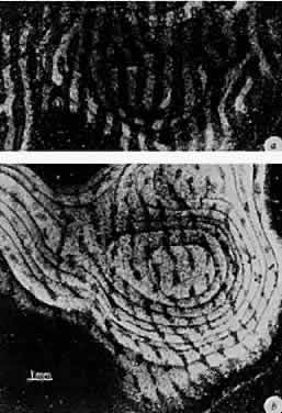
| Fig. 15. Autoradiographs of monkey visual cortex 2 weeks after injection of a radioactive tracer into the vitreous of one eye. Each figure represents a montage of tangential sections through the cortex. A. Normal monkey. The light stripes, representing labeled eye columns, are separated by gaps of the same width representing the other eye. B. Monocularly deprived monkey, who had the right eye closed at 2 weeks for 18 months. Input from the normal eye is in the form of expanded bands, which in places coalesce, obliterating the narrow gaps that represent the columns connected to the closed eye. (Wiesel TN. Postnatal development of the visual cortex and the influence of environment. Nature 1982;299:583.) |