Chapter 62
Genetics of Retinoblastoma
CAROL L. SHIELDS and JERRY A. SHIELDS
Main Menu   Table Of Contents

Search

THE RETINOBLASTOMA GENE
GENETIC COUNSELING
REFERENCES

THE RETINOBLASTOMA GENE

CLINICAL EVIDENCE

Retinoblastoma affects approximately one infant in 15,000 to 20,000 live births in the United States each year.1–4 The incidence of retinoblastoma among the various geographic populations is relatively constant indicating that environmental influences play little role in the development of this malignant intraocular tumor. Before the 1860s when the role of enucleation in the management of retinoblastoma became known, most cases of retinoblastoma proved fatal. At that time little was suspected about the inheritance patterns of this tumor because few patients, if any, survived to reproductive age. Later, as more patients survived and had children of their own, more evidence arose suggesting the hereditary nature of retinoblastoma.5 It is now known that retinoblastoma can be inherited as a familial tumor in which the affected child has a positive family history of retinoblastoma or as a nonfamilial (sporadic) tumor in which the family history is negative for retinoblastoma. Approximately 6% of newly diagnosed retinoblastoma cases are familial and 94% are sporadic. All patients with familial retinoblastoma are at risk to pass the predisposition for the development of the tumor to their offspring.

Retinoblastoma is generally classified in three different ways: familial or sporadic, bilateral or unilateral, and heritable or nonheritable. Clinically, the first two classification schemes are fused.6 Thus, a case may be classified as unilateral sporadic, bilateral sporadic, unilateral familial, or bilateral familial. About two thirds of all cases are unilateral and one third are bilateral. Genetically it is simpler to discuss retinoblastoma with the latter classification of heritable or nonheritable. The three classification schemes, however, are interrelated. It is recognized that bilateral and familial retinoblastoma is caused by a germline mutation and is, thus, a heritable tumor. Unilateral retinoblastoma is usually not heritable. Sporadic retinoblastoma is heritable in approximately 25% of cases, especially if it is a sporadic bilateral retinoblastoma.

In 1971, Knudson7 proposed the “two hit” hypothesis to explain the events that are necessary for both heritable and nonheritable retinoblastoma. His theory was based on a comparative analysis of unilateral and bilateral retinoblastoma cases. He proposed that the development of any retinoblastoma was caused by two complementary chromosomal mutations. Each of these genetic events can occur randomly with a frequency of 2 × 10⁻⁷ per year. In the case of familial retinoblastoma, the initial event or “hit” is a germinal mutation that is inherited and found in all cells of the offspring. The second hit occurs sometime during development, and if it occurs in a somatic cell such as a retinal cell then retinoblastoma develops. Therefore, in familial cases of retinoblastoma, all cells in the body are predisposed to possible tumor development because germline mutation (“first hit”) has been inherited in all cells of the body, including the ovaries and testes. This may help to explain the high incidence of second nonocular tumors, such as osteosarcoma, seen in patients with familial retinoblastoma or bilateral sporadic retinoblastoma.8–11 The offspring in cases of familial retinoblastoma will likewise be predisposed because their germinal mutation will be passed on. By contrast, in most cases of unilateral sporadic retinoblastoma, the two hits occur during development of the retina and both hits are somatic mutations.12 The rest of the body theoretically carries no higher risk to develop other tumors because these patients presumably have normal chromosomal structure elsewhere in the body.

Knudson's theory provides an explanation for the similarities and differences between heritable and nonheritable retinoblastoma. Ophthalmoscopically and histopathologically, heritable and nonheritable retinoblastoma are indistinguishable.13 The major differences between heritable and nonheritable retinoblastoma are that the heritable tumor usually occurs at a younger age,1 is more likely to be bilateral and multicentric,1 and the affected patient is at higher risk for nonocular tumors than is the nonheritable case.8–11 The rationale for this finding is that the probability for the one hit necessary for tumor formation in heritable cases is 2 × 10⁻⁷, whereas the probability for two independent hits to occur as in the nonheritable retinoblastoma case is 2 × 10-7 times 2 × 10-7. It undoubtedly takes longer for the two unlikely events to occur in nonheritable cases, and it is highly unlikely that these two events will occur again at another retinal location, thereby explaining the older age at clinical presentation and the lack of multicentricity in nonheritable retinoblastoma. The reason for the higher likelihood of second nonocular tumors in heritable retinoblastoma is that all of the cells of the body already have inherited a single hit or germinal mutation on chromosome 13, which may predispose them to other cancers found in association with chromosome 13 defects.14 These patients are predisposed to nonocular tumors if the second hit occurs. In nonheritable retinoblastoma, both hits need to occur in a solitary cell and this is statistically less likely.

Excess cancer in relatives of patients with heritable retinoblastoma and advanced paternal age support the findings of the genetic influence in retinoblastoma and other solid childhood tumors.15–17 Excess nonocular cancer in relatives provides strong evidence that the gene that controls retinoblastoma is a generalized cancer gene.15 Fathers of children with bilateral sporadic retinoblastoma are generally older than are fathers of patients with unilateral sporadic cases.16

PHYSICAL PROPERTIES

The first clue as to the location of the retinoblastoma gene was recognized by Stallard18 in 1962 when he reported the case of an infant with retinoblastoma that had a deletion in one of the D-group chromosomes. The D-group chromosomes include numbers 13, 14, and 15. It was difficult to distinguish these three chromosomes without the cytogenetic techniques that evolved in the 1960s. Additional cases of D-group deletion retinoblastoma were recognized, and retinoblastoma became known as a part of the D-deletion syndrome. In the 1970s, high-resolution chromosomal banding showed that the affected chromosome in patients with retinoblastoma was chromosome 13.19 Because of this, the syndrome was renamed the 13-deletion syndrome. In 1978, the locus of the deletion was found to be the q14 band, that is the 14 band on the long arm (q) of the 13th chromosome.

The retinoblastoma locus on chromosome 13 encodes for a large gene of 4.73 kilobase message.20 An intact gene protects against expression of retinoblastoma. It is believed that the gene is a recessive suppressor gene and may play a role in cell growth and development.21 In order for retinoblastoma to develop, both copies of the gene at the 13q14 locus must be lost, deleted, mutated, or inactivated.21 If either the maternal or paternal copy of the gene that is inherited by an individual is defective, then that individual is heterozygous for the mutant allele. Tumor formation requires both alleles of the gene to be mutant or inactive. These two mutations correlate to the two hits theorized by Knudson.7,12

The retinoblastoma gene comprises at least 27 exons over a genomic locus of 200 kilobases. The coding portion of the gene consists of 4.73 kilobases. The retinoblastoma gene has been demonstrated to be missing or rearranged on approximately 40% to 100% of retinoblastoma tumor tissue.14,22,23 Even when no deletion is found, the message from the gene has been found to be altered or absent. In these cases, it is likely that some minute structural changes on the gene are present and are too submicroscopic to be identified with routine karyotyping methods.

Only 5% to 6% of patients with retinoblastoma are found to have a visible deletion in chromosome 13 when studied by peripheral blood sampling.24,25 Approximately 6% of our patients with retinoblastoma have been found to have a chromosomal abnormality involving 13q, and one half were unilateral cases and one half were bilateral cases.24 It is hypothesized that many more patients have chromosomal deletions that are not detected by normal banding chromosomal studies because the deletions are too small for identification. High-resolution banding studies may detect chromosomal mosaicism, that is, the deletion on some but not all cells. More recently, newer techniques of analysis of the deoxyribonucleic acid (DNA) of chromosome 13 has enabled investigators to identify very small deletions that would have been otherwise too small to detect on chromosomal banding.26,27

The increasing cytogenetic technology and the development of DNA probes for loci in the vicinity of the retinoblastoma gene locus (RB1) has allowed detection of subtle genomic rearrangements. The RB1 gene encodes a nuclear phosphoprotein p110RB1 with a molecular weight of 105 to 110 kilodaltons. Evidence suggests that this protein regulates cell proliferation and differentiation. In a review of 192 patients with retinoblastoma studied with DNA analysis, various germline mutations in the RB1 gene were found including nonsense mutation in 43%, frameshift in 35%, intron mutation in 12%, missense mutation in 6%, in-frame deletion in 3%, and promoter mutation in 2%.28 Mutations were distributed throughout 24 of the 27 exons of the RB1 gene with no singe hotspot of mutation. Based on this knowledge, the spectrum of germline RB1 mutations in patients with retinoblastoma is broad. Large chromosomal alterations are detected by cytogenetic analysis in 7% to 8%, and smaller DNA rearrangements are identified by DNA fragment analysis in 16% of patients.28 Despite the sophistication of current techniques, approximately 20% of patients with bilateral retinoblastoma have an existing mutation that is not found.28 It is suggested that a series of complementary tests to rapidly detect mutations is most beneficial for patients with retinoblastoma.

13Q14 SYNDROME

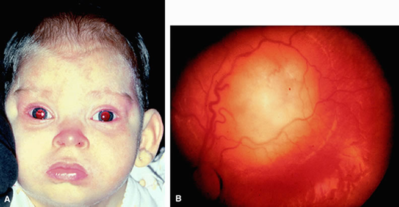
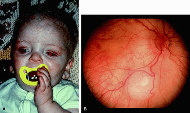
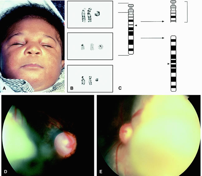
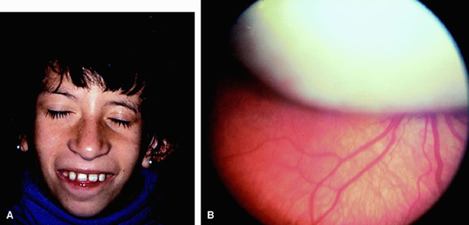
The 13q deletion syndrome may manifested by several phenotypic abnormalities. Many patients have minimal or no visible abnormality.29 The characteristic findings include some degree of the following dysmorphic features: microcephaly, broad prominent nasal bridge, hypertelorism, microphthalmos, epicanthus, ptosis, protruding upper incisors, micrognathia, short neck with lateral folds, large prominent low set ears, facial asymmetry, imperforate anus, genital malformations, perineal fistula, hypoplastic or absent thumbs, toe abnormalities, and psychomotor and mental retardation.30–32 The midface of patients with 13q deletion are notable for prominent eyebrows, broad nasal bridge, bulbous tipped nose, large mouth, and thin upper lip32–34 (Figs. 1, 2, 3, and 4). We recently reported a case of severe midline facial and central nervous system abnormalities in a child with 13q abnormalities that manifested retinoblastoma and holoprosencephaly.35

Fig. 1. Dysmorphic features of the 13q deletion syndrome showing the flat broad nasal bridge, bulbous tip of the nose, low set ears, and prominent eyebrows. A. (Case #1) Sixteen-month-old child with manifested developmental delay and characteristic facies of 13q deletion syndrome. B. (Case #1) Unilateral retinoblastoma was later discovered in this patient. The tumor was successfully treated with plaque radiotherapy.

Fig. 2. Failure to thrive as the initial manifestation of 13q syndrome. A. (Case #2) Eighteen-month-old child with severed failure to thrive and abnormal facies. Genetic screening at age 3 months revealed 13q deletion. B. (Case #2) Fundus examination at age 3 months revealed multifocal bilateral retinoblastoma. The tumors in both eyes were successfully treated with thermotherapy and cryotherapy.

Fig. 3. Failure to thrive and severe developmental delay as the initial manifestations of 13q syndrome. A. (Case #3) A 4-month-old child with slightly abnormal facies and low set ears. B. (Case #3) Cytogenetic analysis revealed mosaic ring chromosome 13. The chromosomes are shown in order from left to right as normal chromosome 13, derivative (13), and ring (13) with different staining methods. Top. G-banded chromosomes showing the normal chromosome 13, derivative (13), and ring (13). Middle. C-banded chromosomes showing positive staining for the normal 13 and ring (13). Bottom. BRdU stain of late replicating chromosomes. No difference is observed between the mirror images of der(13) chromosome. C. (Case #3) Ideogram of chromosome 13 rearrangement in which break occurred at the 13q14 locus resulting in ring (13) and derivative (13). The derivative (13) does not contain a centromere and would be expected to be lost in future cell divisions unless a neocentromere is formed. D. (Case #3) Examination of the right eye revealed multifocal retinoblastoma and enlarged cup to disc ratio consistent with additional congenital glaucoma. E. (Case #3) Examination of the left eye showed advanced retinoblastoma and optic disc changes to suggest congenital glaucoma. The eyes were successfully treated with chemotherapy and radiotherapy, but the child later died of intracranial neuroblastic malignancy (trilateral retinoblastoma). (B and C from Morrissette JJD, Celle L, Owens NL et al: Boy with bilateral retinoblastoma and an unusual ring chromosome 13 with activation of a latent centromere. Am J Med Genetics 99:21, 2001)

Fig. 4. Follow-up of child with 13q syndrome. A. (Case #4) This patient presented with developmental delay and was found by genetic screening at age 4 months to have 13q syndrome and multifocal retinoblastoma. In this photo, she is age 10 years with short stature and the developmental delay is noted. B. (Case #4) Fundus examination at age 4 months shows large retinoblastoma that was successfully treated with external beam radiotherapy.

Karyotype analysis of children with these or other dysmorphic features may allow earlier detection of retinoblastoma. We have seen several cases of retinoblastoma that were initially suspected based on the recognition of the previously described dysmorphic features that prompted a karyotype analysis that revealed a deletion in chromosome 13. This finding subsequently prompted a retinal examination, which revealed unilateral multifocal tumors in both cases.32

LINKAGE MARKERS

Before the identification of the retinoblastoma gene, linked markers such as esterase D were used to identify individuals who had the heritable form of retinoblastoma. Esterase D is an enzyme expressed in all cells that is coded on chromosome 13. If both of its alleles are fully active, then the enzyme is measured as 100% activity. If one allele is missing, then the activity drops to 50%. Esterase D has been found to be closely linked to the retinoblastoma gene on the 13th chromosome. In fact it has been found to lay in the proximal portion of the 13q14 locus.36 Because of this tight association with retinoblastoma, screening for the retinoblastoma gene by testing for esterase D levels has been used in the past but is rarely used today. If esterase D is 50% or less than its normal activity, then it suggests that a chromosome 13 deletion and possible retinoblastoma gene deletion is present.

Back to Top
GENETIC COUNSELING

FUTURE OFFSPRING

One of the more important steps in the management of a patient with retinoblastoma is genetic counseling.1,2,37 Unfortunately the ophthalmologist who manages an infant with retinoblastoma may eventually lose follow-up of the patient over the years after the eyes are successfully treated. The parents may be reluctant to inform the child that he or she had cancer during infancy and may even lead him to believe that the eye was removed because of trauma or infection. The patient may grow up and have children without realizing that there is a possibility of transmitting a malignant tumor to them.

When counseling the patient and family about the possibility of future children developing retinoblastoma, it is critical to know if the child or family carries a germinal mutation for the retinoblastoma gene. There are several clinical features that help identify those families that may carry the retinoblastoma gene. Those patients with bilateral retinoblastoma and those patients with a positive family history of retinoblastoma can be assumed to have a germinal mutation for the retinoblastoma gene; therefore, these children are at a 50% risk of passing this gene to future children. The retinoblastoma gene is about 80% penetrant so that only 40% of their offspring will manifest the clinical findings of the gene and some offspring may only be carriers of the gene without developing retinoblastoma.38

Only 6% of newly diagnosed retinoblastoma patients will have a family history of retinoblastoma (familial) and 94% will have no family history (sporadic). Approximately 155 to 20% of unilateral sporadic retinoblastomas are caused by germinal mutations that by chance affect only one eye. The remaining 80% to 85% are somatic mutations that occur only in the retina and are nonhereditary so that the patient cannot transmit the disease39 (Table 1). Bilateral or multifocal unilateral retinoblastomas represent germinal mutations in theoretically 100% of cases. These patients have a 40% to 50% chance of passing the disease to their offspring.


TABLE 1. Retinoblastoma Type and Laterality


 BilateralUnilateralTotal
Hereditary25%–30%15%–20%40%–50%
Nonhereditary050%–60%50%–60%
Total25%–30%70%–75%100%


Based on genetic studies, the general percentages for predicting the chance of future children inheriting and developing retinoblastoma are listed in Tables 2 and 3.40,41 Table 2 refers to families in which there is no prior history of retinoblastoma, whereas Table 3 refers to families in which at least one family member had retinoblastoma.


TABLE 2. Risk for Future Offspring to Develop Retinoblastoma when There is a Negative Family History*


If the Affected Patient hasUnilateralBilateral
Then the chances for retinoblastoma in the offspring of the following family members are
Parent of affected patient1%6%
Affected patient8%40%
Normal sibling of affected patient1%<1%

*Assumes an 80% penetrance.


TABLE 3. Risk for Future Offspring to Develop Retinoblastoma when There is a Positive Family History*


If the Affected Patient hasUnilateralBilateral
Then the chances for retinoblastoma in the offspring of the following family members are
Parents of affected patient40%40%
Affected patient40%40%
Normal sibling of affected patient7%7%

*Assumes an 80% penetrance.


SECOND PRIMARY TUMORS

Another important aspect of genetic counseling concerns the development of new unrelated cancers in survivors of bilateral or heritable retinoblastoma. It is now recognized that a child with retinoblastoma has approximately a 5% chance of developing another malignancy during the first 10 years of follow-up, 18% during the first 20 years, and 26% within 30 years.10 The 30-year cumulative incidence is about 35% for those patients who received radiation therapy (external beam therapy) as compared with an incidence rate of 6% for those patients who did not receive radiation. Therefore, patients with bilateral retinoblastoma have an increased incidence of second tumors, and this rate is further increased in those treated with external radiation therapy.10 Osteogenic sarcoma, often involving the femur, is most common, but other tumors such as spindle cell sarcoma, chondrosarcoma, rhabdomyosarcoma, neuroblastoma, glioma, leukemia, sebaceous cell carcinoma, squamous cell carcinoma, and malignant melanoma have also been recognized.9,10,42,43 The mean latency period for the appearance of the second primary is approximately 13 years.10

NEUROBLASTIC INTRACRANIAL MALIGNANCY (TRILATERAL RETINOBLASTOMA)

It has recently been recognized that there is a high incidence of neuroblastic intracranial malignancy in patients with the hereditary form of retinoblastoma, most often manifesting as pinealoblastoma or other parasellar tumors.44 The pinealoblastoma is identical to retinoblastoma from an embryologic, pathologic, and immunologic standpoint.45–48 This association of midline intracranial pineal tumors and suprasellar and parasellar neuroblastic tumors with bilateral retinoblastoma has been termed trilateral retinoblastoma.49,50 The retinoblastoma gene is believed to confer an increased susceptibility to developing these intracranial tumors.49,50 Trilateral retinoblastoma is found in approximately 3% of all children with retinoblastoma.47,48 Those patients with bilateral or familial disease are at greatest risk, with 5% to 15% developing these tumors.47,48 In one case from our series, the intracranial tumor preceded the diagnosis of retinoblastoma by 5 months.47 It is possible that many cases of pinealoblastoma were previously misinterpreted as metastatic retinoblastoma to the brain.46,49 Unlike the other second tumors mentioned previously, the pinealoblastoma usually occurs during the first4 years of life.48 It is usually fatal. The possibility of pinealoblastoma should be included in the genetic counseling of patients with hereditary retinoblastoma. Newer evidence suggests that recent treatment methods of systemic chemoreduction for retinoblastoma may prevent trilateral retinoblastoma.51,52 In a study of nearly 100 patients with hereditary retinoblastoma, trilateral retinoblastoma was found in no patient who received chemoreduction and it would be expected for 5 to 15 patients in this group to manifest the associated brain tumor.52 Thus, prevention of trilateral retinoblastoma may be possible.

Back to Top
REFERENCES

1. Shields JA, Shields CL: Intraocular Tumors: A Text and Atlas. Philadelphia, WB Saunders, 1992, pp 305–392

2. Shields JA, Shields CL: Atlas of Intraocular Tumors. Philadelphia, Lippincott, Williams & Wilkins, 1999, pp 207–232

3. Devesa SS: The incidence of retinoblastoma. Am J Ophthalmol 80:263, 1975

4. Friend SH, Bernards R, Rogelj S et al: A human DNA segment with properties of the gene that predisposes to retinoblastoma and osteosarcoma. Nature 323:643, 1986

5. Donoso LA, Shields CL, Lee EY: Immunohistochemistry of retinoblastoma: A review. Ophthal Paediatr Genet 10:3, 1988

6. Shields CL, Shields JA: Recent developments in the management of retinoblastoma. J Pediatr Ophthalmol Strabismus 36:8, 1999

7. Knudson AG: Mutation and cancer: Statistical study of retinoblastoma. Proc Natl Acad Sci U S A 68:820, 1971

8. Abramson DH, Ellsworth EM, Kitchin FD et al: Second non-ocular tumors in retinoblastoma survivors: Are they radiation induced? Ophthalmology 91:1351, 1984

9. Abramson DH, Ellsworth RM, Zimmerman LE: Nonocular cancer in retinoblastoma survivors. Trans Am Acad Ophthalmol Otolaryngol 81:454, 1976

10. Abramson DH, Ronner HJ, Ellsworth RM: Nonocular cancer in nonirradiated retinoblastoma. Am J Ophthalmol 87:624, 1979

11. Roarty JD, McClean IW, Zimmerman LE: Incidence of second neoplasms in patients with bilateral retinoblastoma. Ophthalmology 95:1583, 1988

12. Gallie BL, Phillips RA: Retinoblastoma: A model of oncogenesis. Ophthalmology 91:666, 1984

13. Zimmerman LE: Retinoblastoma and retinocytoma. In Spencer WH, Font RL, Green WR et al (eds): Ophthalmic Pathology. Philadelphia, WB Saunders, 1985, pp 1292–1351

14. Horowitz JM, Park SH, Bogenmann E et al: Frequent inactivation of the retinoblastoma anti-oncogene is restricted to a subset of tumor cells. Proc Natl Acad Sci U S A 87:2775, 1990

15. Cohen MS, Augsburger JJ, Shields JA et al: Cancer in relatives of retinoblastoma patients. Jpn J Ophthalmol 33:173, 1989

16. Pellie C, Briard ML, Feingold J et al: Parental age in retinoblastoma. Humangenetik 20:59, 1973

17. Strong LC, Herson J, Haas C et al: Cancer mortality in relatives of retinoblastoma patients. J Natl Cancer Inst 73:303, 1984

18. Stallard HB: The conservative treatment of retinoblastoma. Trans Ophthalmol Soc U K 82:473, 1962

19. Yunis JJ, Ramsay N: Retinoblastoma and subband deletion of chromosome 13. Am J Dis Child 132:161, 1978

20. Fung YKT, Murphree AL, T'Ang A et al: Structural evidence for the authenticity of the human retinoblastoma gene. Science 236:1657, 1987

21. Murphree AL, Benedict WF: Retinoblastoma: Clues to human oncogenesis. Science 223:1028, 1984

22. Balaban G, Gilbert F, Nichols W et al: Abnormalities of chromosome #13 in retinoblastomas from individuals with normal constitutional karyotypes. Can Genet Cytogenet 6:213, 1982

23. Fung YKT, Murphree AL, T'Ang A et al: Structural evidence for the authenticity of the human retinoblastoma gene. Science 236:1657, 1987

24. Bunin GR, Emanuel BS, Meadows AT et al: Frequency of 13q abnormalities among 203 patients with retinoblastoma. J Natl Can Inst 81:370, 1989

25. Knudson AG: Genetics of human cancer. J Cell Physiol 4(Suppl):7, 1986

26. Wiggs J, Nordenskjold M, Yandell D et al: Prediction of the risk of hereditary retinoblastoma, using DNA polymorphisms within the retinoblastoma gene. N Engl J Med 318:151, 1988

27. Wiggs JL, Dryja TP: Predicting the risk of hereditary retinoblastoma. Am J Ophthalmol 106:346, 1988

28. Harbour JW: Overview of RB gene mutations in patients with retinoblastoma. Implications for clinical genetic screening. Ophthalmology 105:1442, 1998

29. Knudson AG, Meadows AT, Nichols WW et al: Chromosomal deletion in retinoblastoma. N Engl J Med 295:1120, 1976

30. Allderdice PW, Davis JG, Miller OJ et al: The 13q- deletion syndrome. Am J Hum Genet 21:499, 1969

31. Niebuhr E, Ottosen J: Ring chromosome D(13) associated with multiple congenital malformations. Ann Genet 16:157, 1973

32. Seidman DJ, Shields JA, Augsburger JJ et al: Early diagnosis of retinoblastoma based on dysmorphic features and karyotype analysis. Ophthalmology 94:663, 1987

33. Keith CG, Webb GC: Retinoblastoma and retinoma occurring in a child with a translocation and deletion of the long arm of chromosome 13. Arch Ophthalmol 103:941, 1985

34. Montegi T, Kaga M, Yanagawa Y: A recognizable pattern of the midface of retinoblastoma patients with interstitial deletion of 13q. Hum Genet 64:160, 1983

35. Desai VN, Shields CL, Shields JA et al: Retinoblastoma associated with holoprosencephaly. Am J Ophthalmol 109:355, 1990

36. Sparkes RS, Murphree AL, Lingua RW et al: Gene for hereditary retinoblastoma assigned to human chromosome 13 by linkage to esterase D. Science 219:971, 1983

37. Albert DA, Dryja TP: Recent studies of the retinoblastoma gene. What it means to the ophthalmologist. Arch Ophthalmol 106:181, 1988

38. Gallie BL: Gene carrier detection in retinoblastoma. Ophthalmology 87:591, 1980

39. Reese AB: Tumors of the Eye. New York, Harper and Row, 1976, p 127

40. Carlson EA, Letson RD, Ramsay NKC et al: Factors for improved genetic counseling for retinoblastoma based on a survey of 55 families. Am J Ophthalmol 87:449, 1979

41. Gordon H: Family studies in retinoblastoma. Birth Defects 10:185, 1974

42. Benedict WF, Fung YKT, Murphree AL: The gene responsible for the development of retinoblastoma and osteosarcoma. Cancer 62:1691, 1988

43. Tucker MA, D'Angio GJ, Boige JD et al, for the Late Effects Study Group: Bone sarcomas linked to radiotherapy and chemotherapy in children. N Engl J Med 317:588, 1987

44. Zimmerman LE: Trilateral retinoblastoma. In Blodi FC (ed): Retinoblastoma. New York, Churchill Livingstone, 1985, pp 185–210

45. Donoso LA, Rorke LB, Shields JA et al: S Antigen immunoreactivity in trilateral retinoblastoma. Am J Ophthalmol 103:57, 1987

46. Pesin SR, Shields JA: Seven cases of trilateral retinoblastoma. Am J Ophthalmol 107:121, 1989

47. DePotter P, Shields CL, Shields JA: Clinical variations of trilateral retinoblastoma: A report of 13 cases. J Pediatr Ophthalmol Strabismus 31:26, 1994

48. Kivela T: Trilateral retinoblastoma: A meta-analysis of hereditary retinoblastoma associated with primary ectopic intracranial retinoblastoma. J Clin Oncol 17:1829, 1999

49. Bader JL, Meadows AT, Zimmerman LE et al: Bilateral retinoblastoma and ectopic intracranial retinoblastoma. Trilateral retinoblastoma. Cancer Genet Cytogenet 5:203, 1982

50. Bader JL, Miller RW, Meadows AT et al: Trilateral retinoblastoma. Lancet 2:582, 1980

51. Shields CL, Shields JA, Needle M et al: Combined chemoreduction and adjuvant treatment for intraocular retinoblastoma. Ophthalmology 104:2101, 1997

52. Shields CL, Meadows AT, Shields JA et al: Chemoreduction for retinoblastoma may prevent intracranial neuroblastic malignancy (trilateral retinoblastoma). Arch Ophthalmol 119:1269–1272, 2001

Back to Top