Chapter 61
Vitreoretinal Dystrophies
HERMANN D. SCHUBERT and WILLIAM TASMAN
Main Menu   Table Of Contents

Search

X-LINKED HEREDITARY RETINOSCHISIS
FAMILIAL EXUDATIVE VITREORETINOPATHY
DEGENERATIVE MYOPIA
WAGNER'S SYNDROME
GOLDMANN-FAVRE SYNDROME
STICKLER'S SYNDROME
ROBIN'S SYNDROME
CLEFTING SYNDROMES
SKELETAL DYSPLASIAS
CONCLUSION
REFERENCES

Vitreoretinal dystrophies may present on routine ophthalmologic examination as high myopia, strabismus, retinal detachment, vitreous hemorrhage, or abnormal fundus pigmentation. The ocular findings are either isolated1–4 or part of a syndrome such as Robin's syndrome (also known as the Pierre Robin syndrome),5 one of the clefting syndromes,6 or skeletal dysplasia.7 Although the “gestalt” of retinoschisis,1 familial exudative retinopathy (FEVR),2 or original Wagner's disease4 emerges with ease, lengthy deliberation is needed for conceptual separation of some of the syndromes that are not restricted to the eye.7–12 Whereas identification of specific genes is rapidly emerging,13–22 it is not always clear which of the polymorphous signs to include at this mostly descriptive stage of nosology, with subsets and syndromes based on somewhat arbitrary definitions.

An ocular dystrophy is a congenital bilateral degeneration of ocular tissues, here predominantly involving the vitreous and retina; the degeneration may be either stationary or progressive. Dystrophic vitreal changes involve liquefaction (synchysis) of the gel and collapse (syneresis) of the collagenous structure. Thus, destruction of the vitreous gel is superimposed on the aging process.12,23 Microscopically, the liquefied vitreous appears optically empty; collapse of the vitreous collagen causes it to appear veil-like. Vitreoretinal adhesions may be absent or abnormally firm. Dystrophic retinal changes include intraretinal splitting, atrophy, hole formation, and migration of pigment from the pigment epithelium. In addition, extraretinal changes sometimes occur. These changes present as selective atrophy of the retinal pigment epithelium, which appears as choroidal tesselation. Independent of this, choroidal stromal atrophy has been described.8

Vitreoretinal dystrophies are found with greater ease by the prepared examiner. It is the purpose of this review to present the basic pathology, as well as the genetic and clinical features, of nine vitreoretinopathies in which primary retinal involvement is accompanied by passive secondary vitreal changes. The habit of thinking of vitreoretinal dystrophies, especially in children, may lead to earlier recognition of patients at risk for retinal detachment and consequent visual loss.

Back to Top
X-LINKED HEREDITARY RETINOSCHISIS
Although both peripheral and macular retinoschisis were first described in 1898,24 the other vitreoretinal dystrophies have been described only more recently. The history of the recognition and naming of the retinoschisis is particularly interesting, mirroring the progressive characterization of the lesion, and thus may be helpful in understanding it:

  1913: Pagenstecher noted the inheritance of “solutio retinae” in hypermetropic children.25
  1932: Anderson localized the “anterior dialysis” to the inferotemporal quadrant in nonmyopic young males.26
  Thomson emphasized macular involvement.27
  1938: Mann and MacRae theorized that the “congenital vascular veils” in the vitreous are the result of retinal splitting.28
  1948: Juler described retinal detachment in children with thinning and ruptures of the inner linings of the cysts.29 It was then assumed that the pathology is retinal but involves secondary changes in the vitreous.
  1950: Sorsby and colleagues recognized the sex-linked inheritance of retinoschisis.1 Combining this finding with the previous views, they postulated that the veils represent ruptured retinal cysts, which, at a later stage, probably progress to congenital falciform detachment and pseudoglioma.
  1953: Jager coined the term “retinal schisis.”30
  1953/4: Kleinert31 and MacRae32 agreed with the concept of central and peripheral splitting of the retina.
  1960/1: Balian and Falls33 and Gieser and Falls34 emphasized that the congenital vascular veils in the vitreous are combined with macular changes.
  1964/6: Sarin and associates35 and Sabates36 identified members of the same families who had “hereditary retinal schisis.”
  1973: Forsius and colleagues confirmed the X-linkage of retinal schisis and found that the maculopathy is present in virtually all cases.37
  1976: Harris and Yeung classified retinoschisis into stages based on its degree of severity with respect to the occurrence of striation and atrophic and pigmentary changes.38
  1983: Wieacker and coworkers assigned the gene to band Xp 22.2 of the human X chromosome by linkage analysis.13

X-linked retinoschisis1 can be called a retinovitreal dystrophy because retinal schisis, not vitreous syneresis, is the predominant sign. The common denominator of the retinal changes is thought to be Mueller cell degeneration,39 the sequelae of which are most visible in the fovea and in the periphery of the inferotemporal fundus.40 Peripheral changes only may be present in the rare autosomal dominant form.41 The wide range of biomicroscopic findings changes with the duration of the condition and age of the patient.1 The posterior vitreous adhesion (that is, premacular and prepapillary, as well as perivascular) is well developed in the young.

Vitreous attachments to the inner layer of the schisis can cause progression of schisis through traction, particularly if vitreous contraction is accelerated by vitreous hemorrhage. Vitreous traction may lead to nasal dragging of the retina.42 The inner schisis layer consists of glia, nerve fiber layer, and vessels.40 Ultrastructural studies confirm the split along the ganglion cell and nerve fiber layer separating cell processes from their bodies in the inner nuclear layer and resulting in a picture of “decapitated and degenerated Mueller cells.”43 Whereas the inner layer of the schisis consists mostly of glial cells, the outer layer also provides a glial lining of the schisis cavity.43 Because vascularization of the inner retinal layers extends to the middle limiting membrane,44 schisis, however, takes place internal to the inner nuclear layer; the deep retinal capillaries may be disrupted, appearing angiographically as areas of nonperfusion.45 Inner layer atrophy may also ensue as a result of chronic inner layer ischemia and glial cell decapitation. Atrophic holes in the inner layer and perivascular gliosis may be the biomicroscopic corollaries. The outer layer may undergo atrophy or may be invaded by proliferations of the pigment epithelium.39,40

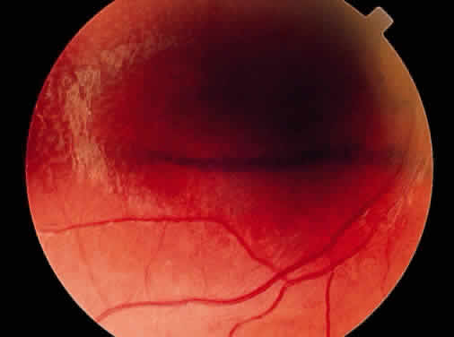
Retinal schisis involves a delicate balance between vitreous traction on the inner layer and the opposing strength of residual intraretinal bridges of tissue and adhesions of the outer layer of schisis to the pigment epithelium. Intraretinal strands of tissue that bridge the schisis cavity have been shown in vivo using the retinal thickness analyzer.46 If the vitreous gel is collapsed, the inner wall of the schisis is found in the retrolental space.33,40 Not surprisingly, such bullous schisis is more common in the young (i.e., in children younger than 10 years),47–49 often accompanied by vitreous and/or intraschisis hemorrhage.47 Visual loss in the young patient is often associated with contraction of adherent vitreous, bullous schisis, and its complications. It is a dynamic process; therefore, with increasing age bullous schisis tends to flatten spontaneously in the vast majority of cases.47,49 Application of a laser beam to the area of schisis has been shown to disrupt the equilibrium of vitreous traction and retinal adhesions and to produce rhegmatogenous detachments resulting from outer layer breaks as well as from full-thickness retinal breaks.50

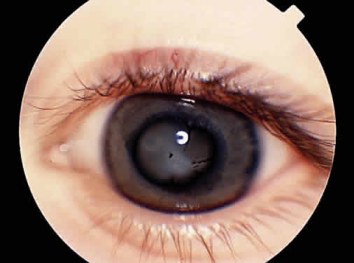
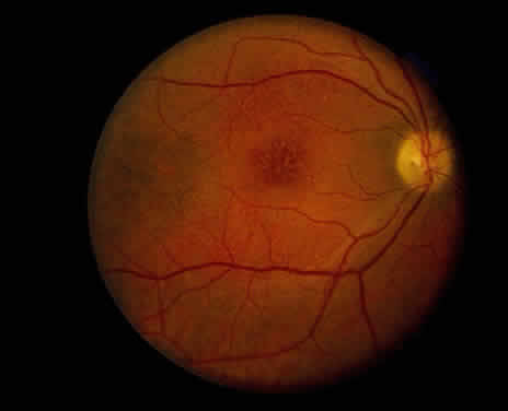
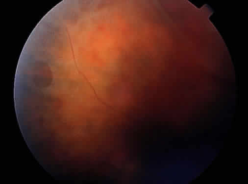
Clinically, retinal schisis presents with decreased central visual acuity, absolute scotomas, or both.1,33,38 Children may have strabismus and nystagmus.25,47 As in FEVR, but in contrast to Wagner and Stickler syndrome, the refraction tends to be hypermetropic (“hypermetropic amblyopia”).1,25,26,31–34,40,48 On biomicroscopic examination, edema and cystic central macular schisis of the retina are apparent in virtually all cases.37,38 Loss of macular reflex, which may be an early sign,34,35 is followed by wheel-like formation,37 a multicystic radiate appearance (Fig. 1),38 pigmentary degeneration, and cystic macular atrophy (Fig. 2).38 These macular changes, not bullous schisis per se, determine visual loss in patients often in the fourth or fifth decade of life.51

Fig. 1. Petaloid maculopathy in a patient with X-linked schisis.

Fig. 2. Pigmented macular demarcation line in X-linked schisis.

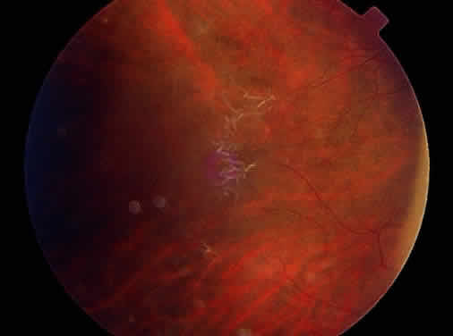
Peripheral retinal schisis is present in half of the patients and in 96% is located in the inferotemporal quadrant.48 At least initially, the inner layer of the schisis is attached to the vitreous and features vessels, internal limiting membrane, and nerve fiber layer. These tissues undergo progressive atrophy as well as reactive gliosis, which in a perivascular location may appear as vascular sheathing (Fig. 3). Total atrophy may result in dehiscences and peripheral arborization of the retinal vasculature.33,34,45 Optic atrophy may follow.40 The outer layer of the schisis cavity also undergoes atrophy, reactive gliosis, intralayer pigment migration, and hole formation. A rhegmatogenous detachment may be the sequela in 4% to 22% of all patients.30,42,49,51–53 With time, the vitreous undergoes syneresis and detachment.31,33,40 Vitreous detachment actually may improve the schisis by releasing traction. Adhesions to retinal vessels are responsible for frequently found vitreous hemorrhages.29,31,40

Fig. 3. Nerve fiber layer defect in X-linked schisis.

Because progression of the disease cannot be predicted and regression sometimes occurs, patients should be observed regularly, particularly those younger than 10 years of age.47 Indications for surgical intervention are rhegmatogenous retinal detachment and repeated vitreous hemorrhages.

Photocoagulation has not been shown to be beneficial in that it creates external wall holes.50 Such prophylactic treatment has led to retinal detachment in 14% to 43% of cases.48,49 This failure may be related to the fact that most of the mechanical cohesiveness of the retina lies in the inner layer near its basement membrane. The usual energy levels for coagulation may be too intense for the thin outer layer.

Similarly, scleral buckling has been shown to be marginally effective in treating the detachments. Redetachments after buckling were reported in up to 40%.48,53 In cases of vitreous hemorrhage and vitreous attachment to the inner layer of schisis, scleral buckling combined with pars plana vitrectomy techniques may be beneficial. Recent approaches to surgery have centered on removal of the inner layer of the schisis cavity,52,53 if possible by lens-sparing technique.54 The position of the inner layer is determined by vitreous forces in its degree of separation and topography (dragging). In many cases it may be impossible to separate the cortical vitreous from the inner layer of schisis. Inner layer retinectomy most definitely eliminates traction that might lead to redetachment and may account for the superior long-term results reported with this method.54

Back to Top
FAMILIAL EXUDATIVE VITREORETINOPATHY
This entity, first described by Criswick and Schepens in 1969,2 is autosomal dominantly inherited,55 even though rare sporadic and X-linked recessive modes have been reported.56 Linkage analysis has located the gene for the autosomal dominant form to the long arm of chromosome 11.17 FEVR can be found in patients of any age and is usually bilateral with asymmetric expression.57 The refraction tends to be emmetropic or myopic and is not of much help in distinguishing it from retinitis of prematurity (ROP).58 As in retinal schisis, the primary changes are in the retina, here characterized by a nonperfused inner retinal layer. The vitreal changes are secondary to chronic exudation into the vitreous, leading to contraction and early posterior detachment of this passive extracellular tissue.59 Symptoms and clinical presentation are related to the extent of nonperfusion, which in turn determines the degree of the ocular response. Three stages are generally distinguished: the first stage of nonperfusion, the second stage of localized tractional detachment, exudation, and possibly neovascularization, and the third stage of tractional and exudative retinal detachments.57,60 Neovascularization, if severe, portends a poor prognosis and appears much like ROP, whereas subretinal exudation of lipid may be prominent enough to suggest a diagnosis of Coats' disease. However, in FEVR, the infrequent exudative detachment usually is combined with a tractional fibrovascular preretinal membrane, whereas in Coats' disease abnormal intraretinal vessels and compressed vitreous predominate. Not surprisingly, histologic studies are concerned with the later stages of disease, showing absence of intraretinal vessels57 and nonspecific fibrovascular preretinal membranes.61 For the early stages of FEVR, angiography shows V-shaped temporal retinal nonperfusion posterior to the ora serrata (i.e., near the posterior vitreous base).60 The peripheral retinal vasculature features supernumerous vascular branchings, arteriovenous shunt formation, and patterns of incomplete arteriovenous interdigitation.62,63 The peripheral retinal vessels are hyperpermeable and leak underneath the retina and into the vitreous base. Subretinal and peripheral retinal fibrosis may also be noted.62 Vitreous collapse induces peripheral traction, retinal detachments, and elevated neovascularization. The newly formed vessels tend to leak and bleed, thus fueling a vicious circle of vitreous contraction and fibrosis. Further, nonperfusion corresponds to ischemia of the inner layer of the retina and thus involves Mueller cells, the nuclei of which are located in the inner nuclear layer. The combination of vitreous base traction, Mueller cell ischemia, and response to exudation sets the stage for retinal schisis, hole formation in the inner and outer layer of the retina, granulomatous proliferations, and vitreous hemorrhage.62,63 Although a high incidence of vitreous detachment was described in early publications, later writers have not found a high incidence.63 The vitreal changes of syneresis and synchysis, contraction, partial or complete detachment, opacification, and traction-rhegmatogenous detachment seem to be secondary to the retinal vasculopathy. The histologic vascular changes are indistinguishable from those of ROP; however, FEVR patients are not myopic and have no history of prematurity or neonatal oxygen exposure.58,59

Mild forms of the disease that produce no symptoms predominate. They are characterized by areas of peripheral nonperfusion, cystoid degeneration, and condensation of the temporal vitreous base and vitreous membranes. Moderate forms of FEVR with visual blurring, floaters, or distortion involve vitreous base traction, flat or elevated neovascularization, and retinal or subretinal exudates. The severe forms of FEVR, which usually cause visual loss, are similar to cicatricial ROP, featuring macular heterotopia, falciform retinal folds, total retinal detachment, iris atrophy, neovascular glaucoma, and cataracts (Fig. 4).64,65 Therefore, angiography and a detailed family history are more important than vitreous findings in making the diagnosis of the uncomplicated early stages. These patients, if asymptomatic and without exudate, can be observed (Fig. 5). Treatments include cryotherapy or photocoagulation, directly applied to areas of nonperfusion, as well as vitrectomy and scleral buckling procedures.66 These interventions were often complicated by nonattachment and redetachment related to proliferative vitreoretinopathy. In one series, all operations performed on patients younger than 19 years of age were unsuccessful, in contrast to the successful reattachments in older patients.57 A more recent publication on the results of surgery in patients younger than 9 years of age reported anatomic reattachment in six of six and visual success in five of six patients.67 Reproliferation and amblyopia were the main problems, emphasizing the need for early surgery and aggressive patching therapy.

Fig. 4. Leukocoria resulting from chronic retinal detachment in a 2-year-old with FEVR.

Fig. 5. Fibrillar border demarcating the avascular periphery in a patient with FEVR.

Without surgical intervention, the natural history of FEVR is one of progressive severe visual loss, often mitigated by asymmetry. FEVR is a lifelong disease. Early onset of symptoms portends a particularly poor visual prognosis, if associated with ocular findings of exudate or traction. Retinal detachment, macular dragging, and cataract are common late complications that can occur at any time, even after many years of apparent remission.57

Back to Top
DEGENERATIVE MYOPIA
The inheritance of degenerative myopia is autosomal recessive,68 autosomal dominant,69 or X-linked recessive.70 If one accepts genetic transmission of degenerative myopia, then it may be a very common tapetoretinal dystrophy.71 Degenerative myopia may be apparent during the first decade of life, characterized by an increase in volume of the posterior segment. The sclera is stretched, resulting in thinning and scleral ectasia.72 In the choroid there are breaks in Bruch's membrane and thinning of both the choroid and the retinal pigment epithelium, as well as peripapillary staphylomas.73 Biomicroscopically, lacquer cracks and gyrate atrophy of the retinal pigment epithelium and choroid may be noted.74 The retina is also stretched, resulting in thinning and peripheral degeneration. In addition, the stretching of the vitreous gel leads to internal schisis of the vitreal collagenous matrix, syneresis, and synchysis.75 Degenerative myopia may be complicated by glaucoma, retinal detachment, and macular hemorrhage with or without choroidal neovascular membrane.76,77 Visual acuity, color vision, and electroretinography (ERG) may be subnormal, depending on the extent of degenerative changes. Myopia may be an isolated finding or part of the syndromes described in the following sections.
Back to Top
WAGNER'S SYNDROME
This autosomal dominant hyaloideoretinal degeneration is characterized by vitreous liquefaction that corresponds to the optically empty vitreous cavity seen biomicroscopically.4,8–12,78–82 The vitreous collagen is collapsed, resulting in threadlike avascular membranes. There may be a peripheral ring-shaped vitreous detachment. Typically, the vitreal veils run circumferentially. The membranes are attached to the retina in areas of equatorial intraretinal pigmentation often grouped around vessels.79,81 The pigment epithelium may atrophy and unmask the choroid. When this occurs, the pigmented intervascular choroidal stroma becomes visible, appearing as choroidal sclerosis or midperipheral tesselation. The walls of the retinal vessels become sheathed and thickened or alternatively become attenuated, particularly anterior to the equator. Perivascular pigment migration may be present. Although all of these pathologic signs are apparent on ophthalmoscopy, there is no universal agreement regarding the definitive ones on which a diagnosis of Wagner's syndrome can be based.8–12 According to most authors, the vitreous changes are the primary and principal signs of this disease. Some authorities, however, distinguish two types of Wagner's syndrome, one in which the vitreous findings predominate and another in which the chorioretinal changes are dominant.9,80 Still others argue that Wagner's disease more likely represents what has been described as Stickler's syndrome.12 In addition to the pathologic characteristics previously described, there can be premature cataracts, white with pressure, lattice degeneration, meridional folds, and optic atrophy. In another point of controversy, some authors have included Robin's syndrome, genua valga and cleft palate,80 chondrodysplasia, flat face, and other physiognomic alterations as signs of Wagner's disease.

Unlike patients with X-linked retinal schisis or FEVR, those with Wagner's or Stickler's disease tend to be myopic. Their color vision and dark adaptation are normal or only slightly subnormal. The ERG is moderately diminished and visual fields can be concentrically contracted.8,9,79,81 In contrast to Goldmann-Favre syndrome, there is no nyctalopia in Wagner's disease, which is seen relatively frequently in a referral retinal practice. The complication of retinal detachment is treated by scleral buckling and vitrectomy83; however, it should be remembered that in Wagner's original series, none of the patients had retinal detachment.

Back to Top
GOLDMANN-FAVRE SYNDROME
Unlike Wagner's syndrome, this hyaloideotapetoretinal degeneration is recessively inherited.84–86 As also occurs in X-linked retinoschisis, peripheral and foveal schisis may be present. Vision is initially normal but then deteriorates as macular schisis, edema, and/or atrophy develops. Angiographically, the involved retina shows leakage of dye or nonperfusion.87 The vitreous undergoes advanced liquefaction, as it does in Wagner's disease, and features preretinal membranes with holes. Goldmann-Favre syndrome also has similarities to retinitis pigmentosa. These include retinal pigmentation and chorioretinal atrophy, posterior subcapsular cataract, annular scotoma, and nyctalopia. On the ERG, the rod response is undetectable and the cone response to long-wavelength stimuli is reduced; this was formerly interpreted as “extinction” of the ERG.85,86,88 Employing more recent computer-averaged and spectral ERG technology, a large and abnormally negative response to short wavelengths and white light can be measured. This has been termed the enhanced S cone syndrome.89 Unlike in retinitis pigmentosa, the ERG can be largely stationary, which led to a first description of the syndrome.90 The similar ERG response suggests a similar gene defect for these entities with variable phenotypic expression: patients at the mild end of the spectrum have near-normal fundi and vision, but at the more severe end of the spectrum the disease combines variable elements of retinoschisis, Wagner's syndrome, retinitis pigmentosa, and the enhanced S cone syndrome.91 The spectral ERG is the most important diagnostic test, particularly in formes frustes of the phenotype.89,92,93 The findings are restricted to the eye.
Back to Top
STICKLER'S SYNDROME
Stickler's hereditary progressive ophthalmopathy is autosomal dominantly inherited.94 Linkage studies have shown linkage of Stickler's disease and the type 2 procollagen gene in some but not all families, suggesting genetic heterogeneity.18–22 In contrast to retinoschisis, Wagner's syndrome, and Goldmann-Favre syndrome, Stickler's syndrome has many nonocular manifestations, most prominently orofacial and skeletal abnormalities. Abnormal collagen fibrils might be the link between vitreous, retinal, and skeletal changes. The ocular pathology resembles that of Wagner's syndrome, consisting of myopia, vitreoretinal degeneration, perivascular pigmentary retinopathy of both circumferential anterior and radial posterior orientation, cataract, and retinal detachments.95 According to some authors, the circular epiretinal line of vitreous condensation overlying the retinal midperiphery is thought to be pathognomonic for Stickler's syndrome.96 The orofacial manifestations include flattening and hypoplasia of the midface, high-arched palate, and palatoschisis, which may overlap with Robin's syndrome, characterized by palatoschisis, glossoptosis, and micrognathia.10 The most common skeletal findings include arthritis, arthropathy, spondyloepiphysial dysplasia, kyphosis, pectus carinatum, genua valga, dysplastic hips, and arachnodactyly. Sensorineural hearing loss or peripheral neuropathy may also be present.96

It is apparent that the inheritance factor and the ocular effects of Wagner's syndrome and Stickler's syndrome may overlap.11,12 In fact, some patients initially diagnosed as having Wagner's have been reclassified as having Stickler's syndrome. Because of the genetic heterogeneity found with linkage analysis and the variable expression of these two syndromes, some members of the same family may have Wagner's, whereas others, in addition, have extraocular symptoms that are more consistent with Stickler's syndrome. Mutations in the procollagen gene limit its usefulness in establishing the diagnosis.21 Apart from variations in expressivity, age might be an important modifying factor.97 Posterior vitreous detachment has been implicated in causing rhegmatogenous detachments, and encircling buckles have been proposed as a preventive measure.98 Stickler's syndrome should be ruled out in all cases of Robin's syndrome, dominantly inherited myopia, deafness, cleft palate, and spondyloepiphysial dysplasia.

Back to Top
ROBIN'S SYNDROME
The constellation of clinical findings in Stickler's syndrome can overlap with the manifestations of Robin's syndrome.99,100 High myopia and the vitreal retinopathy of Wagner's or Stickler's syndrome may be found in combination with micrognathia, a high-arched palate, glossoptosis, and other skeletal abnormalities.5,11 Therefore, patients with micrognathia, cleft palate, and glossoptosis may have the ocular complications of Stickler's syndrome. However, because of the feeding problems related to the orofacial findings in Robin's syndrome, the diagnosis is obvious. The ocular signs are more subtle and need to be searched for.
Back to Top
CLEFTING SYNDROMES
The prominent association between palatoschisis and hereditary vitreoretinal degeneration was first noted in 1961.101 Clefting abnormalities may be part of both Robin's syndrome and Stickler's syndrome.5,11 Moreover, some definitions of Wagner's syndrome also include clefting abnormalities.9,11,81 Any child with the ocular signs of Stickler's syndrome should have an examination of the palate. The palate may be high-arched or there may be palatoschisis, which may be incomplete, ranging from submucous schisis or bifid uvula to rhinolalia aperta.102–104 Other oral facial abnormalities such as maxillary hypoplasia may be encountered. Patients may also have a flat and poorly developed nasal bridge, widely spaced eyes, and a receding chin.6 In a nonselected sample, 10.5% of children in a cleft palate clinic had findings consistent with Stickler's syndrome.99 Because the chance of inheriting Stickler's syndrome is 50%, making the diagnosis would be important for genetic counseling of the parents as well as close observation for symptoms of retinal detachment in affected children.99
Back to Top
SKELETAL DYSPLASIAS
Vitreoretinal degeneration is associated with a wide variety of skeletal dysplasias, most prominently spondyloepiphysial dysplasias.7 These dysplasias are characterized by selective growth retardation; patients have short limbs, but their hands and feet are of normal size. The trunk may be abnormally short and there may be lumbar lordosis. The inheritance of these disorders, which also can include cleft palate and perceptive deafness, is autosomal dominant.
Back to Top
CONCLUSION
Retinal schisis, Goldmann-Favre syndrome, FEVR, and Wagner's syndrome are considered to be restricted to the eye. Subsequently, many clinical signs have been added to the definition of the latter syndrome. Moreover, confusion has arisen with respect to which features distinguish one syndrome with variable gene expression from one that is a genetically separate entity. Molecular genetics and linkage analysis will no doubt provide more answers in the future. Many of the diseases reviewed herein can be separated from one another on the basis of their inheritance pattern, refractive error, and clinical manifestations. For example, only retinoschisis is X-linked, whereas FEVR and Wagner's and Stickler's syndromes are autosomal dominant. GoldmannFavre syndrome is autosomal recessive; few cases have been reported. Therefore, except in cases of retinoschisis and Goldmann-Favre syndrome, it is most likely that one will find an autosomal dominant hyaloideoretinopathy. Retinoschisis and FEVR are associated with hyperopia, Wagner's and Stickler's syndromes with myopia. Vitreous hemorrhages are encountered most frequently in schisis and FEVR, whereas an empty vitreous is more frequently a result of Wagner's or Stickler's syndrome. More problematic in classification are the syndromes that include the ocular findings of Wagner's syndrome, most of which have autosomal dominant inheritance. From a nosologic point of view, it is tempting to subdivide the syndromes.11,12 However, from a clinical viewpoint it is important to realize that patients with congenital myopia, facial clefts, Robin's malformations, arthropathies, or skeletal abnormalities may have associated changes in the retina and vitreous that increase their risk of retinal detachment and blindness. In these patients, inspection and palpation of the palate should routinely follow scleral depression. How many distinct clinical syndromes are needed to distinguish the different aspects of vitreoretinal dystrophy is of less practical importance, given the fact that most of these distinctions are based on arbitrary definitions and speculations. In the absence of more definitive genetic information, the usefulness of vast amounts of descriptive detail must be judged by the reader.
Back to Top
REFERENCES

1. Sorsby A, Klein M, Gann J, Siggins G: Unusual retinal detachment possibly sex-linked. Br J Ophthalmol 35:1, 1950

2. Criswick VG, Schepens CL: Familial exudative vitreoretinopathy. Am J Ophthalmol 68:578, 1969

3. Blach RIC, Jay B, McFaul P: The concept of degenerative myopia. Proc R Soc Med 58:109, 1965

4. Wagner H: Ein bisher unbekanntes Erbleiden des Auges (Degeneratio Hyaloideo-retinalis Hereditaria), beobacht et im Kanton Zuerich. Klin Monatsbl Augenheilk 100:840, 1938

5. Smith J, Cavanaugh JA, Stowe FC: Ocular manifestations of the Pierre Robin syndrome. Arch Ophthalmol 63:984, 1960

6. Knobloch WH, Layer JM: Clefting syndromes associated with retinal detachment. Am J Ophthalmol 73:517, 1972

7. Fraser GR, Friedmann AI, Maroteaux P et al: Dysplasia spondyloepiphysaria congenita and related dysplasias among children with severe visual handicaps. Arch Dis Child 44:490, 1969

8. Pinckers A, Jansen LM: Wagner's syndrome. Doc Ophthalmol 37:245, 1974

9. Hirose T, Lee KY, Schepens CL: Wagner's hereditary vitreoretinal degeneration and retinal detachment. Arch Ophthalmol 89:176, 1973

10. Blair NP, Albert DM, Liberfarb RM, Hirose T: Hereditary progressive arthro-ophthalmopathy of Stickler. Am J Ophthalmol 88:876, 1979

11. Godel V, Nemet P, Lazar M: The Wagner Stickler syndrome complex. Doc Ophthalmol 52:179, 1981

12. Maumenee IH, Stoll HU, Mets MR: The Wagner syndrome versus hereditary arthroophthalmopathy. Trans Am Ophthal Soc 80:349, 1982

13. Wieacker P, Wienker TF, Dallapicolla B et al: Linkage relationships between retinoschisis, Xg and a cloned DNA sequence from the distal short arm of the human X chromosome. Hum Genet 68:143, 1983

14. Dahl N, Pettersson U: Use of linked DNA probes for carrier detection and diagnosis of X-linked juvenile retinoschisis. Arch Ophthalmol 106:1414, 1988

15. Kaplan J, Pelet A, Hentati H et al: Contribution to carrier detection and genetic counseling in X-linked retinoschisis. J Med Genet 28:383, 1991

16. Brown DM, Graeminger RA, Hergersberg M et al: Genetic linkage of Wagner disease and erosive vitreoretinopathy to chromosome 5q13-14. Arch Ophthalmol 113:671, 1995

17. Li Y, Muller B, Fuhrmann C et al: The autosomal dominant familial exudative vitreoretinopathy locus maps on 11q and is closely linked to D11S533. Am J Hum Genet 51:749, 1992

18. Knowlton RG, Weaver EJ, Struyk AF et al: Genetic linkage analysis of hereditary arthro-ophthalmopathy and the type 2 procollagen gene. Am J Hum Genet 45:681, 1989

19. Brunner HG, van Beersum SEC, Warmann ML et al: A Stickler syndrome gene is linked to chromosome 6 near the COL11A2 gene. Hum Mol Genet 3:1561, 1994

20. Ahmad NN, Ala-Kokko L, Knowlton RG et al: Stop codon in the procollagen 2 gene (COL2A1) in a family with Stickler syndrome (arthro-ophthalmopathy). Proc Natl Acad Sci USA 88:6624, 1991

21. Brown DM, Nichols BE, Weingeist TA et al: Procollagen 2 gene mutation in Stickler syndrome. Arch Ophthalmol 110:1589, 1992

22. Francomano CA, Liberfarb RM, Hirose T et al: The Stickler syndrome: Evidence for close linkage to the structural gene for type 2 collagen. Genomics 1:293, 1987

23. Balazs EA, Ozanics V: Functional anatomy of the vitreous. In Jakobiec FA (ed): Ocular Anatomy Embryology and Teratology, pp 425–440. Philadelphia, Harper & Row, 1982

24. Haas J: Ueber das Zusammenvorkommen von Veraenderungen der Retina und Choroidea. Arch Augenheilk 37: 343, 1898

25. Pagenstecher HE: Uber eine unter dem Bilde der Netzhautabloesung verlaufende, erbliche Erkrankung der Retina. Graefes Arch Ophthalmol 86:457, 1913

26. Anderson J: Anterior dialysis of the retina: Disinsertion or avulsion at the ora serrata. Br J Ophthalmol 16:641, 1932

27. Thomson E: Memorandum regarding a family in which neuroretinal disease of an unusual kind occurred only in the males. Br J Ophthalmol 16:681, 1932

28. Mann I, MacRae A: Congenital vascular veils in the vitreous. Br J Ophthalmol 22:1, 1938

29. Juler F: Unusual form of retinal detachment (? cystic) in children. Trans Ophthalmol Soc UK 67:83, 1948

30. Jager GM: A hereditary retinal disease. Trans Ophthalmol Soc UK 73:617, 1953

31. Kleinert H: Eine recessiv-geschlechtsgebundene Form der idiopathischen Netzhautspaltung bei nichtmyopen Jugendlichen. Graefes Arch Ophthalmol 154:295, 1953

32. MacRae A: Congenital vascular veils in the vitreous. Trans Ophthalmol Soc UK 84:187, 1954

33. Balian JV, Falls HF: Congenital vascular veils in the vitreous. Arch Ophthalmol 63:116, 1960

34. Gieser EP, Falls HF: Hereditary retinoschisis. Am J Ophthalmol 51:1193, 1961

35. Sarin LK, Green WR, Dailey EG: Juvenile retinoschisis. Am J Ophthalmol 57:793, 1964

36. Sabates F: Juvenile retinoschisis. Am J Ophthalmol 62:683, 1966

37. Forsius H, Krause U, Helve J et al: Visual acuity in 183 cases of X-chromosomal retinoschisis. Can J Ophthalmol 8:385, 1973

38. Harris GS, Yeung JW: Maculopathy of sex-linked juvenile retinoschisis. Can J Ophthalmol 10:1, 1976

39. Yanoff M, Kertesz Rahn E, Zimmermann LE: Histopathology of juvenile retinoschisis. Arch Ophthalmol 79:49, 1968

40. Manschot WA: Pathology of hereditary juvenile retinoschisis. Arch Ophthalmol 88:131, 1972

41. Yamaguchi K, Hara S: Autosomal juvenile retinoschisis without foveal retinoschisis. Br J Ophthalmol 73:470, 1989

42. Tasman W, Greven C, Moreno R: Nasal retinal dragging in X-linked retinoschisis. Graefes Arch Ophthalmol 229:319, 1991

43. Condon GP, Brownstein S, Wang NS et al: Congenital hereditary (juvenile X-linked) retinoschisis. Arch Ophthalmol 104:576, 1986

44. Tanna AP, Asrani S, Zeimer R et al: Optical cross-sectional imaging of the macula with the retinal thickness analyzer in X-linked retinoschisis. Arch Ophthalmol 116:1036, 1998

45. Hogan MJ, Alvarado JA, Weddel JE: Histology of the Human Eye: An Atlas and Textbook. Philadelphia, WB Saunders, 1971

46. Gass JD: Stereoscopic Atlas of Macular Diseases Diseases, 3rd ed, p 292. St. Louis, CV Mosby, 1987

47. George NDL, Yates YRW, Bradshaw K, Moore AT: Infantile presentation of X-linked retinoschisis. Br J Ophthalmol 79:653, 1995

48. Kellner U, Bruemmer S, Foerster MH, Wessing A: X-linked congenital retinoschisis. Graefes Arch Ophthalmol 228:432, 1990

49. Turut P, Francois P, Castier P, Milazzo S: Analysis of results in the treatment of peripheral retinoschisis in sex-linked congenital retinoschisis. Graefes Arch Ophthalmol 227:328, 1989

50. Brockhurst J: Photocoagulation in congenital retinoschisis. Arch Ophthalmol 84:158, 1970

51. George NDL, Yates JRW, Moore AT: Clinical features in affected males with X-linked retinoschisis. Arch Ophthalmol 114:274, 1996

52. Regillo CD, Tasman WS, Brown GC: Surgical management of complications associated with X-linked retinoschisis. Arch Ophthalmol 111:1080, 1993

53. Ferrone PJ, Trese MT, Lewis H: Vitreoretinal surgery for complications of congenital retinoschisis. Am J Ophthalmol 123:742, 1997

54. Maguire AM, Trese MT: Lens-sparing vitreoretinal surgery in infants. Arch Ophthalmol 110:284, 1992

55. Gow J, Oliver GL: Familial exudative vitreoretinopathy: An expanded view. Arch Ophthalmol 86:160, 1971

56. Plager DA, Orgel IK, Ellis FD et al: X-linked recessive exudative vitreoretinopathy. Am J Ophthalmol 114:145, 1992

57. Benson WE: Familial exudative vitreoretinopathy. Trans Am Ophthalmol Soc 93:473, 1995

58. Campo RV: Similarities of familial exudative vitreoretinopathy and retinopathy of prematurity [letter]. Arch Ophthalmol 101:821, 1983

59. Brockhurst RJ, Albert DM, Zakov N: Pathologic findings in familial exudative vitreoretinopathy. Arch Ophthalmol 99:2143, 1981

60. Canny CL, Oliver GL: Fluorescein angiographic findings in familial exudative vitreoretinopathy. Arch Ophthalmol 94:1114, 1976

61. Boldrey EE, Egbert P, Gass DM, Friberg T: The histopathology of familial exudative vitreoretinopathy: A report of two cases. Arch Ophthalmol 103:238, 1985

62. Ober RR, Bird AC, Hamilton AM, Sehmi K: Autosomal dominant exudative vitreoretinopathy. Br J Ophthalmol 64:112, 1980

63. Miyakubo H, Inohara N, Hashimoto K: Retinal involvement in familial exudative vitreoretinopathy. Ophthalmologica 185:125, 1982

64. van Nouhuys CE: Congenital retinal fold as a sign of dominant exudative vitreoretinopathy. Graefes Arch Ophthalmol 217:55, 1981

65. van Nouhuys CE: Juvenile retinal detachment as a complication of familial exudative vitreoretinopathy. Fortschr Ophthalmol 86:221, 1989

66. van Nouhuys CE: Signs, complications, and platelet aggregation in familial exudative vitreoretinopathy. Am J Ophthalmol 111:34, 1991

67. Glazer LC, Maguire A, Blumenkranz MS et al: Improved surgical treatment of familial exudative vitreoretinopathy in children. Am J Ophthalmol 120:471, 1995

68. Harman BN: An analysis of 300 cases of high myopia in children with a scheme for grading fundus changes in myopia. Trans Ophthalmol Soc UK 33:202, 1913

69. Francois FJ: Heredity in Ophthalmology, p 119. St Louis, CV Mosby, 1961

70. Worth C: Hereditary influence in myopia. Trans Ophthalmol Soc UK 26:141, 1906

71. Blach RK: Degenerative myopia. In Krill AE (ed): Hereditary Retinal and Choroidal Diseases, pp 911–937. Hagerstown, MD, Harper & Rowe, 1977

72. Curtin BJ, Teng CC: Scleral changes in pathological myopia. Am Acad Ophthalmol Otolaryngol 62:777, 1958

73. Heine L: Beitraege zur Anatomie des myopischen Auges. Arch Augenheilk 38:777, 1899

74. Gass JD: Stereoscopic Atlas of Macular Diseases, 3rd ed, p 110. St. Louis, CV Mosby, 1987

75. Curtin BJ: The Myopias—Basic Science and Clinical Management. Philadelphia, Harper & Row, 1985

76. Fuchs E: Der zentrale schwarze Fleck bei Myopie. Z Augenheilk 5:171, 1901

77. Gass JDM: Pathogenesis of disciform detachment of the neuroepithelium. Am J Ophthalmol 63:689, 1967

78. Boehringer HR, Dieterle P, Landolt E: Zur Klinik und Pathologie der Degeneratio Hyaloideo-Retinalis Hereditaria (Wagner). Ophthalmologica 139:330, 1960

79. Jansen LM: Degeneratio hyaloideo-retinalis hereditaria. Ophthalmologica 144:458, 1962

80. Alexander RL, Shea M: Wagner's disease. Arch Ophthalmol 74:310, 1965

81. Frandsen E: Hereditary hyaloideo-retinal degeneration (Wagner) in a Danish family. Acta Ophthalmol 44:233, 1966

82. Hagler WS, Crosswell HH: Radial perivascular chorioretinal degeneration and retinal detachment. Trans Am Acad Ophthalmol Otol 72:203, 1972

83. Brown GC, Tasman WS: Vitrectomy and Wagner's vitreoretinal degeneration. Am J Ophthalmol 86:485, 1978

84. Favre M: Propos de deux cas de degenerescence hyaloideoretinienne. Ophthalmologica 135:604, 1958

85. Ricci A: Clinique et transmission genetique des differentes formes de degenerescences vitreo-retiniennes. Ophthalmologica 139:338, 1960

86. Ricci MA: Clinical aspects and hereditary transmission of vitreoretinal degenerations. Bull Soc Ophthalmol Fr 618:81, 1961

87. Fishman GA, Jampol LM, Goldberg MF: Diagnostic features of the Favre-Goldmann syndrome. Br J Ophthalmol 60:345, 1976

88. Francois J, De Rouck A, Cambie E: Degenerescence hyaloideo-tapeto-retinienne de Goldman Favre. Ophthalmologica 168:81, 1974

89. Marmor MF, Jacobson SG, Foerster MH et al: Diagnostic clinical findings of a new syndrome with night blindness, maculopathy, and enhanced S cone sensitivity. Am J Ophthalmol 110:124, 1990

90. Gouras P: Retinitis pigmentosa, a stationary form in a pedigree with progressive disease [abstr]. Invest Ophthalmol 20:121, 1981

91. Peyman GA, Fishman GA, Sanders DR, Vlchek J: Histopathology of Goldmann-Favre syndrome obtained by full thickness eye-wall biopsy. Ann Ophthalmol 9:479, 1977

92. Jacobson SG, Roman AJ, Roman MI et al: Relatively enhanced S cone function in the Goldmann-Favre syndrome. Am J Ophthalmol 111:446, 1991

93. Kellner U, Zrenner E, Sadowski B, Foerster MH: Enhanced S cone sensitivity syndrome: Long-term follow-up, electrophysiological and psychophysical findings. Clin Vis Sci 8:425, 1993

94. Stickler GB, Belau PG, Farrell FJ et al: Hereditary progressive arthro-ophthalmopathy. Mayo Clin Proc 40:433, 1965

95. Zlotogora J, Sagi M, Schuper A et al: Variability of Stickler syndrome. Am J Med Genet 42:337, 1992

96. Ettl A, Felber S, Kunze C et al: Hereditary vitreoretinal dystrophy associated with peripheral neuropathy. Graefes Arch Ophthalmol 232:330, 1994

97. Wilson MC, McDonald-McGinn, Quinn GE et al: Long-term follow-up of ocular findings in children with Stickler's syndrome. Am J Ophthalmol 122:727, 1996

98. Monin CL, Allagui P, Larricart P et al: Prevention du decollement de retine non traumatique par cerclage chirurgical. J Fr Ophtalmol 16:247, 1993

99. Kronwith SD, Quinn G, McDonald DM et al: Stickler's syndrome in a cleft palate clinic. J Pediatr Ophthalmol 27:265, 1990

100. Schreiner RL, McAlister WH, Marshall RE, Shearer WT: Stickler syndrome in a pedigree of Pierre Robin syndrome. Am J Dis Child 126:86, 1973

101. Edmund J: Familial retinal detachment. Acta Ophthalmol 39:644, 1961

102. Daniel R, Kanski JJ, Glasspool MG: Hyalo-retinopathy in the clefting syndrome. Br J Ophthalmol 58:96, 1974

103. Feiler-Ofry V, Godel V, Nemet P, Lazar M: Retinal detachment in median cleft face syndrome. Br J Ophthalmol 64:121, 1980

104. Van Balen A, Falger EL: Hereditary hyaloideoretinal degeneration and palatoschisis. Arch Ophthalmol 83:152, 1970

Back to Top