Chapter 57
X-Linked Ophthalmic Disorders
KAMMI B. GUNTON, IRFAN ANSARI and LEONARD B. NELSON
Main Menu   Table Of Contents

Search

CHOROIDEREMIA
COLOR VISION DEFECTS
MEGALOCORNEA
NORRIE'S DISEASE
OCULAR ALBINISM
RETINITIS PIGMENTOSA
X-LINKED CATARACTS
X-LINKED JUVENILE RETINOSCHISIS
FUTURE DIRECTIONS
ACKNOWLEDGMENTS
REFERENCES
GLOSSARY

The approach to hereditary ocular disease is undergoing a profound transition with the information provided by modern molecular genetics. The Human Genome Project has begun to provide the human deoxyribonucleic acid (DNA) code for an increasing number of specific diseases, and the application of this data to human disease is still ongoing.1,2 Every aspect of disease management may eventually be affected by decoding its genetic basis. Prenatal diagnosis and carrier state detection should allow early diagnosis so that treatment can be directed at prevention with targeted protein treatments or gene therapy prior to the manifestation of the disease.3,4 A clear understanding of genetic principles begins to allow the applications of molecular genetics to identify genes associated with disease, to clone or isolate those genes, to identify the gene product and the key pathologic events, and finally to design treatment strategies.5

The human genome consists of 22 paired chromosomes and one pair of sex chromosomes, X and Y. Females have two copies of the X chromosome, whereas males exhibit an XY genotype. The Y chromosome carries information that determines secondary male characteristics. Since males have only one X chromosome, recessive genes on the X chromosome in males are expressed phenotypically. Females with one normal allele and one disease allele are called carriers.6 The Lyon hypothesis predicts that one of the X chromosomes in a female is randomly inactivated in somatic cells early in embryogenesis.7 This inactivation may allow the display of some signs of the disease in carrier females. Diseases carried on the X chromosome are, therefore, genetically linked to the sex of the patient.

The X chromosome also has a disproportionately large number of genes coding for ocular traits8 (Table 1, Fig. 1). Given the linkage to the sex of the patient, pedigree analysis may identify some diseases carried on the X chromosome. In X-linked recessive diseases, pedigree analysis will reveal that more males are affected than females, and affected fathers pass the carrier state to 100% of their female offspring and none of their male offspring. Furthermore, both male and female offspring of a carrier female have a 50% possibility of inheriting the affected allele. Carrier females are unaffected, but may show variable signs of the disease9–11 (Fig. 2). X-linked dominant disease is rare, but identifiable by pedigree analysis. Females are affected. There are approximately twice as many affected females as males because females carry a larger percentage of all X chromosomes. The male genotype is often lethal, but if it is viable, affected males transmit the trait to all of their female offspring and to none of their male offspring. Affected females have a 50% probability of passing the disease allele to both their male and female offspring9–11 (Fig. 3). These inheritance patterns may allow the localization of the disease gene to the X chromosome and are important for genetic counseling. Isolation of a specific gene may employ several of the tools of molecular genetics, including restriction endonucleases and gel electrophoresis.3


Table 1. X-linked Diseases With Ocular Manifestations

Aicardi syndrome
Aland Island eye disease
  (ocular albinism type 2—Forsius-Eriksson type)
Albinism, ocular albinism, ocular, with late-onset pensineural deafness (Nettleship-Falls type)
Alport's syndrome
Anophthalmos, X-linked
Arts syndrome
Blue-cone monochromacy
Cataract, congenital
Choroideremia
Color blindness, duetran or protan
Cone dystrophy 1
Cone dystrophy 2
Congenital stationary night blindness
Corneal dermoids, X-linked
Fabry's disease
Familial exudative vitreoretinopathy, X-linked
Fanconi syndrome
Focal dermal hypoplasia (Goltz's syndrome)
Icthyosis with steroid sulfate deficiency
Incontentia pigmenti
Lowe's oculocerebrorenal syndrome
Megalocornea
Menke's syndrome
Microphthalmia
MIDAS (microphthalmia, dermal aplasia, and sclerocornea) syndrome
Myopia 1 (Bornholm eye disease)
Nance-Horan syndrome
Norrie's disease
Nystagmus, congenital X-linked
Optic atrophy, polyneuropathy, and deafness
Pelizacus-Merzbacher syndrome
Retinitis pigmentosa
Retinoschisis
Smith-Lemli-Optiz syndrome
Tapetochoroidal atrophy
Wildervanck syndrome


Fig. 1. Idiogram of the X chromosome showing the approximate locations of linkage markers and several X-linked ocular diseases. RP, retinitis pigmentosa; DXS, F9, known X-chromosome linkage markers. (Modified from Mackey DA, Buttery RG, Wise GM et al: Description of X-linked megalocornea with identification of gene locus. Arch Ophthalmol 109:829, 1991.)

Fig. 2. Pedigree showing typical X-linked recessive inheritance. Generally, asymptomatic female carriers inherit traits from affected males.

Fig. 3. Pedigree showing typical X-linked dominant inheritance. Affected males pass the trait to all their female offspring but to none of their male offspring.

A specific restriction endonuclease enzyme will cleave DNA at a particular recognition site for that enzyme, cutting the DNA into manageable fragments in a predictable and reproducible manner.12 Because mutations in genes may be caused by base pair changes, deletions, or rearrangements of the genetic material, the genetic sequence in a mutation will be modified. If the modification occurs within a recognition site for a restriction endonuclease or includes an insertion or deletion of genetic material adjacent to a recognition site, the fragment lengths produced by the restriction endonuclease will differ from the nonmutated DNA. Restriction endonucleases, therefore, may be used to identify these mutations. The different-size fragments are referred to as restriction fragment length polymorphisms (RFLPs). Gel electrophoresis of the fragments allows the separation of biological molecules based on their size or secondary and tertiary structure. The technique can be tailored to exploit the characteristic of interest by manipulating the components of the gel.

Restriction endonucleases and gel electrophoresis are combined in Southern blot analysis. The total human genome is digested with a particular restriction endonuclease. The fragments are then separated by gel electrophoreses and transferred to a paper medium from the gel. A DNA probe, which is a unique sequence of DNA of interest labeled with either a radioactive or other type of marker, is hybridized with the paper to look for a specific sequence of DNA. Southern blot analysis has many applications, including the search for gene deletions and rearrangements in a pedigree.

These molecular genetic techniques are also employed in determining the location of a gene within the X chromosome. The location of a gene of interest can be estimated by using linkage analysis. Linkage analysis employs specific markers whose position on the chromosome is known relative to each other. These markers must exhibit many different alleles in the population to allow a specific allele to be traced through the pedigree. By comparing the inheritance of these markers to the inheritance of the disease of interest in a pedigree, linkage can be ascertained. Linkage implies that the disease gene is present near the same chromosomal locus as the marker. The closer together two genes are on a chromosome, the less likely they are to recombine during meiosis. Thus, if the disease gene and the marker are close together, they are not likely to separate and will be inherited together. This linkage can be detected with Southern blot analysis. For example, a RFLP is a type of linkage marker. If an endonuclease cleavage site is near a gene of interest, then RFLPs are inherited along with the disease. When screened, patients with the disease will also inherit the same RFLP pattern.13,14 Another type of linkage marker is a variable number of tandem repeats (VNTRs). VNTR polymorphisms are regions of DNA where a particular short sequence of DNA is repeated. The number of repeats is highly variable and can be detected in genetic analysis. A third possible marker is a dinucleotide repeat or “CA” repeat.13 If these sequences are located close to a gene of interest, then both the gene of interest and the number of repeats will be inherited together. This pattern can be detected in offspring. Testing for these linkage markers can be used in prenatal diagnosis and carrier state detection.

Linkage analysis suggests the approximate location of the disease gene on a chromosome. Identification of the specific gene may be by the candidate gene approach or the positional cloning of all the genes in the region until the disease gene is isolated. A candidate gene must either be expressed in the disease tissue or have a known or suspected function in the diseased tissue. In the positional cloning method, there are no assumptions made about the nature of the gene product or its function, but this approach is labor intensive and time consuming. On the other hand, identification of a new gene product may lead to insights into the physiology of diseases.

Upon identifying a gene, the functional coding regions of the gene, or exons, are separated from the noncoding introns. Mutations are alterations in the genetic code. Mutations can be classified in the following categories: missense mutations, nonsense mutations, frame shift mutations, and splice site mutations. Missense mutations change a single amino acid in the protein product due to substitution of a nucleotide in the coding sequence. Nonsense mutations result in a stop codon that leads to a truncated protein. Frame shift mutations are caused by nucleotide deletions or insertions that completely alter the protein product. Splice site mutations affect the transcription of DNA to RNA by altering the exon-intron boundaries of the gene. Base pair mutations may be designated either by the nucleic acid substitutions followed by the numeric position within the gene product, such as GGC-to-GAC base 164, or by the amino acid substitution at the numeric position in the gene product, such as C203R.

Focus on identifying a gene product may lead to a greater understanding of the pathogenesis of the disease in question. Antibody labeled to the gene product can then be used to isolate the location of the protein in the ocular tissue. The localization may help determine the function of the protein. In the future, gene product identification may also allow specific target protein treatment.

The X chromosome carries many genes for ocular diseases as well as for systemic diseases that have ocular manifestations. The systemic diseases are discussed elsewhere in this book. This chapter focuses on the more frequent and severe ophthalmic diseases, including choroideremia, color vision deficits, megalocornea, Norrie's disease, ocular albinism, retinitis pigmentosa, X-linked cataracts, and X-linked juvenile retinoschisis. Discussion focuses on the influence of molecular genetics on diagnosis and treatment.

Back to Top
CHOROIDEREMIA
Choroideremia is a bilateral progressive dystrophy of the retinal pigment epithelium (RPE), photoreceptors and choroid that results in progressive loss of peripheral vision with sparing of central vision until late in the disease.15 Initially described by Mauthner in 1872,16 the disease was thought to be a result of a congenital absence of the choroid. However, it is now apparent that absence of the choroid is a late feature in the disease process and is not congenital. The prevalence varies with the population studied but has been estimated to be 1:100,000.10,17

Most affected males with choroideremia present with symptoms of nyctalopia in the first decade of life.18 Progressive peripheral visual loss ensues. By the third to fourth decade, there is severe constriction of the visual field with anular scotomas or tunnel vision on visual field testing.19,20 Central visual acuity is typically reduced to 20/200 or worse by age 50. Progressive myopia and disorders of color vision have also been reported. Choroideremia is generally isolated to the ocular structures, but sensorineuronal hearing loss has been reported in some affected individuals.17 Choroideremia displays wide variability in disease severity and rate of progression.

Clinically, funduscopic changes are initially apparent in the mid-peripheral fundus, with progression toward the macula and the far-periphery (Fig. 4A). These changes include RPE stippling and atrophy, with focal loss of choriocapillaris and prominence of the underlying choroidal vessels surrounding the optic disc (Fig. 4B). Early in the disease process, areas of skip lesions are clearly evident on fluorescein angiography. Additionally, scattered intraretinal pigment clumps at the equator and midperiphery may be noted. At this early stage, abnormal light and dark adaptation can be detected, and the electroretinogram (ERG) reveals reduced rod responses. Patches of chorioretinal degeneration occur near the equator and eventually advance, with loss of the normal choroidal pattern and diffuse RPE and choroidal atrophy except in the macular region. However, no intraretinal pigment migration occurs, as in retinitis pigmentosa.21 The macular area is the last to be affected and is usually the only area of normal tissue left in the late stages of the disease. In the final stages of choroideremia, the scotopic ERG is unrecordable.16,18

Fig. 4. A. Choroideremia in a male patient. There is loss of the retinal pigment epithelium and prominence of the choroidal pattern in the periphery. There is sparing of the macular region. B. Portions of the retinal pigment epithelium under the fovea still are intact in the late stages of choroideremia. (A and B courtesy of J. M. D. Gass, M.D.)

Female carriers rarely may manifest a milder version of the disease but typically are asymptomatic, although they may exhibit some of the fundoscopic changes in the retina. The typical appearance involves a mottled mosaic fundus with linear retinal pigmentation and punctate areas of pigment epithelial atrophy.16

Histopathologically, loss of the choroid, RPE, and outer retinal layers are evident in eyes of patients with advanced choroideremia.22 In histological samples with intact retinal photoreceptors, the RPE and its basement membrane are duplicated and thickened. One copy is pigmented and the other nonpigmented.23 Female carriers have also shown diffuse abnormalities of the RPE on histopathologic examination. These abnormalities include patchy depigmentation of the RPE, coarse pigment granularity, and peripheral clumps of pigment. Scanning electron microscopy reveals pleomorphic RPE cells with loss of polygonal structure and villi. There is preservation of the underlying choriocapillaris and Bruch's membrane.24 These observations support the hypothesis that the abnormal RPE cell is the primary feature in choroideremia, with secondary effects on the retina and the choroid.24–26 However, controversy exists as to the actual site of pathogenesis, because several other reports have hypothesized an abnormality in the choriocapillaris.27–29 In addition, rod photoreceptors have recently been identified as a possible primary site of degeneration in this disease.30

Choroideremia is inherited in an X-linked recessive pattern.31 The genetic focus for choroideremia, now known as the choroideremia (CHM) gene, has been identified by linkage analysis and cloning to Xq21.2.21,32–34 The CHM gene consists of 15 exons and encodes a ubiquitously expressed protein, the Rab escort protein-1 (REP-1) of geranylgeranyl (GG) transferase.35,36 This enzyme facilitates isoprenylation of cysteine residues in Rab proteins, a family of guanosine triphosphate (GTP)–binding proteins that regulate vesicular traffic. REP-1 is one of the two components of Rab GG transferase. REP-1 binds to Rab proteins and allows the second catalytic component of Rab GG transferase to transfer the GG moiety to the Rab protein. As a consequence of this chemical reaction, the Rab protein becomes more hydrophobic and integrates itself into the cell membrane where it is activated to a GTP-bound state. The Rab protein is then able to perform vesicular transport and membrane regulatory functions.24,37 A marked deficiency in the functional activity of Rab GG transferase was found in lymphoblasts of choroideremia patients. Residual enzyme activity has been detected in patients with choroideremia. This may be due to partial compensation by another gene—choroideremia-like (CHML)/REP-2—located on chromosome 1. It is hypothesized that CHML/REP-2 cannot fully compensate for deficiency of REP-1. The ocular structures, in particular, may be more sensitive to the presence of REP-1 for adequate activity of Rab GG transferase, explaining why the clinical features of choroideremia are confined to the eye.17 The exact site of pathogenesis has not yet been identified.

Mutations that alter the CHM gene and result in the truncation or absence of the CHM/REP-1 protein have been implicated in choroideremia. These mutations include nonsense, frameshift, and splice-site mutations. Missense mutations have generally not been identified, with one possible exception.38 Most mutations eventually lead to the introduction of a premature stop codon which results in a truncated or absent gene product.39–41 No specific exon in the CHM gene is more susceptible than others to mutations.42 Interestingly, the mutations described in European, Canadian and American families vary significantly from those reported in Japanese families. This implies an independent origin for these separate mutations.42

Prenatal diagnosis of choroideremia is performed during the 12th week of prenancy with indirect linkage analysis using a linked polymorphic DNA marker to analyze chorionic villi.43,44 The protein truncation test is another useful diagnostictool. It can be used to detect the alteration causing chorioderemeia n in unrelated patients by detecting truncated protein products.45 Choroideremia can also be confirmed by immunoblot analysis utilizing anti-REP-1 antibody. The absence of the REP-1 protein in peripheral blood samples is diagnostic of choroideremia.46

Despite better understanding of the molecular components of choroideremia, the exact pathogenesis of the disease is unknown and no treatment currently exists. Duncan and coworkers evaluated the effect of lutein supplementation in patients with choroideremia over a 6-month period. Supplementation led to an increase in serum lutein and macular pigment levels; however, no change in central vision was noted.48

Back to Top
COLOR VISION DEFECTS
Three classes of cone photoreceptors encode normal color vision. Red, green, and blue cone opsin pigments are sensitive to long (562 nm), middle (530 nm) and short (420 nm) wavelengths, respectively.48 The absorbance spectrum of these pigments is maximum at the wavelength specified above but is broad enough to cover the entire spectrum of visible light. Each photoreceptor cone contains only one pigment opsin in its outer segment. A color is detected by the mixture of signals from these pigment opsins, allowing the perception of the normal array of colors.

Since the system of color detection is a three-pigment system, normal color vision is trichromatic. If one of the pigments is absent, the person is known as a dichromat. A further descriptor is added to denote the specific opsin absent. For example, a protanopic dichromat lacks the red pigment, whereas a deuteranopic dichromat lacks the green pigment. A tritanopic dichromat, lacking the blue pigment, is rare. Approximately, 8% of Caucasian males and 0.5% of Caucasian females exhibit some abnormality of red/green color detection.49 Color deficits are equal in each eye and nonprogressive. The majority of persons with red/green deficiencies have abnormally functioning pigment rather than complete absence of pigment. The abnormal pigment has an altered absorption spectrum. Deuteranomalous trichromats have abnormal green pigment and are five times more common than protanomalous trichromats with abnormal red pigment. If both red and green pigments are missing, the individual has blue-cone monochromacy and essentially lives in a monochrome world.

There are several different methods for testing color vision. These tests reveal the axis of color deficiency, and the severity of the abnormality. The most common test is the Ishihara pseudoisochromatic plates assay. The 38-plate edition of the Ishihara diagnostic plates correctly classified 98.7% of patients.50 The Rayleigh color match test with an anomaloscope is commonly used for research purposes and can detect not only the axis of color deficiency but also abnormalities in maximum absorption spectrum. The anomaloscope projects a yellow light onto half of a screen. Patients are asked to adjust the ratio of a mixture of red and green light projected on the other half of the screen until both halves of the screen match. Persons with normal color vision use a highly reproducible ratio of red to green light. Dichromats match the yellow standard with any ratio of red and green light, including red or green light alone. Anomalous trichromats require a higher proportion of either red or green light to make the match. Other tests include the Farnsworth D-15 and the Farnsworth-Munsell 100-hue tests. In all test methods, sufficient illumination must be maintained for proper testing.

The understanding of color vision deficits was revolutionized by the cloning of the three visual pigments.51 These pigments are transmembrane proteins that confer color sensitivity to the cone photoreceptors. Color pigments are composed of 348 to 364 amino acids.51 Each pigment has seven hydrophobic sections arranged in seven helices that span the photoreceptor membrane.52 These transmembrane segments are linked by extracellular hydrophilic loops. The helices create a pocket for the binding of retinal. Retinal becomes activated by a photon of light and changes from 11-cis retinal to the all-trans retinal, causing activation of the photoreceptor. The maximum activation depends upon the wavelength sensitivity determined by the particular pigment protein to which the retinal is bound. The red and green opsin pigments differ at only 15 amino acid sites.52 These differences are located within the membrane helices and in close proximity to the retinal binding site. The electronic milieu surrounding the retinal is important in determining the ease of transformation of the retinal.53 This milieu is determined by the charges of the amino acids of the visual pigment. Amino acid substitutions that alter the net charge may promote ease of transformation, which changes the maximum wavelength of activation of the photoreceptor.

The three classes of pigments vary in length, but the helices and the extracellular loop structures are highly conserved. Two cysteine residues within the first and second extracellular loops form a disulfide bridge essential for the proper folding of the protein. These cysteines are highly conserved.52 Alterations in critical amino acid sequences can cause mutations that render a pigment inactive or change the maximum absorbance spectrum of the pigment.

The blue pigment gene is located on chromosome 7, and the red and green pigment genes are located on the X chromosome, Xq22-q28. The red and green pigment genes are arranged in a head-to-tail tandem array. Each gene consists of six exons separated by long noncoding introns. The red gene is approximately 15.1 kilobases (kb) in length and the green gene is 13.2 kb. The primary codons involved in the spectral tuning of these two pigments are located in exon 3 (site 180) and exon 5 (sites 277 and 285).51,54 A single, red pigment gene is located at the 5' position of the pigment array, followed by one or more green pigment genes.55

A locus control region is located upstream of the red pigment gene. This region is essential for the expression of the particular pigment in the cone outer segment. Individuals with multiple green pigments have only one green pigment expressed.56 In one study, photoreceptors of 92% of individuals expressed the first two pigments arranged in the color gene array.57 Although it is generally accepted that the green gene expressed is usually the most proximal one in the gene array,58 other studies present data that conflict with this hypothesis.59

The red and green pigment genes are susceptible to mispairing during meiosis owing to the similarity in sequencing and their close proximity on the X chromosome. This leads to unequal crossing over, with deletion of a gene from one chromosome and its addition to the other. If the crossover occurs within one of the pigment genes, a hybrid gene is formed. These hybrid genes are responsible for anomalous color vision.52 Since sites 277 and 285 are important in the determination of wavelength maximum absorbance and are in close proximity, they are likely to remain together in hybrids. Exon 5, therefore, largely determines the maximum absorption spectrum of the visual pigment.60 Hybrids with the red codons in exon 5 but the green codons in other exons can have a maximum absorption shifted from the 562 nm expected of red pigment opsins to between 545 and 557 nm.61 Protan defects have been associated with red-green hybrid genes, and deutan defects with either green pigment gene deletion or a green-red hybrid gene.62–64

Several point mutations in the pigment genes have been well described. Mutations substituting critical amino acids result in pigments with no absorbance of light or an altered absorbance spectrum.65,66 One mutation, C203R, changes the cysteine required for the disulfide bridge. This destabilizes protein folding and renders the mutated protein unable to function.65 If both the red and green pigment genes are nonfunctional, blue cone monochromacy results.67 Because of the X-linked recessive nature of color blindness, females do not exhibit the color deficiency phenotype, but carrier females can be detected by genetic analysis.68 These female carriers do show some reduced color purity discrimination when tested with more extensive methods69 and may express more than three pigments owing to random inactivation of one X chromosome.70

Despite the better understanding of the specifics of the visual pigment genes, it remains difficult to predict a person's phenotype for color vision. Normal individuals often possess green-red hybrid genes,62 and individuals with color deficits can possess normal pigment genes. Lack of correlation of the genotype with the phenotype found on testing may be due to selective gene expression in the red/green array.58,62,71 The locus control region determines which gene is expressed in each photoreceptor. The exact gene expressed may also depend on the degree of folding or looping of the X chromosome that brings the locus control region into contact with a single promoter region.62 ERG has been proposed to detect the spectral sensitivity of the photoreceptors to more accurately determine which genes are expressed.72 Nevertheless, other factors, such as limitations in testing methods, may influence color detection. Local ocular factors may also influence maximum spectral absorbance, such as preretinal absorbance of light, variation in photopigment optical density, and other optical effects within the photoreceptor.64 At present, although it is not possible to predict an individual's maximum absorbance spectrum for each pigment based on the genotype,56 in general, the principles governing color detection are understood.

Back to Top
MEGALOCORNEA
Megalocornea is a rare congenital condition characterized by bilateral symmetric corneal enlargement, with the horizontal diameter greater than 13 mm by 2 years of age. Although there are both autosomal dominant and autosomal recessive variants of megalocornea, the most common hereditary pattern is X-linked recessive, which was first described in 1914 in a kindred of 17 affected males.73 The differential diagnosis of megalocornea includes congenital glaucoma, ectopia lentis et pupillae,74 congenital miosis,75 and Reiger's anomaly.76

The clinical characteristics of X-linked megalocornea include a cornea with normal central topography and with corneal enlargement occurring at the limbus.77 The corneal horizontal diameter ranges between 13 and 16.5 mm (Fig. 5). The corneal endothelial cell count, density, and morphologic features are normal. The cornea has normal thickness and translucency. Importantly, megalocornea occurs in absence of elevated intraocular pressure. The visual acuity is usually preserved. The other clinical characteristics include enlarged anterior chamber depth, prominent iris processes, increased pigmentation in the anterior chamber angle, Kruckenberg's spindle, iris stromal hypoplasia, iridoensis, miosis,78 posterior subcapsular cataracts, and minimal myopia with less than 2 diopters (D) of with-the-rule astigmatism.79,80

Fig. 5. Megalocornea. (Courtesy of Dr. E Cohen.)

Megalocornea is generally nonprogressive, although in one pedigree the onset of arcus lipoides and mosaic corneal dystrophy occurred in early adulthood.81 The arcus lipoides occurs in the absence of hypercholesterolemia. Female carriers of megalocornea may exhibit a slight increase in corneal diameter,82 although the majority of carriers show no signs of megalocornea.81

Megalocornea is generally an ocular isolated finding. Associations with other genetic syndromes do not occur with this X-linked disease. Various other disorders that exhibit megalocornea include Marfan syndrome,83 Sotos syndrome (cerebral gigantism),84 X-linked ocular albinism,85 ichthyosis,86 nonketotic hyperglycinemia,87 and Rieger-type syndrome.88

Megalocornea is typically diagnosed by its clinical features and pedigree analysis. However, in infants examination under anesthesia maybe required to differentiate megalocornea from congenital glaucoma. Biometric data may be obtained to confirm the large corneas, identify the short radius of the cornea, and measure the anterior chamber and vitreous depth. The anterior chamber depth in megalocornea is greater than the mean plus two standard deviations, and the vitreous depth is short.89 The short corneal radii give a globular shape to the cornea, which is pathognomonic for megalocornea if the cornea is less than 15 mm in diameter. Intraocular pressure is normal and specular microscopy reveals normal endothelial cell density, in contrast to the diminished densities in congenital glaucoma.

The gene locus for X-linked megalocornea (XLM) has been linked to the X chromosome markers DXS87 and DXS94.90 Linkage analysis has isolated the gene locus to the region of Xq21-Xq22.80 XLM has not yet been isolated, nor has its gene product been identified.

The etiology of megalocornea has been proposed to be an abnormal growth rate of the ectoderm of the optic cup embryologically, resulting in enlargement of the ciliary ring.91 This enlargement leads to enlargement of the entire anterior segment of the eye and shortening of the posterior segment, which is seen in megalocornea. The gene product of XLM is, therefore, hypothesized to control the growth of the ectoderm into the optic cup. Further studies are needed to isolate the gene and gene product. Since the disease is proposed to occur in early embryogenesis, carrier state detection and genetic counseling would be the aim of molecular diagnosis.

Back to Top
NORRIE'S DISEASE
Norrie's disease (ND) is a bilateral retinal dysplasia with degenerative and proliferative changes in the retina and vitreous that result in retinal detachment and progressive atrophy of the eyes. It was first described in 1927 in affected males who subsequently developed complete retinal detachments.92 The following ocular manifestations have also been reported: falciform-fold formation in the retina, retinal pigment epithelium proliferation, persistent hyperplastic primary vitreous, vitreous hemorrhage, iris synechiae, glaucoma, and within the first few months of life, the formation of cataracts and corneal opacities.93–95 The ophthalmic manifestations vary, and intrafamilial phenotype variability is high.96

The systemic manifestations of ND include mild to severe mental retardation in one third of cases.93 Another third exhibit a progressive decline in cognitive function, often with psychotic features. The majority of affected infants have normal cognitive development until 2 years of age, and then gradually begin losing skills through adulthood. The remaining third of children are cognitively normal. Warburg reported sensorineuronal hearing loss in one third of all children that began in the third decade of life.93 Although ND has also been reported to have characteristic facies including fine features, narrow nasal bridge, hypotelorism, flattened malar region, thin upper lip, large ears,97 and other features such as microcephaly, cryptorchidism and limb anomalies,98,99 other series of patients with ND have not exhibited these systemic features.100 The differential diagnosis of ND includes all causes of leukocoria in infancy including retinoblastoma, persistent primary hyperplastic vitreous, familial exudative vitreoretinopathy, and Coat's disease. The diagnosis of ND can be confirmed with genetic testing for the ND gene.

Newborn males often present with a vascularized retrolental mass consisting of hemorrhagic vascular and glial tissue (Fig. 6). Arrest in embryonic retinal development during the third to fourth month of gestation has been proposed as a cause of ND.101 Supporting this hypothesis, histopathologic studies revealed neuroblastic inner and outer layers containing rod and cone precursors but complete absence of retinal blood vessels in ND102 (Fig. 7). The vascular nature of the retrolental mass has led others to hypothesize that a primary defect in vascular proliferation causes ND.103 The initial avascular retina could lead to neovascularization that results in the retrolental mass.

Fig. 6. Norrie's disease. A dense, white retrolental mass is present on gross evaluation of the enucleated eye. (Courtesy of J. D. M. Gass, M.D.)10

Fig. 7. Histopathologic section in Norrie's disease. Section shows retinal detachment and some rosettes of immature retinal cells within the hyperplastic vitreous. (Courtesy of J. D. M. Gass, M.D.)10

ND is transmitted in an X-linked recessive pattern. Males are affected, and carrier females either show no retinal or electrophysiologic abnormalities or some degree of the ocular manifestations of ND due to unfavorable X-inactivation.95 The ND gene has been sequenced to Xp11.3.104,105 The ND gene is 28 kb in length and consists of three exons with two polyadenylated signals, a poly A tail, and a 5' region rich in pyrimide sequences that may regulate expression of the gene.106 The ND gene has a promoter region 90 bp upstream.

The ND gene encodes a 133 amino-acid protein called norrin. It is expressed in many human tissues, including the fetal eye, brain, lung, kidney, and cochlea and the adult brain and muscle.107 A signal peptide at the N-terminus of norrin suggests that this protein is secreted. The exact function of norrin is yet unknown. Norrin has a three-dimensional (3D)structure similar to that of transforming growth factor-β.108 The receptor interaction site of norrin is proposed to be in position 58 to 63. Subsequently, mutations in this region have all been reported in ND.96,108 There is a highly conserved “cysteine knot motif.” This motif consists of a series of cysteine amino acids that provide the 3D stability of the norrin protein.109 This region shows homology to proteins with growth factor–like activity. Multiple mutations have been reported in the “cysteine knot motif” region as well.96,110 Other mutations resulting in ND involve the first exon of the ND gene, which is normally not translated.110 Nevertheless, the first exon is hypothesized to alter the splicing of the gene or influence the stability of transcription or translation efficiency. Mutations in the first exon, therefore, would be expected to result in ND. Mutations have been described in more than 90 patients with ND. Missense mutations are most common (50%), followed by deletions (15–20%), and nonsense mutations (13%). Although many mutations in the ND gene have been identified, there is currently no direct correlation between the genotype and phenotype of ND,96 except that mutations at position 121 of the gene are proposed to give rise to a less severe phenotype.107

Mutations in the ND gene are implicated not only in Norrie's disease, but also in X-linked familial exudative vitreoretinopathy (FEVR),111 retinopathy of prematurity, and Coat's disease.107 FEVR is characterized by abnormal neovascularization of the peripheral retina, which can progress to an exudative process with macular traction or retinal detachments. FEVR is bilateral and symmetric but has variable expressivity, similar to ND. FEVR is typically transmitted in an autosomal dominant pattern, but X-linked cases have been reported.112 The X-linked FEVR gene was shown by linkage analysis to be close to the ND gene.113 Retinopathy of prematurity occurs in premature infants due to abnormal development of the retinal vasculature postnatally. Coat's disease is also characterized by abnormal development of the retinal vasculature, although this disease occurs unilaterally.

Further understanding of the exact role of norrin may help elucidate the biological pathways of all of these associated diseases. Norrin may have an important function in vascularization of the inner retina.Through its role as a growth factor, it may stimulate migration of vascular mesoderm cells, leading to normal retinal vascular development.107 In the absence of norrin, ganglion cells in the retina may degenerate, leading to massive gliosis, as is seen in ND. Alternatively, norrin may be involved in the differentiation or maintenance of ganglion cells. Additional information about the function of norrin may result from ongoing experiments identifying the additional components of the molecular pathway involving the norrin gene.

Back to Top
OCULAR ALBINISM
Albinism refers to a heterogeneous group of disorders characterized by hypopigmentation. Two general forms of albinism are recognized: oculocutaneous and ocula. In oculocutaneous albinism, the eye, skin, and hair manifest the phenotypic abnormalities; in ocular albinism, only the eye manifests the phenotype. Four types of ocular albinism have been described.82 Three of these are X-linked and are discussed here: ocular albinism type 1 (Nettleship-Falls), ocular albinism with late-onset sensorineural deafness , and ocular albinism type 2 (Aland Island eye disease).

OCULAR ALBINISM TYPE 1 (NETTLESHIP-FALLS TYPE)

The most common type of ocular albinism is type 1 (OA1), with an incidence of 1:50,000 to 1:150,000 live births.82 Individuals with OA1 exhibit reduced visual acuity (typically 20/100 to 20/200), nystagmus, photophobia, strabismus, and reduced stereoacuity.114 Ophthalmic signs of the disease include hypopigmentation of the retina, foveal hypoplasia, and iris transillumination (Figs. 8, 9, and 10). It is estimated that the cone density of the central retina is reduced, with cones spaced three or four times further apart than in the normal fovea. Cutaneous changes are minimal in OA1, with only mild hypopigmentation of the skin in rare cases. Visually evoked potential (VEP) testing exhibits a nonsymmetric pattern consistent with the misrouting of the optic fibers at the optic chiasm. Temporal retinal fibers, which would normally be routed ipsilaterally through the chiasm, cross in the chiasm to synapse in the contralateral geniculate nucleus. In patients with albinism, approximately 90% of all optic nerve fibers desiccate in the chiasm. This results in a loss of stereoscopic depth perception and the development of alternating suppression scotomas. Amblyopia does not develop in most cases because of the alternating quality of the scotomas. The ERG in OA1 is typically normal, although a supranormal wave pattern may appear owing to the additional transcleral illumination due to lack of pigmentation. OA1 is a X-linked recessive disease, and therefore affected males show the complete phenotype. Carrier females may show minor signs of OA1, such as a mosaic pattern of depigmentation in the fundus and iris transillumination without other signs or symptoms of the disease. This mosaic pattern suggests the presence of X-inactivation in the female. The diagnosis of OA1 is made on the basis of classic findings on the ocular examination and a nonsymmetric pattern on VEP testing.115

Fig. 8. Ocular albinism with hypopigmentation of the peripheral fundus. (Courtesy of Wills Eye Resident Slide Collection.)

Fig. 9. Ocular albinism with extensive hypopigmentation of the posterior pole. (Courtesy of Wills Eye Resident Slide Collection.)

Fig. 10. Ocular albinism demonstrating foveal hypoplasia. (Courtesy of Wills Eye Resident Slide Collection.)

Ocular melanocytes are relatively dormant, with low rates of melanogenesis after fetal development. Normal melanocytes contain several melanosomes with small pigment granules. In contrast, skin biopsy of patients with OA1 may reveal melanocytes with macromelanosomes. A macromelanosome is a large organelle of a melanocyte that contains giant pigment granules. RPE cell melanocytes are also affected. The presence of macromelanosomes is not specific to ocular albinism116 but does help distinguish between OA1 and other types of ocular albinism.117 Macromelanosomes may represent abnormal growth of a single melanosome or the fusion of many normal melanosomes.118,119 In one histologic sample, each RPE melanocyte contained only one macromelanosome,118 implying that each macromelanosome was formed at the expense of normal melanosomes.120 Other studies have found a decreased number of normal melanosomes in the epidermis and RPE, supporting this theory.121 Although several hypotheses exist, the definitive etiology of macromelanosome formation is unknown.

The OA1 gene has been isolated on the X chromosome at position Xp22.3 through linkage122 and mapping.123–125 The gene consists of 9 exons that transcribe a protein that is 404 amino acids in length, with several transmembrane domains.126 OA1 is an integral membrane glycoprotein that is melanocyte specific and, within the melanocyte, localizes to the late endolysosomal compartment that is destined for melanosome formation.127,128 The OA1 protein is hypothesized to be a member of the G protein–coupled receptors because of weak similarities to this class of proteins.127 OA1 is exclusively localized in intracellular organelles, perhaps transducing a lumen-to-cytosolic signal with a yet unidentified ligand.110 Its exact function in the melanosome and in the formation of a melanosome is yet unknown.129

There are two different hypotheses explaining the function of OA1. One theory stipulates that OA1 plays a pivotal role in melanosome sorting or trafficking.129 Proteins destined for melanosomes are thought to exit the endoplasmic reticulum, undergo further modification in the trans-Golgi network, and proceed to a melanosome via an endosomal compartment.130 Both melanosomal and lysosomal proteins share this preliminary pathway until the branch point to their respective organelles,131–133 and OA1 is hypothesized to control the branch point to the melanosome. RPE cells have more lysosomal activity than skin cells because they constantly degrade the shedded tips of the photoreceptor outer segments. Therefore, this model may explain why the eyes are more severely affected than the skin in OA1.

The second hypothesis for the function of OA1 is that of a sensor of melanin or melanosomal maturity. Mutations in OA1 would, therefore, explain the formation of macromelanosomes. Unsensed melanin may also inhibit RPE cell growth,134,135 which may in turn alter retinal maturation. This would not explain the optic nerve misrouting, because developmental models have shown that macromelanosomes have not developed when the first retinal axons enter the chiasm, suggesting that macromelanosomes have no effect on the neural pathway of retinal axons through the chiasm.129 Future studies must determine the downstream effector function of OA1 and the identity of a ligand for OA1 if applicable.

Several mutations of OA1 have been identified.117, 136–143 These mutations include large deletions and missense substitutions that are located in the central coding region or infrequently in the region corresponding to the N-terminus of the OA1 protein. The mutations have been divided into three main classes.120 Class I mutations have gene product retained in the endoplasmic reticulum and include the missense mutations of the N-terminus, G35D, and L39R.13 Class II mutations include D78V, G84R, C116R, G118E, A173D, and W292G.140 These gene products are retained in late endosomes in addition to the endoplasmic reticulum. Finally, class III mutations, W133R, A138V, S152N, T232K, and E235K, localize to the late endosome/lysosome compartment similar to the nonmutated OA1 gene product.139 As yet, no clear correlation between genotype and phenotype has emerged.143 Further study of these mutations may begin to providegreater understanding of OA1 function and may provide data for therapeutic options following prenatal diagnosis.

ALBINISM WITH LATE-ONSET SENSORINEURAL DEAFNESS

Another type of ocular albinism is ocular albinism with late-onset sensorineural deafness. This entity has been described in a large African kindred.144 The ocular features are similar to those of OA1, with hypopigmentation of the fundus. Skin biopsy reveals macromelanosomes in affected patients and carriers. There is abnormal misrouting of the optic nerve fibers similar to OA1. Deafness is moderately severe by mid-adulthood. The disease is linked to Xp22.3.145 This entity has been proposed to be an allelic variant of OA1.

ALAND ISLAND EYE DISEASE (OCULAR ALBINISM TYPE 2)

Another entity that has been classified as X-linked ocular albinism is ocular albinism type 2 or Aland Island eye disease (AIED), also known as Forsius-Erikkson-Miyake syndrome. AIED is characterized by reduced visual acuity, nystagmus, protanomalous color blindness, axial myopia, reduced dark adaptation, hypoplasia of the fovea, and variable hypopigmentation of the fundus.146 The condition may be mildly progressive until early adulthood. Unlike OA1, in AIED, ERG testing characteristically reveals a preserved A wave and a strongly reduced B wave in scotopic conditions. This is referred to as a negative ERG. There is no demonstrable misrouting of the visual pathways on VEP testing and no macromelanosomes are seen on skin biopsy. In fact, the classification of AIED as a type of ocular albinism is in question.147,148 Many of the its features are shared with incomplete congenital stationary night blindness (CSNB), and AIED has been suggested to be a variant of CSNB.147 CSNB is a group of disorders characterized by reduced visual acuity, impaired dark adaptation, nystagmus, and various degrees of rod dysfunction. CSNB also exhibits a negative ERG. AIED has been localized to Xp11.3-p11.22 through linkage analysis.147,148 No candidate gene has been isolated. One form of CSNB also maps to a location inclusive of the AIED region. Identification of the gene responsible for AIED may clarify the relationship between AIED and CSNB.

SUMMARY

The diagnosis and appropriate classification of ocular albinism requires careful characterization of the ocular findings, ERG analysis, VEP testing, and possible skin biopsy. In the future, genetic screening tests may clarify the diagnosis as well.

Back to Top
RETINITIS PIGMENTOSA
Retinitis pigmentosa (RP) is a neurodegenerative disease of the retina. It is a relatively common retinal disorder with an incidence of 1 in 3500.149 Patients present with symptoms of night blindness, then progressive mid-peripheral visual field loss, and finally decreased central visual acuity that can lead to blindness. Clinically, RP is characterized by retinal pigment granules in perivascular clusters called bone-spicules (Fig. 11). The majority of the fundus is depigmented. Accompanying signs include attenuation of the retinal vessels and optic nerve head pallor150 (Fig. 12). The ERG, which reveals reduction or complete loss of patterns, can identify patients before any signs or symptoms of the disease occur. No patient with a normal ERG at age 6 has been reported to subsequently develop RP.151 ERG can also be used to identify female carriers. Typical rod-cone degeneration underlies this disorder, although a small number of families exhibit cone-rod dystrophy.152 Apoptotic cell death of the photoreceptors is the final common pathway of RP.153

Fig. 11. Retinitis pigmentosa. Bone spicule pattern retinal pigment epithelium changes in the periphery. (Courtesy of Wills Eye Resident Slide Collection.)

Fig. 12. Retinitis pigmentosa demonstrating optic disc pallor and attenuation of the blood vessels. (Courtesy of Wills Eye Resident Slide Collection.)

RP can be inherited in autosomal dominant, autosomal recessive, mitochondrial, and X-linked patterns. Over 30 different loci for RP have been mapped or cloned. X-linked RP accounts for 6% to 17% of familial cases154 and is clinically more severe than other types of RP, with earlier onset and rapid progression. Often, severe visual impairment occurs by age 30 to 40.155 X-linked RP is generally nonsyndromic, with rod-cone degeneration, but a few cases have been reported with associated deafness, cone-rod dystrophy, and abnormalities in the respiratory cilia.

X-linked RP was initially identified by linkage studies to X chromosome markers DXS7156 and OTC.157 Subsequent linkage analysis revealed two different loci for X-linked RP: RP2 and RP3.158–160 Recently, three additional loci for X-linked RP have been identified: RP6,161 RP23 on Xp22,162 and RP24 on Xq26-27.163 RP3 has been estimated to account for 56% to 90% of X-linked RP, and RP2 for 10 to 25%.

RP2 was cloned to Xp11.23.164 The RP2 gene is composed of 5 exons encoding a ubiquitously expressed protein 350 amino acids in length. The N-terminus of the protein is homologous to tubulin-specific chaperone cofactor C, which is is involved in beta-tubulin folding. Most of the mutations in RP2 are in the cofactor C–like domain and result in a truncated protein.164,165 The exact function of RP2 is unknown. The remaining residues of the RP2 protein show similarities to a microtubule-associated protein and the nucleoside dephosphate kinase family.166 The protein is localized to the plasma membrane of photoreceptors, suggesting that it is a membrane- or tubulin-associated signaling protein.167

RP3 was mapped to Xp21.1 in a gene called the retinitis pigmentosa guanosine triphosphatase (GTPase) regulator, RPGR.168–170 RPGR consists of 19 exons. Thirty-nine different polymorphisms exist in the gene that do not result in RP. Transcriptional studies have revealed extensive alternative splicing of RPGR, further complicating analysis.171,172 It appears that exons 1-14 and alternative 3'-terminal exon ORF15 are essential for function of RPGR in the retina. The gene product is 815 amino acids in length and contains a domain homologous to RCC1, a guanine nucleotide exchange factor for the GTPase Ran. This RCC1-like portion is coded by exons 1-10.

The function of RPGR is unknown. RCC1 domains may have guanine nucleotide exchange factor activity for small GTPases.173,174 Therefore, RPGR may be an exchange factor for an unknown GTPase.175 Two proteins have been identified that may interact with RPGR, phodiesterase delta subunit,176,177 and a new protein called RPGRIP1.177–179 In one study, RPGR was localized to rod, but not cone, outer segments as well as to the cilia of the retina.179 Another study found that RPGR was localized to rod and cone photoreceptor cilia, but not to the outer segments. RPGRIP1 also localizes to the cilia of rod and cone outer segments. It may anchor RPGR to the cilia.177 The photoreceptor outer segments connect with the inner segment via a cilium, which contains nine pairs of microtubules and a basal body. RPGR may be vital to the function of the cilium. Ciliary abnormalities may explain the associated finding in RP, including hearing loss due to kinociliary abnormalities in the cochlea.180

RPGR has been shown to have relatively ubiquitous expression. Because most X-linked RP has manifestations confined only to the retina, the possibility of a retina-specific transcript exists.171 Exon ORF-15 appears to be preferentially expressed in the retina. Alternative splicing could lead to mutated RPGR expressed only in the retina. Interestingly, RPGR or its associated proteins may also affect other retinal disorders such as Leber's congenital amaurosis and X-linked cone dystrophy.181,182

Seventy-seven different mutations have been identified in RPGR.183 The vast majority of mutations have been found in single families, suggesting a high rate of new mutation.184 Thirty-five percent of the mutations are single amino acid substitutions that result in missense or nonsense mutations. Deletions or insertions that result in frameshift mutations account for 43% of the mutations reported, and the remaining mutations are splice site mutations.175 The exon ORF-15 is often implicated in mutations.185,186 No mutations have been reported in exons 16 through 19.

There is no distinct correlation between genotype and clinical severity of RP in RP3 or RP2. One study did reveal that patients with specific RPGR mutations in RP3 have smaller visual fields and more severely reduced ERG amplitudes,187 but other studies find no significant difference.188,189 An animal model for RP3 was created that is RPGR deficient.190 The animals display the clinical characteristics of RP. This model may allow clarification of the extent of RP loci on the X-chromosome and the role of RPGR. No animal model for RP2 exists as yet.

Currently, the high rate of novel mutations in RPGR complicates the use of molecular genetics for diagnostic screening. Future studies are needed to fully identify the function and interactions of RPGR as well as RP2. The identification of new loci on the X chromosome involved in X-linked RP should help to explain the intricacies of this disease.

Back to Top
X-LINKED CATARACTS
Congenital cataracts are one of the major causes of childhood blindness.1 The etiology of congenital cataract formation includes metabolic disorders, environmental influences, infectious causes, and hereditary disorders.191 Hereditary congenital cataracts can present as an isolated anomaly, as part of a generalized ocular development disorder, or as a component of a multisystem syndrome.

As an isolated entity, congenital cataracts most commonly present as a bilaterally disorder with an autosomal dominant inheritance pattern.192 Isolated X-linked recessive cataracts have also been described.82,193,194 In a recent series, X-linked cataracts accounted for 10% of the pedigrees encountered in an Australian hospital.195 X-linked cataracts begin with opacification of the posterior Y suture of the lens. The opacification may progress through the lamellar region of the lens, leading to total lens opacity causing visual impairment.196 X-linked cataracts may also be associated with microcornea and microphthalmos.197 Female carriers may reveal nonvisually significant opacities within the Y suture of the lens. The gene associated with this isolated X-linked cataract has not yet been identified, although linkage studies indicate that a cataract locus may exist within the region Xp22.3-p21.1.198

Fifteen genes involved in cataract formation have been isolated.198 These genes reside on numerous chromosomes and encode for proteins involved in alpha, beta, and gamma crystalline protein formation (major constituents of the lens), membrane proteins (such as gap-junctions), cytoskeletal proteins, proteins involved in coordinating gene activity within cells (PAX6), and others.192 The genes isolated on the X-chromosome are associated with two X-linked recessive syndromes in which cataracts are a prominent feature, the oculocerebrorenal syndrome of Lowe (OCRL) and Nance-Horan syndrome (NHS).

OCRL is characterized by bilateral congenital cataracts, mental retardation, and renal dysfunction and failure.199 OCRL is also associated with aminoaciduria, Vitamin D–resistant rickets, muscular hypotonia, dwarfism, and congenital glaucoma.200 Opacification of a small lens that lacks differentiation into cortex and nuclear components results in cataracts. Carrier females may have punctate posterior cortical opacities.

The gene responsible for OCRL, (OCRL1), is located at Xq25-26 and contains 24 exons.201 OCRL1 encodes an inositol polyphosphate 5-phosphatase that hydrolyzes water-soluble and lipid inositol polyphosphate substrates. Two domains, involved in substrate binding and catalysis, characterize the inositol 5-phosphatases.202 The exact mechanism by which the OCRL1 protein results in cataract formation is unknown. Fifty-eight mutations of OCRL1 have been described in the literature.203 The majority of these mutations lead to a truncated protein product. Almost 70% of reported mutations include splice-site mutations, deletions, and insertions that result in frameshift, leading to a premature stop codon. Twenty-eight percent of mutations are missense mutations, and the remainder are in-frame deletions. The mutations are not uniformly distributed in the gene. Base pair mutations occur in the 3' portion of the gene, and deletions occur n the 5' portion within exons 1-8. Twenty percent of families with OCRL have a mutation in exon 15, which influences screening strategies to identify mutations. Sporadic cases with new mutations are frequently observed.204 Sporadic mutations must therefore be taken into account regarding genetic counseling of families.203

Another X-linked recessive syndrome associated with cataracts is the Nance-Horan syndrome (NHS). NHS is characterized by severe congenital cataracts, crown-shaped anomalies of the permanent teeth, wide spacing of the teeth, evocative facial features, and mental impairment.205,206 The cataracts involve the fetal nucleus, posterior Y suture of the lens, and zonular extensions into the posterior cortex.207 Microcornea or microphthalmia may also be associated. Carrier females may have thin posterior Y suture opacities. Through linkage analysis, the gene for NHS has been localized to the Xp22.31-p22.13 region.208–211 This is the same region as the isolated X-linked cataract loci, and this implies that the two are synonymous or closely related. Several diseases with similar features have been mapped to the region that coincides with NHS, including oral-facial-digital syndrome and X-linked mental retardation, suggesting that these disease entities may be allelic with NHS.

No candidate gene for NHS has yet been identified. However, a mouse model for NHS, Xcat, has revealed possible candidate genes.212,213 The order of genes in the Xp22.31-p22.13 region in humans and the equivalent region in the mouse is relatively conserved. In the mouse model, the candidate genes for Xcat includes a connexin gap-junction protein and a gastrin-releasing peptide receptor. The latter is a neurotransmitter and growth factor receptor. The genetic recombination data supports the hypothesis that NHS results from various molecular defects in a single gene.

Cataracts are described in multiple other X-linked disorders, indicative of the complex collection of proteins and signals involved in normal lens development. The exact number of genes involved in congenital cataract development is unknown. Further research should continue to reveal information to better understand this process. Current treatments remain based on early surgical extraction of visually significant cataracts, with treatment of amblyopia as applicable.

Back to Top
X-LINKED JUVENILE RETINOSCHISIS
X-Linked juvenile retinoschisis (XLJRS) is a bilateral vitreoretinal degeneration characterized by splitting of the retina at the level of the nerve fiber layer. The incidence of XLJRS varies between 1:5000 to 1:30,000.214,215 The disease presents with decreased visual acuity of 20/40 to 20/60 in young boys between 4 and 8 years of age. Progressive visual loss continues for the first two decades of life. The visual acuity stabilizes until the fifth to sixth decade, when it is again reduced with the onset of macular degeneration-like changes.216 Final visual acuity is typically reduced to 20/100, although reports range from 20/25 to light perception. The diagnosis of XLJRS is made by a combination of factors, including the clinical appearance, ERG patterns, and more recently genetic analysis.217

Spoke-like cystic lesions in the macula representing foveal schisis are the classic initial retinal manifestation (Fig. 13). Fifty percent of affected patients also have peripheral schisis cavities.215 Patients with XLJRS may also manifest the following posterior pole findings: a golden-white fundus reflex,218 RPE hypertrophy, vitreous veils, and congenital vascular veils. Hyperopic astigmatism, posterior subcapsular cataracts, and strabismus may also be associated findings.219 Retinal detachments and vitreous hemorrhages are rare complications of peripheral schisis cavities and unsupported retinal vessels in the inner schisis layer.215 Although cases with the classic spoke-like cystic lesions are easily diagnosed, those lacking this feature present clinical difficulties, especially in the absence of a familial X-linked recessive hereditary pattern. Carrier females have no specific retinal abnormalities, further complicating identification of hereditary patterns.

Fig. 13. X-linked juvenile retinoschisis demonstrating foveal schisis with cystic spoke-like pattern in the fovea. (Courtesy of Wills Resident Slide Collection.)

The ERG is a useful tool in diagnosis. The classic dark-adapted ERG demonstrates a reduction in the B-wave amplitude with a preserved A-wave amplitude. The B-/A-wave ratio is reduced to less than 1.0. In severe XLJRS, the A wave may also be reduced. In some cases, the 30 Hz–flicker ERG may reveal a prolonged implicit time as well.220 Fluorescein angiography initially shows a normal vascular pattern without evidence of leakage from the spoke-like cystic changes in the macular region.221 In later life, the fluorescein angiogram reveals hyperfluorescence in the areas of RPE defects through the macula.222

The gene causing XLJRS has been identified on the X chromosome Xp22.2.223 The gene, XLRS1 (also referred to as RS1), has six exons, which range in size from 26 to 196 base pairs. Because the degree of heterogeneity in the clinical appearance can lead to difficulties with diagnosis, and the XLRS1 gene is small in size, direct sequencing of the gene can aid in the diagnosis.217

The XLJRS1 gene encodes a 224-amino acid protein called retinoschisin expressed exclusively in the retina.223 Retinoschisin has a discoidin domain that is found in the family of cell-cell adhesion proteins. The discoidin domain is encoded in exons 4-6. Retinoschisin also has a leader sequence that labels it for secretion from the cell. It is hypothesized to play a key role during retinal development.224 Retinoschisin is thought to be expressed and assembled in photoreceptors and bipolar cells as a disulfide-linked oligomeric protein complex.225 As yet, the exact role of retinoschisin in the pathogenesis of XLJRS is unknown.

Several theories have been proposed regarding the pathogenesis of XLJRS. Prior to the discovery of the gene for XLJRS, histologic and electrophysiolog analysis suggested a defect in the Müller cells.226–228 Histologic samples revealed marked splitting of the inner nuclear layer of the retina, overall disorganization of the retinal cell layers with irregular displacement of cells, and late degeneration of the photoreceptors.226 Müller cells are the principal glial cell of the retina and play an important role in the structural integrity of the macula. Müller cells, along with bipolar cells, are important in the generation of the B wave. Müller cells also regulate the extracellular potassium concentration in the retina. Excessive potassium in the retina may cause the golden-white fundus reflex.

With the discovery of XLRS1, theories of the pathogenesis of XLJRS havefocused on the cell-cell adhesion properties of retinoschisin. Patients with XLJRS lack normal functioning retinoschisin. In the absence of this adhesion property, schisis cavities are predicted to form.229 Residual radial septa in the macula would account for the spoke-like cystic cavities in the initial stages of the disease. With time, the septa break down, resulting in a schisis cavity in the macula. RPE changes beneath the cystic cavity result in the macular degeneration–like changes seen later in life.

The exact location of retinoschisin in the retina is controversial. All in vitro evidence suggests that retinoschisin is only expressed in photoreceptors and bipolar cells.225,230 It is unclear, however, if the retinoschisin remains on the surface of photoreceptor and bipolar cells or if it is transported directly by Müller cells.225,231,232 The location on the surface of photoreceptors and bipolar cells could stabilize the association of the extracellular matrix and these cells.225 The expression in the bipolar cells could also account for the ERG findings.225 On the other hand, other in vitro evidence indicates that retinoschisin is taken up and transported in a direction-specific manner by Müller cells, implying that Müller cells are important in the pathogenesis.232 Finally, retinoschisin is found in high concentration at photoreceptor synapses Failure to maintain synaptic connections could also lead to photoreceptor death.233 Further studies should continue to elucidate the role of retinoschisin and identify the key retinal cell involved.

One hundred and twenty-five independent mutations have been identified in XLRS1.233 Mutations occur in all six exons, but the majority occur in exons 4, 5, and 6. These exons encode the discoidin domain important for cell-cell adhesion properties. The most common type of mutations includes missense and nonsense mutations, followed by frameshift mutations involving insertions or deletions of genetic codes. Nine splice site nucleotide substitution mutations have also been identified. As yet, no clear link has been established between disease severity and mutation type or location.234 Even within families with the same mutation, fundus variation and disease severity vary markedly.

Currently, the genetic information about XLJRS is used for diagnosis of clinically equivocal cases as well as for carrier state detection. Pretest counseling is recommended to weigh the impact of carrier state detection or diagnosis in asymptomatic individuals on the emotional health of the patient and family. Furthermore, the impact of this information on issues of health care insurance or employability needs to be addressed prior to molecular diagnosis. Further studies are needed to identify the key pathologic events that lead to XLJRS before treatment strategies can be designed.

Back to Top
FUTURE DIRECTIONS
Molecular genetics has furthered the understanding of several X-linked ocular diseases. Carrier recognition and prenatal diagnosis are already possible for several diseases. Nevertheless, lack of correlation between genotype and phenotype and limitations in the application of understanding the gene product remain areas of active research. Phenotype may be affected by other genetic factors not yet identified or by environmental influences. Gene therapy in ocular disease is still theoretical. Perhaps the further identification of genes involved in ocular diseases and characterization of gene products may provide new insights on treatment strategies, with the possibility of preventing visual loss in the future.
Back to Top
ACKNOWLEDGMENTS
We, the authors and editors, gratefully acknowledge the contributions of the following authors of this chapter in previous editions: Ming X. Wang, Andrea J.R. Carlsen, and Judy C. Liu. Some of the text and illustrations in previous editions have been used in this revision. We also thank the following colleagues for their advice and suggestions and for their help in obtaining photographs for this chapter: Elizabeth J. Cohen, Brian Connelly, Hope H. Punnett, and Carol Anderson.
Back to Top
Back to Top
EFERENCES

1. Carr G: The human genome project. Nat Genet 26:21, 2000.

2. Joshi VR: Impact of the human genome project on medical practice. J Assoc Physicians India 50:856, 2002.

3. Wiggs JL: Molecular genetics and ocular disease. In Jakobiec FA, Lamkin JC (eds): International Ophthalmology Clinics: Advances in ophthalmic genetics and heritable eye diseases. Chap 1. Boston, Little, Brown and Company, 1993.

4. Wald KJ, Hirose T: Clinical methods for detecting the carrier state of X-chromosome-linked retinal disorders. In Jakobiec FA, Lamkin JC (eds): International Ophthalmology Clinics: Advances in ophthalmic genetics and heritable eye diseases. pp 203–217. Boston, Little, Brown and Company, 1993.

5. Heckenlively JR: Discussion of juvenile retinoschisis: model for molecular diagnostic testing of X-linked ophthalmic disease. Trans Am Ophthalmol Soc 67:464, 1999.

6. Barsh GS, Epstein CF: Gene structure and function in eukaryotic organisms. In Thompson MW, McInnes RR, Willard HF (eds): Principles and Practice of Medical Genetics, pp 7–29, Philadelphia, WB Saunders, 1999.

7. Lyon M: Gene action in the X-chromosome of the mouse. Nature 190:372, 1961.

8. Maumenee IH, Murphree AL: Genetic Disease of the Eye. New York, Oxford University Press, 1998.

9. Thompson MW, McInnes RR, Willard HF: Genetics in Medicine. 5th ed. Philadelphia, WB Saunders, 1991.

10. Wang MX, Carlsen AJR, Liu JC: X-linked opthalmic disorders. In Tasman W Jaeger EA (eds): Duane's Foundations of Clinical Ophthalmology. Vol 3, Chap 57. Philadelphia, Lippincott-Raven, 1999.

11. Fatt HV, Griffin JR, Lyle WM (eds): Genetics for Primary Eye Care Practitioners, pp 1–11, Boston,Butterworth-Heinemann, 1992.

12. Botstein D, White RL, Skolnick M et al: Construction of a genetic linkage map using restriction fragment length polymorphisms. Am J Hum Genet 32:314, 1980.

13. Wiggs JL: Molecular Genetics of Ocular Disease, pp 1–29, New York, Wiley-Liss, 1995.

14. Gorin MB, Wright AF: Background to molecular genetic principles and techniques. In Wright AF, Barrie J (eds): Molecular Genetics of Inherited Eye Disorders. pp 1–28, Chur, Switzerland, Harwood Academic Publishers, 1994.

15. Seabra MC, Brown MS, Goldstein JL: Retinal degeneration in choroideremia: Deficiency of rab geranylgeranyl transferase. Science 259:377, 1993.

16. Mauthner H: Ein Fall von Choroideremia. Ber Naturw-med Ver Insbruck 2:191, 1872.

17. Cremers FPM, Ropers H: Choroideremia. In Scriver CR, Beaud et al, Sly WS, Valle D (eds): The etabolic and Molecular Basis of Inherited Disease, pp 5935-59. 8th ed. New York, McGraw-Hill, 2001.

18. Kanski JJ: Clinical Ophthalmology: A Systematic Approach. Oxford, UK: Butterworth-Heinemann, 1999.

19. Hirakawa H, Iijima H, Gohdo T et al: Progression of defects in the central 10-degree visual field of patients with retinitis pigmentosa and choroideremia. Am J Ophthalmol 127:436, 1999.

20. Szlyk JP, Seiple W, Laderman DJ et al: Use of bioptic amorphic lenses to expand the visual field in patients with peripheral loss. Optom Vis Sci 75:518, 1998.

21. Lewis AR, Nussbaum RL, Ferrell R: Mapping X-linked ophthalmic diseases: Provisional assessment of the locus for choroideremia to Xq3-q29. Ophthalmology 92:800, 1985.

22. Yannuzzi LA, Guyer DR, Green R: The Retina Atlas. Mosby–Year Book, Inc 1995.

23. Ghosh M, McCulloch JC: Pathological findings from two cases of choroideremia. Can J Ophthalmol 15:147, 1980.

24. Ghosh M, McCulloch C, Parker JA: Pathological study in a female carrier of choroideremia. Can J Ophthalmol 23:181, 1988.

25. MacDonald IM, Chen MH, Addison DJ et al: Histopathology of the retinal pigment epithelium of a female carrier of choroideremia. Can J Ophthalmol 32:329, 1997.

26. Flannery JG, Bird AC, Farber DB et al: A histopathologic study of a choroideremia carrier. Invest Ophthalmol Vis Sci 31:229, 1990.

27. Cameron JG, Fine BS, Sharpiro I: Histopathologic observations in choroideremia with emphasis on vascular changes of the uveal tract. Ophthalmology 94:187, 1987.

28. Rodrigues MM, Ballintine EJ, Wiggert BN et al: Choroideremia: A clinical, electron microscopic, and biochemical report. Ophthalmology 91:873, 1984.

29. van Dorp DB, van Balen ATM: Fluorescein angiography in potential carriers of choroideremia. Ophthalmic Paediatr Genet 5:25, 1985.

30. Syed N, Smith JE, John SK et al: Evaluation of retinal photoreceptors and pigment epithelium in a female carrier of choroideremia. Ophthalmology 108:711, 2001.

31. Goedbloed J: Mode of inheritance of choroideremia. Ophthalmology 104:308, 1942.

32. Shastry BS: Recent developments in certain X-linked genetic eye disorders. Biochim Biophys Acta 1182:119, 1993.

33. Nussbaum RL, Lewis RA, Lesko JG et al : Choroideremia is linked to restriction fragment length polymorphism DXYS1 at Xq13. Am J Hum Genet 37:473, 1985.

34. Lasko JG, Lewis RA, Nussbaum RL: Multipoint linkage analysis of loci in the proximal long arm of the human X chromosome : Application to mapping the choroideremia locus. Am J Hum Genet 40:303, 1987.

35. Beaufrere L, Rieu S, Hache JC et al: Altered rep-1 expression due to substitution at position +3 of the IVS13 splice-donor site of the choroideremia (CHM) gene. Curr Eye Res 17:726, 1998.

36. Seabra MC, Goldstein JL, Sudhof TC et al: Rab geranylgeranyl transferase. A multisubunit enzyme that prenylates GTP-binding proteins terminating in Cys-X-Cys or Cys-Cys. J Bio Chem 267:14497, 1992.

37. Seabra MC, Ho YK, Anant JS: Deficient geranylgeranylation of Ram/Rab27 in choroideremia. J Bio Chem 270:24420, 1995.

38. van den Hurk JA, Swartz M, van Bokhoven H et al: Molecular basis of choroideremia (CHM): Mutations involving the Rab escort protein-1(REP-1) gene. Hum Mutat 9:110, 1997.

39. Sankila EM, Tolvanen R, van den Hurk JA et al: Aberrant splicing of the CHM gene is a significant cause of choroideremia. Nat Genet 1:109, 1992.

40. van Bokhoven H, Schwartz M, Andersson S et al: Mutation spectrum in the CHM gene of Danish and Swedish choroideremia patients. Hum Mol Genet 3:1047, 1994.

41. Schwartz M, Rosenberg T, van den Hurk JA et al: Identification of mutations in Danish choroideremia families. Hum Mutat 2:43, 1993.

42. Fujiki K, Hotta Y, Hayakawa M et al : REP-1 gene mutations in Japanese patients with choroideremia. Graefes Arch Clin Exp Ophthalmol 237:735, 1999.

43. van den Hurk JA, van Zandvoort PM, Brunsmann F: Prenatal exclusion of choroideremia. Am J Med Genet 44:822, 1992.

44. Schwartz M, Rosenberg T: Prenatal diagnosis of choroideremia. Acta Ophthalmol Scand 74:33, 1996.

45. Beaufrere L, Tuffrey S, Hamel C et al : The protein truncation test (PTT) as a method of detection for choroideremia mutations. Exp Eye Res 65:849, 1997.

46. MacDonald IM, Mah DY, Ho YK et al: A practical diagnostic test for choroideremia. Ophthalmology 195:9, 1998.

47. Duncan JL, Aleman TS, Gardner LM et al: Macular and lutein supplementation in choroideremia. Exp Eye Res 74:371, 2002.

48. Darnall HJA, Bowmaker JK, Mollon JD: Human visual pigments: Microspectrophotometric results from the eyes of seven persons. Proc R Soc Lond B Biol Sci 220:115, 1983.

49. Kalmus H: Types of defective color vision. In Kalmus H: Diagnosis and Genetics of Defective Color Vision, pp 14–21. New York, Pergamon Press, 1965.

50. Birch J: Efficiency of the Ishihara test for identifying red-green colour deficiency. Ophthalmic Physiol Opt 17:403, 1997.

51. Nathans J, Thomas D, Hogness DS: Molecular genetics of human color vision: The genes incoding blue, green and red pigments. Science 232:193, 1986.

52. Nathans J, Piantanida TP, Eddy RL et al: Molecular genetics of inherited variations in human color vision. Science 232:203, 1986.

53. Haynie GD, Mukai S: Genetic Basis of Color Vision. In Jakobiec FA, Lamkin JC (eds): International Ophthalmology Clinics: Advances in Ophthalmic Genetic and Heritable Eye Diseases, pp 141–152, Boston, Little, Brown and Company, 1993.

54. Neitz M, Neitz J, Jacobs GH: Spectral tuning of pigments underlying red-green color vision. Science. 252:971, 1991.

55. Wolf S, Sharpe LT, Schmidt HJ et al: Direct visual resolution of gene copy number in the human photopigment gene array. Invest Ophthalmol Vis Sci 40:1585, 1999.

56. Bowmaker JK: Visual pigments and molecular genetics of color blindness. News Physiol Sci 13:63, 1998.

57. Jagla WM, Jagle H, Hayashi T et al. The molecular basis of dichromatic color vision in males with multiple red and green pigment genes. Hum Mol Genet 11:23, 2002.

58. Winderickx J, Battisti L, Motulsky AG, Deeb SS: Selective expression of human X chromosome–linked green opsin genes. Proc Natl Acad Sci U S A 89:9710, 1992.

59. Sjoberg SA, Neitx M, Balding SD, Neitz J: L-cone pigment genes expressed in normal colour vision. Vision Res 38:3213, 1998.

60. Zhang Q, Kensei M: Detection of congenital color vision defects using heteroduplex-SSCP analysis. Jpn J Ophthalmol 40:79, 1996.

61. Merbs SL, Nathans J: Absorption spectra of the hybrid pigments responsible for anomalous color vision. Science 258:464, 1992.

62. Deeb SS, Lindsey DT, Hibiya Y et al: Genotype-phenotype relationships in human red/green color-vision defects: Molecular and psychophysical studies. Am J Hum Genet 51:687, 1992.

63. Zhang Q, Mao W, Ma Q et al: Molecular basis of congenital color vision defects in Chinese patients. Jpn J Ophthalmol 36:479, 1992.

64. Sharpe LT, Stockman A, Jagle H et al: Red, green, and red-green hybrid pigments in the human retina: Correlations between deduced protein sequences and the psychophysical measured spectral sensitivities. J Neurosci 18:10053, 1998.

65. Winderickx J, Sanocki E, Lindsey DT et al: Defective colour vision associated with a missense mutation in the human green visual pigment gene. Nat Genet 1:251, 1992.

66. Ueyama H, Kuwayama S, Imai H et al: Novel missense mutations in red/green opsin genes in congenital color-vision deficiencies. Biochem Biophys Res Commun 294:205, 2002.

67. Reyniers E, Van Thienen NM, Meire F et al: Gene conversion between red and defective green opsin gene in blue cone monochromacy. Genomics 29:323, 1995.

68. Oda S, Ueyama H, Tanabe S et al: Detection of female carriers of congenital color-vision deficiencies by visual pigment gene analysis. Curr Eye Res 21:767, 2000.

69. Lang A, Good GW: Color discrimination in heterozygous deutan carriers. Optom Vis Sci 78:584, 2001.

70. Nagy AL, MacLeod DI, Heyneman NE, Eisner A: Four cone pigments in women heterozygous for color deficiency. J Opt Soc Am 71:719, 1981.

71. Zhang Q, Xiao X, Shen H et al: Correlation of gene structure and psychophysical measurement in red-green color vision deficiency in Chinese. Jpn J Ophthalmol 44:596, 2000.

72. Crognale MA, Teller DY, Motulsky AG, Deeb SS: Severity of color vision defects: Electroretinographic (ERG), molecular and behavioral studies. Vision Res 38:3377, 1998.

73. Kayser B: Megalokornea oder hydrophthalmus? Klin Monatsbl Augenheilkd 52:226, 1914.

74. Goldberg MF: Clinical manifestations of ectopia lentis et pupillae in 16 patients. Ophthalmology 95:1080, 1988.

75. Meire FM, Delleman JW: Autosomal dominant congenital miosis with megalocornea. Ophthalmic Paediatr Genet 13:123, 1992.

76. De Luise VP, Anderson DR: Primary infantile glaucoma (congenital glaucoma). Surv Ophthalmol 28:1, 1983.

77. Yanoff M, Fine B: Ocular Pathology: A Color Atlas, p 90. Philadelphia: JB Lippincott, 1988.

78. Vail DT: Adult hereditary anterior megalophthalmos sine glaucoma: A definite disease entity. Arch Ophthalmol 6:39, 1931.

79. Meire FM: Megalocornea: Clinical and genetic aspects. Doc Ophthalmol 87:1, 1994.

80. Meire FM, Bleeker-Wagemakers EM, Oehler M et al: X-linked megalocornea: Ocular findings and linkage analysis. Ophthalmic Paediatr Genet 12:153, 1991.

81. Mackey DA, Buttery RG, Wise GM et al: Description of X-linked megalocornea with identification of gene locus. Arch Ophthalmol 109:829, 1991.

82. McKusick VA: Mendelian Inheritance in Man, A Catalog of Human Genes and Genetic Disorders. 12th ed. Baltimore, The Johns Hopkins University Press, 1998.

83. Meire FM, Van Egmond J, Hanssens M: Congenital Marfan syndrome with contractures: A clinicopathological report. Bull Belg Ophtalmol 245:91, 1992.

84. Koenekoop RK, Rosenbaum KN, Traboulsi EI: Ocular findings in a family with Sotos syndrome (cerebral gigantism). Am J Ophthalmol 119:657, 1995.

85. Shiono T, Tsunoda M, Chida Y et al: X linked ocular albinism in Japanese patients. Br J Ophthalmol 79:139, 1995.

86. Mansour AM, Traboulsi EI, Frangieh GT et al: Unilateral megalocornea in lamellar ichthyosis. Ann Ophthalmol 17:466, 1985.

87. Hoyt CS, Brown RA: Megalocornea in nonketotic hyperglycinemia. J Pediatr Ophthalmol Strabismus 15:85, 1978.

88. Anton M, Vrba M, Rehurek J et al: Mesodermal dysgenesis. Cesk Oftalmol 46:69, 1990.

89. Meire FM, Delleman JM: Biometry in X linked megalocornea: Pathognomonic findings. Br J Ophthalmol 78:781, 1994.

90. Chen JD, Mackey D, Fuller H et al: X-linked megalocornea: Close linkage to DXS87 and DXS94. Hum Genet 83:292, 1989.

91. Mann I: Developmental Abnormalities of the Eye, pp 352. London, JB Lippincott, 1957.

92. Norrie G: Causes of blindness in children. Acta Ophthalmol 5:357, 1927.

93. Warburg M: Norrie disease. Birth defects 7:117, 1971.

94. Warburg M: Norrie disease: Differential diagnosis and treatment. Acta Ophthalmol 53:217, 1975.

95. Shastry BS, Hiraoka M, Trese DC, Trese MT: Norrie disease and exudative vitreoretinopathy in families with affected female carriers. Eur J Ophthalmol 9:238, 1999.

96. Schuback DE, Chen Z-Y, Battinelli EM et al: Mutations in the Norrie disease gene. Hum Mutat 5:285, 1995.

97. Donnai D, Mountford RC, Read AP: Norrie's disease resulting from a gene deletion: Clinical features and DNA studies. J Med Genet 25:73, 1988.

98. Holmes LB: Norrie's disease: An X-linked syndrome of retinal malformation, mental retardation and deafness. J Pediatr 89:89, 1971.

99. Moreira-Filho CA, Neustein I: A presumptive new variant of Norrie's disease. J Med Genet 16:125, 1979.

100. Goodyear HM, Sonksen PM, McConachie H: Norrie's disease: A prospective study of development. Arch Dis Child 64:1587, 1989.

101. Warburg M: Norrie's disease: A congenital progressive oculo-acoustico-cerebral degeneration. Acta Ophthal Suppl 89:1, 1966.

102. Enyedi LB, Juan E: Ultrastructural study of Norrie disease. Am J Ophthalmol 111:439, 1991.

103. van Nouhuys CE: Ultrastructural study of Norrie's disease [letter comment]. Am J Ophthalmol 111:439, 1991.

104. Berger W, Meindl A, van de Pol TJR et al: Isolation of a candidate gene for Norrie disease by positional cloning. Nature Genet 1:199, 1992.

105. Chen ZY, Hendirks RW, Jobling MA et al: Isolation and characterization of a candidate gene for Norrie disease. Nat Genet 1:204, 1992.

106. Zhu D, Maumenee IH: Mutations in the ND gene in families with Norrie disease. Am J Hum Genet 53:1260, 1993.

107. Berger W, Ropers H: Norrie Disease. In Scriver CR, Beauet AL, Sly WS et al (eds): The metabolic and molecular basis of inherited disease, pp 5977–5985. 8th ed. New York, McGraw-Hill, 2001.

108. Meindl A, Berger W, Meitinger T et al: Norrie disease is caused by mutations in an extracellular protein resembling C-terminal globular domain of mucins. Nature Genet 2:139, 1992.

109. Meitinger T, Meindl A, Bork P et al: Molecular modeling of the Norrie disease protein predicts a cysteine knot growth factor tertiary structure. Nature Genet 5:376, 1993.

110. Fuchs S, van de Pol D, Beudt U et al: Three novel and two recurrent mutations of the Norrie disease gene in patients with Norrie syndrome. Hum Mut 8:85, 1996.

111. Shastry BS, Hejtmancik JF, Trese MT: Identification of novel missense mutations in the Norrie disease gene associated with one X-linked and four sporadic cases of familial exudative vitreoretinopathy. Hum Mut 9:396, 1997.

112. Fuchs S, Kellner U, Wedemann H, Gal A: Missense mutation (Arg 121 Trp) in the Norrie disease gene associated with X-linked exudative vitreoretinopathy. Hum Mutat 6:257, 1995.

113. Shastry BS, Hejtmanick JF, Plager DA et al: Linkage and candidate gene analysis of X-linked familial exudative vitreoretinopathy. Genomics 27:341, 1995.

114. King RA, Hearing VJ, Creel DJ et al: Albinism. In Scriver CR, Beaudet AL, Sly WS et al (eds): The metabolic and molecular basis of inherited disease, pp 5587–5627. 8th ed. New York, McGraw-Hill, 2001.

115. Charles SJ, Green JS, Grant JW et al: Clinical features of affected males with X-linked ocular albinism. Br J Ophthalmol 77:222, 1993.

116. Apkarian P: A practical approach to albino diagnosis: VEP misrouting across the age span. Ophthalmic Paediatr Genet 13:77, 1992.

117. Worobec-Victor SM, Bain MAB: Oculocutaneous genetic diseases. In Renie WA (ed): Goldberg's Genetic and Metabolic Eye Disease, pp 489–541. Boston, Little, Brown and Company, 1986.

118. Schnur RE, Wick PA, Bailey C et al: Phenotypic variability in X-linked ocular albinism: Relationship to linkage genotypes. Am J Hum Genet 55:484, 1994.

119. Wong L, O'Donnell Jr FE, Green WR: Giant pigment granules in the retinal pigment epithelium of a fetus with X-linked ocular albinism. Ophthalmic Paediatr Genet 2:47, 1983.

120. Nakagawa H, Hori Y, Sato S et al: The nature and origin of the melanin macroglobule. J Invest Dermatol 83:134, 1984.

121. Shen B, Samaraweera P, Rosenberg B et al: Ocular albinism type 1: More than meets the eye. Pigment Cell Res 14:243, 2001.

122. Garner A, Jay BS: Macromelanosomes in X-linked ocular albinism. Histopathology 4:243, 1980.

123. Schnur RE, Nussbaum RL, Anson-Cartwright L et al: Linkage analysis in X-linked ocular albinism. Genomics 9:605, 1991.

124. Ballabio A, Andria G: Deletions and translocations involving the distal short arm of the human X chromosome: Review and hypotheses. Hum Mol Genet 1:221, 1992.

125. Schaefer L, Ferrero GB, Grillo A et al: A high resolution deletion map of the human chromosome Xp22. Nat Genet 4:272, 1993.

126. Bassi MT, Schiaffino MV, Renieri A et al: Cloning of the gene for ocular albinism type 1 from the distal short arm of the X chromosome. Nat Genet 10:13, 1995.

127. Schiaffino MV, Baschirotto C, Pellegrini G et al: The ocular albinism type 1 (OA1) gene product is a membrane glycoprotein localized to melanosomes. Proc Natl Acad Sci U S A 93:9055, 1996.

128. Schiaffino MV, d'Addio M, Alloni A et al: Ocular albinism: Evidence for a defect in an intracellular signal transduction system. Nat Genet 23:108, 1999.

129. Samaraweera P, Shen B, Newton JM et al: The mouse ocular albinism 1 gene product is an endolysosomal protein. Exp Eye Res 72:319, 2001.

130. Incertl B, Cortese K, Pizzigoni A et al: Oa1 knock-out: New insights on the pathogenesis of ocular albinism type 1. Hum Mol Genet 9:2781, 2000.

131. Honing S, Sandoval LV, von Figura K: A di-leucine-based motif in the cytoplasmic tail of LIMP-II and tyrosinase mediates selective binding of AP-3. EMBO J 17:1304, 1998.

132. Orlow SJ: Melanosomes are specialized members of the lysosomal lineage or organelles. J Invest Dermatol 105:3, 1995.

133. Jimbow K, Park JS, Kato F et al: Assembly, target-signaling and intracellular transport of tyrosinase gene family proteins in the initial stage of melanosome biogenesis. Pigment Cell Res 13:222, 2000.

134. Setaluri V: Sorting and targeting of melanosomal membrane proteins: Signals, pathways, and mechanisms. Pigment Cell Res 13:128, 2000.

135. Akeo K, Tanaka Y, Okisaka S: A comparison between melanotic and amelanotic retinal pigment epithelial cells in vitro concerning the effects of L-dopa and oxygen on the cell cycle. Pigment Cell Res 7:145, 1994.

136. Wick MM, Byers L, Frei E: L-Dopa: Selective toxicity for melanoma cells in vitro. Science 197:468, 1977.

137. O'Donnell Jr FE, Green WR, Fleischman JA et al: X-linked ocular albinism in blacks, ocular albinism cum pigmento. Arch Ophthalmol 96:1189, 1978.

138. Shiono T, Tsunoda M, Chida Y et al: X-linked ocular albinism in Japanese patients. Br J Ophthalmol 79:139, 1995.

139. Szymanski KA, Boughman JA, Nance WE et al: Genetic studies of ocular albinism in a large Virginia kindred. Ann Ophthalmol 16:183, 1984.

140. Schiaffino MV, Bassi MT, Galli L et al: Analysis of the OA1 gene reveals mutations in only one-third of patients with X-linked ocular albinism. Hum Mol Genet 4:2319, 1995.

141. Rosenberg T, Schwart M: X-linked ocular albinism: Prevalence and mutations—a national study. Eur J Hum Genet 6:570, 1998.

142. Oetting WS, King RA: Molecular basis of albinism: Mutations and polymorphisms of pigmentation genes associated with albinism. Hum Mutat 13:99, 1999.

143. Schnur RE, Gao M, Wick PA et al: OA1 mutations and deletions in X-linked ocular albinism. Am J Hum Genet 62:800, 1998.

144. Winship I, Gericke G, Beighton P: X-linked inheritance of ocular albinism with late-onset sensorineural deafness. Am J Med Genet 19:797, 1984.

145. Winship IM, Babaya M, Ramesar RS: X-linked ocular albinism and sensorineural deafness: Linkage to Xp22.3. Genomics 18:444, 1993.

146. Rosenberg T, Schwartz M, Simonsen SE: Aland eye disease (Forsius-Eriksson-Miyake syndrome) with probability established in a Danish family. Acta Ophthalmologica 68:281, 1990.

147. Weleber RG, Pillars DM, Powell BR et al: Aland Island eye disease (Forsius-Eriksson syndrome) associated with contiguous deletion syndrome at Xp21: Similarity to incomplete congenital stationary night blindness. Arch Ophthalmol 107:1170, 1989.

148. Glass IA, Good P, Coleman MP et al: Genetic mapping of a cone and rod dysfunction (Aland Island eye disease) to the proximal short arm of the human X chromosome. J Med Genet 30:1044, 1993.

149. Bunker CH, Berson EL, Bromley WC, et al: Prevalence of retinitis pigmentosa in Main. Am J Ophthalmol 97:357, 1984.

150. Newsome DA: Retinitis pigmentosa, Usher's syndrome, and other pigmentary retinopathies. In Newsome DA (ed). Retinal Dystrophies and Degenerations, pp 161–194. New York, Raven Press, 1988.

151. Berson EL: Retinitis pigmentosa and allied diseases: Application of electroretinographic testing. Int Ophthalmol 4:7, 1981.

152. Fishman GA, Farber MD, Derlacki DJ: X-linked retinitis pigmentosa. Profile of clinical findings. Arch Ophthalmol 106:369, 1988.

153. Stone J, Maslim J, Valter-Kocsi K, et al: Mechanism of photoreceptor death and survival in mammalian retina. Prog Retin Eye Res 18:689, 1999.

154. Fishman GA: Retinitis pigmentosa. Genetic percentages. Arch Ophthalmol 96:822, 1978.

155. Berson EL, Sandberg M, Rosner B et al: Natural course of retinitis pigmentosa over a three-year interval. Am J Ophthalmol 99:240, 1985.

156. Bhattacharya SS, Wright AF, Clayton JF et al: Close genetic linkage between X-linked retinitis pigmentosa and a restriction fragment length polymorphism identified by recombinant DNA probe L1.28. Nature 309:253, 1984.

157. Musarella MA, Burghes A, Anson-Cartwright L et al: Localization of the gene for X-linked recessive type of retinitis pigmentosa (XLRP to Xp21) by linkage analysis. Am J Hum Genet 43:484, 1988.

158. Musarella MA, Anson-Cartwright L, Leal SM et al: Multipoint linkage analysis and heterogeneity testing in 20 X-linked retinitis pigmentosa families. Genomics 8:286 1990.

159. Ott J, Bhattacharya S, Chen JD: Localizing multiple X chromosome-linked retinitis pigmentosa loci using multilocus homogeneity tests. Proc Natl Acad Sci U S A 87:701, 1990.

160. Dahl N, Sundvall M, Pettersson U et al: Genetic mapping of the loci for X-linked retinitis pigmentosa. Clin Genet 40:435, 1991.

161. Breuer DK, Affer M, Andreasson S et al: X-linked retinitis pigmentosa:current status. In Anderson RE, LaVail MM, Hollyfield JG (eds): New Insights Into Retinal Degenerative Diseases, pp 11–12. New York, Kluwer Academic/Plenum Publishing, 2001.

162. Hardcastle AJ, Thiselton DL, Zito I et al: Evidence for a new locus for X-linked retinitis pigmentosa (RP23). Invest Ophthalmol Vis Sci 4:2080, 2000.

163. Gieser L, Fujita R, Goring HH et al : A novel locus (RP24) for X-linked retinitis pigmentosa maps to Xq26-27. Am J Hum Genet 63:1439, 1998.

164. Schwahn U, Lenzner S, Dong J et al: Positional cloning of the gene for X-linked retinitis pigmentosa 2. Nat Genet 19:372, 1998.

165. Mears AJ, Gieser L, Yan D et al: Protein-truncation mutations in the RP2 gene in a North American cohort of families with X-linked retinitis pigmentosa. Am J Hum Genet 64:897, 1999.

166. Miano MG, Testa F, Filippini F et al: Identification of novel RP2 mutations in a subset of X-linked retinitis pigmentosa families and prediction of new domains. Hum Mutat 18:109, 2001.

167. Chappel JP, Hardcastle AJ, Grayson C et al: Delineation of the plasma membrane targeting domain of the X-linked retinitis pigmentosa protein RP2. Invest Ophthalmol Vis Sci 42:2015, 2002.

168. Meindl A, Dry K, Herrmann K et al: A gene (RPGR) with homology to the RCC1 guaning nucleotide exchange factor is mutated in X-linked retinitis pigmentosa (RP3). Nat Genet 13:35, 1996.

169. Roepman R, Bauer D, Rosenberg T et al: Identification of a gene disrupted by a microdeletion in a patient with X-linked retinitis pigmentosa (XLRP). Hum Mol Genet 5:827, 1996

170. Roepman R, van Duijnhoven G, Rosenbert T et al: Positional cloning of the gene for X-linked retinitis pigmentosa 3: homology with the guanine-nucleotide-exchange factor RCC1. Hum Mol Genet 5:1035, 1996.

171. Kirschner R, Rosenberg T, Schultz-Heienbrok R et al: RPGR transcription studies in mouse and human tissues reveal a retina-specific isoform that is disrupted in a patient with X-linked retinitis pigmentosa. Hum Mol Genet 8:1571, 1999.

172. Vervoort R, Lennon A, Bird AC et al: Mutational hot spot within a novel RPGR exon in X-linked retinitis pigmentosa. Nat Genet 25:462, 2000.

173. Bischoff FR, Ponstingl H: Catalysis of guanine nucleotide exchange on Ran by the mitotic regulator RCC1. Nature 354:80, 1991.

174. Rosa JL, Casaroli-Marano RP, Buckler AJ et al: p619, a giant protein related to the chromosome condensation regulator RCC1, stimulates guanine nucleotide exchange on ARF1 and Rab proteins. EMBO J 15:4262, 1996.

175. Vervoort R, Wright AF: Mutations of RPGR in X-linked retinitis pigmentosa (RP3). Hum Mutat 19:486, 2002.

176. Linari M, Ueffing M, Manson F et al: The retinitis pigmentosa GTPase regulator RPGR interacts with the delta subunit of rod cyclic GMP phosphodiesterase. Proc Natl Acad Sci U S A 96:1315, 1999.

177. Hong DH, Yue G, Adamian M et al: Retinitis pigmentosa GTPase regulator (RPGR)-interacting protein is stably associated with the photoreceptor ciliary axoneme and anchors RPGR to the connecting cilium. J Biol Chem 276:12091, 2001.

178. Boylan JP, Wright AF: Identification of a novel protein interacting with RPGR. Hum Mol Genet 9:2085, 2000.

179. Roepman R, Bernoud-Hubac N, Schick DE et al: The retinitis pigmentosa GTPase regulator (RPGR) interacts with novel transport-like proteins in the outer segments of rod photoreceptors. Hum Mol Genet 9:2095, 2000.

180. Zito I, Downes SM, Patel RJ et al: Evidence for a new X-linked syndrome involving retinitis pigmentosa. Invest Ophthalmol Vis Sci 42:A3447, 2001.

181. Dryja TP, Adams SM, Grimsby JL et al: Null RPGRIP1 alleles in patients with Leber congenital amauosis. Am J Hum Genet 68:1295, 2001.

182. Yang Z, Peachey NS, Moshfeghi DM et al: Mutations in the RPGR gene cause X-linked cone dystrophy. Hum Mol Genet 11:605, 2002.

183. Liu L, Chen H, Liu M et al: Two novel mutations of the retinitis pigmentosa GTPase regulator gene in two Chinese families with X-linked retinitis pigmentosa. Chin Med J 115:833, 2002.

184. Fujita R, Buraczynska M, Gieser L et al: Analysis of the RPGR gene in 11 pedigrees with the retinitis pigmentosa type 3 genotype: Paucity of mutations in the coding region but splice defects in two families. Am J Hum Genet 61:571, 1997.

185. Rabe VW, Sharon D, Berson EL et al: The muation spectrum of RPGR-ORF15 in North-American patients with X-linked retinitis pigmentosa (XLRP). Invest Ophthalmol Vis Sci 42:A3450, 2001.

186. Miano MG, Vervoort R, Conte I et al: Mutational analysis of the RPGR exon ORF15 in South European patients with X-linked retinitis pigmentosa. Invest Ophthalmol Vis Sci 42:A3446, 2001.

187. Sharon D, Bruns GA, McGee TL et al: X-linked retinitis pigmentosa: Mutation spectrum of the RPGR and RP2 genes and correlation with visual function. Invest Ophthalmol Vis Sci 41:2712, 2000.

188. Flaxel CJ, Jay M, Thiselton DL et al : Difference between RP2 and RP3 phenotypes in X linked retinitis pigmentosa. Br J Ophthalmol 83:1144, 1999.

189. Bailey CC, Qureshi SH, Hardcastle AJ et al: Phenotype/genotype correlations in X-linked retinitis pigmentosa. Invest Ophthalmol Vis Sci 42:A417, 2001.

190. Hong DH, Pawlyk BS, Shang J et al: A retinitis pigmentosa GTPase regulator (RPGR)–deficient mouse model for X-linked retinitis pigmentosa (RP3). Proc Natl Acad Sci U S A 97:3649-3654, 2000

191. Buckley EG: Pediatric cataracts and lens anomalies. In Nelson LB (ed): Harley's Pediatric Ophthalmology, pp 258-282.4th ed. Philadelphia, W.B. Saunders, 1998.

192. He W, Li S: Congenital cataracts: Gene mapping. Hum Genet 106:1, 2000.

193. Goldberg MF, Hardy J, Hussels I: X-linked cataract: Two pedigrees. Birth Defects 7:164, 1971.

194. Francois J: Genetics of cataract. Ophthalmolgica 184:61, 1982.

195. Wirth MG, Russell-Eggitt IM, Craig JE et al : Aetiology of congential and paediatric cataract in an Australian population. Br J Ophthalmol 86:782, 2002.

196. Drill AE, Woodbury G, Bowman JE: X-chromosomal-linked sutural cataracts. Am J Ophthalmol 68:867, 1969.

197. Capella JA, Kaufman HE, Lill FJ: Hereditary cataracts and microphthalmia. Am J Ophthalmol 56:454, 1963.

198. Francis PJ, Berry V, Hardcastle AJ et al: A locus for isolated cataract on human Xp. J Med Genet 39:105, 2002.

199. Abbassi V, Lowe CU, Calcagno PL: Oculo-cerebro-renal syndrome: A review. Am J Dis Child 115:145, 1968.

200. Bateman JB, Harley RD: Genetics of Eye Disease. In Nelson LB (ed): Harley's Pediatric Ophthalmology, pp 15. 4th ed.Philadelphia, W.B. Saunders, 1998.

201. Attree O, Olivos I, Okabe I et al: The Lowe oculocerebrorenal syndrome gene encodes a novel protein highly homologous to inositol polyphosphate-5-phosphatase. Nature 358:239, 1992.

202. Jefferson AB, Majerus PW: Properties of type II inositol polyphosphate 5-phosphatase. J Biol Chem 2:9370, 1995.

203. Monnier N, Satre V, Lerouge E et al: OCRL1 mutation analysis in French Lowe syndrome patients: Implications for molecular diagnosis strategy and genetic counseling. Hum Mutat 16:157, 2000.

204. Satre V, Monnier N, Berthoin F et al: Characterization of a germline mosaicism in familes with Lowe syndrome and identification of seven novel mutations in the OCRL1 gene. Am J Hum Genet 65:68, 1999.

205. Nance W, Warburg M, Bixler D et al: Congenital X-linked cataract, dental anomalies and brachymetacarpalia. Birth Defects 10:285, 1974.

206. Horan M, Billson F: X-linked cataract and Hutchinsonian teeth. Aust Paediatr J 10:98, 1974.

207. Bixler D, Higgins M, Hartsfield J: The Nance-Horan syndrome: A rare X-linked ocular dental trait with expression in heterozygous females. Clin Genet 26:30, 1984.

208. Stambolian D, Lewis RA, Buetow K et al: Nance-Horan syndrome: Localization within the region Xp21.1-Xp22.3 by linkage analysis. Am J Hum Genet 47:13, 1990.

209. Zhu D, Alcorn DM, Antonarakis SE et al: Assignment of the Nance-Horan syndrome to the distal short arm of the X chromosome. Hum Genet 86:54, 1990.

210. Bergen AAB, Ten Brink J, Schuurman EJM et al: Nance-Horan syndrome: Linkage analysis in a family from the Netherlands. Genomics 21:238, 1994.

211. Toutain A, Ronce N, Dessay B et al: Nance-Horan syndrome: Linkage analysis in 4 families refines localization in Xp22.21-p22.13 region. Hum Genet 99:256, 1997.

212. Favor J, Pretsch W: Genetic localization and phenotypic expression of X-linked cataract (Xcat) in Mus musculus. Genet Res (Camb) 56:157, 1990.

213. Stambolian D, Favor J, Silvers W et al: Mapping of the X-linked cataract (Xcat) mutation, the gene implicated in the Nance Horan syndrome, on the mouse X chromosome. Genomics 22:377, 1994.

214. George ND, Yates JR, Moore AT: X-linked retinoschisis. Br J Ophthalmol 79:697, 1995.

215. De la Gaoelle A, Aitalo T, Forsisus H: X-linked juvenile retinoschisis. In Wright A, Jav B. (eds): Molecular Genetics of Inherited Eye Disorders, pp 339–357. Chur, Switzerland: Harwood Academic Publishers, 1994.

216. Forsius H, Krause U, Helve J et al: Visual acuity in 183 cases of X-chromosome retinoschisis. Can J Ophthalmol 8:385, 1973.

217. Sieving PA, Yashar BM, Ayyagari R: Juvenile retinoschisis: A model for molecular diagnostic testing of X-linked 
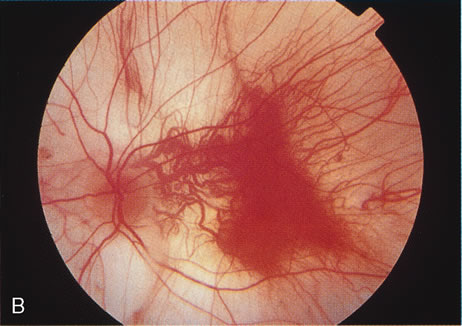
ophthalmic disease. Trans Am Ophthalmol Soc 157:451, 1999.

218. Deutman AF: Sex-linked juvenile retinoschisis. In The Hereditary Dystrophies of the Posterior Pole of the Eye, pp 48–99. The Netherlands, Van Gottum, 1971.

219. Primo SA, Tomasino SF: Progressive retinal changes observed in juvenile X-linked retinoschisis. J Am Optom Assoc 61:548, 1990.

220. Dalmer B, Brummer S, Foerster MH et al: X-linked congenital retinoschisis. Arch Clin Exp Ophthalmol 228:432, 1990.

221. Hardy RA, Crawford JB: Retina. In Vaughan D, Asbury T, Riordan-Eva P (eds): General Ophthalmology, pp 178–199 Stamford, CT, Appleton & Lange, 1999.

222. Ewing CC, Cullen AP: Fluorescein angiography in X-chromosomal maculopathy with retinoschisis (juvenile hereditary retinoschisis). Can J Ophthalmol 7:19, 1972.

223. Sauer CG, Oehrig A, Warneke-Wittstock K et al: Positional cloning of the gene associated with X-linked juvenile retinoschisis. Nat Genet 17:164, 1997.

224. Retinoschisis Consorium: Functional implications of the spectrum of mutations found in 234 cases with X-linked juvenile retinoschisis (XRLS). Hum Mol Genet 7:1185, 1998.

225. Molday LL, Hicks D, Sauer CG et al: Expression of X-linked reinoschisis protein, RS1 in photoreceptors and bipolar cells. Invest Ophthalmol Vis Sci 42:816, 2001.

226. Manchot WA: Pathology of hereditary juvenile retinoschisis. Arch Ophthalmol 88:131, 1972.

227. Peachey NS, Fishman GA, Derlaci DJ et al: Psychophysical and electroretinographic findings in X-linked juvenile retinoschisis. Arch Ophthalmol 105:513, 1987.

228. Condon G, Arnwostern S, Wang NS et al: Congenital hereditary (juvenile X-linked) retinoschisis: Histopathological and ultrastructural findings in three cases. Arch Ophthalmol 104:576, 1986.

229. Gass JD: Müller cell cone, an overlooked part of the anatomy of the fovea centralis: Hypothesis concerning its role in the pathogenesis of macular hole and foveomacular schisis. Arch Ophthalmol 117:821, 1999.

230. Mooy CM, van den Born LI, Baarsma S et al: Hereditary X-linked juvenile retinoshcisis: A review of the role of Müller cells. Arch Ophthalmol 120:979, 2002.

231. Grayson G, Rend SN, Ellis JA et al: Retinoschisin, the X-linked retinoschisis protein, is a secreted photoreceptor protein and is expressed and released by Wert-Rb 1 cells. Hum Mol Genet 9:1873, 2000.

232. Reid SN, Farber DB: Transport of a photoreceptor-secreted protein, retinoschisin, by Müller cells. Invest Ophthalmol Vis Sci 42:S647, 2001.

233. Weber BHF, Schrewe H, Molday L et al: Inactivation of the murine X-linked juvenile retinoschisis gene, Rs1h, suggests a role of retinoschisin in retinal cell layer organization and synaptic structure. Proc Natl Acad Sci 99:6222, 2002.

234. Eksandh LC, Ponjavie V, Ayyagari R et al: Phenotypic expression of juvenile X-linked retinoschisis in Swedish families with different mutations in the XLRS1 gene. Arch Ophthalmol 119:1098, 2000.

Back to Top