Chapter 54B
Neurometabolic Disease Affecting the Eye
LOIS J. MARTYN
Main Menu   Table Of Contents

Search

THE MUCOPOLYSACCHARIDOSES
THE GANGLIOSIDOSES
NIEMANN-PICK DISEASE
GAUCHER DISEASE (GLUCOSYLCERAMIDE LIPIDOSIS)
METACHROMATIC LEUKODYSTROPHY (SULFATIDE LIPIDOSIS)
KRABBE DISEASE (GALACTOSYLCERAMIDE LIPIDOSIS, GLOBOID CELL LEUKODYSTROPHY)
FABRY DISEASE (α-GALACTOSIDASE A DEFICIENCY)
FARBER LIPOGRANULOMATOSIS (CERAMIDE DEFICIENCY)
MUCOLIPIDOSES
BATTEN DISEASE (NEURONAL CEROID-LIPOFUSCINOSIS)
DISORDERS OF COPPER METABOLISM
REFERENCES

Ophthalmologists traditionally have played a significant role in the delineation and diagnosis of a broad spectrum of metabolic diseases, particularly those affecting the nervous system. Because the body of knowledge concerning these diseases is massive and rapidly evolving, this chapter is designed to summarize the salient features of the major neurometabolic disorders that have significant effects on the eye and visual system, emphasizing an understanding of the disease processes and their clinical signs.
Back to Top
THE MUCOPOLYSACCHARIDOSES
The group of conditions generically referred to as the mucopolysaccharidoses results from deficiency of specific lysosomal enzymes involved in the metabolic degradation of dermatan sulfate, heparan sulfate, or keratan sulfate, either singly or in combination. The incompletely degraded mucopolysaccharides (glycosaminoglycans) accumulate in various tissues and organs throughout the body and are excreted in the urine.

As a group, the mucopolysaccharidoses are characterized by a rather distinctive spectrum of clinical manifestations. Skeletal deformity, resulting from changes in both the bones and the joints, is a prominent feature. There is also a characteristic facies with coarse, often somewhat grotesque features. Visceromegaly, cardiac disease, respiratory problems, deafness, and mental deficiency occur in certain of the syndromes. The principal ocular manifestations of the various mucopolysaccharidoses are progressive corneal clouding, pigmentary retinal degeneration, optic atrophy, and in some cases glaucoma (Table 1).


TABLE ONE. The Systemic Mucopolysaccharidoses


DesignationMetabolic FeaturesGeneral Clinical ManifestationsOphthalmologic Manifestations
MPS IH: Hurler SyndromeProfound deficiency of α-L-iduronidaseMPS prototype. Coarse facial features. Severe dysostosis multiplex. Moderate dwarfism, contractures; semi-crouched habitus, kyphoscoliosis, gibbus, claw-hand deformity. Visceromegaly, protruberant abdomen, hernias. Pulmonary and cardiovascular disease. Mental deficiency. Hearing impairment. Early death, usually before age 10 years.Prominent wide-set eye (shallow orbits, hypertelorism), prominent supraorbital ridges, heavy brows, puffy lids. Prominent corneal clouding, of early onset, progressive and severe. Retinal degeneration; arteriolar attenuation, pigmentary changes, abnormal ERG. Nerve head swelling. Optic atrophy. Progressive vision impairment. In some cases, megalocornea and/or glaucoma.
 MPS accumulation in virtually every system of the body, producing marked somatic and visceral abnormalities  
 Urinary excretion of dermatan sulfate and heparan sulfate  
 Autosomal recessive  
MPS IS: Scheie Syndrome (formerly MPS V) α-L-iduronidase deficiencyMinimal to moderate somatic and visceral signs of MPS, less severe than in Hurler prototype. Somewhat coarse facial features. Stature, habitus relatively normal. Prominent joint stiffness, claw-hand deformity, carpal tunnel syndrome, and aortic valve disease. Intellect normal or nearly normal. Hearing impairment common. Life span relatively normal.Corneal clouding, early and progressive, often more dense peripherally. “Retinitis pigmentosa-like” retinal degeneration, with progressive vision impairment and reduced ERG. In some cases glaucoma.
 Urinary excretion of dermatan sulfate and heparan sulfate  
 Autosomal recessive  
MPS IH/S: Hurler-Scheie Compoundα-L-iduronidase deficiencyPhenotype intermediate between that of Hurler and Scheie syndrome. Dwarfing, progressive joint stiffness, claw-hand deformity, hypertelorism, progressive coarsening of facial features, micrognathia. Hepatosplenomegaly. Cardiovascular disease. Intellectual impairment. Hearing impairment. Survival into teens or twenties.Progressive corneal haze and vision impairment. In some cases retinal degeneration. Nerve head swelling. Optic atrophy. Possibly glaucoma.
 Urinary excretion of dermatan sulfate and heparan sulfate  
 Autosomal recessive  
MPS II: Hunter SyndromeIduronate sulfatase deficiencyCoarse facial features. Dwarfing and skeletal deformities, similar to but less severe than in Hurler prototype. Hepatosplenomegaly, cardiac and respiratory disease. Hydrocephalus. Motor paralysis in some. Hearing impairment. Nodular or pebbly skin lesions. Rapid psychomotor and physical deterioration and early death (often by age 15 years) in severe form. Slower deterioration and longer survival (even into fifth or sixth decade) in milder form.Progressive retinal degeneration, usually severe, with pigmentary changes, arteriolar attentuation, disc pallor, vision impairment, and abnormal ERG. Often nerve head swelling. Possibly glaucoma. Clinically clear cornea, but microscopic changes reported.
 Urinary excretion of dermatan sulfate and heparan sulfate.  
 X-linked recessive  
MPS III: Sanfilippo SyndromeFour biochemically different forms:Clinical manifestations similar in all four forms. Early and severe progressive mental deterioration. Less severe somatic changes. Coarse facial features, megalocephaly, hypertelorism. Moderate skeletal changes. Hepatosplenomegaly.Corneas clinically clear; some microscopic changes reported. Attentuation of retinal arterioles, some pigmentary changes, vision impairment, and ERG changes documented. Possibly optic atrophy. Rarely nerve head swelling.
 Type A: Heparan N-sulfatase deficiencySurvival into third decade. 
 Type B: α-N-Acetylglucosaminidase deficiency  
 Type C: Acetyl-Co: α-Glucosaminide-N-acetyl transferase deficiency  
 Type D: N-Acetyl- glucosamine 6-sulfatase deficiency  
 Urinary excretion of heparan sulfate in all four forms  
 Autosomal recessive  
MPS IV: Morquio SyndromeTwo biochemically different formsIn classic form (IV A), severe dwarfing, skeletal dysplasia, with kyphosis, sternal bulging. Joint laxity rather than stiffness. Odontoid hypoplasia, atlantoaxial instability; spinal cord and medullary compression, long tract signs and respiratory paralysis may occur. Prominent joints, knock-knees. Semicrouching stance, waddling gait. Somewhat coarse facial features. Hypoplastic dental enamel. Occasionally hepatomegaly. Protruberant abdomen. Cardiopulmonary complications. Hearing impairment. Intellect normal or mildly impaired. In milder variant (IV B) findings similar to those of classic form, but often less severe dwarfism, less tendency to atlantoaxial instability, and usually normal dental enamel.Corneal clouding, usually mild or fine haze, in A and B. Subcortical lens opacities in A. Fundi usually normal; in some cases optic atrophy, disc blurring, arteriolar narrowing, reduced scotopic ERG.
 N-acetylgalactosamine 6-sulfatase deficiency in classic form (MPS IV A)  
 β-galactosidase deficiency in later onset variant (MPS IV B)  
 Urinary excretion of keratan sulfate in both forms  
 Autosomal recessive  
MPS V: (Vacant, Formerly Scheie)   
MPS VI: Maroteaux-Lamy SyndromeN-acetylgalactosamine-4-sulfatase (arylsulfatase B) deficiencyStriking dwarfism and skeletal deformities.Progressive corneal clouding, often dense, usually evident within first few years of life. Graft may accumulate MPS. Papilledema, abducent palsy secondary to hydrocephalus. Optic atrophy. Retinal vascular tortuosity. Typically no signs of retinal degeneration, but pigmentary and ERG-VEP changes noted in milder variant.
 Urinary excretion of dermatan sulfate Coarse facial features. Visceromegaly and cardiac disease. Atlantoaxial subluxation, spinal cord compression, hydrocephalus in some cases. Normal intellect. Granular inclusions in circulating leukocytes. Milder variants occur. 
 Autosomal recessive  
MPS VII: Sly Syndromeβ-glucuronidase deficiencyVariable, often moderate, clinical manifestations. Coarse facial features. Short stature and skeletal deformity. Hepatosplenomegaly, hernias, cardiovascular and respiratory problems. Intellectual impairment. Inclusions in circulating lymphocytes.Corneal clouding in some cases. Nerve head swelling also documented.
 Urinary excretion of dermatan sulfate and heparan sulfate  
 Autosomal recessive  


To date, deficiency of ten specific lysosomal enzymes has been demonstrated in the various mucopolysaccharidoses. All are recessively inherited; one mucopolysaccharidosis, MPS type II (Hunter syndrome), is X-linked; the others are autosomal.

The diagnosis of the various mucopolysaccharidoses is made on the basis of the distinguishing clinical features, the presence of excessive mucopolysaccharide substances in tissue and urine, and demonstration of the enzyme deficiency using fibroblasts, leukocytes, or serum. Prenatal diagnosis is also possible by analysis of cultured amniotic fluid cells or chorionic villi. Identification of heterozygotes is becoming more available. There has been some success in altering the course of certain of the mucopolysaccharidoses by bone marrow transplantation. Some improvement in the ocular findings after marrow transplantation has been documented.1

In reviewing the mucopolysaccharidoses, reference should be made to comprehensive discussions of the clinical, pathologic, biochemical, and genetic features of these disorders.2–5

MPS TYPE IH: HURLER SYNDROME

Hurler syndrome (MPS IH) is the prototype of the mucopolysaccharidoses. The disorder is severe and progressive. There is accumulation of acid mucopolysaccharide in virtually every system of the body, producing both somatic and visceral abnormalities and leading to early death, usually by age 10 years.

In MPS IH there is profound deficiency of α-L-iduronidase, with excessive urinary excretion of both dermatan sulfate and heparan sulfate in a ratio of approximately 70:30. The disorder is autosomal recessive. It occurs in many races and is probably the most frequent of the mucopolysaccharidoses. The gene encoding α-L-iduronidase, previously assigned to chromosome 22, has been localized to chromosome 4 p 16.3.

Manifestations develop in infancy and early childhood and become more apparent with increasing age. The head tends to be large and misshapen. Scaphocephaly due to premature closure of the sagittal suture is common; there is often a prominent longitudinal ridge along the sagittal suture. The facial features characteristically are coarse and the expression dull (Fig. 1). Hypertelorism is usual and the orbits are shallow; the eyes appear wide-set and prominent. The lids tend to be puffy, the brows prominent. The nose is broad, with wide nostrils and a flat bridge. The ears may be large and low-set. The lips usually are patulous; the tongue is large and protuberant. The teeth generally are small, stubby, and widely spaced; the gums are hyperplastic.

Fig. 1. A. Coarse facial features and distorted body habitus in a patient with Hurler syndrome. B. Face profile of a patient with Hurler syndrome. C. Full facial view.

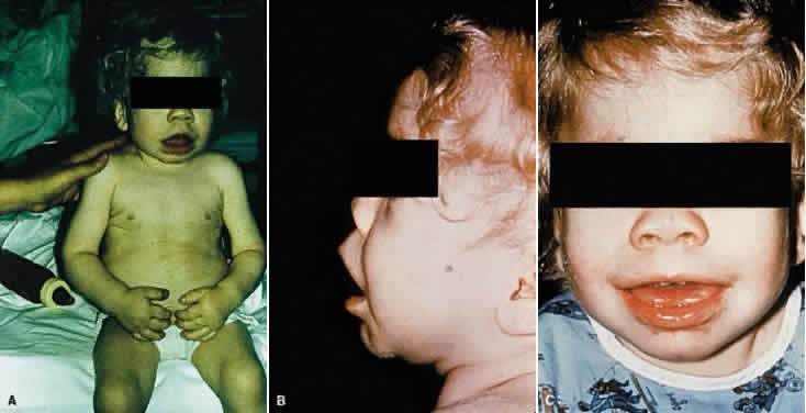
Characteristically there are marked skeletal changes. Moderate dwarfism, short neck, kyphoscoliosis, and gibbus are typical. On plain film examination, the vertebral bodies (particularly those of the lower dorsal and upper lumbar region) are wedge-shaped with an anterior hooklike projection referred to as beaking. The extremities are short, the hands and feet are broad, and the phalanges are short and stubby. Radiologically, the tubular bones show expansion of the medullary cavity and thinning of the cortex. The terminal phalangeal bones commonly appear hypoplastic. The joints are stiff and flexion contractures develop; clawlike deformity of the hands is especially characteristic. The posture is semicrouching and the gait is awkward.

Thoracic deformity is another regular feature of the syndrome; the chest appears large and wide with flaring of the lower ribs over the abdomen. On plain film examination, the ribs appear spatulate or saber-shaped. Typically, the medial end of the clavicle is widened. The many radiologic findings in this condition are commonly described by the term dysostosis multiplex.

The abdomen is protuberant owing to abnormalities in supporting tissues and to visceromegaly. As a rule there is enlargement of both the liver and spleen. Diastasis recti, umbilical hernia, and inguinal hernias are common.

The skin tends to be thick. There is usually generalized hypertrichosis.

Manifestations of cardiac involvement, including murmur, angina, myocardial infarction, and congestive heart failure, are common. Pathologic changes in the heart due to mucopolysaccharide deposition can be extensive. The great vessels and peripheral vessels also are affected.

Respiratory problems develop in virtually every patient. Recurrent upper respiratory tract infection, bronchitis, and chronic nasal congestion are common, and the patients almost always are noisy mouth breathers. A number of factors, including deformity of the facial and nasal bones, narrowing of the passages, abnormalities of the tracheobronchial cartilage, and deposition of mucopolysaccharide in the lungs, contribute. In addition to the abnormalities of the respiratory passages and lungs, cardiac disease and thoracic deformity may contribute to respiratory difficulties.

The principal neurologic manifestation is mental deficiency. There may also be motor signs. Pathologic changes can be found throughout the nervous system. Hydrocephalus may develop. A special feature in some cases is the presence of leptomeningeal cysts. Shoe-shaped deformity of the sella is common. The optic foramina also may be enlarged.

Deafness is frequent; it may be of mixed or sensorineural type. Middle ear infections are common.

The course is one of progressive mental and physical deterioration. Death most frequently results from cardiac or respiratory disease.

The principal ophthalmologic manifestations of MPS IH are progressive corneal clouding, retinal degeneration, optic atrophy, and vision loss. It appears that the corneal and retinal changes relate somewhat to the pattern of mucopolysacchariduria—that is, corneal changes are greater in those conditions characterized by higher levels of dermatan sulfate in the urine, as in Hurler syndrome. The retinal degeneration appears to correlate with the degree of heparan sulfaturia; the retinal changes are more severe in Hunter and Sanfilippo syndromes, less in Hurler syndrome.6,7

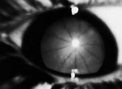
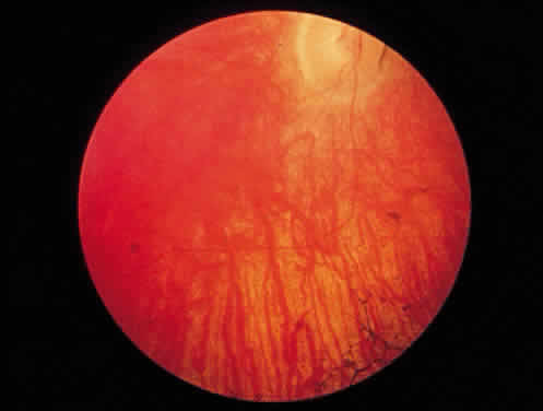
Corneal clouding was recognized early to be an important feature of this disorder, and classic clinical descriptions of the corneal changes are to be found in the older literature.8–12 Clouding of the cornea is usually evident by age 2 to 3 years, often by age 1 year; in some cases it is seen at birth.12,13 Photophobia is a common early symptom. With time there is progression from a generalized haziness or steamy appearance to a dense, milky ground-glass opacification (Fig. 2). On slit-lamp examination, one sees fine granular opacities in the corneal stroma, often increasing in density from the anterior stroma and subepithelial region to the posterior stromal layers.10–12

Fig. 2. Ground-glass corneal haze of Hurler syndrome.

In 1939, Berliner provided what is probably the first significant histopathologic study of the eye in Hurler syndrome, describing the corneal changes in detail. He found large vacuolated cells under Bowman's layer, fragmentation of Bowman's layer, separation of the corneal lamellae, and deposits of granular material in the stromal spaces.10 Subsequent reports confirmed these findings, and in 1944 Hogan and Cordes noted in addition fine granules in the cytoplasm of the corneal corpuscles.14–16 Later studies documented these histopathologic changes and provided further evidence for mucopolysaccharide accumulation in the cornea.13,17–19 The epithelium may be intact or may show edema and cytoplasmic vacuolization, with accumulation of metachromatic material in and around cells. Bowman's layer usually shows thinning and lamellar splitting or fragmentation, with infiltration of vacuolated cells containing metachromatic material. In the stroma there is swelling and vacuolization of keratocytes, intracellular and extracellular deposition of metachromatic material, and lamellar separation. Descemet's membrane and endothelium usually are described as normal, although cytoplasmic vacuolization and metachromatic staining of the endothelium have been noted. Histochemical techniques provide evidence for mucopolysaccharide accumulation in the vacuolated cells. In their ultrastructural study of the eyes in two cases of MPS IH, Chan and coworkers documented the presence of numerous fibrillogranular inclusions in corneal epithelium, keratocytes, and endothelium, the presence of multimembranous inclusions in keratocytes, and the presence of extracellular fibrillogranular material in corneal stroma.20

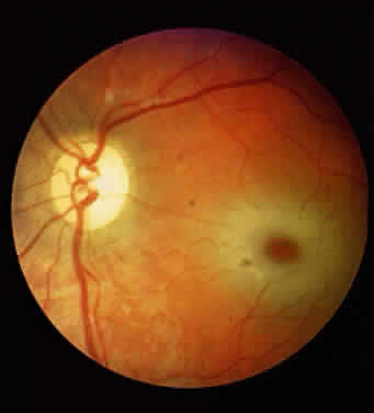
Progressive corneal clouding may prevent visualization of the fundus, but signs of retinal involvement and optic atrophy have been documented in Hurler syndrome. Gills and coworkers reported the absence of the foveal reflex and optic atrophy in several patients.21 In addition, the fundus appeared “albinoid” in one, and the retinal arterioles were narrowed in another. The electroretinogram (ERG) is abnormal, usually markedly reduced, in Hurler syndrome.21–23

Mailer also emphasized the association of optic atrophy in Hurler syndrome and reviewed the possible causes.19 It would seem that the optic atrophy can be secondary to any of the following, singly or in combination: mucopolysaccharide infiltration, hydrocephalus, retinal degeneration, and even glaucoma. Papilledema also has been observed, in some cases in association with hydrocephalus.16,24

With regard to retinal degeneration and optic atrophy, related histopathologic findings include enlargement and vacuolization of cells of the nuclear layer of the retina, vacuolization of the ganglion cells, atrophy of the optic nerve, and thickening and infiltration of the arachnoid with foam cells.13,17,19 By electron microscopy, Chan and coworkers documented the presence of fibrillogranular inclusions in retinal pigment epithelium and ganglion cells and multimembranous inclusions in retinal ganglion cells and optic nerve astrocytes.20

In addition to corneal, retinal, and optic nerve changes, there may be histopathologic evidence of mucopolysaccharide accumulation in the epithelium of the ciliary body, in the walls of the iris capillaries, in the sclera, and in the conjunctiva.13,17,19 Ultrastructural changes have been found in uveal melanocytes and fibrocytes, ciliary epithelium, smooth muscle cells of ciliary body, pericytes, trabecular endothelium, lens epithelium, and sclerocytes.20 Conjunctival biopsy had been used as a diagnostic procedure. The characteristic ultrastructural changes and histochemical reactions of mucopolysaccharidosis have been well documented in conjunctival specimens.20,25 The findings include the presence of single membrane-limited cytoplasmic vacuoles containing fibrogranular material in epithelium, vacuoles containing predominantly fibrogranular material and occasionally membranous lamellar inclusions in fibroblasts and histiocytes in subepithelial connective tissue, vacuolization of the lymphatic endothelium and perithelial cells, and membranous lamellar vacuolization of the Schwann cells of the conjunctival nerves. There is evidence that the fibrogranular vacuoles and the membranous lamellar vacuoles are lysosomes that contain, respectively, accumulated acid mucopolysaccharide and glycolipid.

Megalocornea has been described in many cases; in most cases intraocular pressure has been normal, but glaucoma has been documented in some patients with MPS IH.9–13,26,27 Electron microscopy of the trabeculectomy specimen from a child with Hurler syndrome and open-angle glaucoma showed membrane-bound cytoplasmic inclusions consistent with mucopolysaccharide accumulation in cells of the corneoscleral junction and iris stroma.28

Progressive impairment of vision is usual, secondary to corneal clouding, retinal degeneration, and optic atrophy, singly or in combination; glaucoma, the effects of cerebral mucopolysaccharide accumulation, and the development of hydrocephalus may also contribute. In view of the extensive systemic and neurologic abnormalities in MPS IH, the poor prognosis for life, and the probability of concurrent retinal degeneration and optic atrophy, corneal transplant in an effort to improve vision had not often been recommended in the past. However, the possibility of altering the course of Hurler syndrome with treatment may alter the prospects for successful keratoplasty. Corneal transplantation several years after successful bone marrow transplantation in a child with MPS IH has been reported; Huang and colleagues in 1996 documented the ultrastructural changes in the corneal specimen.29

Whereas Rosen and coworkers described keratoplasty and ultrastructural changes of the cornea in a patient with “Hurler's disease,” it is doubtful that the patient had MPS type IH.30 In the case of a successful keratoplasty in a patient with “atypical mucopolysaccharidosis” reported by Gollance and D'Amico, it is difficult to determine the type of mucopolysaccharidosis involved.31

As mentioned in the description of the facies, the eyes tend to be wide-set and prominent owing to hypertelorism and shallow orbits, the lids tend to be puffy, and the brows are heavy and the lashes coarse. These features are seen to some degree in other mucopolysaccharidoses also.

MPS TYPE IS: SCHEIE SYNDROME

MPS type IS was first described by Scheie, Hambrick, and Barness in their classic study of ten patients reported in 1962.18

In the Scheie syndrome, as in the Hurler syndrome, there is deficiency of the lysosomal enzyme α-L-iduronidase and urinary excretion of both dermatan sulfate and heparan sulfate. The condition is autosomal recessive. The predominant manifestations are corneal clouding, joint stiffness, claw-hand deformity, carpal tunnel syndrome, and aortic valve disease, principally aortic stenosis and regurgitation. The facial features are coarse; the mouth is broad. Other somatic and visceral changes characteristic of mucopolysaccharidosis tend to be minimal. Stature is normal, and the patients do not develop the distorted habitus characteristically seen in Hurler syndrome. Intellect is normal or nearly normal, although psychiatric disturbances have been reported. There may be hearing impairment. The life span is relatively normal.

Histopathologic changes are similar to those of the prototype MPS IH, but in MPS IS the cortical neurons appear normal.

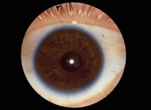
Corneal clouding is a prominent manifestation of the Scheie syndrome.18 Developing early in life, sometimes present at birth, the corneal clouding tends to worsen with age and may ultimately interfere with vision. The corneal involvement is diffuse but tends to be most dense peripherally. Clinically the hazy cornea may appear enlarged, edematous, and thickened, initially raising suspicion of glaucoma, particularly when telltale somatic signs of mucopolysaccharidosis are minimal.

Scheie and coworkers showed that the pathologic corneal and conjunctival changes were similar to those in Hurler syndrome and presented evidence for the presence of acid mucopolysaccharide in the abnormal vacuolated cells of these tissues.18 Subsequently, Quigley and Goldberg and others described the ultrastructural changes of the conjunctiva in Scheie syndrome, documenting the presence of single membrane-limited vacuoles containing granulofibrillar material and occasional membranous inclusions in the conjunctival fibroblasts and similar vacuolization of epithelial cells.32 Kenyon and associates showed similar changes and demonstrated histochemical reactions for acid mucopolysaccharide in several of the mucopolysaccharidoses, including Scheie syndrome.25

A study by Quantock and colleagues suggests that variation in collagen fibril diameter in corneal stroma, in addition to light scattering from glycosaminoglycan deposits, may contribute to corneal clouding in Scheie syndrome.33

Corneal transplants have been tried with little success.18

In most reported cases, the ocular pressure has been normal or in the upper range of normal, but in some cases glaucoma has been documented.34,35

Although retinal changes have not been documented in all reported cases,18,36 retinal degeneration is a recognized feature of the Scheie syndrome.2,21–23,32,35 Manifestations include vision impairment, particularly progressive night blindness, field changes such as ring scotoma, retinal pigmentary changes (“RP-like”), and subnormal or extinguished ERG responses. Reduced corneal sensitivity also has been noted.34

MPS TYPE I H/S: HURLER-SCHEIE SYNDROME

A number of patients having features intermediate between those of the Hurler and the Scheie syndromes have been reported.2,37–39 As in MPS IH and MPS IS, in MPS I H/S there is deficiency of α-L-iduronidase and urinary excretion of both dermatan sulfate and heparan sulfate. The histopathologic changes are those of mucopolysaccharide accumulation in connective tissue throughout the body, as well as in parenchymal cells of the liver and brain.

The prominent clinical manifestations are skeletal changes (dysostosis multiplex) with dwarfing and progressive joint stiffness, scaphocephaly, hypertelorism, and progressive coarsening of facial features. In addition, a receding chin (micrognathia) appears to be a distinctive feature. Other manifestations include hepatosplenomegaly, pulmonary and cardiovascular involvement, mental retardation, and hearing impairment. Significant manifestations (destruction of the sella, cerebrospinal fluid rhinorrhea, and loss of vision) related to the presence of arachnoid cysts have also been reported in MPS I H/S. Patients with the Hurler-Scheie syndrome may survive into the teens or twenties.

As in both MPS IH and MPS IS, corneal clouding occurs in MPS I H/S.2,37–39 The corneal haze is diffuse (sometimes more dense peripherally) and progressive; it may be evident in childhood and ultimately may interfere with vision. Keratoplasty has been tried; a lamellar graft in one patient with MPS I H/S had remained clear for 4 years at the time of the report.38

Retinal degeneration also occurs in MPS I H/S, although the true incidence is unknown because corneal clouding may obscure the fundus findings. Chijiiwa and associates reported two patients with night blindness, decreased visual acuity, constricted visual fields, and reduced ERG; both had retinal pigmentary changes with scattered spicules and arteriolar attenuation.40 Jensen and associates also had documented fundus and ERG abnormalities.41 In their ERG study, Caruso and colleagues found variable abnormalities in MPS I H/S.23

Blurring of the disc margins has been noted, and in one case this finding was associated with increased intracranial pressure, with dilatation of the ventricles, enlargement of the sella, and pathologic documentation of mucopolysaccharide accumulation in the central nervous system.42

Mullaney and coworkers reported chronic angle-closure glaucoma in an 11-year-old boy with MPS I H/S.43 Electron microscopy of tissues obtained at trabeculectomy showed the presence of vacuoles containing fibrillogranular material consistent with mucopolysaccharide deposition; there was marked intracellular and extracellular vacuolar formation in the iris, scattered vacuolar formation in sclera and Tenon's capsule, and little extracellular or intracellular vacuolar formation in trabecular meshwork.

MPS TYPE II: HUNTER SYNDROME

In Hunter syndrome (MPS II), the metabolic defect is deficiency of the lysosomal acid hydrolase iduronate sulfatase. There is urinary excretion of both dermatan sulfate and heparan sulfate in a ratio of approximately 1:1. In contrast to the other mucopolysaccharidoses, MPS II is X-linked recessive. Its locus has been mapped to Xq27-28.

Phenotypically Hunter syndrome closely resembles the Hurler prototype. The manifestations of MPS II, however, are generally less severe than those of MPS IH, and Hunter syndrome is distinguished clinically by longer survival and the absence of gross corneal clouding.

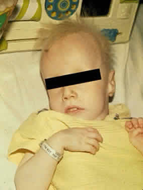
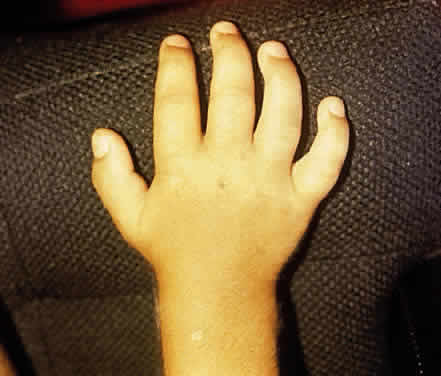
In Hunter syndrome, the facial features are coarse, the supraorbital ridges tend to be prominent, the tongue is large, and the teeth are widely spaced. Dwarfing and stiffness of the joints are prominent features. Claw-hand deformity is common (Fig. 3). Lumbar gibbus may develop but is usually not severe. As a rule there is hepatosplenomegaly. The abdomen is protuberant. Hernias are common. Cardiac involvement is a regular feature of the syndrome; congestive heart failure and coronary artery disease are major causes of death. Respiratory disability also is evident in most patients. Neurologic manifestations vary. Spastic quadriplegia may develop from impingement on the cervical spinal cord. Hydrocephalus may develop. Mental deterioration occurs, but the severity and the rate of regression vary. Progressive deafness occurs in most patients.

Fig. 3. Claw-hand deformity of Hunter syndrome.

A distinctive feature of Hunter syndrome is the occurrence of nodular or pebbly ivory-colored skin lesions, most frequently on the back extending from the inferior angle of the scapula toward the axillary line, less often in the pectoral area, nape of the neck, and lateral aspect of the upper arms and thighs. Adults with Hunter syndrome also tend to have a rosy or ruddy complexion.

Within Hunter syndrome there is a broad spectrum of severity.44,45 At least two major clinical forms, differentiated primarily on the basis of central nervous system involvement, are recognized. Patients with the more severe form, type A, show more rapid neurologic deterioration and usually die before age 15 years. The milder form, type B, is characterized by slower mental deterioration and is compatible with survival into the fifth or sixth decade of life.

In contrast to MPS IH, obvious corneal clouding is not a regular feature of MPS II.46,47 However, slight corneal changes may be detected by slit-lamp examination in older patients with Hunter syndrome, and histologic evidence of corneal mucopolysaccharide accumulation has been reported.48,49 Spranger and colleagues documented clinically visible corneal opacities in a child with severe MPS II, in addition to fine corneal opacities in a young adult with mild MPS II.50

The principal ophthalmologic manifestation of MPS II is progressive retinal degeneration with attendant impairment of vision.21,51 Night vision problems and visual field defects are common. The disorder may lead to blindness. The fundus signs include retinal pigmentary changes, sometimes spicule formation, retinal arteriolar attenuation, and optic disc pallor. The ERG is usually reduced or extinguished21,22,52; in some cases it is normal.22 In addition, bilateral epiretinal membranes with tortuosity of the retinal vessels has been reported as an unusual finding in two brothers with Hunter syndrome type B.53

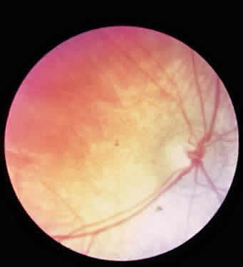
Swelling of the nerve head is a frequent finding in Hunter syndrome; it may be due to increased intracranial pressure or mucopolysaccharide deposition in and around the optic nerve.44,54–56

In their light and electron microscopic study of the eye in type A Hunter syndrome, McDonnell and coworkers found single membrane-bound structures containing fibrillogranular and multimembranous material in conjunctival epithelium, pericytes, and fibrocytes; corneal epithelium, keratocytes, and endothelium; trabecular endothelium; iris pigment epithelium, smooth muscle, and fibrocytes; ciliary pigmented and nonpigmented epithelium and fibrocytes; retinal pigment epithelium and ganglion cells; optic nerve astrocytes and pericytes; and sclerocytes.57

In their histopathologic study of the eye by light microscopy in type B Hunter syndrome, Goldberg and Duke found few corneal abnormalities.48 The corneal epithelium and Bowman's layer were intact, except peripherally where Bowman's layer was split and where eosinophilic material was present beneath the epithelium. Descemet's membrane and endothelium were intact, although eosinophilic granules were present in the endothelial cytoplasm. Fine granular deposits were present in the corneal stroma, chiefly in interlamellar spaces. The nonpigmented epithelium of the ciliary processes appeared foamy. There were significant retinal changes, including pigment migration, paucity of pigment epithelial cells, diminution of rods and cones, reduction in number of ganglion cells, and gliosis of the nerve fiber layer. The sclera was thickened.

In their electron microscopy study of the same patient, Topping and coworkers found fibrillogranular vacuoles and membranous lamellar vacuoles in various tissue of the eye, although the ultrastructure of the cornea was relatively unaltered.49 The nonpigmented epithelium of the ciliary processes was engorged with membranous fibrillogranular vacuoles, and similar vacuoles were present in choroid and scleral fibroblasts. Similar but fewer inclusions of this type were found within the corneal keratocytes and the pigmented epithelium of the ciliary processes. There were membranous lamellar vacuoles in the retinal ganglion cells and in migrated pigment epithelial cells. Some membranous lamellar vacuoles were also present in keratocytes, choroidal and scleral fibroblasts, and nonpigmented epithelium of the ciliary processes. The content of the fibrillogranular vacuoles is probably mucopolysaccharide; that of the membranous vacuoles is probably glycolipid.

Kenyon and coworkers found similar ultrastructural changes in their study of conjunctiva and by histochemical techniques confirmed acid mucopolysaccharide in several of the mucopolysaccharidoses, including Hunter syndrome.25 Kaiden and associates reported chronic angle-closure glaucoma in an adult patient with Hunter syndrome.58 Spranger and colleagues also documented glaucoma in MPS II type B.50

MPS TYPE III: SANFILIPPO SYNDROME

The Sanfilippo syndrome (MPS III), sometimes referred to as polydystrophic oligophrenia, is a mucopolysaccharidosis in which there is severe mental retardation and relatively less severe somatic abnormalities. Four biochemically different but clinically indistinguishable forms of the syndrome occur: in type A there is deficiency of heparan N-sulfatase; in type B there is deficiency of α-N-acetylglucosaminidase; in type C there is deficiency of acetyl-CoA: α-glucosaminide-N-acetyltransferase; and in type D there is deficiency of N-acetyl glucosamine 6-sulfatase, the gene for which has been localized to chromosome 12q14. In all forms there is urinary excretion of heparan sulfate. All forms are autosomal recessive. Mental retardation, the predominant clinical manifestation of MPS III, usually becomes evident in the first few years of life. With increasing age there is progressive deterioration of intellect and behavior. Because the patients usually are strong, management often becomes a problem as they regress; many require institutionalization.

Somatic abnormalities typical of mucopolysaccharidosis tend to be mild or inconspicuous. There is some coarseness of facial features. Synophrys is usual. Generalized hirsutism may be marked. Dwarfing, joint stiffness, and claw-hand deformity are usually evident but are not as severe as in the Hurler prototype. Radiologically, the skeletal changes of dysostosis multiplex are relatively mild. Slight to moderate hepatosplenomegaly develops, and the abdomen tends to be protuberant. Respiratory difficulties are common. Heart involvement may occur but tends to be less severe than in other mucopolysaccharidoses. Hearing loss is common in moderate to severely affected patients. Hydrocephalus also may develop.59

Corneal clouding does not occur in MPS III, although microscopic changes were noted in one of Sanfilippo's patients, and histologic corneal and scleral changes were reported in another patient subsequently.60,61 Jensen found vacuoles and accumulation of granular material in sclera and, to a lesser degree, in cornea and ciliary body; the histochemical findings were believed to be consistent with accumulation of acid mucopolysaccharide.61 In their histopathologic study of the conjunctiva in the various mucopolysaccharidoses, Kenyon and coworkers also found the characteristic light and electron microscopic changes in Sanfilippo syndrome and by histochemical techniques demonstrated accumulation of acid mucopolysaccharide.25

Retinal involvement and progressive vision loss may occur in MPS III. Narrowing of the retinal vessels and pigmentary changes have been noted.21,22,25 Subnormal ERG responses have been recorded in both types A and B.21–23 Optic atrophy may develop.22,62 Significant histopathologic changes of the retina and perineural connective tissue, in addition to signs of mucopolysaccharide accumulation in many other parts of the eye, in Sanfilippo syndrome type A were first documented by Del Monte and colleagues.63 Phase contrast and electron microscopy showed intracellular accumulation of fibrillogranular and membranous lamellar vacuoles in cornea, trabecular meshwork, iris, lens, ciliary body, sclera, retinal ganglion cells, retinal pigment epithelium, and optic nerve glia. There was retinal pigment epithelial hyperplasia and hypopigmentation, vascular attenuation, and marked photoreceptor loss, closely resembling that found in inherited retinitis pigmentosa. Clinically the patient had signs of pigmentary retinal degeneration and optic atrophy, with vision loss and no recordable ERG; the corneas were clear.

Ceuterick and associates also found membrane-bound electron-lucent inclusions in the retinal ganglion cells and photoreceptors of a 22-week-old fetus with type A Sanfilippo disease; there were also vacuoles containing granular material in other cells and tissues of the eye.64

In their study of the histopathologic and ultrastructural changes in the eye in Sanfilippo type B, Lavery and coworkers found cytoplasmic single membrane-bound vacuoles containing the major storage product, acid mucopolysaccharide, in virtually every ocular tissue.65 There were also lamellar cytoplasmic membranous bodies of complex lipid, the minor storage product, mainly in retinal ganglion cells and lens epithelium. In addition, many tissues contained inclusions of an intermediate type, composed of combined fibrillogranular and lamellar membranous material. There was photoreceptor cell degeneration similar to that seen in some forms of retinitis pigmentosa.

MPS TYPE IV: MORQUIO SYNDROME

The syndrome that bears Morquio's name is characterized by severe dwarfism and skeletal deformity, often with neurologic complications, and a number of extraskeletal abnormalities such as corneal clouding and aortic valve disease. In this mucopolysaccharidosis (MPS IV), there is defective degradation of keratan sulfate. There is excessive urinary excretion of keratan sulfate, although the amount of keratan sulfate in the urine tends to diminish as affected patients grow older.

As with Sanfilippo syndrome, clinically similar but enzymatically different forms of Morquio syndrome occur. The designation MPS IV-A is used to denote classic Morquio syndrome, in which the enzyme defect is a deficiency of N-acetylgalactosamine 6-sulfatase; the gene encoding this enzyme has been localized to chromosome 16q24. The designation MPS IV-B is used to denote a later-onset variant of Morquio syndrome in which the enzyme defect is a deficiency of β-galactosidase. Both mild and severe forms of MPS IV-A and MPS IV-B occur. All forms are autosomal recessive.

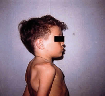
Patients with Morquio syndrome appear normal in the first months of life, although radiographic signs may be present early. With growth during the first years of life, abnormalities such as retarded growth, knock knees, flat feet, prominent joints, dorsal kyphosis, sternal bulging, flaring of the rib cage, and awkward gait become evident. The deformities worsen with age. Affected persons characteristically are markedly dwarfed and develop a semicrouching stance. Joint stiffness is not a feature, however; rather, joints may be excessively loose, leading to instability. Barrel chest and pigeon breast deformity are common (Fig. 4). The neck typically is short. The face is abnormal, with somewhat coarse features, a broad-mouthed appearance, prominent jaw, and widely spaced teeth. The dental enamel is often thin, giving the teeth a grayish appearance and leading to flaking and fracturing of the enamel and multiple cavities. In some cases aortic regurgitation develops. Progressive hearing loss occurs in almost all patients. Invariably there is absence or severe hypoplasia of the odontoid process, and there is usually ligamentous laxity of the spinal column. Atlantoaxial subluxation and spinal cord and medullary compression are frequent complications; manifestations may be acute, subacute, or chronic, and subtle or severe, ranging from minimal long tract signs to spastic paraplegia, respiratory paralysis, and death. In general the course is one of progressive incapacitation. Intelligence usually is normal or mildly impaired.

Fig. 4. Skeletal and thoracic deformities of Morquio syndromes.

Corneal clouding is a feature of Morquio syndrome,66 although some of the earlier literature would suggest otherwise.26 The corneal clouding in Morquio syndrome is relatively mild, having the appearance of a fine haze rather than the dense ground-glass opacification common to Hurler syndrome. The changes may not become clinically evident to the unaided eye for several years, often not before age 10 years. In the early stages the corneal involvement may be overlooked unless careful slit-lamp examination is performed. The biomicroscopic appearance is that of diffuse involvement of the stroma with punctate or granular opacities but usually sparing of the epithelium, Bowman's layer, and endothelium. Depending on the density of the corneal haze, there may be some impairment of vision but usually not of a severe degree. Corneal clouding is seen in MPS IV-B as well as in MPS IV-A.67,68

In their histologic examination of the cornea in Morquio syndrome, Ghosh and McCulloch found intracytoplasmic vacuolization of keratocytes and the presence of fibrillar, granular, and lamellar substance having histochemical properties consistent with those of acid mucopolysaccharide.69 Subsequently, Iwamoto and associates documented the presence of fibrillogranular and multimembranous membrane-bound inclusions distributed primarily in the cornea and trabecular meshwork, to a milder degree in the conjunctiva and sclera, and sparsely in the retinal pigment epithelium.70 Intracytoplasmic vacuoles limited by single-unit membranes containing fine fibrillogranular material characteristic of the mucopolysaccharidoses have also been documented in conjunctival biopsy.68

Olsen and coworkers found cataracts in addition to corneal stromal clouding in three siblings with Morquio syndrome (MPS IV-A).71 On slit-lamp examination there were innumerable small spherical grayish opacities of about identical size subcortically, in a zonular arrangement; the nuclei and lens capsules were clear. The lenses and corneas of the parents and healthy siblings were clear.

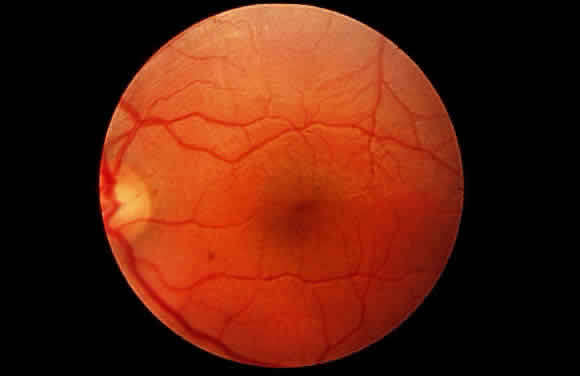
Fundus abnormalities have been reported infrequently in Morquio syndrome. Optic atrophy has been noted.72 In a review of reported fundus changes in Morquio syndrome, Dangel and Tsou made reference to blurring of the discs.73 They described narrowing of the retinal arterioles in an adult with Morquio syndrome and documented electrophysiologic changes: the light-adapted ERG was normal, but the dark-adapted ERG was reduced and the electro-oculogram was slightly abnormal in one eye.73 Abraham and coworkers also found slight scotopic abnormalities.74 In most cases the fundi and light-adapted ERG are normal in patients with MPS IV.21,22 Von Noorden and coworkers documented mesodermal anomalies in one patient with MPS IV.66 Mydriasis attributed to sympathetic involvement in Morquio syndrome has also been mentioned.73

MPS TYPE VI: MAROTEAUX-LAMY SYNDROME

The Maroteaux-Lamy syndrome (MPS VI) is characterized by severe dwarfism, visceromegaly, cardiac lesions, and progressive corneal clouding. In some cases hydrocephalus and spinal cord compression develop. Resembling the prototype mucopolysaccharidosis in many ways, the Maroteaux-Lamy syndrome is distinguished by retention of normal intellect, the pattern of mucopolysacchariduria, and the enzyme defect. In MPS VI there is deficiency of N-acetylgalactosamine-4-sulfatase (arylsulfatase B), with urinary excretion of predominantly dermatan sulfate. The gene encoding N-acetylgalactosamine 4-sulfatase has been localized to chromosome 5q 13-q14. Metachromatic granulation of circulating leukocytes is a characteristic finding in MPS VI. The disorder is autosomal recessive. In addition to the classic form of Maroteaux-Lamy, milder variants associated with the same enzyme deficiency are described.

In the severe or classic form of MPS VI, growth retardation affecting both the trunk and limbs is usually evident by age 2 or 3 years. Genu valgum, lumbar kyphosis, and anterior sternal protrusion develop. The lower ribs are flared. Joint movement is restricted. Claw-hand deformity develops; carpal tunnel syndrome is common. The head appears relatively large. The facial features tend to be coarse. There is often mild hypertrichosis. As a rule, hepatomegaly develops in patients older than 6 years of age, splenomegaly develops in about half the patients, and the abdomen usually is protuberant. Cardiac involvement, particularly valve lesions, similar to that of Hurler syndrome may develop. Deafness occurs in some patients.

The principal neurologic complications are hydrocephalus and spinal cord compression secondary to atlantoaxial subluxation consequent to hypoplasia of the odontoid process. Survival is variable; most patients with the severe form of MPS VI die by the second or third decade.

The principal ophthalmologic manifestation of MPS VI is progressive corneal clouding, usually evident within the first few years of life. The appearance is that of ground-glass haze distributed diffusely throughout the stroma, sometimes denser peripherally, and usually of sufficient degree to be seen grossly.75,76 In addition to the stromal opacities, some epithelial and endothelial changes may be seen on slit-lamp examination.76 Corneal opacities have been documented in the mild variant of MPS VI as well in the severe form.77 A decrease in corneal clouding after bone marrow transplantation has been noted.78 In what appears to be the first reported histopathologic study of the eye in Maroteaux-Lamy syndrome, Kenyon and coworkers in 1972 described changes typical of mucopolysaccharidosis.76 On light microscopy they found cytoplasmic vacuolization of corneal epithelium, interruption of Bowman's layer with accumulation of foamy histiocytes, swelling of keratocytes with foamy cytoplasm and separation of stromal lamellae, some cytoplasmic vacuolization of corneal endothelium, but essentially no alteration of Descemet's membrane. Other findings included thickening of sclera with vacuolated cells between the fibers, vacuolated cells in the trabecular meshwork, ballooned histiocytes, vacuolated fibrocytes in connective tissue stroma of the ciliary body, involvement of the basal portion of nonpigmented ciliary epithelium, and some changes in the choroid. By histochemical techniques, they documented accumulation of acid mucopolysaccharide in the affected cells and tissues. The retina appeared normal except for the macular area, where reduction of the ganglion cell population and thinning of the nerve fiber layer were noted. The optic nerve showed atrophy and secondary gliosis. Electron microscopy confirmed the presence of single membrane-limited vacuoles containing predominantly fibrillogranular material, some containing polymorphous material and membranous lamellae, in the cornea, sclera, trabecular meshwork, and uveal tract, but not in the retina.

Similar corneal changes have been found in the mild phenotype of MPS VI.79 Schwartz and associates documented the recurrence of mucopolysaccharide accumulation in the repeat corneal graft specimens from two patients with the mild form of MPS VI.80

Goldberg and coworkers reported the occurrence of papilledema and abducent palsy secondary to the increased intracranial pressure of hydrocephalus in a child with Maroteaux-Lamy syndrome.75 In addition, they documented tortuosity of the retinal vessels not only in the child with papilledema but also in her siblings, whose optic discs were normal. Sheridan and Johnston also documented the occurrence of papilledema secondary to hydrocephalus in Maroteaux-Lamy.56

Except for the above-mentioned disc changes and retinal vascular tortuosity, the fundi in MPS VI generally are normal. As a rule, patients with Maroteaux-Lamy syndrome do not develop ophthalmoscopic signs of pigmentary retinal degeneration, and the ERG usually is normal.76,79 However, in an adult patient with a mild variant of Maroteaux-Lamy with typical diffuse corneal clouding, DiFerrante and colleagues documented alternating areas of hypopigmentation and hyperpigmentation in the parapapillary region, with reduced A-wave response on ERG and increased latency on visual-evoked responses.81

MPS TYPE VII: SLY SYNDROME

In the Sly syndrome (MPS VII), there is deficiency of β-glucuronidase, leading to a block in the degradation of dermatan sulfate and heparan sulfate, with urinary excretion of both dermatan and heparan sulfate. The disorder is autosomal recessive. The human gene encoding β-glucuronidase is localized on chromosome 7q 21.1-q22.

Clinical manifestations within the syndrome vary. The spectrum includes many of the characteristic features of mucopolysaccharidosis, including short stature, progressive skeletal deformity and radiologic signs of dysostosis multiplex, coarse facial features, hypertelorism, hepatosplenomegaly, diastasis recti, protuberant abdomen, hernias, intellectual impairment, cardiovascular involvement, and respiratory problems. Reported patients have shown coarse inclusions in circulating leukocytes. A severe neonatal form associated with fetal hydrops has been reported.

In some patients with Sly syndrome the corneas are clear.82–86 Within the phenotypic variation of this disorder, however, corneal clouding may occur; this may be evident grossly or only on slit-lamp examination.87 The patient initially reported by Sly as having clear corneas subsequently developed progressive corneal clouding.82,88 In their postmortem study of this patient, Vogler and associates found vacuolated cytoplasm in nonpigmented ciliary epithelium, in corneal fibrocytes, and in lens epithelium; the retinal pigment epithelium was not vacuolated.88

Back to Top
THE GANGLIOSIDOSES
The gangliosidoses are metabolic neurodegenerative diseases involving defects in ganglioside degradation. Gangliosides are glycosphingolipids that contain sialic acid in their oligosaccharide chain. These lipids are present in most cell types of the body. They are found in greatest concentration in neurons and are of highest content in the brain. Disorders of ganglioside degradation resulting in abnormal accumulation and storage of these glycolipids are of two major types: GM1 and GM2 (Table 2).


TABLE TWO. The Major Gangliosidoses


DesignationMetabolic and Genetic FeaturesGeneral ManifestationsOphthalmologic Manifestations
GM1 type 1: infantile GM1 gangliosidosis: generalized gangliosidosisProfound deficiency of β-galactosidase.Signs evident at birth or soon thereafter.Macular cherry-red spot in approximately 50% of cases. Retinal vascular tortuosity, retinal hemorrhages also reported. Optic atrophy. Early vision loss. Strabismus, nystagmus. Mild corneal clouding in some cases. Also vascular abnormalities of conjunctiva.
  Marked accumulation of GM1 ganglioside in nervous system. Accumulation of galactosyloligosaccharides and keratan sulfate degradation products in somatic cells. Dysmorphism, prominent skeletal abnormalities resembling those of Hurler syndrome. 
 Autosomal recessive Visceromegaly. Severe psychomotor retardation, rapid neurologic deterioration leading to decerebrate rigidity, spastic quadriplegia, blindness, and deafness. Death usually by age 2 years. 
GM1 type 2: late infantile/juvenile GM1 gangliosidosisDeficiency of β-galactosidase. Moderate accumulation of GM1 ganglioside in nervous system. Accumulation of galactosyloligosaccharides and keratan sulfate degradation products in somatic cells less prominent than in type 1.Onset later, somatic abnormalities milder, and course slower than in type 1. Progressive mental and motor deterioration in time. Average life span 3 to 10 years.Absence of cherry-red spot, but histologic evidence of retinal lipidosis and optic atrophy reported. Corneas clear. Strabismus, nystagmus. Loss of vision in later stages.
 Autosomal recessive  
GM1 type 3: adult/chronicDeficiency of β-galactosidase. Neuronal accumulation of GM1 ganglioside predominately in basal ganglia, less in cortex and white matter. Accumulation of galactosyl oligosaccharides and keratan sulfate degradation products in somatic cells less prominent than in type 1.Later onset and slower course than type 1.Absence of cherry-red spot, corneas usually clear, vision usually not impaired.
GM1 gangliosidosisAutosomal recessive Progressive spasticity and ataxia. Mild intellectual impairment. Absence of dysmorphism and organomegaly; mild if any bony abnormalities. 
   Survival to adulthood. 
GM2 gangliosidosis: acute infantile type: Tay-Sachs diseaseProfound deficiency of hexosaminidase A activity; HEXA defect. Abnormal accumulation of GM2 throughout central, peripheral, and autonomic nervous system.Onset in infancy with listlessness, weakness, or irritability. Exaggerated startle response.Classic macular cherry-red spot in virtually all cases. Optic atrophy. Progressive loss of vision. Nystagmus. Deterioration of eye movements.
  Autosomal recessive. Predominantly in infants of Ashkenazic Jewish descent. Psychomotor retardation. Seizures. Megancephaly. 
   Progressive mental and motor deterioration in second year, leading to decerebrate rigidity, blindness, deafness. Death usually by age 2 to 4 years. 
GM2 gangliosidosis: infantile type: Sandhoff variantDeficiency of hexosaminidase A and hexosaminidase B activity; HEXB defect. Neuronal accumulation of ganglioside and visceral accumulation of globoside.Neurologic manifestations similar to those of Tay-Sachs prototype; psychomotor retardation, progressive deterioration. Also liver, spleen, renal, and cardiac involvement. Death usually by age 2 to 4 years.Macular cherry-red spot in some cases.
 Autosomal recessive  Optic atrophy. Progressive loss of vision.
    Corneas slightly opalescent in one case.
GM2 gangliosidosis: subacute forms: late infantile and juvenile onset formsHEXA or HEXB defect. Neuronal GM2accumulation without visceral histiocytosis.Onset between 2 and 10 years of age. Progressive spasticity. Seizures. Marked developmental regression and dementia. Deterioration to vegetative state by age 10 to 15 years, followed by death within a few yearsVision loss in later stages. Macular cherryred spot in some cases. Sometimes pigmentary retinal changes. Optic atrophy. Strabismus.
 Autosomal recessive  
GM2 gangliosidosis: chronic formsHEXA or HEXB defect. Hexosaminidase A deficiency more common than combined A and B deficiency. Neuronal lipidosis.Spectrum of clinical pictures: progressive dystonia; cerebellar dysfunction; motor neuron disease, weakness; psychiatric disorders. Variable age of onset from juvenile to adult. Usually slowly. progressive with long-term survivalVision rarely affected. Fundi generally normal. Ocular motor abnormalities, including defects in convergence, horizontal and vertical gaze.
  Autosomal recessive  


The GM1 gangliosidoses are due to deficiency of acid β-galactosidase activity. A number of subtypes occur, with wide phenotypic variability. For clinical purposes, the GM1 gangliosidoses generally are classified into infantile, late infantile/juvenile, and adult/chronic types. The human β-galactosidase gene has been mapped to chromosome 3.

The GM2 gangliosidoses are due to a deficiency of the isoenzymes hexosaminidase A, B, or both, or to a deficiency of an activator factor that stimulates hexosaminidase A to cleave the ganglioside GM2. The underlying cause is a defect in one of three genes: HEXA, which encodes the α subunit of hexosaminidase A; HEXB, which encodes the β subunit of hexosaminidases A and B; or GM2A, which encodes the GM2 activator. HEXA has been mapped to chromosome 15, and HEXB and GM2A have been mapped to chromosome 5. The GM2 gangliosidoses also show considerable phenotypic variability, ranging from infantile-onset, rapidly progressive disease to later-onset, subacute or chronic forms. The most familiar of this group is Tay-Sachs disease.

The GM1 and GM2 gangliosidoses are autosomal recessive disorders. Diagnosis of these disorders can be confirmed by enzyme assay of tissue, body fluids, and cells. The heterozygote state also can usually be detected by these methods. Prenatal diagnosis is possible by assay of cultured amniotic fluid cells and chorionic villi. At this time no specific treatment for the gangliosidoses is known; enzyme replacement therapy is not yet available.

For a comprehensive description of these disorders, the reader is referred to the reviews of Suzuki and Gravel and their colleagues.89,90

GM1 TYPE 1: INFANTILE GM1 GANGLIOSIDOSIS: GENERALIZED GANGLIOSIDOSIS

In GM1 type 1, commonly referred to as generalized gangliosidosis, there is deficiency of acid β-galactosidase activity with both neuronal lipidosis and visceral histiocytosis. The disease is characterized by infantile onset, rapid neurologic degeneration, and prominent bony abnormalities, although some patients present with neurodegenerative signs without marked physical changes.

Signs may be evident at birth or soon thereafter. Morphologic abnormalities include frontal bossing, depressed nasal bridge, large low-set ears, coarse facial features, and hirsutism of the forehead and neck. There may be gingival hypertrophy and macroglossia. There is often edema of the extremities. Hepatosplenomegaly usually develops within the first months of life. Macrocephaly may develop.

Dorsolumbar kyphoscoliosis is common. The hands are broad, the fingers short and stubby. The joints are stiff, with generalized contractures. Hard, nontender enlargements of the epiphyseal joints due to cartilaginous hypertrophy may be prominent. Radiologic findings are those of dysostosis multiplex with deformities of the vertebral bodies and long bones; signs include rarefaction, anterior beaking of the vertebral bodies, periosteal cloaking of long bones, spatulate deformity of the ribs, shoe-shaped deformity of the sella, and modeling deformities of the bones of the pelvis, hands, and feet.

Affected infants initially tend to be hypotonic and hypoactive with a weak sucking reflex. Respirations tend to be irregular and labored. Clonic-tonic convulsions often develop. The course is one of progressive neurologic deterioration and increasing rigospasticity, often to a state of decerebrate rigidity with spastic quadriplegia, blindness, deafness, and unresponsiveness. Death, commonly due to bronchopneumonia, usually occurs by age 2 years.

The pathologic changes are those of neuronal lipidosis and visceral histiocytosis. Neurons are ballooned with storage material and contain numerous cytoplasmic membranous bodies. Foamy histiocytes are found in the bone marrow, liver, spleen, lymph nodes, and most visceral organs. In addition, there is cytoplasmic ballooning of renal glomerular epithelial cells. Suzuki demonstrated visceral accumulation of keratan sulfate and related compounds in addition to accumulation of ganglioside.91

Ophthalmologic findings in this condition are significant. Cherry-red spots of the maculae, clinically resembling those seen in Tay-Sachs disease, occur in approximately 50% of patients with infantile generalized gangliosidosis.92 Tortuosity of the retinal vessels, retinal hemorrhages, and optic atrophy have also been noted on ophthalmoscopic examination.93

The cornea usually is clear, but mild diffuse corneal clouding has been reported in some cases.93,94 Loss of vision occurs early in the course of the disease. Strabismus and nystagmus are common.92

Evidence for ganglioside accumulation in the retina and mucopolysaccharide accumulation in the cornea has been well documented.93 In a case study reported by Emery and associates, the retinal ganglion cells appeared distended and had foamy cytoplasm that stained intensely with periodic acid-Schiff (PAS); numerous intracytoplasmic inclusion bodies similar to those found in cerebral ganglion cells in GM1 gangliosidosis were present.93 The cytoplasm of the corneal epithelial cells, the cytoplasm of histiocytes in the region of Bowman's layer, and the cytoplasm of keratocytes had a foamy appearance and stained positively for mucopolysaccharide. Intracytoplasmic vacuoles were also present within the pigmented and nonpigmented layers of the ciliary epithelium, ciliary fibroblasts, and sclerocytes. There also appeared to be extracellular mucopolysaccharide pooled between collagen lamellae of the cornea.

In their histopathologic study of an affected 22-week-old fetus aborted when the diagnosis of GM1 gangliosidosis was made by amniocentesis, Cogan and coworkers found early retinal changes; electron microscopy showed numerous lamellar (lipid) inclusions in the retinal ganglion cells.95

An additional manifestation of GM1 gangliosidosis in some cases is the presence of vascular abnormalities of the conjunctiva. Tortuosity of the conjunctival vessels and saccular microaneurysms may be noted clinically, and histopathologic study of the conjunctiva has shown narrowing of the lumen of the vessels due to ballooning of endothelial cells with PAS-positive foamy material. By electron microscopy, the endothelial cells are filled with membranous cytoplasmic vesicles containing fibrillogranular material.96

GM1 TYPE 2: LATE INFANTILE/JUVENILE GM1 GANGLIOSIDOSIS

In GM1 type 2, as in GM1 type 1, there is a deficiency of β-galactosidase with neuronal lipidosis and visceral histiocytosis. In type 2, however, the onset is later, the course is slower, and the bony abnormalities are milder than those of type 1.

In the first year, development may be normal. Coarsening of facial features is not evident. Hepatosplenomegaly is usually absent. Radiologic signs such as beaking of vertebral bodies, proximal pointing of metacarpal bones, and modeling deformities of pelvic bones may be present early but tend to be mild. Neurologic manifestations usually begin at about 1 year of age.

Locomotor ataxia, generalized weakness of the upper and lower extremities, strabismus, and loss of speech are common early signs. Progressive mental and motor deterioration, spasticity, and in time decerebrate rigidity follow. Seizures also develop. Recurrent infections, particularly bronchopneumonia, are a problem. The average life span is only 3 to 10 years.

Ophthalmologic manifestations are not prominent in patients with the juvenile form of GM1 gangliosidosis.97 Strabismus and nystagmus may be present. Blindness may occur late in the course of the disease.90 The corneas usually are clear. The fundi usually appear normal as well. Cherry-red spot of the macula is not a feature of this condition, though atypical cherry-red spots have been described in one case.90,98

Microscopic changes of the retina have been reported. Goebel and coworkers found membranous cytoplasmic storage lysosomes in the retinal ganglion cells of a 6-year-old child who died of GM1 gangliosidosis type 2.99 They also documented atrophy of the optic nerves histopathologically and clinically.

GM1 TYPE 3: ADULT/CHRONIC GANGLIOSIDOSIS

This rare form of GM1 gangliosidosis is characterized by slowly progressive neurologic disease, beginning in the juvenile to adult years. The course is protracted, with long survival. The major neurologic manifestation is dystonia. Gait or speech disturbance is usually the first sign. Dystonic posture develops gradually. Intellectual impairment is not prominent. Dysmorphism is not obvious or is absent. Slight vertebral dysplasia usually is evident. Ocular changes do not appear to be a feature of this gangliosidosis. Cherry-red spots are not observed, but corneal clouding has been recorded in some cases.90 Vision is usually not impaired.90

GM2 GANGLIOSIDOSIS ACUTE INFANTILE FORM: TAY-SACHS DISEASE

The British ophthalmologist Warren Tay first described this neurodegenerative condition in 1881.100 In Tay-Sachs disease there is a nearly total deficiency of hexosaminidase A activity, resulting in abnormal accumulation of ganglioside GM2 due to HEXA mutation. Pathologic changes of lipidosis can be seen throughout the nervous system but are most conspicuous in cortical, autonomic, and rectal mucosal neurons. The cytoplasm of neurons is distended and ballooned. There is marked accumulation of membranous cytoplasmic storage bodies. Axonal degeneration, demyelination, and gliosis occur. Pathologic changes of viscera are not a feature of this disorder, although an occasional lipid inclusion body may be found.

The clinical picture is that of progressive motor and mental deterioration beginning in infancy. The onset is often insidious, with listlessness, apathy, irritability, or feeding difficulties. Psychomotor development is retarded. The startle reaction, an extension response to sudden sharp sounds, is a characteristic early sign. By age 3 to 6 months, motor weakness becomes evident. In time paralysis develops. After age 18 months, spasticity appears, convulsions develop, and there is progression to a state of decerebrate rigidity, deafness, and blindness. Macrocephaly may develop in time as the result of cerebral gliosis. Death usually occurs by age 3 years, commonly as the result of bronchopneumonia. Many of these patients have a doll-like facial appearance with pale translucent skin, delicate pink coloring, fine hair, and long eyelashes.

Ophthalmologic manifestations are an important feature of Tay-Sachs disease. Macular cherry-red spots develop in virtually all cases (Fig. 5). As a rule, the macular sign is evident by the time other neurologic signs appear in infancy. Clinically, this appears as a bright to dull red spot surrounded by a well-defined hazy gray, creamy white, or yellowish halo. The halo is the result of loss of transparency of the multilayered ganglion cell ring of the macula. The red spot is the normal blush of the more transparent central region of the macula, its color accentuated by the creamy halo surrounding it. The so-called cherry-red spot is the focal ophthalmoscopically visible sign of generalized retinal involvement in this disease.

Fig. 5. Characteristic cherry-red spot in child with Tay-Sachs disease.

Pathologic changes occur throughout the retina similar to those in the brain. There is lipid loading and degeneration of ganglion cells.101 The lipids stored in the retina were early shown to be like those of the brain, and deficiency of hexosaminidase A in the retina and optic nerve has been documented.102 There is also demyelination and degeneration of the optic nerves, chiasm, and tracts.101 Clinical evidence of optic atrophy is common. In the first reported electron microscopic studies of the eye in Tay-Sachs disease, Cogan and Kuwabara in the United States and Harcourt and Dobbs in Great Britain documented the presence of membranous cytoplasmic bodies in the retinal ganglion cells, similar in all respects to those present in cerebral ganglion cells in Tay-Sachs disease.103,104 The presence of membranous cytoplasmic inclusion bodies has also been demonstrated in the retina of affected fetuses, and it appears that the retinal abnormalities may antedate those of the cerebral cortex.95,105

Progressive loss of vision is the rule; vision loss commences early and blindness is usually complete by age 2 years. There is evidence that much of the loss of visual function is of central rather than peripheral origin; the pupil reaction to light may be retained in blind patients, even in terminal stages of the disease, and the ERG may not become abnormal until late in the course of the disease. Ocular motor changes may also occur in Tay-Sachs disease. There may be regression of eye movements in reverse order of their normal ontogenetic development.106 In addition, the enzyme defect of Tay-Sachs disease can be detected in tears.107,108

GM2 GANGLIOSIDOSIS INFANTILE FORM: SANDHOFF VARIANT

The clinical manifestations and pathologic changes of Sandhoff disease are like those of Tay-Sachs disease. In this gangliosidosis, however, there is a severe deficiency of both hexosaminidase A and hexosaminidase B activity, with neuronal lipidosis of GM2, and some visceral accumulation of a globoside; this is due to HEXB mutation.

As in Tay-Sachs disease, the principal ophthalmic manifestations of Sandhoff disease are macular cherry-red spot, optic atrophy, and progressive vision loss leading to early blindness.98 Evidence of ganglioside storage in the retina and optic nerves has been well documented by histopathologic and ultrastructural examination.109–112 By light microscopy, Brownstein and associates found swelling, vacuolization, and dropout of retinal ganglion cells, a moderate decrease in the number of optic nerve axons, a marked decrease in myelin, and some thickening of pial septae.112 Electron microscopy showed large numbers of concentric membranous cytoplasmic bodies in the retinal ganglion cells and similar storage material in the inner nuclear layer, the inner segments of the photoreceptors, and the endothelium and pericytes of the retinal blood vessels. Astrocytes of the optic nerve were distended with numerous pleomorphic cytosomes. In addition, storage cytosomes have been found in corneal keratocytes.111,112 The corneas usually are clear clinically in Sandhoff disease, but in one case they appeared slightly opalescent.111 Changes have also been documented in the fetal retina.113

GM2 GANGLIOSIDOSIS SUBACUTE FORMS

This category includes late-infantile and juvenile-onset forms of GM2 gangliosidosis. Patients may have HEXA or HEXB mutations. The pathologic changes are those of neuronal lipidosis with abnormal storage of GM2. Neurons contain cytoplasmic inclusions of mixed type: cytoplasmic membranous bodies like those seen in Tay-Sachs and Sandhoff disease and other inclusions called pleomorphic lamellar bodies are present. Visceral histiocytosis is not a feature of this disorder.

Clinical manifestations appear between 2 and 10 years of age. The usual presenting signs are locomotor ataxia and incoordination. Progressive spasticity, athetoid posturing, seizures, and loss of speech follow. Developmental regression and dementia are prominent features. With time there is deterioration to a vegetative state with decerebrate rigidity usually by age 10 to 15 years, followed by death within a few years, usually due to intercurrent infection.

The ophthalmologic signs vary somewhat from those of the infantile-onset GM2 gangliosidoses. Vision loss occurs later in the course of subacute GM2 gangliosidosis, in contrast to the early blindness characteristic of Tay-Sachs and Sandhoff disease.90,114,115 Macular cherry-red spot, which is characteristic of Tay-Sachs and Sandhoff disease, may develop in subacute GM2 but is not a constant feature.115 The fundus may appear normal; in time there may be loss of the macular light reflex.114 Pigmentary retinal changes may occur.98,115 Optic atrophy develops in some cases.114,115 There may be strabismus.115

GM2 GANGLIOSIDOSIS CHRONIC FORMS

The spectrum of GM2 gangliosidosis includes a group of later-onset, more slowly progressive variants that are generally compatible with long-term survival. Several different phenotypes have been described, each having clinical manifestations reflecting predominant involvement of one or another part of the central nervous system, although there is evidence of widespread involvement of the central nervous system in most cases, with overlap between the different phenotypes. In some patients, slowly progressive dystonia dominates the clinical picture; psychomotor regression is less prominent or absent. The age of onset is variable, ranging from juvenile to adult onset. In other patients, cerebellar signs dominate the clinical course. Dysarthria, ataxia, incoordination, and abnormalities of posture develop in childhood. Mentation remains intact. In some cases the prominence of cerebellar signs combined with spasticity, muscle wasting, and pes cavus suggests an atypical form of spinocerebellar degeneration. In another variant, the clinical picture is that of motor neuron disease, with progressive muscle wasting and weakness. Neuroimaging of the brain generally shows cerebellar atrophy, often with some cerebral atrophy. Muscle biopsy shows denervation atrophy.

In many patients with late-onset GM2 gangliosidosis, psychiatric abnormalities are the predominant feature. Manifestations include acute hebephrenic schizophrenia associated with marked disorganization of thought, agitation, delusions and hallucinations, paranoia, and recurrent psychotic depression. Dementia usually is not a prominent feature of the late-onset variants of GM2 gangliosidosis presenting in adolescence or adulthood, although many patients show evidence of organic brain syndrome.

In the slowly progressive chronic forms of GM2 gangliosidosis, vision is rarely affected and the fundi generally are normal.116,117 Ocular motor abnormalities have been noted. Rapin and coworkers reported poor convergence and loss of optokinetic nystagmus and vestibular nystagmus.116 Subsequently, Musarella and associates documented defects of horizontal smooth pursuit movements, varying defects in vertical gaze (hypometric and hypermetric saccades), and inability to suppress the vestibulo-ocular reflex by fixation.117

The chronic variants of GM2 gangliosidosis are more commonly due to hexoaminidase A deficiency (HEXA mutations) than to combined hexosaminidase A and B deficiency (HEXB mutations); GM2 activator defects in this group are rare.

Back to Top
NIEMANN-PICK DISEASE
The eponym Niemann-Pick disease (NPD) has long been used to describe a group of lipid storage disorders characterized by abnormal accumulation of predominantly sphingomyelin and cholesterol, the presence of foamy hystiocytes in affected tissues and organs, and a broad spectrum of visceral and neurologic manifestations. The current classification of NPD encompasses two biochemically distinct entities: the primary sphingomyelin lipidoses, designated NPD types A and B, and a cholesterol lipidosis, designated NPD type C (Table 3).


TABLE THREE. Niemann-Pick Disease


DesignationMetabolic FeaturesGeneral ManifestationsOphthalmologic Manifestations
Type AProfound deficiency of acid sphingomyelinase activity (less than 5% of normal). Sphingomyelin lipidosis. Characteristic foam cells in affected tissues and organs. Autosomal recessive. More common in patients of Ashkenazic Jewish descent.Onset in infancy, with hypotonia, weakness, feeding difficulties, failure to thrive, hepatosplenomegaly, psychomotor retardation. Progressive neurodegeneration and debilitation. Sometimes respiratory involvement. Commonly osteoporosis. Death usually by age 2 to 3 years.Macular cherry-red spot in 30–50% of cases, often with extension of retinal opacification beyond the peripheral region, even mild generalized retinal haze. Optic atrophy. Diffuse stromal haze of cornea in some cases. Lens changes including brownish granular “deposits” on or in anterior capsule, and scattered white spots on or in posterior capsule.
    Vision loss late in course. ERG abnormal.
Type BLess severe deficiency of acid sphingomyelinase (5–10% of normal).Onset in childhood to adolescenceTrue macular cherry-red spot infrequent. Macular changes ranging from grayness of macular region to perifoveal halo of punctate or granular opacities— “macula halo syndrome”. Lid fullness.
  Sphingomyelin lipidosis. NP foam cells in affected tissues and organs. Autosomal recessive. Panethnic.Phenotypic variability 
  Hepatosplenomegaly. Little or no neurologic involvement in most patients. Progressive pulmonary involvement in more severely affected patients. Death in childhood or adulthood. 
Type CAbnormality of cholesterol transport.Presents in childhood. Slowly progressive variable neurodegenerative course with ataxia, dysarthria, dysphagia, drooling, dystonia, seizures. Hepatosplenomegaly, less than in types A and B NPD; may present with jaundice. Death in teens, commonly from inanition, aspiration. Possible survival into adulthood.Progressive supranuclear vertical gaze palsy affecting upward and downward gaze; sometimes horizontal gaze. Occasionally opalescence of perifoveal retina, disc pallor.
  Primary metabolic defect not yet identified.  
  Secondary reduction in acid sphingomyelinase activity. Lysosomal accumulation of cholesterol and sphingomyelin. Characteristic inclusions; NP foam cells. Autosomal recessive. Panethnic. (Includes those of Nova Scotia descent previously classified as type D.)  


TYPES A AND B NIEMANN-PICK DISEASE

These lipid storage diseases result from deficient activity of acid sphingomyelinase, a hydrolase important in the normal metabolic degradation of sphingomyelin. The phospholipid sphingomyelin is a common constituent of plasma membranes, subcellular organelles, endoplasmic reticulum, and mitochondria, and a major lipid of myelin sheaths and erythrocyte stroma. The enzyme defect leads to lysosomal accumulation of sphingomyelin, cholesterol, and other metabolically related lipids throughout the body. For a detailed description of these primary sphingomyelin lipidoses, the reader is referred to the review of Schuchman and Desnick.118

The pathologic hallmark of NPD types A and B is the presence of histochemically distinctive lipid-laden foamy cells referred to as Niemann-Pick cells, found particularly in tissues and organs of the monocyte-macrophage system. Infiltration of spleen and lymph nodes, marrow, liver, lungs, and kidneys occurs in both types A and B; the endocrine glands and heart also may be involved. In addition, neuronal lipidosis characteristically occurs in NPD type A, but not in type B. In NPD type A, foam cells and/or lipid-laden glial cells are found in the brain and connective tissues surrounding cerebral vessels. There is swelling of ganglion cells, often with cytoplasmic vacuolization, and there may be swelling of dendrites with loss of normal fibrillae and severe deficiency of myelin. The cerebellum generally is more severely affected than the cerebrum. Similar changes also may be seen in the spinal cord, autonomic nuclei, and sympathetic nerve cells of the adrenal medulla. Peripheral neuropathy also may occur in NPD type A.

The clinical manifestations of NPD type A differ significantly from those of type B. This may be explained by the fact that there is a profound deficiency of sphingomyelinase activity (usually less than 5% of normal) in type A, and somewhat more residual sphingomyelinase activity (5% to 10% of normal) in type B. In type A, there are severe neurologic and visceral manifestations. Hepatomegaly and splenomegaly develop in infancy. Early neurologic signs include hypotonia and muscle weakness. Feeding difficulties and recurrent vomiting are common. There is failure to thrive. Psychomotor retardation is evident by age 6 months. Progressive neurologic deterioration and debilitation follow. The infant loses contact with the environment. In time spasticity and rigidity develop. The child eventually becomes emaciated, with a protuberant abdomen and thin extremities. The skin may take on a brownish-yellow hue. There may be respiratory problems. Osteoporosis is common. The course is rapidly progressive, leading to death usually by age 2 to 3 years.

In contrast to type A, type B NPD is characterized by reticuloendothelial and visceral involvement without neurologic involvement. The presentation and course of type B are variable. Manifestations may develop in infancy or childhood, sometimes later. Enlargement of the liver and/or spleen usually is the first sign; there may be little if any clinical evidence of dysfunction. Severely affected patients may develop cirrhosis, portal hypertension and ascites, or pancytopenia due to hypersplenism. There is often pulmonary infiltration; dyspnea is common, and increased susceptibility to respiratory infection can be a problem. Patients may survive to adulthood in reasonably good health. Although neurologic involvement is not a feature of type B, some patients having mental retardation, and others having cerebellar ataxia, have been reported.

Significant ocular manifestations develop in many patients with type A NPD. The most frequent sign, noted in approximately 30% to 50% of reported cases, is a macular cherry-red spot.118–120 In most cases this is characterized by definite gray to white opacification of the macular ring, surrounding and accentuating the red blush of the central foveal region. A distinguishing feature noted in patients with NPD, however, is extension of the retinal opacification beyond the parafoveal region, in some cases involving the whole posterior pole and in others extending farther peripherally as a generalized mild retinal haze (Table 4).120


TABLE FOUR. Cherry-red Spot and Related Macular Changes in Metabolic Neurodegenerative Diseases


 DiseasesMacular Findings
GangliosidosesInfantile GM1 gangliosidosis: generalized gangliosidosisClassic cherry-red spot in approximately 50% of cases
 Late infantile/juvenile GM1 gangliosidosisAtypical cherry-red spot in one reported case
 Acute infantile GM2 gangliosidosis: Tay-Sachs diseasePrototype cherry-red spot in virtually all cases. White, yellow, or grayish opacification of macular ganglion cell ring surrounding and accentuating the red blush of the fovea.
  Appearance may change with progressive degeneration of ganglion cells.
 Infantile GM2 gangliosidosis: Sandhoff variantClassic cherry-red spot like that of Tay-Sachs
 Subacute GM2 gangliosidosisIn some cases, cherry-red spot or just loss of macular light reflex
Niemann-Pick DiseaseType A, acute neuronopathic sphingomyelin lipidosisCherry-red spot in 30% to 50% of patients.
  Distinguishing feature is extension of retinal opacification beyond parafoveal region; sometimes widespread generalized retinal haze.
 Type B, sphingomyelin lipidosis“Macula halo syndrome”: ring of crystalloid or granular opacities around fovea
 Type C, chronic neuronopathic NPDIn some cases, cherry-red—like spots
Gaucher Disease (Glycosylceramide Lipidosis)Type 3: Subacute neuronopathicMacular grayness in some patients but not true cherry-red spot. Discrete white retinal spots in some cases.
Metachromatic Leukodystrophy (Sulfatide Lipidosis)Infantile formMacular grayness or cherry-red—like spot
Krabbe Disease (Galactosyl-Ceramide Lipidosis) Subtle cherry-red spot reported in a child with unusual variant of Krabbe disease (infantile onset)
Farber Disease (Lipogranulomatosis) Macular grayness or cherry-red-like spot in some cases
SialidosisType I and type IICherry-red spot
GalactosialidosisEarly infantile, late infantile, and juvenile formsCherry-red spot
Neuronal Ceroid-LipofuscinosisJuvenile form: Spielmeyer-SjögrenBull's eye maculopathy
 Fucosidosis Bull's eye maculopathy


Corneal and lenticular changes may also be found clinically in some patients. Walton and coworkers noted a mild diffuse stromal haze of the cornea, evident on hand-light or on slit-lamp examination in several patients, without epithelial abnormalities or signs of glaucoma.120 They also described a definite brownish discoloration of the anterior lens surface in some cases; by slit-lamp examination, there were fine and granular brownish changes (“deposits”) on or in the anterior lens capsule, densest centrally. In addition, there were scattered white spots on or in the posterior capsule.

Vision loss occurs late in the course of type A NPD, in contrast to the early blindness that occurs in Tay-Sachs disease. Histopathologic changes of the eye in patients with type A NPD have been well documented. Evidence for lipid accumulation in retinal ganglion cells was provided in early pathologic reports.121,122 In a complete study of the eye of an infant with type A NPD published in 1973, Robb and Kuwabara described widespread ocular changes.123 Clinically, the infant had bilateral macular cherry-red spots. Electron microscopy confirmed the widespread distribution of storage material in the form of membranous cytoplasmic bodies, of predominantly lamellar architecture. There was marked involvement of retinal ganglion cells and retinal pigment epithelium; moderate involvement of corneal stromal cells, lens epithelium, corneal endothelium, vascular endothelium, and sphincter muscle of the iris; and some involvement of Muller cells, scattered glial cells, and inner segments of rods and cones. The morphology of the membranous cytoplasmic bodies found in the eye corresponded closely to the previously reported ultrastructure of lipid stored in the brain and viscera of patients with NPD.

Libert and coworkers subsequently confirmed the widespread distribution of lipid inclusions in the eye in type A NPD and distinguished two types of cytosomes: membranous cytoplasmic bodies resembling those of Tay-Sachs disease, found predominantly in ganglion cells and axons of the retina; and those with a less uniform lamellar architecture, of wider distribution, including conjunctival and corneal epithelial cells, keratocytes, nonpigmented epithelium of ciliary processes, choroidal and iris fibrocytes, endothelial cells, and pericytes of retinal and choroidal vessels.124 In addition, they documented optic atrophy, both clinically and pathologically; they found many inclusions in the demyelinated optic nerve and in Schwann cells of the ciliary nerves. Membranous cytoplasmic bodies characteristic of lipid storage disease have also been found already present in the eye of a 23-week-old affected fetus aborted after sphingomyelinase deficiency was documented by enzyme assay of cultured amniotic fluid cells. Many cells of the retina were involved; interestingly, there was no selectively greater accumulation of cytosomes in the ganglion cells, particularly in the primitive macular region. In addition, there was involvement of cornea, lens, choroid, sclera, and extraocular muscles.125

Ocular changes in type B NPD are infrequent. Cogan and coworkers, however, brought to attention the significance of what they termed “macula halo syndrome” in this disorder.126 They characterized the retinal abnormality as a ring of crystalloid or granular opacities around the fovea. Subsequently, a number of observers confirmed the occurrence of this distinctive retinal sign in patients with type B NPD.127 In most cases there was no attendant impairment of visual function. Whereas Cogan and colleagues initially thought the opacities probably were located in or beneath Henle's layer, by retinal angiography Matthews and associates showed masking of the perifoveal vessels by the halo, localizing the accumulated material to the ganglion cell layer.128 In addition to macular changes, periorbital fullness, often affecting both lids, has been noted in patients with type B NPD.129

The inheritance of types A and B NPD is autosomal recessive. Type A is more common in Ashkenazic Jewish individuals. The human acid sphingomyelinase gene has been mapped to the chromosomal region 11p 15.1-p15.4, and 12 mutations have been identified that cause types A and B NPD. The diagnosis is confirmed by assay of acid sphingomyelinase activity in cells or tissue extracts. Heterozygote detection requires molecular studies. Prenatal diagnosis by enzymatic molecular analysis of cultured amniotic cells or chorionic villi is possible. Currently there is no specific treatment for type A or B NPD, although the possibility of enzyme replacement and somatic gene therapy has been under investigation.

TYPE C NIEMANN-PICK DISEASE

This form of NPD is distinguished by a unique cellular disorder of cholesterol processing that is associated with accumulation of unesterified cholesterol and other lipids in lysosomes. The primary molecular defect, however, has not yet been identified. Although partial sphingomyelinase deficiency can be found in cultured cells as a variable secondary consequence of lysosomal cholesterol storage, tissue levels of sphingomyelinase activity usually are normal in type C. For a detailed review of this complex cholesterol lipidosis, see the chapter by Pentchev and colleagues.130

Cardinal pathologic features of type C are those of visceral and neuronal storage. There is usually splenomegaly with or without hepatomegaly. Inclusion-laden histiocytes referred to as foam cells and sea-blue histiocytes are found in many tissues and organs, particularly spleen, liver, marrow, lymph nodes, tonsils, and lung. On ultrastructural examination, histiocytes containing abnormal inclusions can be found also in skin, skeletal muscle, and ocular tissues. Cytoplasmic ballooning of neurons and a variety of inclusions are found throughout the nervous system. In the brain in particular there is involvement of cortical neurons, but ballooned neurons are found also in the basal ganglia, thalamus, substantia nigra, and locus ceruleus. There may be demyelination of white matter. The cerebellum is variably affected. Biochemical assays of lipid storage in liver and spleen show accumulation of unesterified cholesterol, sphingomyelin, phospholipids, and glycolipids. In the brain only glycolipid levels are elevated.

Clinical manifestations of type C are heterogenous. The “classic” phenotype is characterized by progressive dementia, ataxia, dystonia, supranuclear vertical gaze paresis, and variable hepatosplenomegaly. Manifestations appear in late childhood. Physical and intellectual disabilities gradually increase through late childhood and adolescence, eventually leading to incapacitation. Dysarthria and dysphasia contribute to communication, nutrition, and airway problems. Seizures may develop in childhood or later. Spasticity or rigidity can add to the burden of care. Death from inanition or aspiration occurs in the teen years.

In other cases, type C can present with fetal ascites, self-limited or rapidly fatal liver disease in the newborn, or organomegaly or signs of neurologic impairment in infancy. Later-onset variants with progressive neurologic deterioration and cognitive and psychiatric disturbances presenting in adolescents and adults also occur.

The ophthalmologic hallmark of type C is progressive supranuclear gaze vertical palsy.119,131–134 This begins with subtle slowing of vertical saccades. Early in the course, the first sign may be blinking or head thrusting on attempted vertical gaze (upward or downward). Older patients may first complain of difficulty in negotiating stairs, or their eyes becoming “stuck” in extremes of vertical gaze. Later in the course, voluntary vertical gaze can be completely paralyzed. In some cases there is impairment of vertical pursuit. Oculogyric reflexes are preserved. There is loss of vertical optokinetic nystagmus. Horizontal gaze movements also may be affected. There may be impairment of convergence.

Macular cherry-red-like spots occasionally occur in patients with type C NPD (Fig. 6).135,136 The eye may appear normal. In their pathologic study of the eye of a child with type C NPD and clinically normal fundi, Emery and coworkers found no convincing microscopic, histochemical, or ultrastructural evidence for lipid storage within the retina or other tissues of the eye.135 There were unusual triangular cornea-like patches of thin sclera adjacent to the limbus in the interpalpebral region, but any relationship of this abnormality to NPD is questionable. As the authors pointed out, however, Rabinowicz and associates had reported ballooning of the retinal ganglion cells in a patient who probably had type C NPD.137 Subsequently, Palmer and coworkers documented evidence of lipid storage throughout much of the eye in a patient with opaque grayness of the perifoveal region, disc pallor, and supranuclear vertical gaze paresis.138 Pleomorphic membranous inclusions were found in conjunctival fibrocytes, endothelial cells and pericytes, keratocytes, lens epithelium, retinal ganglion cells, retinal pigment epithelium, fibrocytes of the uveal tract, and optic nerve astrocytes. Ultrastructural changes of the conjunctiva had been documented previously in type C, as well as in type A and B.136,139 Conjunctival biopsy may be of value in the diagnosis of the disease. Higgins and associates mentioned VEP abnormalities in their study of patients with NPD type C.140

Fig. 6. Subtle macular changes in child with Niemann-Pick disease with vertical gaze paresis.

The heredity of type C NPD is autosomal recessive. The disorder is panethnic, but genetic isolates have been described in the French Acadians of Nova Scotia (formerly type D) and in Spanish Americans in Southern Colorado. Type C is as frequent as types A and B combined. Linkage of type C to an 18p genomic marker, D 185 40, has been found in some patients.

The diagnosis of type C requires demonstration of abnormal intracellular cholesterol esterification, and documentation of intralysosomal accumulation of unesterified cholesterol, as evidenced by intense perinuclear fluorescence in filipin-stained fibroblasts. Generalized screening for type C is not yet available. Antenatal diagnosis is currently restricted to families in which the index case has very low cholesterol esterification levels.

Currently there is no specific treatment for type C. Attempts at replacement therapy by liver transplantation have been disappointing. Treatment strategies to reduce intracellular cholesterol accumulation have been under investigation. Symptomatic treatment of seizures, dystonia, and cataplexy may be helpful.

Back to Top
GAUCHER DISEASE (GLUCOSYLCERAMIDE LIPIDOSIS)
Gaucher disease is an inherited metabolic disorder in which there is abnormal accumulation of the glycolipid glucosylceramide (glucocerebroside), a normal intermediate in the catabolism of globoside and gangliosides. It is due to subnormal activity of the lysosomal enzyme β-glucosidase (glucocerebrosidase). The gene encoding this enzyme has been localized to chromosome 1. A variety of mutations have been found to cause Gaucher disease. The lipid accumulates primarily in cells of the reticuloendothelial system. The presence of histologically distinctive lipid-laden storage cells, referred to as Gaucher cells, in affected organs and tissues is a hallmark of the disease.

This condition affects predominantly the spleen, liver, and bone marrow, and in certain forms of the disease the central nervous system. Many patients develop ocular manifestations. Based on variations in the clinical presentation of the disorder, three major forms of Gaucher disease are described: type 1 is the chronic nonneuronopathic form, type 2 is the acute neuronopathic form, and type 3 is the subacute neuronopathic form (Table 5). All forms of Gaucher disease are autosomal recessive. The disease is panethnic, but type 1 is more frequent in Ashkenazic Jews.


TABLE FIVE. Gaucher Disease (Glucosylceramide Lipidosis)


DesignationGeneral Clinical FeaturesOphthalmologic Manifestations
Type 1: Nonneuronopathic form (previously “adult” form)Onset in childhood or adult years. Broad spectrum of severity. Enlargement and dysfunction of liver and, spleen. Skeletal involvement leading to infarctions and fractures. Pulmonary infiltration, sometimes pulmonary failure. Occasionally yellow-brown pigmentation of skin. Variable course, rapidly progressive or protracted. Normal life span in most: sometimes death in first or second decade. Characteristic absence of primary neurologic manifestations.Brownish triangular or cuneiform areas of thickening of bulbar conjunctiva, nasal and temporal to limbus, resembling pingueculae. Occasionally retinal hemorrhage, edema. Possibly macular, perimacular changes.
Type 2: Acute neuronopathic form (“infantile” form)Onset in infancy. Severe neurologic involvement. Signs of cranial nerve nuclei and extrapyramidal tract involvement. Progressive spasticity, dysphasia. Classic triad of trismus, strabismus, retroflexion of head. Seizures. Sometimes choreoathetoid movements. Hepatosplenomegaly. Osseous lesions. Rapidly progressive course. Death usually by age 2 years, often from pneumonia or apnea.Paralytic strabismus; often a presenting sign. Progressive impairment of conjugate gaze movements in some cases.
Type 3: Subacute neuronopathic form (so-called “juvenile” form)Variable age of onset, usually later in first decade. Severity intermediate between that of types 1 and 2. Hepatosplenomegaly, usually preceding neurologic signs. Osseous lesions. Neurologic involvement usually beginning in childhood or adolescence. Ataxia, spastic paraparesis, seizures, slowly progressive dementia. More chronic course than type 2.Ocular motor abnormalities, frequently the presenting manifestation. Progressive supranuclear conjugate gaze palsies. Signs of ocular motor apraxia. Paralytic strabismus. Multiple discrete small white spots of retina posteriorly. Grayness of macular region. Possibly corneal opacities in variant of type 3. Myopia common.


The diagnosis can be made on the basis of the demonstration of Gaucher cells in bone marrow aspirates, the measurement of glucosylceramide in tissue samples, and the measurement of β-glucosidase activity in leukocytes or cultured skin fibroblasts of affected persons. Heterozygotes also can be identified by enzyme assay, and prenatal detection by amniocentesis is possible.

The authoritative review of Gaucher disease by Beutler and Grabowski is recommended.141

TYPE 1 GAUCHER DISEASE (CHRONIC NONNEURONOPATHIC GAUCHER DISEASE)

In type 1 Gaucher disease, previously designated the adult form, age of onset and severity vary widely. Symptoms may appear in childhood, as early as the first few months of life, or in the adult years, even as late as the seventh or eighth decade. The course may be rapid or slowly progressive and protracted. The initial sign is usually splenomegaly. There is frequently hypersplenism with thrombocytopenia, anemia, and leukopenia. There may be hepatomegaly. There may be evidence of moderate hepatic dysfunction. Skeletal involvement is frequent. Osteoporotic erosions of bone, aseptic necrosis of the femoral head, vertebral collapse, and pathologic fractures of long bones are common. Some patients suffer episodic bone pain, sometimes accompanied by fever. Pulmonary involvement may occur. Some patients develop pulmonary hypertension and cor pulmonale. The pulmonary involvement may predispose to pneumonia, a major cause of death in young patients with this condition. In older patients, yellow pallor and yellow-brown pigmentation of the skin of the face and lower extremities may be noted. Primary neurologic manifestations are not a feature, but occasionally there are secondary neurologic signs due to vertebral collapse, fat emboli, and coagulopathies. There is an increased incidence of neoplastic disease.

Ocular lesions resembling pingueculas have been noted frequently in patients with type 1 Gaucher disease.142–144 Clinically the appearance is that of a yellow or brownish triangular area of infiltration and thickening of the bulbar conjunctiva adjacent to the limbus nasally and temporally. These lesions have been reported to contain Gaucher-like foamy histiocytes.142,143 Chu and colleagues found no Gaucher cells in their microscopic study of the pingueculas in ten patients with Gaucher disease.145

Some reference has been made to macular or perimacular abnormalities in the chronic form of Gaucher disease, but these are difficult to substantiate.144,146 According to Petroehelos and colleagues, histologic examination of the choroid has disclosed Gaucher cells.144 An adolescent with chronic nonneuronopathic Gaucher disease reported by Carbone and Petrozzi had retinal hemorrhages and retinal pallor.143 With the anemia and thrombocytopenia of Gaucher disease, retinal hemorrhages and edema may occur.

Progressive retinal degeneration with optic atrophy and vision loss has been reported in one adult with type 1 Gaucher disease; the question is whether the ocular changes are causally related to the metabolic disease.147

Miller and coworkers reported impairment of eye movements in association with seizures and mental deterioration in two adult siblings with Gaucher disease, although neurologic involvement characteristically does not occur in type 1 Gaucher disease.148 One patient also had cystic changes of the macula.

TYPE 2 GAUCHER DISEASE (ACUTE NEURONOPATHIC GAUCHER DISEASE)

In type 2 Gaucher disease, also referred to as the classic, infantile, or cerebral form, the average age of onset of symptoms is 3 months, with a range from birth to 18 months. Hepatosplenomegaly, invariably a presenting sign, develops early. Within a few months, usually by age 6 months, neurologic manifestations appear. Most patients show signs of involvement of the cranial nerve nuclei and extrapyramidal tracts. The triad of trismus, strabismus, and retroflexion of the head is common. Feeding problems and difficulty handling secretions develop. Progressive spasticity, hyperreflexia, and pathologic reflexes develop. Seizures occur in some patients. Late in the course, the patients tend to become hypertonic and apathetic. The disease progresses rapidly. Death occurs early, between age 1 month and 2 years, usually as the result of pulmonary infection or anoxia.

The principal ophthalmologic manifestation of type 2 Gaucher disease is paralytic strabismus due to involvement of cranial nerves.141 A progressive impairment of conjugate gaze movements, like that described in the subacute neuronopathic form of Gaucher disease (type 3), also may develop.134,149

Corneal opacities were mentioned in one case, but no further description was given.150 Salgado-Borges and associates found cytoplasmic vacuolization of the keratocytes in a corneal button from a carrier of Gaucher disease who had keratoconus.151 They also documented reduced β-glucosidase activity in the corneal specimen. Two of the patient's siblings had died from Gaucher disease before age 2 years. The patient's mother and younger brother, both Gaucher disease carriers, also had signs of keratoconus.

TYPE 3 GAUCHER DISEASE (SUBACUATE NEURONOPATHIC GAUCHER DISEASE)

In type 3 Gaucher disease, sometimes referred to as the juvenile form, there is hepatosplenomegaly as in types 1 and 2. Signs may appear early in life, but the course is protracted. Major manifestations are spasticity, ataxia, retardation, and seizures. A progressive disorder of horizontal conjugate gaze movements has been described. The findings may simulate those of congenital ocular motor apraxia, including impairment of voluntary horizontal saccades with retention of slow pursuit movements, compensatory head thrusting, and contraversive deviation of the eyes during rotation of the body.134,152 In some patients with type 3 Gaucher disease, ocular motor abnormalities are the only or primary neurologic manifestation of the disorder.153

Retinal lesions also have been reported. Cogan and colleagues described multiple discrete small white spots situated superficially in the retina or on the surface of the retina in the posterior region of the fundus.154 Similar spots previously reported by Ueno and coworkers were found at autopsy to consist of “polymorphonuclear giant cells” within and on the surface of the retina. The retinal changes have also been well documented by Rodriguez et al.155

Cherry-red spots of the macula are not a feature of Gaucher disease, although grayness of the macular region may be noted.154 It is possible that patients reported to have had macular changes of Gaucher disease had another disorder, such as the cherry-red spot/myoclonus syndrome.156 Discrete stromal opacities of the cornea have been reported in a group of patients who may have a variant of type 3 Gaucher disease.157–159 Myopia has been noted as a finding in many patients with Gaucher disease.160

The quality of life for patients with Gaucher disease can be improved with a variety of treatment modalities, including splenectomy and joint replacement. Accumulation of glucosylceramide and the associated clinical manifestations can be reversed by repeated infusions of modified acid β-glucosidase (Aglucerase). Response to bone marrow transplantation has been encouraging in some cases.

Back to Top
METACHROMATIC LEUKODYSTROPHY (SULFATIDE LIPIDOSIS)
Sulfatide lipidosis is an inherited disorder of myelin metabolism characterized by lysosomal storage of glycolipids, predominantly sulfatides, in the white matter of the central and peripheral nervous system, with progressive degeneration of myelin and progressive deterioration of mental and motor function. In histologic preparations, the accumulated lipids exhibit metachromasia, giving rise to the descriptive term metachromatic leukodystrophy.

Pathologic studies of the brain show reduction of the central white matter with moderate to severe loss of myelin sheaths, a diminished number of oligodendrocytes, and striking accumulation of metachromatic granules. The deposits are found within macrophages in perivascular spaces and within oligodendrocytes; they may also appear to be free-lying within tissues and may also be found within neurons in the cerebellum, brain stem, hypothalamus, basal ganglia, pons, anterior horns, and spinal root ganglia. There is atrophy and severe demyelination of the cerebellum. Nerve cells of the cerebral cortex usually are spared; there may be some loss of axis cylinders. There may also be involvement of the visceral organs, particularly kidney, gallbladder, and liver.

Several forms are described, based on variation in clinical and biochemical features (Table 6); these disorders are described in detail by Kolodny and Fluharty.161


TABLE SIX. Metachromatic Leukodystrophy Variants


DesignationBiochemical AbnormalitiesGeneral ManifestationsOphthalmologic Manifestations
CongenitalUnknownEvident at birth. Apnea, cyanosis, generalized weakness, seizures. Death in the newborn period or early infancy. 
Late infantileArylsulfatase A deficiency (little or no activity). Elevated urinary sulfatide excretion.Evident by age 1–2 or 3 years.Macular grayness or cherry-red-like spot. Optic atrophy common. Loss of vision, nystagmus, strabismus.
  Developmental delay, weakness, ataxia, loss of speech, progressive spastic quadriparesis, mental regression, occasionally seizures. 
  Death usually by age 2–6 years. 
JuvenileArylsulfatase A deficiency (some residual activity). Elevated urinary sulfatide excretion.Usually evident by age 4–6 years, sometimes later. Mental confusion and abnormal behavior, ataxia, clumsiness, incontinence, postural abnormalities, progressive spastic quadriparesis. Protracted course; survival sometimes 20 years or more.Optic atrophy, vision loss, nystagmus
AdultArylsulfatase A deficiency (somewhat more residual activity than in other forms). Elevated urinary sulfatide excretion.Usually manifest by age 16–18 years, sometimes after age 20 years. Dementia, psychotic behavior, incontinence, clumsiness, ataxia, progressive spastic quadriparesis.Optic atrophy, vision loss, nystagmus
  Survival of 5–10 years or longer. 
Multiple sulfatase deficiency (mucosulfatidosis)Multiple sulfatase deficiency: in addition to deficiency of A, deficiency of activity of B and C and other sulfatases. Elevated urinary sulfatide excretion and excess urinary mucopolysaccharide excretion.Evident in first or second year of life. Signs like those of late infantile MLD, plus features of MPS. Coarse facial features, deafness, skeletal anomalies, hepatosplenomegaly, also ichthyosis.Macular grayness, cherry-red-like spot, pigmentary retinopathy, optic atrophy, vision impairment, nystagmus, strabismus, possibly corneal clouding, capsular lenticular opacity
MLD without arylsulfatase deficiencyDeficiency of cerebroside sulfate activator factor (saposin B). Arylsulfatase A normal or mildly reduced. Elevated urinary sulfatide excretion.Evident by age 4–6 years. Signs like those of juvenile MLD including behavioral abnormalities and psychomotor deterioration.Possibly optic atrophy


The most common is the late infantile form. In this type, clinical manifestations usually appear by 1 to 2 years of age. The course is one of relatively rapid deterioration, leading to death within 1 to 7 years. Major manifestations are developmental retardation and regression, generalized weakness, ataxia, progressive spastic quadriparesis, and bulbar and pseudobulbar palsies. Ocular changes develop as the disease progresses. Optic atrophy is a common finding.162 There may be grayness of the macular region and in some cases even a cherry-red—like spot.162,163 Progressive loss of vision and impairment of the pupillary response to light have been documented. Strabismus and nystagmus may be present.

The juvenile form usually presents at age 4 to 6 years, sometime later. Early signs include decline in school performance, confusion and abnormal behavior, incontinence, and clumsiness. Progressive ataxia, spasticity, pseudobulbar palsy, and seizures develop. Optic atrophy is a common finding. The pupillary response to light may be diminished, but macular changes have not been documented clinically in the juvenile form.162 The course may be protracted, but most patients do not survive their teens.

Adult-onset disease manifests in the late teens, twenties, thirties, or later decades. This form is characterized by progressive mental deterioration, progressive pyramidal and extrapyramidal signs, and in some cases seizures. Optic atrophy and nystagmus may develop, and in time vision may decrease.164,165

In all of these forms, the principal enzymatic abnormality is a deficiency of arylsulfatase A, the heat-labile component of cerebroside sulfate sulfatase. There is accumulation of galactosyl sulfatide (cerebroside sulfate), a constituent of myelin and cellular membranes, in the white matter of the central nervous system and peripheral nerves. Galactosyl sulfatide and to a lesser extent lactosyl sulfatide also accumulate in the kidney, gallbladder, and other organs and are excreted in excessive amounts in urine.

A congenital form has also been described, with signs in the newborn period. Manifestations include apnea, generalized weakness, seizures, and early death. Information concerning the biochemical features in these cases is lacking.

The classification also includes a rare variant resembling the infantile form but having in addition features of a mucopolysaccharidosis, including coarse facial features, deafness, hepatosplenomegaly, skeletal abnormalities, and mucopolysacchariduria. Degenerative pigmentary changes of the retina, macular changes, optic atrophy, and vision loss have been reported in this variant.162,163,166–169 Corneal clouding has been reported, but this feature is uncommon.163,170 A circumferential opacity of the peripheral region of the anterior lens capsule has been noted in one case.167 In this variant, at least nine different sulfatases are deficient. The condition is referred to as mucosulfatidosis or multiple sulfatase deficiency.

In addition, several patients having many features of the juvenile form, including sulfatiduria, but having normal arylsulfatase A activity have been described. Persons with this variant have deficiency of the cerebroside activator protein, saposin B, necessary in the metabolic hydrolysis of sulfatide.

All forms are autosomal recessive. In the major forms, heterozygotes can be identified by assay of leukocytes or cultured skin fibroblasts for arylsulfatase or cerebroside sulfate sulfatase activity. Prenatal diagnosis can be made by enzyme assay of cultured amniotic fluid cells or chorionic villus cells. Arylsulfatase A deficiency may also be detected in tears.162 The human arylsulfatase A gene is located near the end of the long arm of chromosome 22. A number of disease-related mutations have been identified.

Histopathologic studies of the eye have documented primarily optic nerve and retinal abnormalities. In the infantile form, there is evidence of lysosomal storage of complex lipids within the retinal ganglion cells and in glial cells of the inner retinal layer. Abnormal accumulation of metachromatic material in ballooned ganglion cells can be seen by light microscopy, and the presence of membrane-bound inclusions in retinal ganglion cells and glial cells has been documented by electron microscopy.162,163,171,172 Atrophy of the optic nerve with degeneration of the myelin sheaths is well documented. Accumulation of metachromatic material in glial cells and between fibers of the optic nerve can be seen by light microscopy, and a variety of inclusions in the cytoplasm of glial cells have been found by electron microscopy.162,163,172 Changes have also been noted in the Schwann cells of the ciliary nerves.162,163 In addition, swelling and vacuolization of nonpigmented epithelial cells of the ciliary body have been noted.162 In addition, Scott and colleagues have documented the presence of inclusions within corneal epithelial and endothelial cells, keratocytes, lens fibers and epithelial cells, and trabecular meshwork and peripheral iris macrophages.173

In the juvenile form, some depletion of retinal ganglion cells was noted, but the cells were of normal morphology and the retina stained normally. The optic nerves and ciliary body showed changes similar to those seen in the infantile form.

In the adult-onset form, reported histopathologic changes include loss of ganglion cells and nerve fibers and the presence of lysosomes in some ganglion cells in the retina, loss of axons and myelin sheaths in the optic nerve, and the presence of membrane-bound inclusions in glial cells in the optic nerve.164,165

Reported histopathologic changes in mucosulfatidosis include polysaccharide staining of storage inclusions in ganglion cells of the retina, in glial cells of the optic nerves, and in Schwann cells of the ciliary nerves.162 Cogan and associates described a patient with a probable diagnosis of mucosulfatidosis in whom pathologic examination of the retina showed the presence of metachromatic material in macular ganglion cells, in the interstices between optic nerve fibers, and around blood vessels of the optic nerve. The optic nerve was partially demyelinated.163 The ciliary nerves showed macrophages filled with metachromatic material. Electron microscopy confirmed the presence of numerous inclusions in ganglion cells, amacrine cells, and glial cells of the retina. In addition, conjunctival biopsy in mucosulfatidosis has shown vacuoles and inclusions consistent with storage of mucopolysaccharides.162,168

Currently there is no specific effective treatment. Vigabatrin, an inhibitor of GABA-aminotransferase, can be used to reduce spasticity and ataxia in children, but the drug does not alter the progression of the disease process.

Back to Top
KRABBE DISEASE (GALACTOSYLCERAMIDE LIPIDOSIS, GLOBOID CELL LEUKODYSTROPHY)
Krabbe globoid cell leukodystrophy is a rare degenerative disease of the nervous system. It affects predominately the white matter of the brain, leading to mental and motor deterioration. Long tract signs are prominent. Vision and hearing loss are common. In most cases the disease is rapidly progressive and fatal in infancy or early childhood, but later-onset variants have been described. The reader is referred to Suzuki and associates for their comprehensive review of the disorder.174

The underlying defect is profound deficiency of galactosylceramidase (galactocerebroside-β-galactosidase). This lysosomal enzyme normally acts in the metabolic degradation of galactosylceramide (galactocerebroside), a sphingoglycolipid involved in myelination. A number of related galactolipids, including galactosylsphingosine (psychosine), also are substrates for the same enzyme. It is postulated that accumulation of a toxic metabolite (psychosine) leads to destruction of oligodendroglia, the cells that produce myelin. Characteristically there is widespread loss of myelin and oligodendrocytes, degeneration of axons, and severe gliosis of white matter in the brain, with less change in gray matter. Peripheral nerves also are commonly affected. In the spinal cord, the pyramidal tracts are more severely affected than the dorsal columns. The pathologic hallmark of Krabbe disease is the presence of numerous globoid cells in white matter; these are distinctive multinucleated macrophages that contain undegraded galactosylceramide.

Clinical manifestations of Krabbe disease usually appear between ages 3 to 6 months, sometimes earlier. In some cases the clinical onset is in late infancy, childhood, or even adulthood. The clinical course of classic infantile-onset Krabbe disease is often described as occurring in three stages. Stage I is characterized by irritability, hypersensitivity to external stimuli, and some stiffness. Episodic fevers, feeding difficulties, and seizures may develop. Retardation or regression of psychomotor development may be evident. Stage II is characterized by rapidly progressive mental and motor deterioration, marked hypertonicity, and seizures. In stage III, described as the “burnt-out stage,” the infant is decerebrate, often blind and deaf. The final stage may last years, but patients rarely survive more than 2 years.

Optic atrophy, attendant impairment of the pupillary response to light, and progressive loss of vision are prominent clinical manifestations of Krabbe disease. Loss of the foveal reflex may be detected ophthalmoscopically.175,176 In addition, subtle cherry-red spots have been documented in a child with an unusual variant of Krabbe disease.177 Studies of the optic nerve in Krabbe disease have shown loss of axons, loss of myelin, gliosis, and the presence of numerous multinucleated globoid cells containing fibrillar structures and tubules consistent with accumulation of galactocerebroside.103,176,178,179

In their 1972 report of histopathologic and ultrastructural changes of the eye in Krabbe disease, Emery and associates described extensive loss of the ganglion cell and nerve fiber layers; no abnormal inclusions were found in the remaining ganglion cells of the retina, and the other layers of the retina were normal on both light and electron microscopy.176 In this disease, the nerve fiber and ganglion cell layer changes in the retina appear to be due to retrograde degeneration of the optic nerve related to the disorder of myelin metabolism. Of special interest with regard to the optic atrophy of Krabbe disease is the fact that apparent enlargement of the optic nerves, possibly related to extensive gliosis, has been documented by serial magnetic resonance imaging in a child with infantile-onset disease.180

Progressive loss of vision results from the degenerative changes in the afferent visual pathways and brain in infantile-onset and later-onset forms of Krabbe disease. Baker and associates have emphasized the importance of considering the diagnosis of Krabbe disease not only in infants with vision loss and optic atrophy but also in older children with visual symptoms or disc pallor.181 Patients with Krabbe disease also may show an abnormality of saccadic eye movements.182

Krabbe disease is an autosomal recessive disorder. Antemortem diagnosis is confirmed by assay of galactosylceramidase activity in leukocytes or cultured fibroblasts. Carriers also can be detected by enzyme assay. Prenatal diagnosis is possible by galactosylceramidase assay of amniotic fluid cells or chorionic villi. The galactosylceramidase gene has been mapped to chromosome 14.

Bone marrow transplantation may offer some hope in treatment of the disease.

Back to Top
FABRY DISEASE (α-GALACTOSIDASE A DEFICIENCY)
Fabry disease, also referred to as angiokeratoma corporis diffusum universale, is an X-linked disorder of glycosphingolipid catabolism characterized by angiectatic lesions of the skin, cerebrovascular abnormalities, peripheral neuropathy, and autonomic symptoms related to lipid deposits throughout the body. Vascular lesions of the eye and distinctive opacities of the cornea and lens are important manifestations. There is a profound deficiency of the lysosomal hydrolase α-galactosidase A (ceramide trihexosidase) in the plasma and tissues of hemizygous males and a partial deficiency of enzyme activity in heterozygous females, resulting in progressive systemic accumulation of neutral glycosphingolipids with terminal β-galactosyl moieties, predominantly trihexosyl ceramide globotriaosylceramide and to a lesser degree galabiosylceramide and blood group B substances, in most tissues, organs, and fluids of the body. Birefringent lipid crystals are found primarily in the lysosomes of endothelial, perithelial, and smooth muscle cells of blood vessels; in reticuloendothelial, connective tissue, and myocardial cells; and in epithelial cells of the kidney, cornea, and adrenal gland. In the nervous system, lipid accumulates in ganglion cells of the brain and peripheral nervous system and in the peripheral cells of the autonomic system. Vascular changes are prominent throughout the nervous system.

The gene encoding α-galactosidase A has been localized to the Xq 22 region of the X chromosome. Clinical manifestations of Fabry disease usually develop in hemizygous males during childhood or adolescence. Heterozygous females may be asymptomatic or show attenuated manifestations of the disease.

Paroxysmal episodes of severe burning pain in the extremities are characteristic. Crises may last minutes to days and may be triggered by exercise, fatigue, emotional stress, or rapid changes in temperature and humidity. Episodes may be accompanied by low-grade fever. Patients may also experience paresthesias of the hands and feet. Hypohidrosis is common. Characteristic skin lesions, referred to as angiokeratomas, develop early as clusters of angiectasias in the superficial layers of the skin, increasing in number and size with age. The lesions may be flat or slightly elevated. They do not blanch with pressure. Larger lesions may show slight hyperkeratosis. The lesions tend to be most numerous between the umbilicus and the knees. Mucosal areas, particularly the oral mucosa and conjunctiva, are commonly involved.

With increasing age there is progressive involvement of the cardiovascular and renal systems. Angina, myocardial ischemia, infarction, arrhythmias, valvular disease, congestive heart failure, and cardiac enlargement may develop. Albuminuria, uremia, and systemic hypertension are common. Cerebrovascular complications, including aneurysms, thrombosis, and hemorrhage, are frequent; patients may develop seizures, hemiplegias, aphasia, and personality and behavior changes. Death usually results from renal failure or cardiovascular or cerebrovascular complications. Desnick and associates have provided a comprehensive review of the biochemical, pathologic, clinical, and genetic aspects of this disorder.183

Distinctive corneal opacities resulting from accumulation of lipid in the corneal epithelial cells are the ocular hallmark of Fabry disease.184–187 These corneal changes are seen in almost all affected males and in many carrier females. They may develop early in childhood or infancy. The typical appearance is that of a fine stippling of intraepithelial or subepithelial opacities arranged in a whorl-like pattern of radiating lines, often more prominent inferiorly. On slit-lamp examination the opacities may appear brown, tan, or cream-colored. The corneal opacities do not seem to interfere with vision.185,186

Distinctive lenticular opacities also occur in Fabry disease. Granular anterior capsular or subcapsular opacities arranged in a radiating wedge-shaped or propeller pattern may be seen in affected males.185,186 In addition, fine whitish opacities arranged in a linear spokelike pattern on or near the posterior lens capsule may be seen in affected males and in some carrier females (Fig. 7).184–186

Fig. 7. Spokelike lens changes in Fabry disease. (Courtesy of George L. Spaeth, MD.)

Other important ocular signs are those of vascular involvement. In the conjunctiva one may see aneurysmal dilatations, vessel tortuosity, sludging, and telangiectasias.184–186 In the retina, varying degrees of vessel tortuosity, sometimes corkscrew-like, and segmental vascular dilatations are common (Fig. 8).184–186 In addition, there may be retinal edema, characteristic retinovascular signs of hypertension, and in some patients papilledema.184,185 Central retinal artery occlusions may occur as a complication of Fabry disease.186,188 Central retinal artery occlusion and ischemic optic neuropathy have been documented in female carriers as well as in males with Fabry disease.189,190 In addition, an instance of internuclear ophthalmoplegia, presumably related to cerebrovascular involvement, has been described.191 Other neuro-ophthalmic manifestations, including nystagmus, oculomotor palsy, and strabismus, have also been reported.185 In some cases optic atrophy has been documented.186 Some patients develop orbital and lid edema.184,186

Fig. 8. Corkscrew-like tortuosity and dilatation of conjunctival vessels in Fabry disease. (Courtesy of George L. Spaeth, MD.)

Histopathologic studies have documented changes in the blood vessels of the eye and orbit, in the smooth muscle of the ciliary body and iris, and in the epithelial cells of the cornea, iris, conjunctiva, and lens. In their pathologic study of the eye of a female carrier, Weingeist and Blodi showed the characteristic whorl-like corneal lesion to consist of a series of subepithelial ridges composed of bands of reduplicated basement membrane with deposits of amorphous material between the basement membrane and Bowman's membrane.192 Granular deposits corresponding to lysosomes and lamellar bodies seen by electron microscopy were also present within corneal epithelial cells. Intracytoplasmic inclusion bodies were observed by light and electron microscopy in smooth muscle cells and in vascular endothelial cells throughout the globe, as well as in connective tissue fibroblasts.

Subsequent studies confirmed the widespread distribution of lamellar intracytoplasmic lipid inclusions in the eye, including the epithelium of the cornea and conjunctiva; the fibrocytes of the sclera; the smooth muscle and endothelial cells of blood vessels of conjunctiva, choroid, ciliary body, iris, and retina; the pigment epithelium of the iris; and the epithelium and cortex of the lens, but not in the ganglion cells of the retina.193

The typical pathologic changes are readily detected in conjunctiva, making conjunctival biopsy useful for diagnosis of the disease and the carrier state.194–196 Assay of α-galactosidase A enzyme activity in tears also can be used in the diagnosis of Fabry disease and in the detection of heterozygous carriers.195,197

Management of Fabry disease includes the use of diphenylhydantoin or carbamazepine for the pain and discomfort, oral anticoagulants for stroke-prone patients, and renal dialysis and transplantation for end-stage renal disease. Enzyme replacement therapy is being investigated.

Back to Top
FARBER LIPOGRANULOMATOSIS (CERAMIDE DEFICIENCY)
Farber disease is a rare disorder of lipid metabolism characterized by disseminated lipogranulomatosis. Subcutaneous and periarticular nodules, progressive arthropathy, hoarseness, pulmonary failure, and cachexia are prominent manifestations. There also may be impairment of neurologic function and significant involvement of the eye. In Farber disease there is a deficiency of lysosomal acid ceramidase, with abnormal tissue accumulation of ceramides and glycolipids. It should be recalled that ceramides, N-acyl fatty acid derivatives of sphingosine, are intermediates in the formation of more complex sphingolipids, such as gangliosides, cerebrosides, and sphingomyelin. The disorder is inherited as an autosomal recessive condition. The diagnosis can be confirmed by demonstration of the enzyme defect in cultured skin fibroblasts or in white blood cells. Obligate heterozygotes usually show reduced ceramidase activity. Prenatal diagnosis by assay of cultured amniotic fluid cells is possible.

The distribution of ceramide accumulation is variable. In some cases almost all tissues, including those of the nervous system, are involved. In other cases there is relative sparing of certain organs, such as the brain or lung. The characteristic histopathologic findings are accumulation of lipid-laden macrophages and formation of granulomas. In early stages, the lesions contain PAS-positive fibroblasts and fibrocytes. In later stages, the granulomas are populated by foam cells replete with lysosomal structures containing pleomorphic profiles called curvilinear tubular bodies. The granulomas may be found in skin, subcutaneous tissue, periarticular and synovial tissues, lymph nodes, thymus, and to a lesser extent viscera such as liver, lungs, and heart. Other inclusions may be seen, including zebra bodies in distended neurons of the central and autonomic nervous system, endothelial cells, pericytes, and epidermal cells.

Several variants of Farber disease have been described. Moser's 1995 update of the general manifestations and biochemical aspects of the disease is recommended.198

In the classic form, clinical manifestations usually develop in infancy. Common early signs are painful joint swelling and palpable nodules. In time flexion contractures develop. Another early sign is hoarseness, which may progress to aphonia. Also common are respiratory problems, intermittent fever, swallowing and feeding difficulties, vomiting, and poor weight gain. There may be generalized lymphadenopathy, hepatomegaly, splenomegaly, or heart murmur. Neurologic manifestations include retardation or psychomotor deterioration, seizures, hypotonia, and muscular atrophy. The disease is progressive, often leading to death within a few years; however, in the mild variant, the course is protracted. The usual cause of death is pulmonary disease.

A number of ocular abnormalities, including xanthoma-like conjunctival lesions, nodular corneal opacities, lenticular opacities, retinal changes, and vision loss, have been reported.199 In the first detailed report of the ocular findings in Farber disease, Cogan and coworkers described diffuse grayish opacification of the parafoveal area with a “mild” cherry-red center in an 8-month-old child.200 They indicated that the macular changes were similar to, but less striking than, those of Tay-Sachs disease. There was also mild pallor of the discs. Clinically the child appeared to have normal visual function; the eye movements were normal and the corneas were clear. At age 10 months, some retinal pigmentary changes also were noted. On histologic examination of the eyes after the child's death at age 11 months, the authors found deposits of birefringent lipid granules within the ganglion cells of the retina, most conspicuous in the macular region. There was also irregularity in the thickness of the retinal pigment epithelium. Histologically, the optic nerve appeared normal.

Subsequently, Zarbin and coworkers confirmed the presence of accumulated lipid in the retina and also documented involvement of other tissues of the eye in Farber disease.199 They described in detail inclusions of various morphologic types in retinal ganglion cells, glia of the optic nerve, fibrocytes of the sclera, epithelial cells and keratocytes of the cornea, endothelial cells of the trabecular meshwork, fibrocytes of the iris, nonpigmented epithelial cells of the ciliary body, some epithelial cells of the lens, epithelial and stromal cells of the conjunctiva, and in some nonmuscle cells of the extraocular muscles. Gross inspection of the eyes postmortem had revealed punctate subepithelial corneal opacities and grayish whiteness of the macula. Clinically, the patient, a child who died at age 35 months, had cherry-red spots of the maculae, apparently more striking than those previously reported by Cogan and associates.199,200

Although no specific therapy is available, corticosteroids may provide some relief, and surgery for some of the granulomas may be useful. The possible benefit of bone marrow transplantation has been under investigation.

Back to Top
MUCOLIPIDOSES
In 1970, Spranger and Wiedemann introduced the term mucolipidosis to describe a number of hereditary disorders having clinical features common to both the mucopolysaccharidoses and the sphingolipidoses.201 Originally included were Spranger disease or lipomucopolysaccharidosis (ML I), I-cell disease (ML II), and pseudo-Hurler polydystrophy (ML III). Sialolipidosis (ML IV) was later added to the group. Over the years, other disorders such as mannosidosis, fucosidosis, aspartylglucosaminuria, juvenile sulfatidosis, generalized gangliosidosis, and even Farber's lipogranulomatosis have been included by various authors.201–204 As biochemical and genetic understanding of metabolic disease has progressed, most of the so-called mucolipidoses and related disorders have been reclassified; many are oligosaccharidoses (Table 7).205 The descriptive term persists, however, and is once again included for historic and clinical purposes. Significant ocular changes occur in these disorders.


TABLE SEVEN. Principal Oligosaccharidoses


DiseaseGeneral Clinical ManifestationsOcular Findings
α-Mannosidosis 235–239  
Deficiency of α-mannosidase; tissue accumulation and urinary excretion of mannose-rich oligosaccharidesHurler-like clinical features, including facial coarsening, skeletal dysplasia, psychomotor retardation, hearing impairment, hepatomegaly, splenomegaly, heart disease, hernia, recurrent infectionsCataracts: distinctive spoke-wheel pattern of opacities in posterior lens cortex, seemingly more common in type I, or scattered punctate cortical opacities, seemingly more common in type II
Autosomal recessive  
 Some phenotypic variability: 
  More severe infantile form (type I) with rapid mental deterioration, obvious hepatosplenomegaly, more severe dysostosis multiplex; death often in childhoodPunctate corneal opacities in some patients
  Retinal vascular tortuosity and optic nerve head abnormalities (elevation, grayness) also reported
  Milder juvenile-adult form (type II) with appearance of mental retardation in childhood and adolescence; milder dysostosis multiplex; hearing loss particularly prominent; in some cases hydrocephalus, spastic paraplegia, pancytopenia; survival into adult years possibleConjunctival biopsy potentially useful in diagnosis
β-Mannosidosis 235  
Deficiency of β-mannosidase with increased storage and excretion of Man (B1-4) Gl n NacClinical variability. Mental retardation in all; mild facial dysmorphism in some; skeletal dysplasia infrequent; recurrent infections, hearing loss common. In most severe involvement, status epilepticus, quadriplegia, and death by age 15 months. In milder cases (two adults), mental retardation, mild dysmorphism, angiokeratomas. 
Autosomal recessive  
Fucosidosis 235,240–245  
Deficiency of α-fucosidase, resulting in accumulation and excretion of a variety of glycoproteins, glycolipids, and oligosaccharides containing fucoside moietiesWide spectrum of clinical severity:Vascular abnormalities including dilatation and tortuosity of retinal veins; dilatation, tortuosity, and aneurysmal changes of conjunctival vessels; telangiectatic lesions of eyelids
  In more severe infantile form (type I) psychomotor retardation, coarse facies, dysostosis multiplex, growth retardation, neurologic deterioration within first year of life; increased sweat chloride; sometimes hepatosplenomegaly, cardiomegaly, seizures 
Autosomal recessive Papilledema
  In milder form (type II) onset of psychomotor retardation between 1 and 2 years of age; coarse facies, dysostosis multiplex, growth retardation, neurologic signs similar to or somewhat milder than type I; angiokeratomas; more normal sweat chloride; sometimes anhidrosis; longer survivalRetinal pigmentary changes, bull's-eye maculopathy
  Slight corneal clouding, corneal opacities
  Inclusions in conjunctival biopsy specimens and enzyme deficiency in tears
Aspartyl-Glucosaminuria 235,246  
Deficiency of aspartylglucosaminidase with tissue accumulation and excess urinary excretion of aspartylglucosamineProgressive clinical manifestations: recurrent infections, diarrhea and hernias in first year of life. Facial coarsening, sagging skin folds, short stature, joint laxity, mild skeletal dysplasia later in childhood. Sometimes macroglossia, hoarseness. Increased acne and sun sensitivity. Mental deterioration by age 6–15 years.Crystal-like lens opacities in some patients older than 10 years
Autosomal recessive, occurring primarily in Finland  
Sialidosis 235,247  
Isolated neuraminidase deficiency with increased amounts of oligosaccharides and glycopeptides in tissues and fluidsTwo major forms: 
  Type I: Milder form characterized by macular cherry-red spots and generalized myoclonus, with onset usually in second decade of lifeIn type I: Progressive visual impairment, optic atrophy, sometimes punctate lens opacities
Autosomal recessive  
  Type II: Infantile onset form characterized by progressive mucopolysaccharidosis-like phenotype with visceromegaly, dysostosis, multiplex, mental retardation. Survival to second decade. (A congenital form characterized by ascites and fetal hydrops also occurs.)In Type II: Cherry-red spots and myoclonus in older children, optic atrophy, also cataracts, corneal opacities, and strabismus
Galactosialidosis 248  
Combined deficiency of neuraminidase and β-galactosidase resulting in intracellular accumulation of sialic acid-containing oligosaccharidesThree phenotypic forms: 
  Early infantile type: Associated with fetal hydrops, edema, ascites, visceromegaly, coarse facies, skeletal dysplasia, hernias, telangiectases, early deathIn early infantile type, corneal clouding, cherry-red spots, grayish disc
Autosomal recessive Late infantile type: Characterized by hepatosplenomegaly, growth retardation, dysostosis multiplex, coarse facies, cardiac valvular disease, little if any neurologic involvementIn late infantile type, cherry-red spots and/or corneal clouding
  Juvenile/adult form: Characterized by myoclonus, ataxia, angiokeratoma, coarse facies, vertebral changes, mental retardation, and neurologic deterioration without visceromegalyIn juvenile/adult form, cherry-red spots, corneal clouding, punctate lens opacities, vision impairment
I-Cell Disease  
A disorder of lysosomal phosphorylation x acetylglucosamine phosphotransferase, acetylglucosamine phosphotransferase, resulting in deficiency of multiple lysosomal enzymes in cultured fibroblasts and increased levels of lysosomal enzymes in serum and other body fluids, with tissue storage of oligosaccharides, mucopolysaccharides and lipidsEarly-onset facial dysmorphism, skeletal dysplasia, growth retardation, visceromegaly; impaired psychomotor development; death usually by age 5–8 yearsCorneal clouding a prominent feature
Autosomal recessive  
Pseudo-Hurler Polydystrophy  
Deficiency of N-acetylglucosamine phosphotransferase with abnormal lysosomal transportClinical manifestations milder, appear later, and progress more slowly than those of I-cell disease; signs include dysostosis multiplex, joint stiffness, moderate growth retardation, coarse features, mildly impaired mental develoment.Progressive corneal clouding, mild. In some cases disc swelling, retinal vascular tortuosity, surface wrinkling maculopathy.
Autosomal recessive  
Generalized Gangliosidosis  
Deficiency of acid β-galactosidase with neuronal and visceral histiocytosis; GM1 gangliosidosisInfantile onset. Rapid neurologic deterioration. Dysmorphic features, prominent skeletal abnormalities, visceromegaly. Death usually by age 2 years.Macular cherry-red spots in 50% of patients. In some cases retinal vascular tortuosity, retinal hemorrhages, optic atrophy.
Autosomal recessive  Occasionally mild corneal clouding. Early vision loss.


MUCOLIPIDOSIS I (DYSMORPHIC SIALIDOSIS)

Commonly referred to as Spranger disease, the condition once classified as mucolipidosis I is a sialidosis, specifically sialidosis type II or dysmorphic sialidosis. The sialidoses are inborn errors of metabolism in which there is intracellular accumulation and excessive urinary excretion of syalylated glycoproteins and oligosaccharides due to deficiency of the lysosomal enzyme α-N-acetylneuraminidase. The defect is inherited as an autosomal recessive condition. Diagnosis is confirmed by enzyme assay of fibroblasts and white blood cells, and prenatal diagnosis is possible by enzyme assay of cultured amniotic fluid cells. Leroy's review of these conditions is recommended.205

Manifestations appear in infancy and early childhood. Patients have coarse facial features, with a depressed nasal bridge, broad maxilla, widely spaced teeth, gingival hypertrophy, and large tongue. They develop Hurler-like skeletal changes (dysostosis multiplex), with thoracolumbar kyphosis or dorsolumbar gibbus, barrel chest, pectus excavatum, spatulate ribs, and short trunk. Growth and psychomotor development are slow. Hepatomegaly may develop; splenomegaly is rare. There may be sensorineural hearing impairment. Later in childhood, progressive ataxia, nystagmus, muscle wasting, and ophthalmologic signs appear. Subsequently, coarse tremor and myoclonic jerks develop. Patients may survive into the second decade.

The principal ophthalmologic finding in sialidosis II (ML I) is cherry-red spot of the macula. There may be attendant optic atrophy and progressive loss of vision. Carta and coworkers documented variation in the appearance and severity of these abnormalities in siblings of various ages with dysmorphic sialidosis.206 The youngest of the three children had a well-defined cherry-red spot without optic atrophy or vision impairment. The middle child had cherry-red spots with some optic atrophy and visual impairment. The oldest child had diffuse grayness of the macula without a distinct white ring or discrete central red spot; optic atrophy and vision loss were more advanced.

Corneal clouding and cataracts also have been reported in sialidosis II (ML I). Cibis and associates documented the progressive development of deep stromal and epithelial corneal opacities and the development of posterior sutural spokelike cataracts, evident by age 21 months.207 They also documented aneurysmal dilatation of the conjunctival vessels and tortuosity and dilatation of the retinal vessels in the same child. By electron microscopic examination of the eyes postmortem, they documented the presence of single membrane-bound inclusions containing fibrillogranular material and occasional membranous lamellar bodies in conjunctival and corneal epithelium, in conjunctival and corneal stromal cells, and in endothelial cells of conjunctival and retinal blood vessels; some were also present in nuclear layers of the retina.

Cibis and associates also described the pathologic changes in lamellar corneal buttons after keratoplasty in a second child with sialidosis II (ML I).207 The epithelial cells were ballooned with numerous membrane-bound vacuoles containing fine granuloamorphous material and some membranous bodies. There was some vacuolization of keratocytes also.

MUCOLIPIDOSIS II (I-CELL DISEASE)

In 1967, DeMars and Leroy described unusual inclusion-filled cells in cultured skin fibroblasts from a patient thought to have Hurler syndrome.208 Later the disease was shown to be distinct from Hurler syndrome, and it was named I-cell disease for the phenotype of the cultured cells.209 I-cell disease is a disorder of lysosomal phosphorylation and localization. There is deficiency of N-acetylglucosamine phosphotransferase, a key enzyme in the pathway by which mannose-6-phosphate, a recognition marker, is added to lysosomal enzymes. Newly synthesized lysosomal enzymes are secreted into the extracellular medium instead of being targeted correctly to the lysosomes. Affected patients show deficiency of multiple lysosomal enzymes in cultured fibroblasts, with increased levels of lysosomal enzymes in culture medium, serum, and other body fluids. Characteristically there are numerous membrane-bound vacuoles containing electron-lucent or fibrillogranular material in the cytoplasm of mesenchymal cells, especially fibroblasts. The storage material includes oligosaccharides, mucopolysaccharides, and lipids.

I-cell disease is an autosomal recessive condition. Heterozygote identification and prenatal diagnosis are possible by enzyme assay. As yet there is no specific treatment for the disease. The review of I-cell disease by Kornfeld and Sly is recommended.210

Patients with I-cell disease exhibit many of the clinical and radiologic abnormalities seen in Hurler syndrome, but they do not exhibit mucopolysacchariduria. Clinical manifestations of I-cell disease appear early; signs may be evident at birth. Patients have coarse facial features with puffy eyelids, prominent epicanthal folds, flat nasal bridge, anteverted nostrils, and macroglossia. They develop severe skeletal abnormalities including kyphoscoliosis, lumbar gibbus, anterior beaking and wedging of vertebral bodies, widening of ribs, and joint contractures. Retardation of growth is severe. There may be firm subcutaneous nodules. Hernias are common. There is striking gingival hypertrophy. There may be prominent hepatomegaly, sometimes splenomegaly; cardiomegaly and aortic insufficiency are not infrequent. Respiratory infections are a problem. Psychomotor development is markedly impaired. Death usually occurs by age 5 to 8 years.

The principal ocular abnormality observed in I-cell disease is corneal clouding.211–213 On slit-lamp examination, the opacities are fine and granular. Corneal clouding and megalocornea in the absence of glaucoma have also been reported.212,213 Macular cherry-red spots are not a feature of I-cell disease. In 1971, Kenyon and Sensenbrenner first reported the ultrastructural findings in a conjunctival biopsy specimen.212 The subepithelial connective tissue was hypercellular; numerous histiocytes and fibrocytes showed extensive vacuolization with single membrane-limited inclusions containing fibrillogranular and membranous lamellar material. Schwann cells and axonal processes of the nerves and the perithelial cells of the vessels were similarly affected. Conjunctival biopsy may be useful in the diagnosis of the disease.

Subsequently, Libert and Borit and their coworkers documented histopathologic changes of the cornea.214,215 In 1977, Libert and coworkers provided the first complete pathologic study of the eye in I-cell disease.213 The keratocytes, sclerocytes, and conjunctival and choroidal fibroblasts were distended by granular inclusions. Electron microscopy documented the presence of membrane-bound inclusions containing fibrillogranular and lamellar material in keratocytes and sclerocytes and in the corneoscleral trabeculum, iris, ciliary body, and choroid. Some inclusions were also found in the myoid portion and perikaryon of the retinal receptors and in the endothelial cells of the retinal capillaries. The retina otherwise was normal. The optic nerve was unaffected.

MUCOLIPIDOSIS III (PSEUDO-HURLER POLYDYSTROPHY)

Mucolipidosis III is a rare genetic disorder of lysosomal function biochemically and clinically related to I-cell disease. Patients have features suggestive of mucopolysaccharidosis, but urinary excretion of acid mucopolysaccharides is within normal range. As in I-cell disease, there is deficiency of N-acetylglucosamine phosphotransferase with abnormal lysosomal enzyme transport in cells of mesenchymal origin; newly synthesized lysosomal enzymes are secreted into the extracellular medium instead of being targeted correctly to lysosomes. Affected cells contain dense inclusions filled with storage materials, and the level of lysosomal enzymes in serum and body fluids is elevated, whereas levels of lysosomal enzymes within cultured fibroblasts are deficient.

The disorder is autosomal recessive. Heterozygote identification and prenatal diagnosis by enzyme assay are possible. No definitive treatment is available. The comprehensive descriptions of pseudo-Hurler polydystrophy by Kornfeld and Sly and by McKusick are recommended.210,216

Clinical manifestations are milder, appear later, and progress more slowly than those of I-cell disease. The patient usually presents with joint stiffness appearing by age 2 to 4 years. Growth is moderately retarded. Facial features become coarse. Radiologic findings are those of dysostosis multiplex. Heart murmur or aortic involvement may develop. Mental development is usually mildly impaired. Patients may survive into the third decade or beyond.

The principal ocular manifestation is mild corneal clouding, often evident clinically as a faint ground-glass haze.210,216,217 Kelly and coworkers found fine peripheral opacities in all 12 of their patients examined by slit lamp.217 Increasing with age, corneal changes may be detectable grossly by age 7 or 8 years but never become as striking as in MPS I or MPS VI. A patient described by McKusick also developed papilledema and decreased vision secondary to increased intracranial pressure, presumably a result of meningeal infiltration.216 Patients with pseudo-Hurler polydystrophy studied by Leung and associates had normal fundi and normal ERG recordings.22 Ultrastructural examination of conjunctival biopsies from patients with pseudo-Hurler polydystrophy have shown the presence of single membrane-limited vacuoles containing membranous lamellar material within fibroblasts and histiocytes of the stroma. The ultrastructural and histochemical findings were consistent with accumulation of both acid mucopolysaccharide and glycolipid.218 In their 1986 report of eight patients with pseudo-Hurler polydystrophy, Traboulsi and Maumenee documented surface wrinkling maculopathy and significant hyperopic astigmatism as well as corneal clouding, optic nerve head swelling, and retinal vascular tortuosity.219

MUCOLIPIDOSIS IV (SIALOLIPIDOSIS)

Mucolipidosis IV, also referred to as Berman syndrome, is a rare storage disease characterized by severe psychomotor retardation and early corneal clouding.220–222 It differs from mucolipidosis I, II, and III in its lack of skeletal abnormalities. It is not associated with visceromegaly, and patients do not exhibit abnormal mucopolysacchariduria. The primary metabolic defect is not yet known. A characteristic finding is the presence of cytoplasmic inclusions of both single membrane-bound vesicles filled with granular material consistent with mucopolysaccharide and lamellar concentric bodies consistent with phospholipids. It has been suggested that mucolipidosis IV may be a ganglioside sialidosis caused by catalytically defective ganglioside neuraminidase.223,224

The disorder is inherited as an autosomal recessive condition and occurs predominantly in persons of Ashkenazic Jewish origin. Prenatal diagnosis is possible by ultrastructural examination of cultured amniotic fluid cells.225

Affected patients show developmental delay and progressive psychomotor deterioration. Survival is variable. Corneal clouding may be present at birth or develop later. The clouding is due primarily to epithelial involvement.226 There may be marked corneal surface irregularities. Some patients experience bouts of pain, tearing, photophobia, and conjunctival injection, possibly related to recurrent corneal erosions. Lubrication may be helpful.227

Histopathologic studies have shown engorgement and vacuolization of the epithelial cells of the cornea and of the conjunctiva. On electron microscopy, the epithelial cells contain fine granular material consistent with acid mucopolysaccharide and concentric lamellar bodies consistent with complex lipids.222,226–230 Similar inclusions are found in macrophages, plasma cells, ciliary epithelial cells, Schwann cells, retinal ganglion cells, and vascular endothelial cells.

Keratoplasty and surgical removal of corneal epithelium have been attempted, but opacification recurs with re-epithelialization of the cornea.226,229,230 Keratoplasty is not recommended, although conjunctival grafting may be of some value.229,231 Conjunctival biopsy may be useful in the diagnosis.

In addition to corneal clouding, retinal degeneration also occurs in this disorder. Pigmentary changes, arteriolar attenuation, progressive optic atrophy, and vision impairment have been documented clinically.228,232,233 The characteristic inclusions have been found in retinal ganglion cells, Schwann cells, and vascular endothelial cells.233 The ERG may be reduced or extinguished.233,228,230 There may be associated nystagmus and strabismus. Lens opacities also have been reported.233 In addition to the classic form of this disorder (Berman syndrome), a milder variant with corneal clouding, retinal dystrophy, and vision loss appearing in the teens has been described.234

Back to Top
BATTEN DISEASE (NEURONAL CEROID-LIPOFUSCINOSIS)
The term neuronal ceroid-lipofuscinosis (NCL) is used to describe a group of hereditary metabolic disorders in which the characteristic pathologic findings are (1) marked accumulation of autofluorescent lipopigments, ceroid and lipofuscin, in neural and nonneural cells, (2) distinctive membrane-bound cytoplasmic inclusions having granular, curvilinear, or fingerprint patterns, and (3) progressive neuronal degeneration, particularly of the cerebral cortex and cerebellum; there is also demyelination of white matter with reactive gliosis.

The eponym commonly applied to this group of disorders is Batten disease. The major NCL syndromes of childhood are the infantile, late infantile, and juvenile forms, referred to respectively as Santavuori-Haltia disease, Jansky-Bielschowsky disease, and Spielmeyer-Sjögren disease. There is also an adult form of NCL, referred to as Kufs' disease. Occurring worldwide, the neuronal ceroid-lipofuscinoses are probably the most frequent of the hereditary progressive neurodegenerative disorders of childhood. These disorders are autosomal recessive.

The principal neurologic manifestation of the childhood forms of NCL are developmental retardation and progressive psychomotor deterioration, ataxia, seizures, and progressive vision loss with signs of retinal degeneration and optic atrophy (Fig. 9). Atrophy of the brain is often evident on computed tomography and magnetic resonance imaging.

Fig. 9. Optic atrophy and pigmentary peripheral retinal changes in a child with Batten disease.

Despite extensive investigation, the biochemical pathogenesis of NCL has not yet been delineated. Diagnosis is usually established by demonstration of the distinctive cytosomes on skin biopsy, sometimes conjunctival biopsy. Prenatal diagnosis can be accomplished in some cases. Reliable heterozygote detection is not yet available. There is no specific treatment for these disorders. Rapola's review is recommended.249

INFANTILE NCL (SANTAVUORI-HALTIA DISEASE)

Beginning by age 12 to 18 months, sometimes as early as 8 months, affected children show mental and motor regression with hypotonia, ataxia, myoclonus, and micrencephaly. There is progressive deterioration to a vegetative state within several years. Death usually occurs by age 5 to 10 years. Vision loss, ultimately leading to blindness, is an early and prominent manifestation. There is retinal degeneration, characterized by pigmentary changes (hypopigmentation and/or pigment aggregation), attenuation of the retinal vessels, and optic atrophy.250,251 The ERG is reduced or extinguished; the visual evoked response is diminished.249,250 On histopathologic study, atrophic changes of the retina and optic nerve have been documented.252

LATE INFANTILE NCL (JANSKY-BIELSCHOWSKY DISEASE)

Clinical signs appear between ages 2 and 4 years, sometimes by age 1 year. Seizures are a prominent manifestation and may be difficult to control. Ataxia develops early, followed by rapidly progressive motor and mental regression. Death usually occurs by age 8 to 12 years. Visual symptoms are not prominent early, but blindness occurs later in the course of the disease. Signs of retinal degeneration may be evident, including pigmentary changes, attenuation of the vessels, optic atrophy, and diminished ERG.249,253 Photoreceptor degeneration and lipopigment storage in the retina have been documented by light and electron microscopy.253

JUVENILE NCL (SPIELMEYER-SJÖGREN DISEASE)

The onset is between 5 and 10 years of age. The course is protracted, with survival into the second or third decade. Early signs include intellectual deterioration, decline in school performance, and behavior changes. Seizures occur later in most patients. Decreasing vision is often the presenting manifestation. In time there is progression to blindness. Maculopathy, frequently described as bull's-eye maculopathy, is an important ophthalmoscopic finding.254 Other signs of retinal degeneration, including pigmentary changes (granularity, clumping, spicule formation), vascular attenuation, and optic atrophy, often develop.254,255 The ERG is reduced or extinguished.249,256 Accumulation of lipopigment inclusions in the retina has been documented.255

ADULT NCL (KUFS' DISEASE)

Mental and motor manifestations may appear in the second or third decade. Personality changes, ataxia, and myoclonus are common. Vision loss is not a prominent feature in this form, but evidence for retinal degeneration and storage of lipopigments has been documented.257

Back to Top
DISORDERS OF COPPER METABOLISM

WILSON'S DISEASE (HEPATOLENTICULAR DEGENERATION)

Wilson's disease is a hereditary disorder of copper metabolism that results in the deposition of copper in a variety of tissues throughout the body. The disease process is characterized primarily by cirrhosis of the liver, progressive degeneration of the central nervous system, and Kayser-Fleischer ring of the cornea.

Copper, an essential micronutrient and an important component of many enzyme systems, is also a toxic ion capable of damaging lipids, proteins, and nucleic acids. In humans, copper homeostasis depends on proper balance between intestinal copper absorption and biliary copper excretion. An important metabolic step within the liver is the incorporation of copper into ceruloplasmin. Although the exact function of ceruloplasmin is not clear, most copper in human plasma is present in this blue α2-globulin glycoprotein. In Wilson's disease, incorporation of copper into ceruloplasmin and biliary excretion of copper are severely impaired; intestinal absorption of copper is normal. The net effect is abnormal accumulation of copper in the liver, causing progressive liver damage, and an increase in nonceruloplasmin copper in plasma, leading to deposition of copper in extrahepatic tissues and organs, particularly the brain, kidney, and eye, and also skeletal and heart muscles, bones, and joints. For a further description of the disease process and related aspects of copper metabolism, the chapter by Danks is recommended.258

Clinical manifestations of liver disease can appear at any age after about 6 years, even as late as 60 years, but this form of presentation is most frequent between 8 and 16 years. Episodes of jaundice, vomiting, and malaise are common; they may spontaneously resolve and recur. Often the course is chronic with progressive hepatic insufficiency, portal hypertension, splenomegaly, gastroesophageal varices, and ascites. In some cases the onset is acute. There may be fulminant hepatic failure with rapidly progressive jaundice, coagulopathy, encephalopathy, and in some cases early death.

Neurologic manifestations are uncommon before age 17 years; they more frequently appear in the adult years and can develop as late as age 60 years. The most frequent signs are dysarthria and incoordination of voluntary movements, often accompanied by involuntary movements and disorders of posture and tone. Pseudobulbar palsy may develop and can lead to death in untreated cases. Cognition and sensory functions usually are preserved, but intellectual and behavioral deterioration may occur. The neurologic manifestations are attributed to involvement of the basal ganglia (lenticular degeneration), deep cerebral cortical layers, cerebellum, and less commonly the brain stem.

Most patients suffer some degree of renal tubular damage; some develop the full picture of Fanconi syndrome, with aminoaciduria, glucosuria, alkaline urine, and rickets. Poor growth and renal stones may be the presenting signs. Many patients develop bone and joint problems; the most frequent are osteomalacia, osteoporosis, spontaneous fractures, osteoarthritis, osteophytes, ligamentous laxity, and joint hypermobility. Cardiac involvement may lead to arrhythmias and congestive heart failure. Some patients develop hypoparathyroidism.

The ocular hallmark of Wilson's disease, the Kayser-Fleischer ring, is due to deposition of copper in Descemet's membrane. Clinically this appears as a band of golden to greenish-yellow, bronze or brownish hue in the peripheral region of the cornea (Fig. 10).259 Although the ring often is visible to the unaided eye, slit-lamp biomicroscopy is essential to accurate diagnosis and localization. In some cases gonioscopy is necessary to detect early or subtle changes. The ring usually begins as a narrow crescent superiorly, and then as a narrow crescent inferiorly. The crescents gradually extend circumferentially, eventually meeting temporally and nasally to form a complete ring. The ring, however, may be incomplete, or attenuated temporally and nasally. In time the band also increases in breadth, spreading inward from Schwalbe's line, and increases in density. The width of the band is variable but rarely exceeds 5 mm. The density usually is greatest peripherally and tends to fade centrally. The sequence of formation of the ring may be related to the flow of aqueous in the anterior chamber.260 (A similar ring can be seen in patients with liver disease other than Wilson's.261–263)

Fig. 10. Typical Kayser-Fleischer ring found in the cornea of patients with Wilson disease.

On light and electron microscopy and histochemical studies, Uzman and Jakus clearly demonstrated that the Kayser-Fleischer ring consists of deposits of copper in granules of unequal size, arranged in parallel zones within the peripheral region of Descemet's membrane, close to the endothelial layer of the cornea.264 The layers were of unequal width, separated by a clear interval. They suggested that it is the optical effect of this arrangement, producing photo-interference, reflection, and scattering of incident light, that explains the variety of colors observed clinically. The pattern of copper distribution in Descemet's membrane, with some variations, was subsequently confirmed by others.265–269

Most clinical series indicate that the Kayser-Fleischer ring occurs in about 95% of all symptomatic patients, and it is found in virtually 100% of patients with neurologic manifestations. However, absence of the Kayser-Fleischer ring in children with acute liver disease may be more frequent, and absence of the Kayser-Fleischer ring does not exclude Wilson's disease as a cause of hepatic symptoms in children or adults.258 Also, the ring frequently is absent in asymptomatically affected siblings of clinical patients.

Another important but less frequent ocular manifestation of Wilson's disease is sunflower cataract (“scheinkataract”), which occurs in only 15% to 20% of affected persons.258,259,270,271 Slit-lamp examination reveals fine deposits immediately beneath the anterior and posterior lens capsule, forming a disclike opacity axially, with tapering spoke- or petal-like extensions radiating peripherally. The opacities appear to be of various colors, including reds, blues, greens, yellows, and browns. The opacities reportedly do not interfere with vision.272 By light and electron microscopy and histochemical studies, Tso and associates documented the presence of copper deposits in the anterior and posterior lens capsule without degenerative changes of the epithelial or cortical layers of the lens.269 They proposed that cellular activity, rather than simple diffusion, was required for the copper deposition. (Similar cataracts can be caused by exogenous copper.)

Other ophthalmologic abnormalities in Wilson's disease are uncommon. In particular, ocular motor functions generally are spared. In their study of ocular motility in Wilson's disease, Goldberg and von Noorden found no ophthalmoplegia, no involuntary eye movements, and no pathologic nystagmus, although three of their patients had exotropia and one had a staircase pattern of jerky pursuit movements of questionable significance.273 They did cite previous reports of gaze paresis, involuntary gaze movements, jerky oscillation of the eyes, and infrequent or absent blinking. Kirkham and Kamin documented impairment of saccadic eye movements in Wilson's disease.274 Gadoth and Liel reported a case of transient ophthalmoplegia, followed by periodic upward gaze movements (resembling oculogyric crisis) during the recovery phase of the ophthalmoplegia.275 Hyman and Phuapradit documented ocular dysmetria in a patient with Wilson's disease presenting with reading difficulties.276 Impairments of accommodation and convergence also have been documented.270,275,277,278 Keane reported apraxia of lid opening in a young man with Wilson's disease,279 and Wiebers and coworkers documented mild blepharoptosis and minimal paresis of orbicularis oculi in one of their patients.270 There have been isolated reports of night blindness and retinal changes in Wilson's disease.273,280–282

Classic diagnostic features of Wilson's disease, namely Kayser-Fleischer ring, low ceruloplasmin concentration, increased nonceruloplasmin copper, and increased urinary copper, are found in all neurologic cases but in only 70% to 90% of hepatic cases. To confirm the diagnosis of Wilson's disease, the definitive test is demonstration of negligible incorporation of copper isotope into ceruloplasmin. Liver biopsy with assay of copper content by graphite furnace atomic absorption spectrometry also is reliable.

Wilson's disease can be effectively treated with penicillamine, a chelating agent that reduces body stores of copper. However, neurologic improvement takes weeks, and it may be months before improvement in liver function is seen. It has been well documented that both the Kayser-Fleischer ring and sunflower cataract regress with treatment, leaving little or no residua, and changes in the eye can be used to monitor the efficacy of treatment and the patient's compliance with treatment.270,283,284 Ocular complications during penicillamine therapy, including optic neuritis and retinal changes, have occasionally been reported.285,286 Treatment alternatives are trientine and orally administered zinc salts.258,287 Liver transplantation has a place in the treatment of patients with advanced liver disease and can be successful in fulminant cases. Disappearance of the Kayser-Fleischer ring after liver transplant also has been documented.288

The heredity of Wilson's disease is autosomal recessive. The gene usually involved is near 13q14, closely linked to the esterase D locus and other loci useful for linkage analysis. The disease occurs in approximately 1 in 100,000 live births. Heterozygotes do not exhibit clinical manifestations; approximately 20% have lowered levels of ceruloplasmin. Prenatal diagnosis is possible, provided DNA is available from the index case.

MENKES DISEASE (TRICHOPOLIODYSTROPHY)

Menkes disease is a genetic disorder of copper metabolism in which there is widespread disturbance in the cellular transport of copper. There is defective intestinal absorption of copper, leading to copper deficiency, and defective synthesis of copper enzymes, with severe neurologic and connective tissue consequences. Major features of the disease are abnormal hair, a distinctive facies, hypopigmentation, progressive neurologic deterioration, lax skin and arterial degeneration, bone changes, urinary tract diverticula, and hypothermia. Important laboratory findings include very low levels of serum copper and ceruloplasmin, grossly reduced copper content in the liver, and greatly increased copper content in intestinal mucosa. Danks' detailed review of this complex disease is recommended.258

Manifestations develop in infancy; some features may be evident in the newborn period. Premature birth, neonatal hypothermia, and hyperbilirubinemia are common. The hair typically is pale, lusterless, brittle, and often stubby, giving rise to the descriptive term “steely hair” (Fig. 11). Microscopic examination of the hair shows twisting (pili torti), segmental narrowing (monilethrix), and fracture of the hair shaft (trichorrhexis nodosa). The facies is characterized by pudgy cheeks and sagging jowls. Growth may be slow. By age 3 months, affected infants show developmental delay and regression. Seizures develop. The course is one of progressive psychomotor deterioration resulting from widespread neuronal destruction and associated gliosis, especially in the cerebral cortex and cerebellum. Vascular complications (thrombosis, rupture) may occur; subdural hematomas are common. Arteriograms show elongation, tortuosity, segmental narrowing, and dilatation of major arteries in the brain, viscera, and limbs. Skeletal x-rays show osteoporosis. Fractures are common. Diverticula of the bladder or ureters may rupture or predispose to infection. In most cases death occurs by age 2 to 3 years, although some patients survive longer, in a severely incapacitated or decerebrate state.

Fig. 11. “Steely hair” of child with Menkes disease.

The eyes may appear sunken owing to the paucity of orbital fat. The eyebrows typically are pale and “steely,” often stubby and sparse. The eyelashes are sometimes better preserved and slightly more pigmented; they may be curly or long and straight. The irides commonly are light blue or gray and appear thin with a delicate stromal pattern, but do not transilluminate. Patients may exhibit photophobia. In most cases there is generalized hypopigmentation of the fundus with increased visibility of the choroidal pattern, often more pronounced peripherally then posteriorly (Fig. 12). In some cases there is attenuation or tortuosity of the retinal arterioles. Often the macular landmarks are poorly defined. The discs appear normal or slightly pale; with time optic atrophy develops. Visual function deteriorates with progression of the disease. Nystagmus and strabismus are common. In addition, there may be signs of blepharitis, dacryostenosis, and possibly tear deficiency.

Fig. 12. Generalized fundus hypopigmentation in a child with Menkes disease.

ERG and visual evoked potential (VEP) abnormalities, specifically progressive deterioration, have been well documented.289–291 Levy and coworkers attempted to correlate ERG and VEP changes with copper levels in a child who showed no visual fixation and whose fundi were normal except for some retinal vascular tortuosity.291 They found a progressive decrease in amplitude of the ERG and VEP over a period of 3 months, corresponding to a fall in serum copper levels; responses were not improved with intravenous copper therapy. On the basis of a mouse study, Watanabe and associates proposed that vision could be preserved by early normalization of copper levels.292 In the first published histopathologic study of the eyes in Menkes disease, Seelenfreund and associates found multiple microcytes in the pigment epithelium of the iris, a paucity of ganglion cells and thinning of the nerve fiber layer of the retina, most evident in the macular region, and a marked decrease in the nerve fibers of the optic nerve, with an increase in the glial elements.293 Wray and colleagues subsequently confirmed degeneration of retinal ganglion cells, loss of nerve fibers, and optic atrophy in their light and electron microscopic study.294 They also found abnormality of the pigment epithelium with small and irregular melanin granules among electron-dense inclusion bodies, and irregularity of the elastica in Bruch's membrane. Sakano and coworkers reported bilateral congenital cataracts, possibly incidental, in two siblings with Menkes disease.295

Menkes disease is an X-linked recessive disorder, characteristically affecting hemizygous males; occasionally heterozygous females show manifestations. The gene has been localized to the X q 13 region. Prenatal diagnosis and heterozygote detection are possible. Disturbances of copper handling in cultured cells provide the most definitive test for the disease. As yet there is no truly effective treatment for Menkes disease. Various forms of copper replacement therapy have been tried. Presymptomatic treatment with copper histidine injections can modify the disease, but no treatment has been found to alter the course significantly once brain damage has occurred.

Back to Top
REFERENCES

1. Summers CG, Purple RL, Krivit W et al: Ocular changes in the mucopolysaccharidoses after bone marrow transplantation. A preliminary report. Ophthalmology 96:977, 1989

2. McKusick VA: The mucopolysaccharidoses. In McKusick VA: Heritable Disorders of Connective Tissue, 4th ed, p 521. St. Louis, CV Mosby, 1972

3. McKusick VA: Genetic nosology: Three approaches. Am J Hum Genet 30:105, 1978

4. McKusick VA, Neufeld EF: The mucopolysaccharide storage diseases. In Stanbury JB, Wyngaarden JB, Fredrickson DS et al (eds): The Metabolic Basis of Inherited Disease, 5th ed, p 751. New York, McGraw-Hill, 1983

5. Neufeld EF, Muenzer J: The mucopolysaccharidoses. In Scriver CR, Beaudet AL, Sly WS, Valle D et al (eds): The Metabolic and Molecular Bases of Inherited Disease, p 2465. New York, McGraw-Hill, 1995

6. Cotlier E: Corneal clouding and retinitis pigmentosa in the mucopolysacharidoses. N Engl J Med 292:812, 1975

7. Kaplan D: Classification of the mucopolysaccharidoses based on the pattern of mucopolysacchariduria. Am J Med 47:721, 1969

8. Helmholtz JF, Harrington ER: A syndrome characterized by congenital clouding of the cornea and by other anomalies. Am J Dis Child II:793, 1931

9. Ellis RWB, Sheldon W, Capon NB: Gargoylism (chondro-osteodystrophy, corneal opacities, hepatosplenomegaly, and mental deficiency). Q J Med 29:119, 1936

10. Berliner ML: Lipin keratitis of Hurler's syndrome (gargoylism or dysostosis multiplex): Clinical and pathologic report. Arch Ophthalmol 22:97, 1939

11. Meyer SJ, Okner HB: Dysostosis multiplex with special reference to ocular findings. Am J Ophthalmol 22:713, 1939

12. Cordes FC, Hogan MJ: Dysostosis multiplex (Hurler's disease; lipochondrodysplasia; gargoylism): Report of the ocular findings in five cases, with a review of the literature. Arch Ophthalmol 27:637, 1942

13. Newell FW, Koistinen A: Lipochondrodystrophy (gargoylism): Pathologic findings in five eyes of three patients. Arch Ophthalmol 53:45, 1955

14. Von Rochat GF: The corneal changes in dysostosis multiplex. Ophthalmologica 103:253, 1942

15. Zeeman WPC: Gargoylism. Acta Ophthalmol 20:40, 1942

16. Hogan MJ, Cordes FC: Lipochondrodystrophy (dysostosis multiplex; Hurler's disease): Pathologic changes in the cornea in three cases. Arch Ophthalmol 32:287, 1944

17. Wexler D: Ocular histology in Hurler's disease (gargoylism). Arch Ophthalmol 46:14, 1951

18. Scheie HG, Hambrick GW, Barness LA: A newly recognized form fruste of Hurler's disease (gargoylism). Am J Ophthalmol 53:753, 1962

19. Mailer C: Gargoylism associated with optic atrophy. Can J Ophthalmol 4:266, 1969

20. Chan CC, Green WR, Maumenee IH et al: Ocular ultrastructural studies of two cases of the Hurler syndrome (systemic mucopolysaccharidosis I-H). Ophthal Pediatr Genet 2:3, 1983

21. Gills JP, Hobson R, Hanley B et al: Electroretinography and fundus oculi findings in Hurler's disease and allied mucopolysaccharidoses. Arch Ophthalmol 74:596, 1965

22. Leung L-S E, Weinstein GW, Hobson RR: Further electroretinographic studies of patients with mucopolysaccharidoses. Birth Defects 7:32, 1971

23. Caruso RC, Kaiser-Kupfer MI, Muenzer J et al: Electroretinographic findings in the mucopolysaccharidoses. Ophthalmology 93:1612, 1986

24. Shinnar S. Singer HS, Valle D: Acute hydrocephalus in Hurler's syndrome. Am J. Dis Child 136:556, 1982

25. Kenyon KR, Quigley HA, Hussels IE et al: The systemic mucopolysaccharidoses: Ultrastructural and histochemical studies of conjunctiva and skin. Am J Ophthalmol 73:811, 1972

26. Veasey CA Jr: Ocular findings associated with dysostosis multiplex and Morquio's disease: Report of a case of the former. Arch Ophthalmol 25:557, 1941

27. Nowaczyk, MJ, Clarke JTR, Morin JD: Glaucoma as an early complication of Hurler's disease. Arch Dis Child 63:1901, 1988

28. Spellacy E, Bankes JLK, Crow J et al: Glaucoma in a case of Hurler disease. Br J Ophthalmol 64:773, 1980

29. Huang Y, Bron AJ, Meek KM et al: Ultrastructural study of the cornea in a bone marrow-transplanted Hurler syndrome patient. Exp Eye Res 62:377,1996

30. Rosen DA, Haust MD, Yamashita T et al: Keratoplasty and electron microscopy of the cornea in systemic mucopolysaccharidosis (Hurler's disease). Can J Ophthalmol 3:218, 1968

31. Gollance RB, D'Amico RA: Atypical mucopolysaccharidosis and successful keratoplasty. Am J Ophthalmol 64:707, 1967

32. Quigley HA, Goldberg MF: Scheie syndrome and macular corneal dystrophy. Arch Ophthalmol 85:553, 1971

33. Quantock AJ, Meek KM, Fullwood NJ, Zabel RW: Scheie's syndrome: The architecture of corneal collagen and distribution of corneal proteoglycans. Can J Ophthalmol 28:266,1993

34. Koshenoja M, Suvanto E: Gargoylism: Report of adult form with glaucoma in two sisters. Acta Ophthalmol 37:234, 1959

35. Quigley HA, Maumenee AE, Stark WJ: Acute glaucoma in systemic mucopolysaccharidosis I-S. Am J Ophthalmol 80:70, 1975

36. Constantopoulos G, Dekaban AS, Scheie H: Heterogeneity of disorders in patients with corneal clouding, normal intellect, and mucopolysaccharidosis. Am J Ophthalmol 72:1106, 1971

37. McKusick VA, Howell RR, Hussel IE et al: Allelism, non-allelism and genetic compounds among the mucopolysaccharidoses: Hypothesis. Lancet 1:993, 1972

38. Kajii T, Matsuda K, Ohsawa T et al: Hurler/Scheie genetic compound (mucopolysaccharidosis I H/IS) in Japanese brothers. Clin Genet 6:394, 1974

39. Stevenson RE, Howell RR, McKusick VA et al: The iduronidase-deficient mucopolysaccharidosis: Clinical and roentgenographic studies. Pediatrics 57:111, 1976

40. Chijiiwa T, Inomata H, Yamana Y et al: Ocular manifestations of Hurler-Scheie phenotype in two sibs. Jpn J Ophthalmol 27:54, 1983

41. Jensen OA, Penderson C, Schwartz M et al: Hurler-Scheie phenotype. Report of an inbred sibship with tapeto-retinal degeneration and electron-microscopic examination of conjunctiva. Ophthalmologica Basel 176:194, 1978

42. Winters PR, Harrod MJ, Molenich-Heetred SA et al: α-L-Iduronidase deficiency and possible Hurler-Scheie genetic compound. Neurology 26:1003, 1976

43. Mullaney P, Abdulaziz HA, Millar L: Glaucoma in mucopolysaccharidosis I-H/S. J Pediatr Ophthalmol Strab 33:127, 1996

44. Lichtenstein JR, Bilbrey GL, McKusick VA: Clinical and probable genetic heterogeneity within mucopolysaccharidosis II: Report of a family with a mild form. Johns Hopkins Med J 131:425, 1972

45. Yatziv S, Erickson RP, Epstein CJ: Mild and severe Hunter syndrome (MPS II) within the same sibship. Clin Genet 11:319, 1977

46. Nja A: A sex-linked type of gargoylism. Acta Pediatr 33:267, 1946

47. Beebe RT, Formel PF: Gargoylism: Sex-linked transmission in nine males. Am Clin Climatol Assoc 66:199, 1954

48. Goldberg MF, Duke J: Ocular histopathology in Hunter's syndrome: Systemic mucopolysaccharidosis type II. Arch Ophthalmol 77:503, 1967

49. Topping TM, Kenyon KP, Goldberg MF et al: Ultrastructural ocular pathology of Hunter's syndrome: Systemic mucopolysaccharidosis type II. Arch Ophthalmol 88:164, 1971

50. Spranger J, Cantz M, Gehler J et al: Mucopolysaccharidosis II (Hunter disease) with corneal opacities. Report on two patients at the extremes of a wide clinical spectrum. Eur J Pediatr 129:II, 1978

51. Hooper JMD: An unusual case of gargoylism. Guy's Hosp Rep 101:222, 1952

52. Abraham FA, Yatziv S, Russel A, Auerbach E: Electrophysiological and psychophysical findings in Hunter syndrome. Arch Ophthalmol 91:181, 1974

53. Narita AS, Russell-Eggitt II: Bilateral epiretinal membranes: A new finding in Hunter syndrome. Ophthalmic Genetics 17:75, 1996

54. Beck M: Papilloedema in association with Hunter's syndrome. Br J Ophthalmol 67:174, 1983

55. Beck M, Cole G: Disc oedema in association with Hunter's syndrome: Ocular histopathological findings. Br J Ophthalmol 68:590, 1984

56. Sheridan M, Johnston I: Hydrocephalus and pseudotumor cerebri in the mucopolysaccharidoses. Child Nerv Syst 10:148, 1994

57. McDonnell JM, Green WR, Maumenee IH: Ocular histopathology of systemic mucopolysaccharidosis, type II-A (Hunter syndrome, severe). Ophthalmology 92:1772, 1985

58. Kaiden JS, Schechter R, Bader BF et al: Angle-closure glaucoma in a patient with Hunter's syndrome. J Ocular Ther Surg I:250, 1982

59. Fowler GW, Sukoff M, Hamilton A, Williams JP: Communicating hydrocephalus in children with inborn errors of metabolism. Brain 1:251, 1975

60. Sanfilippo SJ, Podosin R, Langer L et al: Mental retardation associated with acid mucopolysacchariduria (heparitin sulfate type). J Pediatr 63:837, 1963

61. Jensen OA: Mucopolysaccharidosis type III (Sanfilippo's syndrome): Histochemical examination of the eyes and brain with a survey of the literature. Acta Pathol Microbiol Scand 79:257, 1971

62. Wallace BJ, Kaplan D, Adachi M et al: Mucopolysaccharidosis type III: Morphologic and biochemical studies of two siblings with Sanfilippo syndrome. Arch Pathol 82:462, 1966

63. Del Monte MA, Maumenee IH, Green WR, Kenyon KR: Histopathology of Sanfilippo's syndrome. Arch Ophthalmol 101:1255, 1983

64. Ceuterick C, Martin JJ, Libert J, Farriaux JP: Sanfilippo A disease in the fetus—comparison with pre- and postnatal cases. Neuropediatrie 11:176, 1990

65. Lavery MA, Green WR, Jabs EW et al: Ocular histopathology and ultrastructure of Sanfilippo's syndrome, type III-B. Arch Ophthalmol 101:1263, 1983

66. von Noorden GK, Zellweger H, Ponseti IV: Ocular findings in Morquio-Ullrich's disease: With report of two cases. Arch Ophthalmol 64:585, 1960

67. Trojak JE, Ho C-K, Roesel RA et al: Morquio-like syndrome (MPS IV B) associated with deficiency of a B-galactosidase. Johns Hopkins Med J 146:75, 1980

68. Arbisser AI, Donnelly KA, Scott CI et al: Morquio-like syndrome with beta-galactosidase deficiency and normal hexosamine sulfatase activity: Mucopolysaccharidosis IV B. Am J Med Genet 1:195, 1977

69. Ghosh M, McCulloch C: The Morquio syndrome: Light and electron microscopic findings from two corneas. Can J Ophthalmol 9:445, 1974

70. Iwamoto M, Nawa Y, Maumenee IH et al: Ocular histopathology and ultrastructure of Morquio syndrome (systemic mucopolysaccharidosis IV A). Graefe's Arch Ophthalmol 228:342, 1909

71. Olsen H, Baggesen K, Sjolie AK: Cataracts in Morquio syndrome (mucopolysaccharidosis IV A). Ophthalm Paediatr Genet 14:87, 1993

72. Davis DB, Currier FP: Morquio's disease: Report of two cases. JAMA 102:2173, 1934

73. Dangel ME, Tsou B H-P: Retinal involvement in Morquio's syndrome (MPS IV). Ann Ophthalmol 17:349, 1985

74. Abraham FA, Yatziv S, Russell A et al: A family with two siblings affected by Morquio syndrome (MPS IV): Electrophysiological and psychophysical findings in the visual system. Arch Ophthalmol 91:265, 1974

75. Goldberg MF, Scott CI, McKusick VA: Hydrocephalus and papilledema in the Maroteaux-Lamy syndrome (mucopolysaccharidosis type VI). Am J Ophthalmol 69:969, 1970

76. Kenyon KP, Topping TM, Green WR et al: Ocular pathology of the Maroteaux-Lamy syndrome (systemic mucopolysaccharidosis type VI): Histologic and ultrastructural report of two cases. Am J Ophthalmol 73:718, 1972

77. Paterson DE, Rad M, Harper G et al: Maroteaux-Lamy syndrome, mild form—MPS VI b. Br J Ophthalmol 55:805, 1982

78. Krivit W: Maroteaux-Lamy syndrome (mucopolysaccharidosis type VI). Treatment by allogenic bone marrow transplantation in 6 patients and potential for autotransplantation bone marrow gene insertion. Intl Pediatr 7:47, 1992

79. Quigley HA, Kenyon KR: Ultrastructural and histochemical studies of a newly recognized form of systemic mucopolysaccharidosis (Maroteaux-Lamy syndrome, mild phenotype). Am J Ophthalmol 77:809, 1974

80. Schwartz MF, Werblin TP, Green WR: Occurrence of mucopolysaccharide in corneal grafts in the Maroteaux-Lamy syndrome. Cornea 4:58, 1986

81. DiFerrante N, Hyman BH, Klish W et al: Mucopolysaccharidosis VI (Maroteaux-Lamy disease): Clinical and biochemical study of a mild variant case. Johns Hopkins Med J 135:42, 1974

82. Sly WS, Quinton BA, McAlister WH et al: Beta-glucuronidase deficiency: Report of clinical, radiologic, and biochemical features of a new mucopolysaccharidosis. J Pediatr 82:249, 1973

83. Danes BS, Degnan M: Different clinical and biochemical phenotypes associated with B-glucuronidase deficiency. Birth Defects 10:251, 1974

84. Gehler J, Cantz M, Tolksdorf M et al: Mucopolysaccharidosis VII: B-glucuronidase deficiency. Humangenetik 23:149, 1974

85. deKremer RD, Givogri I, Argaranna CE et al: Mucopolysaccharidosis type VII (B-glucuronidase deficiency): A chronic variant with an oligosymptomatic severe skeletal dysplasia. Am J Med Genet 44:145, 1992

86. Sewell AC, Gehler J, Mittermaier G, Meyer E: Mucopolysaccharidosis type VII (B-glucuronidase deficiency): A report of a new case and a survey of those in the literature. Clin Genet 21:366, 1982

87. Beaudet AL, DiFerrante NM, Ferry GD et al: Variation in the phenotypic expression of B-glucuronidase deficiency. J Pediatr 86:388, 1975

88. Vogler C, Levy B, Kyle JW et al: Mucopolysaccharidosis VII: Postmortem biochemical and pathologic findings in a young adult with β-glucuronidase deficiency. Modern Pathol 7:132,1994

89. Suzuki Y, Sakaraba H, Oshima A: β-Galactosidase deficiency (β-Galactosidosis): GM1 gangliosidosis and Morquio B disease. In Scriver CR, Beaudet AL, Sly WS, Valle D et al (eds): The Metabolic and Molecular Bases of Inherited Disease, p 2785. New York, McGraw-Hill, 1995

90. Gravel RA, Clarke JTR, Kaback MM et al: The GM2 gangliosidoses. In Scriver CR, Beaudet AL, Sly WS, Valle D et al (eds): The Metabolic and Molecular Bases of Inherited Disease, p 2839. New York, McGraw-Hill, 1995

91. Suzuki K: Cerebral GM1 gangliosidosis: Clinical pathology of visceral organs. Science 159:1471, 1968

92. O'Brien J: Generalized gangliosidosis. J Pediatr 75:167, 1969

93. Emery JM, Green WR, Wyllie RG et al: GM1-gangliosidosis: Ocular and pathologic manifestations. Arch Ophthalmol 85:177, 1971

94. Weiss MJ, Krill AE, Dawson G et al: GM1 gangliosidosis type 1. Am J Ophthalmol 76:999,1973

95. Cogan DG, Kuwabara T, Kolodny E et al: Gangliosidoses and the fetal retina. Ophthalmology 91:508, 1984

96. Boniuk V, Ghosh M, Galin MA: Conjunctival eye signs in GM1, type 1 gangliosidosis. Birth Defects 12:543, 1976

97. Wolf LS, Callahan J, Fawcett JG et al: GM1 gangliosidosis without chondrodystrophy or visceromegaly. Neurology 20:23, 1970

98. O'Brien JS, Okada S, Ho MW et al: Ganglioside storage diseases. Fed Proc 30:956, 1971

99. Goebel HH, Fix JD, Zeman W: Retinal pathology in GM1 gangliosidosis, type II. Am J Ophthalmol 75:434, 1973

100. Tay W: Symmetrical changes in the region of the yellow spot in each eye of an infant. Trans Ophthalmol Soc UK 1:55, 1881

101. Cogan DG, Kuwabara T: Histochemistry of the retina in Tay-Sachs disease. Arch Ophthalmol 61:415, 1959

102. Cotlier E: Tay-Sachs' retina: Deficiency of acetyl hexosaminidase A. Arch Ophthalmol 86:352, 1971

103. Cogan DG, Kuwabara T: The sphingolipidoses and the eye. Arch Ophthalmol 79:437, 1968

104. Harcourt RB, Dobbs RH: Ultrastructure of the retina in Tay-Sachs disease. Br J Ophthalmol 52:898, 1968

105. Adachi M, Schneck L, Volk BW: Ultrastructural studies of eight cases of fetal Tay-Sachs disease. Lab Invest 30:102, 1974

106. Jampel RS, Quaglio ND: Eye movements in Tay-Sachs disease. Neurology 14:1013, 1964

107. Singer JD, Cotlier E, Krimmer R: Hexosaminidase A in tears and saliva for rapid identification of Tay-Sachs disease and its carriers. Lancet 2:1116, 1973

108. Carmody PJ, Rattazzi MC, Davidson RG: Tay-Sachs disease: The use of tears for the detection of heterozygotes. N Engl J Med 28:1072, 1973

109. Garner A: Ocular pathology of GM2 gangliosidosis type 2 (Sandhoff's disease). Br J Ophthalmol 57:514, 1973

110. Dolman CL, Chang E, Duke RJ: Pathologic findings in Sandhoff disease. Arch Pathol 96:272, 1973

111. Tremblay M, Szots F: GM2 type 2-gangliosidosis (Sandhoff's disease): Ocular and pathological manifestations. Can J Ophthalmol 9:338, 1974

112. Brownstein S, Carpenter S, Polomeno RC et al: Sandhoff's disease (GM2 gangliosidosis type 2): Histopathology and ultrastructure of the eye. Arch Ophthalmol 98:1089, 1980

113. Norby S, Jensen OA, Schwartz M: Retinal and cerebellar changes in early fetal Sandhoff disease (GM2-gangliosidosis type 2). Metab Pediatr Ophthalmol 4:115, 1980

114. Menkes JH, O'Brien JS, Okada S et al: Juvenile GM2 gangliosidosis: Biochemical and ultrastructural studies on a new variant of Tay-Sachs disease. Arch Neurol 25:14, 1971

115. Brett EM, Ellis RB, Haas L et al: Late-onset GM2-gangliosidosis: Clinical, pathological, and biochemical studies on 8 patients. Arch Dis Child 48:775, 1975

116. Rapin I, Suzuki K, Suzuki K et al: Adult (chronic) GM2 gangliosidosis: Atypical spinocerebellar degeneration in a Jewish sibship. Arch Neurol 33:120, 1976

117. Musarella MA, Raab EL, Rudolph SH et al: Oculomotor abnormalities in chronic GM2 gangliosidosis. J Pediatr Ophthalmol Strabismus 19:80, 1982

118. Schuchman EH, Desnick RJ: Niemann-Pick disease types A and B: Acid sphingomyelinase deficiencies. In Scriver CR, Beaudet AL, Sly WS, Valle D, et al (eds): The Metabolic and Molecular Bases of Inherited Disease, p 2601. New York, McGraw-Hill, 1995

119. Crocker AC, Farber S: Niemann-Pick's disease: A review of 18 patients. Medicine 37:1, 1958

120. Walton DS, Robb RM, Crocker AC: Ocular manifestations of group A Niemann-Pick disease. Am J Ophthalmol 85:174, 1978

121. Goldstein I, Wexler D: Niemann-Pick's disease with cherry-red spots in the macula: Ocular pathology. Arch Ophthalmol 5:704, 1931

122. Larson, HW, Ehlers N: Ocular manifestations in Tay-Sachs' and Niemann-Pick diseases. Acta Ophthalmol 43:285, 1965

123. Robb RM, Kuwabara T: The ocular pathology of type A Niemann-Pick disease: A light and electron microscopic study. Invest Ophthalmol 12:366, 1973

124. Libert J, Toussaint D, Guiselings R: Ocular findings in Niemann-Pick disease. Am J Ophthalmol 80:991, 1975

125. Howes EL, Wood IS, Golbus M et al: Ocular pathology of infantile Niemann-Pick disease: Study of a fetus of 23 weeks' gestation. Arch Ophthalmol 93:494, 1975

126. Cogan DG, Chu FC, Barranger JA, Gregg RE: Macula halo syndrome; variant of Niemann-Pick disease. Arch Ophthalmol 101:1698, 1983

127. Sperl W, Bart G, Vanier MT et al: A family with visceral course of Niemann-Pick disease, macular halo syndrome and low sphingomyelin degradation rate. J Inher Metab Dis 17:93, 1994

128. Matthews JD, Weiter JJ, Kolodny EH: Macular halos associated with Niemann-Pick type B disease. Ophthalmology 93:933, 1986

129. Filling-Kate MR, Fink JK, Gorin MB et al: Ophthalmologic manifestations of type B Niemann-Pick disease. Metabol Pediatr Systemic Ophthalmol 15:16,1992

130. Pentchev PG, Vanier MT, Suzuki K, Patterson MC: Niemann-Pick disease type C: A cellular cholesterol lipidosis. In Scriver CR, Beaudet AL, Sly WS, Valle D, et al (eds): The Metabolic and Molecular Bases of Inherited Disease, p 2625. New York, McGraw-Hill, 1995

131. Norman RM, Forrester RM, Tingey AH: The juvenile form of Niemann-Pick disease. Arch Dis Child 42:91, 1967

132. Grover WD, Naiman JL: Progressive paresis of vertical gaze in lipid storage disease. Neurology 21:896, 1971

133. Neville BGR, Lake BD, Stephens R et al: A neurovisceral storage disease with vertical supranuclear ophthalmoplegia, and its relationship to Niemann-Pick disease: A report of nine patients. Brain 96:97, 1973

134. Cogan DG, Chu FC, Reingold D et al: Ocular motor signs in some metabolic diseases. Arch Ophthalmol 99:1802, 1981

135. Emery, JM, Green WR, Huff DS et al: Niemann-Pick disease (type C): Histopathology and ultrastructure. Am J Ophthalmol 74:144, 1972

136. Merin S, Livni N, Yatziv S: Conjunctival ultrastructure in Niemann-Pick disease type C. Am J Ophthalmol 90:708, 1980

137. Rabinowicz T, Klein D, Tchicaloff M: Juvenile form of Niemann-Pick disease. Pathol Europ 3:154, 1968

138. Palmer M, Green WR, Maumenee IH et al: Niemann-Pick disease type C. Ocular histopathologic and electron microscopic studies. Arch Ophthalmol 103:817, 1985

139. Libert L, Danis P: Diagnosis of type A, B and C Niemann-Pick disease by conjunctival biopsy. J Submicr Cytol 11:143, 1979

140. Higgins JJ, Patterson MC, Dambrosia JN et al: A clinical staging classification for type C Niemann-Pick disease. Neurology 42:2266, 1992

141. Beutler E, Grabowski GA: Gaucher disease. In Scriver CR, Beaudet AL, Sly WS, Valle D et al (eds): The Metabolic and Molecular Bases of Inherited Disease, p 2641. New York, McGraw Hill, 1995

142. East T, Savin LH: A case of Gaucher's disease with biopsy of the typical pingueculae. Br J Ophthalmol 24:64, 1940

143. Carbone AO, Petrozzi CF: Gaucher's disease: Case report with stress on eye findings. Henry Ford Hosp Med J 16:55, 1968

144. Petroehelos M, Tricoulis D, Kotsiras I et al: Ocular manifestations of Gaucher's disease. Am J Ophthalmol 80:1006, 1975

145. Chu FC, Rodrigues MM, Cogan DG, Barranger JA: The pathology of pingueculae in Gaucher's disease. Ophthalmic Paediatr Genet 4:7, 1984

146. Cogan DG, Federman D: Retinal involvement with reticuloendotheliosis of unclassified type. Arch Ophthalmol 71:489, 1964

147. McKeran RO, Bradbury P, Taylor D, Stern G: Neurological involvement in type 1 (adult) Gaucher's disease. J Neurol Neurosurg Psych 48:172, 1985

148. Miller JD, McCluer R, Kanfer JN: Gaucher's disease: Neurologic disorder in adult siblings. Ann Intern Med 78:883, 1973

149. Grover WD, Tucker SH, Wenger DA: Clinical variations in two related children with neuronopathic Gaucher disease. Ann Neurol 3:281, 1978

150. Drukker A, Sachs MI, Path MC et al: The infantile form of Gaucher's disease in an infant of Jewish Sephardi origin. Pediatrics 45:1017, 1970

151. Salgado-Borges J, Silva-Araujo A, Lemos MM et al: Morphological and biochemical assessment of the cornea in a Gaucher disease carrier with keratoconus. Eur J Ophthalmol 5:69, 1995

152. Tripp JH, Lake BD, Young E et al: Juvenile Gaucher's disease with horizontal gaze palsy in three siblings. J Neurol Neurosurg Psych 40:470, 1977

153. Sidransky E, Tsuji S, Stubblefield BK et al: Gaucher patients with oculomotor abnormalities do not have a unique genotype. Clin Genet 41:1, 1992

154. Cogan DG, Chu FC, Gittinger J et al: Fundal abnormalities of Gaucher's disease. Arch Ophthalmol 98:2202, 1980

155. Rodriguez MJG, Conde HP, Nieto CL et al: La participation retinienne dans la maladie de Gaucher. J Fr Ophthalmol 15:185, 1992

156. Rapin I, Goldfischer S, Katzman R: The cherry-red spot-myoclonus syndrome. Ann Med 3:234, 1978

157. Uyama E, Takahashi K, Owada M et al: Hydrocephalus, corneal opacities, deafness, valvular heart disease, deformed toes and leptomeningeal fibrous thickening in adult siblings: A new syndrome associated with β-glucocerebrosidase deficiency and a mosaic population of storage cells. Acta Neurol Scand 86:407, 1992

158. Erduran E, Mocan H, Gedik Y et al: Hydrocephalus, corneal opacities, deafness, left ventricle hypertrophy, clinodactyly in an adolescent patient. A new syndrome associated with glucocerebrosidase deficiency. Genetic Counseling 6:211, 1995

159. Abrahamov A, Elstein D, Gross-Tsur V et al: Gaucher's disease variant characterized by progressive calcification of heart valves and unique genotype. Lancet 346:1000, 1995

160. Erikson A, Wahlberg I: Gaucher disease Norrbotton type. Ocular abnormalities. Acta Ophthalmol 63:221, 1985

161. Kolodny EH, Fluharty AL: Metachromatic leukodystrophy and multiple sulfatase deficiency: Sulfatide lipidosis. In Scriver SR, Beaud et al, Sly WS, Valle D et al (eds): The Metabolic and Molecular Bases of Inherited Disease, p 2693. New York, McGraw-Hill, 1995

162. Libert J, Van Hoof F, Toussaint D et al: Ocular findings in metachromatic leukodystrophy: An electron microscopic and enzyme study in different clinical and genetic variants. Arch Ophthalmol 97:1495, 1979

163. Cogan DG, Kuwabara T, Moser H: Metachromatic leukodystrophy. Ophthalmologica 160:2, 1970

164. Quigley HA, Green WR: Clinical and ultrastructural ocular histopathologic studies of adult-onset metachromatic leukodystrophy. Am J Ophthalmol 82:472, 1976

165. Goebel HH, Shimokawa K, Argyrakis A et al: The ultrastructure of the retina in adult metachromatic leukodystrophy. Am J Ophthalmol 85:841, 1978

166. Raynaud EJ, Escourolle R, Beaumann N et al: Metachromatic leukodystrophy: Ultrastructural and enzymatic study of a case of variant O form. Arch Neurol 32:834, 1975

167. Bateman JB, Philippart M, Isenberg SJ: Ocular features of multiple sulfatase deficiency and a new variant of metachromatic leukodystrophy. J Pediatr Ophthalmol Strabismus 21:133, 1984

168. Guerra WF, Verity A, Fluharty AL et al: Multiple sulfatase deficiency: Clinical, neuropathological, ultrastructural and biochemical studies. J Neuropath Exp Neurol 41:406, 1990

169. Harbord M, Buncic R, Chuang SA et al: Multiple sulfatase deficiency with early severe retinal degeneration. J Child Neurol 6:229, 1991

170. Vamos E, Liebaers I, Bousark N et al: Multiple sulfatase deficiency with early onset. J Inherited Metab Dis 4:103, 1981

171. Goebel HH, Busch-Hettwer H, Bohl J: Ultrastructural study of the retina in late infantile metachromatic leukodystrophy. Ophthalmic Res 24:103, 1992

172. Cogan DG, Kuwabara T, Richardson EP et al: Histochemistry of the eye in metachromatic leucoencephalopathy. Arch Ophthalmol 60:397, 1958

173. Scott IU, Greene WR, Goyal AK et al: New sites of ocular involvement in late-infantile metachromatic leukodystrophy revealed by histopathologic studies. Graefe's Arch Clin Exp Ophthalmol 231:187, 1993

174. Suzuki K, Suzuki Y, Suzuki K: Galactosylceramide lipidosis: Globoid-cell leukodystrophy (Krabbe disease). In Scriver SR, Beaudet AL, Sly WS, Valle D et al (eds): The Metabolic and Molecular Bases of Inherited Disease, p 2671. New York, McGraw-Hill, 1995

175. Suzuki K, Grover WD: Krabbe's leukodystrophy (globoid cell leukodystrophy): An ultrastructural study. Arch Neurol 22:385, 1970

176. Emery JM, Green WR, Huff DS: Krabbe's disease: Histopathology and ultrastructure of the eye. Am J Ophthalmol 74:400, 1972

177. Naidu S, Hofmann KJ, Moser HW et al: Galactosylceramide β-galactosidase deficiency in association with cherry-red spot. Neuropediatrics 19:46, 1988

178. Harcourt B, Ashton N: Ultrastructure of the optic nerve in Krabbe's leucodystrophy. Br J Ophthalmol 57:885, 1973

179. Brownstein S, Meagher-Villemure K, Polomeno RC et al: Optic nerve in globoid leukodystrophy (Krabbe's disease): Ultrastructural changes. Arch Ophthalmol 96:864, 1978

180. Hittmair K, Wimberger D, Wiesbauer P et al: Early infantile form of Krabbe disease with optic hypertrophy: Serial MR examinations and autopsy correlation. AJNR 15:1454, 1954

181. Baker RH, Trautman JC, Younge BR et al: Late juvenile-onset Krabbe's disease. Ophthalmology 97:1176, 1990

182. Harris CN, Shawkat F, Russell-Eggitt I et al: Intermittent horizontal saccade failure (ocular motor apraxia) in children. Br J Ophthalmol 80:151, 1996

183. Desnick RJ, Ioannou YA, Eng CM: α-Galactosidase A deficiency: Fabry disease. In Scriver CR, Beaudet AL, Sly WS, Valle D et al (eds): The Metabolic and Molecular Bases of Inherited Disease, p 2741. New York, McGraw-Hill, 1995

184. Spaeth GL, Frost P: Fabry's disease: Its ocular manifestations. Arch Ophthalmol 74:760, 1965

185. Franceschetti AT: Fabry disease: Ocular manifestations. Birth Defects 12:195, 1976

186. Sher NA, Letson RD, Desnick RJ: The ocular manifestations in Fabry's disease. Arch Ophthalmol 97:671, 1979

187. Macrae WG, Ghosh M, McCulloch C: Corneal changes in Fabry disease: A clinicopathologic case report of a heterozygote. Ophthalmic Pediatr Genet 5:185, 1985

188. Sher NA, Reiff W, Letson RD et al: Central retinal occlusion complicating Fabry's disease. Arch Ophthalmol 96:815, 1978

189. Abe H, Sakai T, Sawaguchi S et al: Ischemic optic neuropathy in a female carrier with Fabry's disease. Ophthalmologica 205:83, 1992

190. Anderson MVN, Dahl H, Fledelius H, Nielsen NV: Central retinal artery occlusion in a patient with Fabry's disease documented by scanning laser ophthalmoscopy. Acta Ophthalmologica 72:635, 1994

191. Ho PC, Feman SS: Internuclear ophthalmoplegia in Fabry's disease. Ann Ophthalmol 13:949, 1981

192. Weingeist TA, Blodi FC: Fabry's disease: Ocular findings in a female carrier. Arch Ophthalmol 85:169, 1971

193. Font RL, Fine BS: Ocular pathology in Fabry's disease: Histochemical and electron microscopic observations. Am J Ophthalmol 73:419, 1972

194. Frost P, Tanaka Y, Spaeth GL: Fabry's disease glycolipid lipidosis: Histochemical and electron microscopic studies of two cases. Am J Med 40:618, 1966

195. Libert J, Tondeur M, Van Hoof F: The use of conjunctival biopsy and enzyme analysis in tears for the diagnosis of homozygotes and heterozygotes with Fabry disease. Birth Defects 12:221, 1976

196. Libert J Toussaint D: Tortuosities of retinal and conjunctival vessels in lysosomal storage diseases. Birth Defects: Original Article Series 18:347, 1982

197. Del Monte MA, Johnson DL, Cotlier E et al: Diagnosis of inherited enzymatic deficiencies with tears: Fabry disease. Birth Defects 12:209, 1976

198. Moser HW: Ceramide deficiency: Farber lipogranulomatosis. In Scriver CR, Beadet AL, Sly WS, Valle D et al (eds): The Metabolic and Molecular Bases of Inherited Disease, p 2589. New York, McGraw-Hill, 1995

199. Zarbin MA, Green WR, Moser HW et al: Farber's disease: Light and electron microscopic study of the eye. Arch Ophthalmol 103:73, 1985

200. Cogan DG, Kuwabara T, Moser H et al: Retinopathy in a case of Farber's lipogranulomatosis. Arch Ophthalmol 75:752, 1966

201. Spranger JW, Wiedemann HR: The genetic mucolipidoses: Diagnosis and differential diagnosis. Humangenetik 9:113, 1970

202. Kenyon KP: Ocular ultrastructure of inherited metabolic disease. In Goldberg MF (ed): Genetic and Metabolic Eye Disease. Boston, Little, Brown & Co, 1974

203. van Hoof F: Mucopolysaccharidoses. In Hers HG, van Hoof F (eds): Lysosomes and Storage Diseases, p 217. New York, Academic Press, 1973

204. Kelly TE: Clinical Genetics and Genetic Counseling, p 245. Chicago, Year Book Medical Publishers, 1980

205. Leroy JG: Oligosaccharidoses. In Rimoin DL, O'Connor JM, Pyeritz RE (EDS): Emery and Rimoin's Principles and Practice of Medical Genetics, p 2081. New York, Churchill Livingstone, 1997

206. Carta F, Tondi M, Carboni F et al: Mucolipidosis I: Ocular signs in three sisters. Metab Ophthal 8:21, 1984

207. Cibis GW, Tripathi RC, Harris DJ: Mucolipidosis I. Birth Defects 18:359, 1982

208. DeMars RI, Leroy JG: The remarkable cells cultured from a human with Hurler's syndrome: An approach to visual selection for in utero genetic studies. In Vitro 2:107, 1967

209. Tondeur M, Vamos-Hurwitz E, Mockel-Pohl S et al: Clinical, biochemical, and ultrastructural studies in a case of chondrodystrophy presenting the I-cell phenotype in culture. J Pediatr 79:366, 1971

210. Kornfeld S, Sly WS: I-cell disease and pseudo-Hurler polydystrophy: Disorders of lysosomal enzyme phosphorylation and localization. In Scriver CR, Beaudet AL, Sly WS, Valle D et al (eds): The Metabolic and Molecular Bases of Inherited Disease, p 2495. New York, McGraw-Hill, 1995

211. Luchsinger U, Buhler EM, Menes K et al: I-cell disease. N Engl J Med 282:1374, 1970

212. Kenyon KR, Sensenbrenner JA: Mucolipidosis II (I-cell disease): Ultrastructural observations of conjunctiva and skin. Invest Ophthalmol 10:555, 1971

213. Libert J, van Hoof F, Farriaux J-P et al: Ocular findings in I-cell disease (mucolipidosis type II). Am J Ophthalmol 83:617, 1977

214. Libert J, Pohl-Mockel S. Toussaint D: Mucolipidosis type II: Etude ultrastructurale de la cornea. Bull Soc Belge Ophthalmol 164:241, 1973

215. Borit A, Sugarman GI, Spencer WH: Ocular involvement in I-cell disease (mucolipidosis II). Light and electron microscopic findings. Albrecht von Graefes Arch Klin Exp Ophthalmol 198:25, 1976

216. McKusick VA: The mucopolysaccharidoses. In McKusick VA: Heritable Disorders of Connective Tissue, 4th ed, p 641. St. Louis, CV Mosby, 1972

217. Kelly TE, Thomas GH, Taylor HA Jr et al: Mucolipidosis III (pseudo-Hurler polydystrophy): Clinical and laboratory studies in a series of 12 patients. Johns Hopkins Med J 137:156, 1975

218. Quigley HA, Goldberg MF: Conjunctival ultrastructure in mucolipidosis III (pseudo-Hurler polydystrophy). Invest Ophthalmol 10:568, 1971

219. Traboulsi EI, Maumenee IH: Ophthalmologic findings in mucolipidosis III (pseudo-Hurler polydystrophy). Am J Ophthalmol 102:592, 1986

220. Berman ER, Livni N, Shapira E et al: Congenital corneal clouding with abnormal systemic storage bodies: A new variant of mucolipidosis. J Pediatr 84:519, 1974

221. Amir N, Zlotogora J, Bach G: Mucolipidosis type IV: Clinical spectrum and natural history. Pediatrics 79:953, 1987

222. Merin S, Livni N, Berman ER, Yatziv S: Mucolipidosis IV: Ocular, systemic, and ultrastructural findings. Invest Ophthalmol 14:437, 1975

223. Bach G, Zeigler M, Schapp T et al: Mucolipidosis type IV: Ganglioside sialidase deficiency. Biochem Biophys Res Comm 90:1341, 1979

224. Ben-Yosepe Y, Momoi T, Hahn LC et al: Catalytically defective ganglioside neuraminidase in mucolipidosis IV. Clin Genet 21:374, 1982

225. Kohn G, Livni N, Beyth Y et al: Prenatal diagnosis of mucolipidosis IV by electron microscopy. Pediatr Res 9:314, 1975

226. Merin S, Nemet P, Livni N et al: The cornea in mucolipidosis IV. J Pediatr Ophthalmol 13:289, 1976

227. Newman NJ, Starck T, Kenyon KP et al: Corneal surface irregularities and episodic pain in a patient with mucolipidosis IV. Arch Ophthalmol 108:251, 1990

228. Newell FW, Matalon R, Meyer S: New mucolipidosis with psychomotor retardation, corneal clouding, and retinal degeneration. Am J Ophthalmol 80:440, 1975

229. Kenyon KP, Maumenee IH, Green WR et al: Mucolipidosis IV: Histopathology of conjunctiva, cornea, and skin. Arch Ophthalmol 97:1106, 1979

230. Lake BD, Milla PJ, Taylor DSI et al: A mild variant of mucolipidosis type 4 (ML 4). Birth Defects 18:391, 1982

231. Dangel ME, Bremer DL, Rogers GL: Treatment of corneal opacities in mucolipidosis IV with conjunctival transplantation. Am J Ophthalmol 99:137, 1985

232. Zwaan J, Kenyon KP: Two brothers with presumed mucolipidosis IV. Birth Defects 18:381, 1982

233. Reidel KG, Zwaan J, Kenyon KP et al: Ocular abnormalities in mucolipidosis IV. Am J Ophthalmol 99:125, 1985

234. Casteels I, Taylor DSI, Lake BD et al: Mucolipidosis type IV. Presentation of a mild variant. Ophthalm Paediatr Genet 13:205, 1992

235. Thomas GH, Beaud et al: Disorders of gycoprotein degradation and structure: α-mannosidosis, β-mannosidosis, fucosidosis, sialidosis, aspartylglucosaminuria, and carbohydrate-deficient glycoprotein syndrome. In Scriver CR, Beaudet AL, Sly WS, Valle D et al (EDS): The Metabolic and Molecular Bases of Inherited Disease, p 2529. New York, McGraw-Hill, 1995

236. Murphree AL, Beaudet AL, Palmer EA et al: Cataract in mannosidosis. Birth Defects 12:319, 1976

237. Arbisser AI, Murphree AL, Garcia CA et al: Ocular findings in mannosidosis. Am J Ophthalmol 82:465, 1976

238. Letson RD, Desnick RJ: Punctate lenticular opacities in type II mannosidosis. Am J Ophthalmol 85:218, 1978

239. Kjellman B, Gamstorp I, Brun A et al: Mannosidosis: A clinical and histopathologic study. J Pediatr 75:366, 1969

240. Gatti R, Borrone C, Trias N et al: Genetic heterogeneity in fucosidosis. Lancet 2:1024, 1973

241. Borrone G, Gatti R, Trias X et al: Fucosidosis: Clinical, biochemical, immunologic, and genetic studies in two new cases. J Pediatr 84:727, 1974

242. Snyder RD, Carlow TJ, Ledman J et al: Ocular findings in fucosidosis. Birth Defects 12:241, 1976

243. Snodgrass MB: Ocular findings in a case of fucosidosis. Br J Ophthalmol 60:508, 1976

244. Libert J, Van Hoof F, Tondeur M: Fucosidosis: Ultrastructural study of conjunctiva and skin and enzyme analysis of tears. Invest Ophthalmol 15:626, 1976

245. Willems PJ, Gatti R, Darby JK et al: Fucosidosis revisited: A review of 77 patients. Am J Med Genet 38:111, 1991

246. Beaudet AL: Disorders of glycoprotein degradation: Mannosidosis, fucosidosis, sialidosis, and aspartylglycosaminuria. In Stanbury JB, Wyngaarden JB, Fredrickson DS et al (eds): The Metabolic Basis of Inherited Disease, 5th ed, p 788. New York, McGraw-Hill, 1983

247. Till JS, Roach ES, Burton BK: Sialidosis (neuraminidase deficiency) types I and II: Neuro-ophthalmic manifestations. J Clin Neuro-Ophthalmol 7:40, 1987

248. d'Azzo A, Andria G, Strisciuglio P, Gelhjaard H: Galactosialidosis. In Scriver CR, Beaudet AL, Sly WS, Valle D et al (eds): The Metabolic and Molecular Bases of Inherited Diseases, p 2825. New York, McGraw-Hill, 1995

249. Rapola J: Neuronal ceroid-lipofuscinosis in childhood. In Landing BH, Haust MD, Bernstein J, Rosenberg HS (eds): Genetic Metabolic Disease, p 7. Basel, Karger, 1993

250. Raitta C, Santavuori P: Ophthalmological findings in infantile type of so-called neuronal ceroid lipofuscinosis. Acta Ophthalmol 51:755, 1973

251. Bateman JB, Philippart M: Ocular features of the Hagberg-Santavuori syndrome. Am J Ophthalmol 102:262, 1986

252. Tarkkanen A, Haltia M, Merenmies L: Ocular pathology in infantile type of neuronal ceroid-lipofuscinosis. J Pediatr Ophthalmol 14:235, 1977

253. Schochet SS, Font RL, Morris HH: Jansky-Bielschowsky form of neuronal ceroid-lipofuscinosis. Ocular pathology of the Batten-Vogt syndrome. Arch Ophthalmol 98:1083, 1980

254. Spalton DJ, Taylor DSI, Sanders MD: Juvenile Batten's disease: An ophthalmological assessment of 26 patients. Br J Ophthalmol 64:726, 1980

255. Goebel HH, Fix JD, Zeman W: The fine structure of the retina in neuronal ceroid-lipofuscinosis. Am J Ophthalmol 77:25, 1974

256. Jaben SL, Flynn JT, Parker JC: Neuronal ceroid lipofuscinosis. Diagnosis from peripheral blood smear. Ophthalmology 90:1373, 1983

257. Dom R, Brucher JM, Ceuterick C et al: Adult ceroid-lipofuscinosis (Kufs' disease) in two brothers. Retinal and visceral storage in one; diagnostic muscle biopsy in the other. Acta Neuropathol 45:67, 1979

258. Danks DM: Disorders of copper transport. In Scriver CR, Beaudet AL, Sly WS, Valle D et al (eds): The Metabolic and Molecular Bases of Inherited Diseases, p 2211. New York, McGraw-Hill, 1995

259. Gartner S: Kayser-Fleischer ring associated with hepato-lenticular degeneration. Arch Ophthalmol 26:595, 1941

260. Cairns JE, Walshe JM: The Kayser-Fleischer ring. Trans Ophthalmol Soc UK 90:187, 1970

261. Fleming CR, Dickson ER, Wahner HW et al: Pigmented corneal rings in non-Wilsonian liver disease. Ann Intern Med 86:285, 1977

262. Frommer D, Morris J, Sherlock S et al: Kayser-Fleischer-like rings in patients without Wilson's disease. Gastroenterology 72:1331, 1977

263. Kaplinsky C, Sternlieb I, Javett N, Rotem Y: Familial cholestatic cirrhosis associated with Kayser-Fleischer rings. Pediatr 65:782, 1980

264. Uzman LL, Jakus MA: The Kayser-Fleischer ring: A histochemical and electron microscope study. Neurology 7:341, 1957

265. Touismis AJ, Adler I: Electron probe x-ray microanalysis study of copper within Descemet's membrane of Wilson's disease. J Histochem Cytochem 2:40, 1963

266. Liebergall GS: Eye in hepatolenticular degeneration. Am J Ophthalmol 55:1260, 1963

267. Harry J, Tripathi R: Kayser-Fleischer ring: A pathological study. Br J Ophthalmol 54:794, 1970

268. Johnson BL: Ultrastructure of the Kayser-Fleischer ring. Am J Ophthalmol 76:455, 1973

269. Tso MOM, Fine BS, Thorpe HE: Kayser-Fleischer ring and associated cataract in Wilson's disease. Am J Ophthalmol 79:479, 1975

270. Wiebers DO, Hollenhorst RW, Goldstein NP: The ophthalmologic manifestation of Wilson's disease. Mayo Clin Proc 52:409, 1977

271. Stevens AC: Image of the month. Gastroenterology 112:6, 1997

272. Walshe JM: The eye in Wilson's disease. In Bergsma D, Bron AJ, Cotlier E (eds): The Eye and Inborn Errors of Metabolism, p 187. New York, Alan R. Liss, 1976

273. Goldberg MF, von Noorden GK: Ophthalmologic findings in Wilson's hepatolenticular degeneration with emphasis on ocular motility. Arch Ophthalmol 75:162, 1966

274. Kirkham TH, Kamin DF: Slow saccadic eye movements in Wilson's disease. J Neurol Neurosurg Psych 37:191, 1974

275. Gadoth N, Liel Y: Transient external ophthalmoplegia in Wilson's disease. Metab Pediatr Ophthalmol 4:71, 1980

276. Hyman NM, Phuapradit P: Reading difficulties as a presenting symptom in Wilson's disease. J Neurol Neurosurg Psych 42:478, 1979

277. Klingele TG, Newman SA, Burde M: Accommodation defect in Wilson's disease. Am J Ophthalmol 90:22, 1980

278. Curran RE, Hedges TR, Boger WP: Loss of accommodation and the near response in Wilson's disease. J Pediatr Ophthalmol Strab 19:157, 1982

279. Keane JR: Lid-opening apraxia in Wilson's disease. J Clin Neuro-Ophthalmol 8:31, 1988

280. Segal P, Ruszkowski M, Berger S, Masiak M: Abortive form of Wilson's syndrome with dark adaptation disturbance. Am J Ophthalmol 44:623, 1957

281. Rossa V: Netzhautveranderungen bei M. Wilson. Fortschr Ophthalmol 88:230, 1991

282. Pillat A: Changes of the eyegrounds in Wilson's disease (pseudosclerosis). Am J Ophthalmol 16:1, 1933

283. Cairns JE, Wilson HP, Walsh JM: “Sunflower cataract” in Wilson's disease. Br Med J 3:95–96, 1969

284. Sussman W, Scheinberg IH: Disappearance of Kayser-Fleischer rings. Effects of penicillamine. Arch Ophthalmol 82:738, 1969

285. Goldstein NP, Hollenhorst RW, Randall RV, Gross JB: Possible relationship of optic neuritis, Wilson's disease, and DL-penicillamine therapy. JAMA 195:734, 1966

286. Dingle J, Havener WH: Ophthalmoscopic changes in patient with Wilson's disease during long-term penicillamine therapy. Ann Ophthalmol 10:1227, 1978

287. Esmaeli B, Burnstine MA, Martonyi CL et al: Regression of Kayser-Fleischer rings during oral zinc therapy: Correlation with systemic manifestations of Wilson's disease. Cornea 15:582, 1996

288. Schoenberger M, Ellis PP: Disappearance of Kayser-Fleischer ring after liver transplantation. Arch Ophthalmol 97:1914, 1979

289. Billings DM, Degnan M: Kinky hair syndrome: A new case and a review. Am J Dis Child 212:447, 1971

290. Singh S, Bresman MJ: Menkes' kinky-hair syndrome (trichopolyodystrophy): Low copper levels in the blood, hair, and urine. Am J Dis Child 125:572, 1973

291. Levy NS, Dawson WW, Rhodes BJ et al: Ocular abnormalities in Menkes' kinky-hair syndrome. Am J Ophthalmol 77:319, 1974

292. Watanabe I, Watanabe Y, Motomura E et al: Menkes' kinky hair disease: Clinical and experimental study. Documenta Ophthalmologica 60:173, 1985

293. Seelenfreund NH, Gartner S, Vinger PF: The ocular pathology of Menkes' disease (kinky hair disease). Arch Ophthalmol 80:718, 1968

294. Wray SH, Kuwabara T, Sanderson P: Menkes' kinky hair disease: A light and electron microscopic study of the eye. Invest Ophthalmol 15:128, 1976

295. Sakano T, Okuda N, Yoshimitsu K et al: A case of Menkes syndrome with cataracts. Eur J Pediatr 138:357, 1982

Back to Top