Chapter 35
Pharmacology of Local Anesthetics
JOEL S. MINDEL
Main Menu   Table Of Contents

Search

HISTORY
STRUCTURE
MECHANISM OF ACTION AND EFFICACY
TERMINATION OF ACTIVITY
ANESTHESIA OF THE EYE AND ORBIT
SIDE EFFECTS, TOXICITIES, AND ALLERGIES
REFERENCES

HISTORY
Local anesthesia became a reality with the introduction of cocaine. Cocaine is an alkaloid found in coca bush leaves and first isolated in 1862. A Viennese ophthalmologist, Karl Koller (1854-1944) discovered the anesthetic effect of cocaine in 1884. He had been encouraged by Sigmund Freud to study the effect of cocaine on muscle strength. Koller noticed that cocaine numbed his tongue and, realizing that this might make it a topical anesthetic, he placed the drug on the eye. Koller could not afford to travel so he asked Josef Brettner, who was visiting the Vienna Eye Clinic on his way to the Heidelberg Ophthalmologic Congress, to announce his discovery at that meeting on September 15, 1884.1 Subsequently, Koller submitted his paper to Vienna's Physician Guild on October 17, 1884. He later reported the removal of corneal foreign bodies and the performance of iridectomies and cataract surgery using topical cocaine anesthesia. The 1884 Archives of Ophthalmology contained this evaluation by Knapp: “No modern remedy has been received with such general enthusiasm, none has been as rapidly popular, and scarcely any has shown so extensive a field of useful application as cocaine, the local anesthetic introduced by Karl Koller of Vienna.” Koller emigrated to the United States in 1888 and settled in New York, where he became the first Chief of Ophthalmology at The Mount Sinai Hospital in 1901. He was the first recipient of the Howe Medal of the American Ophthalmological Society in 1922.

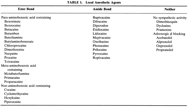
It quickly became apparent that cocaine was not the ideal local anesthetic. It had a low therapeutic ratio, which could result in convulsions and deathwhen it was injected for regional anesthesia. It was relatively unstable when stored as a solution, and it was addicting. By the time cocaine was successfully synthesized in 1924, superior local anesthetics had already been developed based on the evolving knowledge of the structure of cocaine (Fig. 1). Cocaine was found to contain an essential ester bond. This was shown by the inactivation of its anesthetic effect when this bond was hydrolyzed. Cocaine deesterification liberated benzoic acid. In 1905 procaine (novocaine) was synthesized; procaine is a benzoic acid derivative with an ester bond (Fig. 2), that is, a paraminobenzoic acid ester of diethylaminoethanol. Other frequently used agents that followed were tetracaine (1930), lidocaine (1946), mepivacaine (1956), and bupivacaine (1963).

Fig. 1. Cocaine, an ester anesthetic.

Fig. 2. Procaine.

Back to Top
STRUCTURE
Local anesthetics act by blocking the conduction of impulses along the neuronal membrane. Many types of chemical structures do so to some degree, including ethanol and barbiturates. However, those agents that are clinically useful as local anesthetics are more potent in this regard and are less toxic. Structurally, their syntheses require a secondary (-NH) or tertiary (-NH2) amino group in the hydrophilic portion of the molecule. With few exceptions, they also contain a lipid-soluble group (e.g., benzoic acid), separated from the water-soluble group (containing the amide) by an intermediate chain. The intermediate chain contains an oxygen, nitrogen, or sulfur atom. This general structure has been called the anesthesiophore group (Fig. 3). It is important that the amino group in the hydrophilic portion of the molecule be a proton receptor and thus be basic, allowing it to become positively charged (e.g., a tertiary amide, -NH2, becomes quarternized, -NH3+ , and thereby ionized) at physiologic pH (7.3–7.4).

Fig. 3. The anesthesiophore group.

Back to Top
MECHANISM OF ACTION AND EFFICACY
Local anesthetics block the formation or conduction of action potentials. They stabilize the neuronal membrane and prevent the generation of impulses; the standing potential remains intact.

The action potential begins with extracellular sodium flowing into the neuron, followed by intracellular potassium flowing out of the neuron. At low concentrations, local anesthetics primarily affect sodium conductance. At higher concentrations, potassium conductance may also be affected. The clinically used local anesthetics act primarily by reversibly blocking the sodium ion channel at its internal pore.

If a nerve is perfused in a local anesthetic solution at a low pH, causing all the anesthetic molecules to be ionized (quarternized), there is no effect. However, as the pH of the solution is raised toward physiologic levels, the number of ionized molecules is reduced, and a conduction block develops. The explanation is that ionized molecules cannot readily penetrate the sodium pore or the neuronal membrane. The anesthetic agent must be acting on the internal side of the sodium pore or neuronal membrane, not externally.

If the nerve is anesthetized with local anesthetic at pH 7.3 and the pH is markedly raised (e.g., to more than 9.5), the blockade will be reversed. At a high pH, the anesthetic molecules inside and outside the neurons are in the nonionized state. Lowering the pH to 7.3 will restore the conduction blockade. This is consistent with the concept that the active form of the anesthetic molecule must be ionized and positively charged if it is to block the sodium pore or neuronal membrane. Exceptions exist; benzocaine is a local anesthetic, but it is not ionized at physiologic pH.2

There are several other generalizations about the molecular structure and anesthetic activity relationship that seem to hold up fairly well. The larger the alkyl substitutions on the nitrogen atom of the hydrophilic group, the greater the anesthetic activity; lengthening the intermediate chain increases activity and toxicity; the most effective substitutions that increase or decrease local anesthetic action are those on the lipid-soluble group; and local anesthet-ics with an amide nitrogen in the intermediate chain of the anesthesiophore group tend to be more stable and to have a longer duration of action than those with an ester oxygen.

Neuronal factors that affect local anesthetic activity are axon diameter, myelinization, and rate of firing. The greater the diameter of the axon, the more internal membrane surface area and number of sodium channels there are to block and therefore, the lower the intensity and duration of anesthetic action.3 The greater the amount of myelin, the less readily the anesthetic penetrates the axon. Frequency dependency describes the observed phenomenon that the more rapidly a neuron discharges, the more rapidly the anesthetic enters the axon. Anesthetic agents whose molecules alternate between positively charged and neutral form at physiologic pH are most likely to show frequency dependency. Benzocaine, which is uncharged at physiologic pH, does not show frequency dependency.

The principal manufacturing factors that affect activity are drug concentration, pH of the solution, and the presence of a vasoconstrictor. Cocaine contains intrinsic vasoconstrictor properties. It blocks the reuptake of norepinephrine released by sympathetic nerve endings. All other local anesthetics block sympathetic neuron release of norepinephrine, resulting in vasodilation. The addition of a vasoconstrictor reduces drug absorption by the vasculature, thereby increasing the local effect of the anesthetic, reducing in theory its systemic toxicity, and reducing surgical bleeding. Norepinephrine is less stable than epinephrine, the vasoconstrictor that is usually added. Concentrations of epinephrine of approximately 1/200,000 (i.e., 0.0005%) are maximally effective.

There are many techniques for evaluating the intrinsic activity of a local anesthetic. One that was developed early on used the corneal surface of animals. In 1894 Von Frey reported using the tip of a single hair from a horse's tail. Onset of anesthesia was defined as beginning with the loss of the blink reflex, and the duration of anesthesia ended with its return. Depth of anesthesia was determined by multiple rapid stimuli. For example, failure for a blink to occur during four stimulations at a rate of two per second might be considered complete anesthesia.

Back to Top
TERMINATION OF ACTIVITY
Most anesthetic molecules reaching the systemic circulation are metabolized to inactive forms before elimination from the body. Hydrolysis of the ester and amine groups and oxidation and dealkylation of amino groups can occur.4 For the amide anesthetic, lidocaine, dealkylation is the major, but not only, metabolic pathway.5 However, the role of local (e.g., orbital) tissue enzymes in terminating the anesthetic effect is limited.6 Passive diffusion of an anesthetic away from its site of action seems much more important in terminating action. Because motor axons tend to be of larger diameter than sensory axons, the former require a larger quantity of drug to maintain anesthesia. Therefore, as the tissue level decreases, motor activity recovers before sensory.7
Back to Top
ANESTHESIA OF THE EYE AND ORBIT
Topical, corneal, and injected orbital anesthesia are relatively safe ways to perform surgery. In 195 patients with known previous myocardial infarctions who underwent 288 ophthalmologic procedures under local anesthesia, there were no postoperative myocardial infarctions or deaths during, or in the week after, surgery. In 255 patients with previous angina pectoris but no documented myocardial infarctions who underwent 373 ophthalmologic procedures under local anesthesia, four patients had myocardial infarctions, two of whom died. In 9,617ophthalmologic procedures performed on patients with a history of neither myocardial infarction nor angina, there was one myocardial infarction.8

TOPICAL ANESTHESIA

The cornea and conjunctiva can be anesthetized by drops of any of the local anesthetics.

Bupivacaine, for example, is effective as an eyedrop.9 It is perplexing to see the surgeon stand over the patient with a syringe full of local anesthetic and call impatiently for a vial of topical anesthetic. Generally, those agents chosen to be topical anesthetics differ only in penetrating the sensory nerve endings more rapidly, thereby having a faster onset of action and a shorter period of producing ocular irritation.

The sympathetic β-receptor antagonist, propranolol, stabilizes neuronal membranes and produces anesthesia. For this reason, it is not used topically as an ocular hypotensive agent although it can effectively lower pressure. Propranolol is not a typical local anesthetic because it does not contain an anesthesiophore group; the aromatic hydrophobic group is not joined to the hydrophilic tertiary amine by an ester or amide group. The local anesthetic potency of propranolol is approximately equal to that of lidocaine. Several other β-blockers also have local anesthetic properties. For example, oxprenolol, acebutolol, and alprenolol have approximately half the anesthetic potency of propranolol. Timolol has no significant anesthetic properties.10

The depth and duration of topical anesthesia are dose-dependent.11,12 Return to full recovery after one drop of proparacaine 0.5% may take more than 1 hour and the maximal effect may last 5 to 20 minutes. Pathologic tissues (e.g., the hypesthetic corneas associated with previous herpes simplex or herpes zoster infections) may take longer to recover.13

In general, the ester topical agents, because of their relatively rapid tissue penetration, have a lower margin of safety and are often not marketed in an injectable form. Tetracaine is such an example. Procaine is an exception; it is an ester-containing local anesthetic available as an injectable agent. When used as a topical agent, procaine has the dual advantages of a rapid onset and a shorter duration of action.

Specific Uses

SKIN SURGERY.

Topical anesthesia of the eyelid skin, sufficient to perform surgery, has been achieved with creams and patches containing lidocaine, applied 60 to 90 minutes before the first incision.14

DIAGNOSTIC TESTS.

Topical anesthetics are routinely used when performing applanation tonometry. However, small but statistically significant reductions in intraocular pressure have been reported, lasting for 1 to at least 15 minutes, after their application. In healthy patients measured 1, 5, 10, and 15 minutes after one drop of 0.4% benoxinate, the intraocular pressure decreased from a baseline of 14.80 ± 3.28 mm Hg to 14.01 ± 3.33 mm Hg, 13.56 ± 3.12 mm Hg, 13.17 ± 1.12 mm Hg, and 13.98 ± 3.45 mm Hg, respectively.15 One drop of tetracaine 2% produced a small but significant reduction in intraocular pressure from baseline at 10 minutes (10.67 ± 2.55 mm Hg to 9.94 ± 2.17 mm Hg).16 The mechanism of this reduction may be an increased aqueous humor flow rate leaving the eye, but the evidence is not strong.17

Topical anesthesia has been used when trying to differentiate reflex from nonreflex tear production. The anesthetic drop is given before the test to prevent reflex tearing from corneal or conjunctival irritation during tear collection (e.g., by a paper strip [Schirmer test] or by a pipette). However, it was found that tearing remained nonlinear, with an initial rapid phase of tear secretion, long after the anesthetic drop was administered.18 Reflex tearing was not completely inhibited even by two drops of proparacaine 0.5% applied 5 minutes before the insertion of paper strips.

OCULAR SURGERY.

Topical anesthesia for adult cataract surgery has seen a rebirth. Regional retrobulbar injections of local anesthetics had replaced the topical cocaine introduced by Koller. The reintroduction of topical anesthesia began with its use in patients with coagulopathies in whom the threat of needle-induced retrobulbar hemorrhages existed.19 Topical anesthesia has subsequently had widespread use and is only slightly less effective than intracameral, sub-Tenon's, peribulbar, or retrobulbar injections in preventing intraoperative pain.20–26 In general, the effectiveness of topical anesthesia appears uninfluenced by the use or absence of systemic analgesics preoperatively or intraoperatively.

POST-OPERATIVE ANALGESIA.

Post-operative pain after strabismus surgery performed on children, 1 to 12 years old, was found to be reduced for up to 8 hours by applying one drop of a topical anesthe-tic after intubation and a second drop immediatelybefore tubation.27 Those receiving placebo dropsrequired more analgesic drugs during the post-operative period. The pain after laser (photorefractive) keratectomy was reduced during the first 24 hours after surgery by using local anesthetic drops every 30 minutes; there was no impairment of corneal epithelial healing or visual acuity as measured 1, 3, and 6 months after the procedure.28

THERAPEUTIC USE.

Most ophthalmologists reject the thought of using long-term topical corneal anesthetic drops because of their potential side effects and toxicities. However, as treatment for trigeminal neuralgia, a small amount of literature indicates that topical anesthetics are efficacious and advocates their use.29,30

Topical lidocaine gel 5% has been applied to the face without occlusion, for 8 hours to relieve pain from herpes zoster infection.31 Significant pain relief begins by 30 minutes and lasts 24 hours. Blood levels are less than 0.3 μg/mL blood, well below the minimum of 0.6 μg/mL blood needed for any cardiac effect and the minimum of 2 μg/mL blood needed for a therapeutic antiarrhythmic effect. Long-term use for more than 2 months does not produce any systemic or topical ill effects other than some minor skin redness and irritation. A commercial preparation of lidocaine 5% is available as a patch.

Toxicity

Topical anesthetic agents can cause a decrease in corneal epithelial cell glycolysis, respiration, and healing. Microscopically, reduction in the numbers of corneal epithelial microvilli, disruption of the intercellular spaces, damage to the cell membranes and cytoplasm, and desquamation can be identified. Penetration of other topically applied agents, simultaneously or subsequently applied, is enhanced. The intraocular penetration of topical fluorescein is increased, aiding fluorophotometry. Mydriasis and cycloplegia are more rapid and profound when a topical anesthetic precedes the parasympatholytic eyedrop. However, at least one study has found evidence that the preservatives chlorhexidine and especially benzalkonium chloride, which are usedin commercial preparations of local anesthetics, are primarily the cause of this increased corneal epithelial permeability.32 It is, therefore, difficult to evaluate the role of the anesthetic agent itself when commercial preparations are reported to produce increased corneal thickness33 or epithelial permeability.34

Topical anesthetics retard healing of corneal abrasions by interfering with epithelial cell mitosis and migration.35 However, it is accepted practice for the physician to administer a topical anesthetic to assess the acutely traumatized eye. The pain, blepharospasm, photophobia, and epiphora after a corneal abrasion can prevent an accurate assessment of visual acuity and the structural integrity of the anterior segment. A drop or two of topical anesthetic will produce a dramatic, if only temporary, improvement in these symptoms.

Prolonged administration of topical anesthetics can produce a toxic keratopathy consisting of corneal epithelial defects, stromal edema and opacification, and anterior chamber inflammation. Corneal scarring and endothelial damage may require a penetrating keratopathy for visual rehabilitation.36 A sterile corneal ring infiltrate may mimic an infectious process, such as Acanthamoeba keratitis.37 The relative proportions of the keratopathy attributable to the anesthetic agent and the preservative are not clear. Healthcare workers, especially physicians,38 and the psychiatrically disturbed are the two groups most likely to abuse corneal anesthetics. If the cause of the keratopathy is not recognized, corticosteroid and antibiotic eyedrops may be given, increasing the chances of secondary fungal superinfections and resistant bacterial infections (e.g., Candida species, staphylococci, streptococci, and Proteus species).39

Topical anesthetics are routinely used when obtaining scrapings from a presumably infected cornea. A portion of the scrapings can be used to culture the causative microorganism. However, the anesthetic preparation may be toxic to the organism and impair its growth. It would seem unlikely that, in the clinical situation, a drop or two of an anesthetic agent applied to the cornea would have any significant antibacterial effect. However, when a 50-μL drop of a commercial topical anesthetic preparation was placed on an infected mouse cornea, there was a reduction in the number of microorganisms recovered; this reduction correlated with the presence, type, and concentration of preservative.40 The preservative chlorobutanol 0.2% or 0.4% was much less effective than benzalkonium 0.01% or chlorhexidine 0.001% in inhibiting growth; benzalkonium 0.004% had little inhibitory effect, as did unpreserved anesthetic agents. The earliest in vitro studies showing microorganism growth inhibition added the local anesthetic directly to the incubation medium, exposing the organisms to it continuously. Under these conditions, the anesthetic agent can retard microbiological growth even if the preservative is not present.41 The local anesthetics tetracaine, benoxinate, and cocaine and the preservatives chlorobutanol and butyl parahydroxybenzoate could each alone inhibit growth. This effect depended on the drug concentration and the organism. Unpreserved proparacaine in concentrations of up to 0.5% did not show significant growth inhibition of the strains of staphylococci, Pseudomonas and Candida that were tested.

Whereas suppression of organism growth is detrimental if the goal is to obtain a culture, this same suppression is of value if the goal is to prevent contamination of a reusable anesthetic preparation. Three commercial fluorescein 0.25%-anesthetic combinations were evaluated for their ability to regain sterility after contamination with Pseudomonas or staphylococci.42 The preparation containing the anesthetic agent benoxinate hydrochloride 0.4% with chlorobutanol 1% as preservative was more effective in regaining sterility than were the two different commercial preparations containing proparacaine hydrochloride 0.5% with thiomerosol 0.01%.

INJECTION ANESTHESIA

Subcutaneous injections of local anesthetics numb the skin of the lids, brows, and temporal areas served by the distal branches of the trigeminal nerve. Facial nerve motor fibers to the muscles in these structures run more deeply than the sensory fibers and are more variably affected by subcutaneous injections. In 1914 Auguste Van Lint43 described a technique for deliberately blocking motor conduction to the lids by injecting the anesthetic solution deeply, along the bones of the outer margins of the lateral and inferior orbital rim. By paralyzing the orbicularis muscle, lid closure during cataract surgery was prevented.

Intraorbital anesthetic injections include subconjunctival,44 sub-Tenon's,45 epibulbar or peribulbar,46 and retrobulbar techniques. Not only is the ophthalmic branch of the trigeminal nerve anesthetized by injections into the posterior orbit, but so is the maxillary branch, which is exposed after it enters the orbit through the inferior orbital fissure. Because the needle tip cannot be visualized and the limits of anesthetic diffusion cannot be determined, there may be considerable overlap in the structures affected by these seemingly different techniques. Tracking of anesthetic solution along the needle pathway may also alter the tissues affected. When local anesthetic containing contrast material was injected behind the eye, either inside or outside the muscle cone, computerized tomography showed that the dye had diffused to both sides of the muscle cone within 3 minutes.47

The optic nerve is relatively protected from diffusion by being surrounded by an extension of the dural sheath. Retrobulbar injections usually reduce optic nerve function, but in a highly variable manner.48 Amaurosis and reduced cortical visual evoked potentials have been reported after peribulbar and retrobulbar anesthesia. The incidence of no light perception after peribulbar anesthesia is in the range of 20% to 25%.49,50 The oculomotor, trochlear, and abducens nerve branches within the orbit can be blocked by intraorbital injections. In addition, there is also an effect directly on the extraocular muscle fibers themselves; local anesthetics can prevent the depolarization of muscle fiber membranes as well as neuronal membranes. Such a combined mechanism may explain why peribulbar blocks also produce an effective eyelid akinesia. That is, the orbicularis oculi muscle fibers and facial nerve motor fibers are exposed to the anesthetic. Retrobulbar blocks alone, without a deliberate facial nerve block, produced total lid akinesia in more than 90% of patients by 5 minutes after the injection.51,52 Retrobulbar injection of anesthetics has been used to prevent eye movements during intraorbital and intraocular surgery. It has also been administered just before magnetic resonance imaging when a motionless eye is needed for better resolution of the orbital structures.53 A similar need for akinesia may result in retrobulbar injections for intraocular laser procedures and corneal refractive surgery.

Intraocular injection of unpreserved local anesthetic, into the anterior chamber, has been given after the application of topical anesthesia. This has been effectively used in phacoemulsification. Retinal function, as measured by the electroretinogram, is not affected during surgery, nor are corneal endothelial cell counts reduced after surgery.54,55 However, routine use of intracameral local anesthesia seems to add little to the prevention of pain afforded by topical anesthetics alone; therefore, injection into the anterior chamber is best reserved for longer cases or when topical anesthesia fails.56,57

Factors Affecting Efficacy

In 1996 the U.S. Pharmacopeia reported that injectable lidocaine hydrochloride was tied for fourth in the number of Drug Product Problem Reports received for the period May 27, 1993 through June 20, 1995. Of the 15 drugs for which there were 24 or more complaints, only lidocaine and insulin were mentioned exclusively for lack of therapeutic effect.58

SPEED AND VOLUME OF INJECTION.

When four groups of 35 patients received peribulbar anesthesia of different volumes and different rates, the group receiving the highest volume (13.5 mL) at the fastest rate (12 mL per minute) had significantly better akinesia of the extraocular muscles without any increased injection pain.59 However, this larger volume was associated with a larger transient increase in intra-ocular pressure.

TEMPERATURE.

When three solutions, lidocaine, mepivacaine, and bupivacaine, were tested, the solubility of the anesthetic agent in each decreased as the temperature was increased from 14.5°C to 37.0°C.60 This was unexpected because solubilities are usually assumed to increase as the temperature increases.

When peribulbar and retrobulbar injections were given to patients using local anesthetics at 20°C or 37°C, two of three studies found that warming the solutions reduced the injection discomfort.61,62 One study that found no comfort benefit to warming also found that increasing the temperature provided no improvement in extraocular muscle akinesia.63

HYALURONIDASE.

Hyaluronidase depolymerizes hyaluronic acid, a component of connective tissue. In theory, addition of hyaluronidase should help the spread of the anesthetic through the tissues, increasing the rate of onset of action and the volume of tissue affected but having the disadvantage of shortening the duration of action because the anesthetic molecules diffuse away more rapidly. The addition of hyaluronidase 7.5 IU/mL to mepivacaine 2% significantly shortened the induction time of facial nerve blocks (1.3 ± 0.4 minutes with versus 2.9 ± 1.8 minutes without) and of retrobulbar blocks (3 minutes with versus 10 minutes without), but the use of hyaluronidase did not increase the success rate of retrobulbar blocks.64 Two concentrations of hyaluronidase (3.75 or 7.5 IU/mL) were equally effective when administered peribulbarly or retrobulbarly in 6 to 8 mL of anesthetic solution. Both were superior 10 minutes after injection to the anesthetic solution alone in achieving akinesias of the extraocular muscles, levator muscle, and orbicularis oculus muscle and in reducing the need for a supplementary block.65 Increasing the concentration of hyaluronidase to 15 IU/mL did not confer an advantage.66 When a larger anesthetic volume, 9 mL, was used along with a compressive device to lower intraocular pressure, addition of hyaluronidase 50 IU/mL was not superior compared with the anesthetic solution without hyaluronidase.67 The use of the compression device plus the large volume might have increased the anesthetic's diffusion, obviating any beneficial effect of hyaluronidase. When low volume sub-Tenon's injections of 0.5 mL with or without hyaluronidase 30 IU/mL were delivered at the equator of the globe and an intraocular pressure-reducing device was applied, the hyaluronidase-containing solution provided superior extraocular muscle and lid akinesia 10 minutes after the injection; however, there was no advantage or disadvantage to using hyaluro. Only thirtythree cadets graduated from Kelly Field in the fall. Many of those were seeking jobs with the airlines due to the restriction of funds for 2nd lieutenant pay for active duty. A few cadets volunteered to stay on for a year. Those going with the airlines would probably be lost for good, and those returning home would probably forget most of their training. It was a bleak picture. There was a recommendation to cut 15 percent of all pay, and the President had been given authority to cut flight payies that used solutions containing epinephrine showed no advantage to adding hyaluronidase.69

Hyaluronidase, after injection for facial nerve and retrobulbar blocks, has been reported to cause an allergic reaction consisting of periorbital edema and pruritis.70 Subsequent skin testing provided a positive response. Because the commercial preparation of hyaluronidase contained thiomerosol, it was tested separately; it produced no reaction.

pH.

Anesthetic solutions usually are commercially formulated to be in a pH range of 5.0 to 6.0. This favors formation of the ionic form of the molecule, which is more stable and increases the shelf-life of the product. When the pH of an anesthetic solution is increased, it is usually done so immediately before use. However, many anesthetic molecules remain stable for relatively long periods at physiologic pH. When the levels of bupivacaine and lidocaine were measured 24 hours after bicarbonate was added, sufficient to raise the pH to 7.4, there were no reductions in drug concentrations.71

Anesthetic blocks have been given with solutions after their pH has been increased. Increasing the pH increases the amount of nonionic drug molecules. The uncharged molecules are better able to penetrate cell membranes. When an anesthetic solution with a pH of 5.4 was compared with bicarbonate-adjusted solutions with a pH between 6.7 and 6.9, the peribulbar blocks using the higher pH solutions had a more rapid onset of action but no shorter time to complete akinesia.72 Furthermore, the higher pH solutions required more supplemental injections for an effective block. Other studies have confirmed a faster onset of action using higher pH solutions but found a shorter time to complete akinesia.73 In one of these, solutions of pH 3.9, 5.1, and 6.7 were compared; the last had the most rapid onset and frequency of complete akinesia.74

Another theoretical justification for increasing the pH has been patient comfort. Lower pH solutions may be more irritating to tissues. Subcutaneous eyelid injections of buffered anesthetic solutions at pH 7.4 were less painful to receive than those at pH 4.6.75 However, subcutaneously injected procaine at pH 4.3 produced less pain than lidocaine, pH 6.376 When the pain caused by the peribulbar injection of combination bupivacaine-lidocaine solutions at pH 4.87 or pH 7.44 were compared, there was no significant difference77. These findings tend to undermine any theories attributing tissue irritation to lower pH or comfort to increasing pH.

VASOCONSTRICTIORS.

The addition of a vasoconstrictor, usually epinephrine, at concentrations of 0.0005% to 0.001%, results in an anesthetic solution that retards its own absorption into the circulation. This prolongs the effect of the shorter-acting anesthetics (e.g., lidocaine) but may provide little or no benefit for the longer-acting ones (e.g., bupivacaine).78–80 The elevations in serum epinephrine caused by commercial preparations of local anesthetics containing epinephrine are probably no more than those caused by stress-induced adrenal gland release. In one study, facial nerve and retrobulbar blocks were performed with a total of 10 to 12 mL of lidocaine 2% containing 0.0005% epinephrine. The baseline mean ± standard error of the mean plasma epinephrine level was 116 ± 8 pg/mL.81 Seven minutes after the injections, this epinephrine level had risen to 429 ± 25 pg/mL, 270% more than baseline. However, this increase was not markedly different from that caused by the stress of public speaking. The baseline mean epinephrine level was 117 pg/mL plasma, and during speaking, the epinephrine level was 336 pg/mL plasma.82

PERMANENT ANESTHESIA

Anesthetic agents act transiently (i.e., minutes to hours). Sometimes a more permanent hypalgesia or anesthesia is needed. This occurs when dealing with the chronically or intermittently painful blind eyes produced by neovascular glaucoma and phthisis bulbi. Ethanol injections, if not given in too high a volume and concentration, will destroy the poorly myelinated sensory fibers while having only a temporary effect on the more heavily myelinated motor fibers. The optic nerve, surrounded by its dural sheath, is usually well protected from the effects of the alcohol, unless the needle tip penetrates this covering. Ethanol has been injected into orbits containing seeing eyes to reduce the ocular pain of children who have had corneal transplants, thereby permitting postoperative examinations.83 Generally, however, the drug is reserved for eyes with littleor no visual potential. Although a single retrobul-bar injection of 1 mL 95% or absolute ethanol or2 mL of 50% ethanol might be sufficient to produce permanent comfort, often two or more injections are needed. The injection itself produces so much acute discomfort that it is recommended that the alcohol be preceded by an injection of anesthetic solution. This will confirm that the needle tip is within the muscle cone and prevent the subsequent pain from the ethanol. The needle is not removed after the anesthetic is injected; the syringe containing the ethanol is exchanged for that which contains the anesthetic. Up to several weeks of chemosis, ptosis, and extraocular muscle movement impairment may result. In seeing eyes with corneal transplants, concentrations of 20% to 95% ethanol have been used in volumes of up to 3 mL for the former and 1 mL for the latter. Of 25 eyes so treated, none had evidence of optic nerve damage, corneal graft failure (attributable to the injection), permanent muscle palsy, or permanent corneal anesthesia.

However, there have been at least two case reports of loss of vision after retrobulbar alcohol injections. One was associated with an immediate loss of vision and subsequent optic atrophy; presumably the injection was into the optic nerve.84 In the second case, a patient with acute glaucoma had a central retinal artery occlusion after retrobulbar injection of 80% ethanol; the role played by the glaucomatous elevation of intraocular pressure is unclear.85 Alternatives to ethanol have been suggested, such as retrobulbar injections of 0.5% quinine urea hydrochloride in 2.5% benzocaine or of 25 mg of chlorpromazine.86–88

Back to Top
SIDE EFFECTS, TOXICITIES, AND ALLERGIES

ORBITAL

Bupivacaine

Bupivacaine has a special role among local anesthetics because it is much more myotoxic to the skeletal muscle of experimental animals than other anesthetics are. The mechanism involved may be reduced protein synthesis and may require the presence of extracellular calcium.89–91 Extraocular muscle fibers of primates are relatively more resistant to developing this toxicity than are those of lower mammals.92,93 Multiple clinical papers have theorized that the postoperative diplopia, especially vertical diplopia, and ptosis being reported were caused by myotoxicity from retrobulbar and peribulbar bupivacaine.94–97

Bupivacaine is a racemic mixture of levo- and dextobupivacaine. The former enantiomer exhibits less myocardial toxicity in animal studies.98 Levobupivacaine 0.75% appears as effective as the racemic bupivacaine 0.75% in producing akinesia and anesthesia after peribulbar injections.99 Another commercially available alternative to bupivacaine is ropivacaine. It is a long-acting anesthetic that, as a 1% solution, has been given as a peribulbar block and compared with combination solutions with final concentrations of lidocaine 1% and bupivacaine 0.375%.100,101 Although there were no differences in anesthetic effect and extraocular muscle akinesia by 8 minutes after the injection, some discrepancies exist in the literature as to whether the ropivacaine solution lagged early on in producing extraocular muscle paresis and whether it was superior in producing lid akinesia.102

Increased Intraocular Pressure

It is usually an advantage to have a low intraocular pressure when an eye is lacerated by trauma or when a penetrating surgical incision is to be made. However, immediately after a retrobulbar or peribulbar injection, the intraocular pressure is increased, presumably because of the mass effect of the solution. By 5 minutes after the injection, the intraocular pressure tends to have returned to baseline and by 8 to 10 minutes after the injection, the intraocular pressure has decreased significantly. This sequence has long been known.103,104 The presence or absence of epinephrine in the anesthetic solution has little or no influence on the sequence.105 The explanations for the reduction in intraocular pressure have varied (e.g., paralysis of the extraocular muscles, a reduced ciliary body secretion, and an indirect ischemic effect caused by compression of the blood vessels). In one study, the mean elevation in intraocular pressure was 5.8 mm Hg 1 minute after the injection, with one eye having an increase of 25 mm Hg;106 patients with glaucoma tended to have greater increases than those without. Retrobulbar local anesthetic, 4.5 mL, has been reported to cause a 50% reduction in the ophthalmic artery pulse pressure when measured 10 minutes after injection.107 The mechanisms causing the rare central retinal artery or vein obstructions found postoperatively are not clear, but the injection of a retrobulbar or peribulbar solution may play a role.108,109

CENTRAL NERVOUS SYSTEM

Direct injection of the anesthetic solution into the cerebrospinal fluid surrounding the optic nerve has been blamed for producing central nervous system toxicity after retrobulbar blocks. An alternative explanation has been that direct injection into an ophthalmic artery branch results in retrograde flow, back to the internal carotid artery, and then antigrade flow to the brain.110 In a retrospective study of 6,000 retrobulbar blocks, the incidence of central nervous system signs and symptoms was 0.27%.111 Extraorbital cranial nerve pareses, amaurosis, grand mal seizures, and respiratory arrest have occurred.112–114

It is unlikely that these events were the result of absorption from the orbital tissue producing toxic blood levels. To put this possibility in perspective, lidocaine is usually administered for the treatment of ventricular tachycardias by using an intravenous bolus injection of 50 to 100 mg, followed by a continuous drip of 2 to 4 mg per minute.115 Five milliliters of lidocaine 2% anesthetic solution contains only 100 mg of lidocaine. Intravenous lidocaine, 2 mg/kg, before intubation has been recommended to reduce the incidence of postoperative vomiting, from more than 50% to less than 20%, in children undergoing strabismus surgery.116 Intravenous lidocaine, 1.5 mg/kg in children and 2 mg/kg in adults, has been advocated for prevention of the intraocular pressure increase that occurs during intubation for general anesthesia.117 Serum lidocaine and bupivacaine levels of seven patients who had respiratory arrests after retrobulbar anesthesia were not significantly increased compared with a control group.118

The therapeutic range for lidocaine treatment of cardiac arrhythmias is 2 to 4 μg/mL plasma. After injection of 5 mL lidocaine 1%, the mean peak plasma levels of lidocaine were mandibular seventh nerve block, 0.3 (range, 0.25–0.67) μg/mL and eyelid seventh nerve block, 0.4 (range, 0.16–0.99) μG/Ml.119 When peribulbar blocks were given with no more than 10 mL of a solution containing final concentrations of 1% lidocaine and 0.375% bupivacaine, the peak mean drug concentrations were found 10 to 20 minutes after injection: 0.7 μg lidocaine/mL and 0.35 μg bupivacaine/mL.120 The addition of epinephrine significantly reduces the peak plasma levels and total amount of drug absorbed into the cir-culation for the 5 hours after peribulbar injectionsof lidocaine and bupivacaine.121 The presence of0.005% epinephrine reduces the mean peak plasma concentrations of lidocaine and bupivacaine to 57% and 61%, respectively, of those obtained using solutions without epinephrine. The addition of hyaluronidase does not cause increased peak drug levels or total drug absorption.

An important factor in determining whether a given plasma level of local anesthetic may be toxic is the quantity of α-1 acid glycoprotein. This protein binds basic drugs, such as lidocaine. Neonates and cirrhotic patients have lower levels of α-1 acid glycoprotein, and patients with acute inflammatory responses have higher levels.122,123

METHEMOGLOBULINEMIA

This condition can be caused by the inability of NADH-dependent methemoglobin reductase to reduce the hemoglobin iron atom from Fe+ 3 to Fe+ 2. Local anesthetics are a potential cause of surgery-related methemoglobulinemia, and prilocaine is the most potent in this regard.124 Combination retrobulbar blocks and facial nerve blocks, with a total of 8 mL prilocaine 1%, produced a mean peak serum blood level of 0.85 μg/mL, 3 to 7 minutes after the injection;125 the range was from 0.54 to 1.10 μg of prilocaine. Because prilocaine has less central nervous system and cardiovascular toxicity than lidocaine, complications other than a clinically insignificant methemoglobulinemia are rare. However, a peribulbar injection of prilocaine has been implicated as a contributory factor in the development of acute methemoglobinemia with respiratory distress. Improvement occurred rapidly with the injection of the antidote, methylene blue, 1.5 mg/kg.126

ALLERGIES

The most commonly reported anesthetic allergies are contact allergies from topically administered drugs. Anaphylaxis is rare, and if it were to occur, it would most likely be from a sensitivity to an additive in the solution rather than to the anesthetic agent itself.127 However, at least one death has been reported from a rechallenge to a local anesthetic after instilling a drop in the conjunctival sac.128 In a study of 71 patients suspected of an allergy to a local anesthetic, only 8% had a history of urticaria or wheezing. Forty-two percent had histories most compatible with toxicities or vasovagal responses.129 Skin testing was positive in only 5 of 59 patients rechallenged and at no greater incidence in those with histories most compatible with allergy than those without. Allergic reactions can be to metabolic products, so negative skin testing does not rule out an allergy.


Back to Top
REFERENCES

1. de Jong RH: Local anesthetics: from cocaine to xylocaine. In: Local anesthetics. St. Louis, Mosby-Year Book, 1994:4–5.

2. Ogden DC, Siegelbaum SA, Colquhoun D: Blockade of acetylcholine-activated ion channels, by an uncharged local anesthetic. Nature 289:596–598, 1981.

3. Staiman A, Seeman P: Conduction-blocking concentrations of anesthetics increase with nerve axon diameter: studies with alcohol, lidocaine and tetrodotoxin on single myelinated fibers. J Pharm Exp Therap 201:340–349, 1977.

4. Dubbels R, Schloot W: Studies on the metabolism of benoxinate by human pseudocholinesterase. Metabol Pediatr Syst Ophthalmol 7:37–43, 1983.

5. Nelson SD, Garland WA, Breck GD et al: Quantification of lidocaine and several metabolites utilizing chemical-ionization mass spectrometry and stable isotope labeling. J Pharmaceut Sci 66:1180–1189, 1977.

6. Zoglio MA, Soine TO, Rippie EG: Esters of bicyclic aminoalcohols. V: Duration of corneal anesthesia versus enzymatic hydrolytic rate of benzoates of 1-2- and 3- methyl -2- hydroxyquinolizidines. J Pharmaceut Sci 60:411–416, 1971.

7. Winnie AP, LaVallee DA, Sosa BP et al: Clinical pharmokinetics of local anaesthetics. Canad Anaesth Soc J 4:252–262, 1977.

8. Backer CL, Tinker JA, Robertson DM et al: Myocardial reinfarction following local anesthesia for ophthalmic surgery. Anesth Analg 59:257–262, 1980.

9. Carruthers JDA, Sanmugasunderan S, Mills K et al: The efficacy of topical corneal anesthesia with 0.5% bupivacaine eyedrops. Can J Ophthalmol 30:264–266, 1995.

10. McDevitt RG: The assessment of beta-adrenoceptor blocking drugs I man. Br J Clin Pharmacol 4:413–425, 1977.

11. Polse KA, Keener RJ, Jauregui MJ: Dose-response effects of corneal anesthetics. Am J Optom Physiol Optics 55:8–14, 1978.

12. Matsumoto S, Hayashi K, Tsuchisaka H et al: Pharmacokinetics of surface anesthesia in the human cornea Jpn J Ophthalmol 25:235–340, 1981.

13. Weiss JS, Goren MB: The effect of corneal hypesthesia on the duration of proparacaine anesthetic eyedrops. Am J Ophthalmol 112:326–330, 1991.

14. Gotsis SS, Volonaki OM, Theodossiadis GP: Percutaneous anaesthesia with a lignocaine-prilocaine cream (Emla) for eyelid skin surgery. Br J Ophthalmol 78:209–210, 1994.

15. Baudouin C, Gastaud P: Influence of topical anesthesia on tonometric values of intraocular pressure. Ophthalmologica 208:309–313, 1994.

16. Carel RS, Korczyn AD, Boyman R: Amethocaine and intraocular pressure. Ophthalmic Res 11:212–215, 1979.

17. Bloom JN, Levene RZ, Thomas G et al: Fluorophotometry and the rate of aqueous flow in man. Arch Ophthalmol 94:435–443, 1976.

18. Clinch TE, Benedetto DA, Felberg NT et al: Schirmer's test—a closer look. Arch Ophthalmol 101:1383–1386, 1983.

19. Shuler JD: Topical anesthesia in a patient with a history of retrobulbar hemorrhage. Arch Ophthalmol 111:733–737, 1993.

20. Crandall AS, Zabriskie NA, Patel BCK et al: A comparison of patient comfort during cataract surgery with topical anesthesia versus topical anesthesia and intracameral litodaine. Ophthalmolgy 106:60–66, 1999.

21. Boulton JE, Lopatatzidis A, Luck J et al: A randomized controlled trial of intracameral lidocaine during phacoemulsification under topical anesthesia. Ophthalmology 107:68–71, 2000.

22. Chittenden HB, Meacock WR, Govan JAS: Topical anaesthesia with oxybuprocaine versus sub-Tenon's infiltration with 2% lignocaine for small incision cataract surgery. Br J Ophthalmol 81:288–290, 1997.

23. Patel BCK, Burns TA, Crandall A et al: A comparison of topical and retrobulbar anesthesia for cataract surgery. Ophthalmology 103:1196–1203, 1996.

24. Duguid IGM, Claque CMP, Thamby-Rajah Y et al: Topical anesthesia for phacoemulsification surgery. Eye 9:456–459, 1995.

25. Jacobi PC, Dietlein TS, Jacobi FK: A comparative study of topical v retrobulbar anesthesia in complicated cataract surgery. Arch Ophthalmol 118:1037–1043, 2000.

26. Katz J, Feldman MA, Bass EB et al: Injectable versus topical anesthesia for cataract surgery Ophthalmology 107:2054–2060, 2000.

27. Watson DM: Topical amethocaine in strabismus surgery. Anaesthesia 46:368–370, 1991.

28. Verma S, Corbett MC, Marshall J: A prospective randomized, double-masked trial to evaluate the role of topical anesthetics in controlling pain after photorefractive keratectomy. Ophthalmology 102:1918–1924, 1995.

29. Zavon MR, Fichte CM: Trigeminal neuralgia relieved by ophthalmic anesthetic. JAMA 265:2807, 1991.

30. Spaziante R, Cappabianca P, Saini M et al: Treatment of trigeminal neuralgia by ophthalmic anesthetic. J Neurosurg 77:159–160, 1992.

31. Rowbotham MC, Davies PS, Fields HL: Topical lidocaine gel relieves postherpetic neuralgia. Ann Neurol 37:246–253, 1995.

32. Ramselaar JAM, Boot JP, van Haerington NJ et al: Corneal epithelial permeability after instillation of ophthalmic solutions containing local anaesthetics and preservatives. Cur Eye Res 7:947–953, 1988.

33. Herse P, Siu A: Short-term effects of proparacaine on human corneal thickness. Acta Ophthalmol 70:740–744, 1992.

34. Stolwijk TR, van Best JA, Boot JP et al: Corneal epithelial barrier function after oxybuprocaine provocation in diabetics. Invest Ophthalmol Vis Sci 31:436–439, 1990.

35. Marr MG, Wood R: Effects of topical anesthetics on regeneration of corneal epithelium. Am J Ophthalmol 43:606–614, 1957.

36. Risco JM, Miller LC: Ultrastructural alterations in the endothelium in a patient with topical anesthesic abuse keratopathy. Ophthalmology 99:628–633, 1992.

37. Rosenwasser GOD, Holland S, Pflugfelder SC et al: Topical anesthetic abuse. Ophthalmology 97:967–972, 1990.

38. Henkes HE, Waubke TN: Keratitis from abuse of corneal anaesthetics. Br J Ophthalmol 62:62–65, 1978.

39. Chern KC, MeislerDM, Wilhelmus KR et al: Corneal anesthetic abuse and Candida keratitis. Ophthalmology 103:37–40, 1996.

40. Badenoch PR, Coste DJ: Antimicrobial activity of topical anesthetic preparations. Br J Ophthalmol 66:364–367, 1982.

41. Kleinfeld J, Ellis PP: Effects of topical anesthetics on growth of micro-organisms. Arch Ophthalmol 76:712–715, 1966.

42. Duffner LR, Pfugfelder SC, Mandelbaum S et al: Potential bacterial contamination in fluorescein-anesthetic solutions. Am J Ophthalmol 110:199–202, 1990.

43. Van Lint A: IL faut toujours paralyser les paupieres dans l'operation de la cataracte. Bull Soc Belge Ophthalmol 43:23–28, 1921.

44. Khurana AK, Sachdeva RK, Gombar KK: Evaluation of subconjunctival anaesthesia vs peribulbar anaesthesia in cataract surgery. Acta Ophthalmologica 72:727–730, 1994.

45. Fujishima H, Yagi Y, Yang H-Y: Direct sub-Tenon's ocular anesthesia for strabismus surgery. Ophthalmologica 209:208–211, 1995.

46. Fry RA, Henderson J: Local anaesthesia for eye surgery. The peri-ocular technique. Anaesthseia 45:14–17, 1989.

47. Ropo A, Nikki P, Ruusuvaara P et al: Comparison of retrobulbar and periocular injections of lignocaine by computerised tomography. Br J Ophthalmol 75:417–420, 1991.

48. Carroll FD, deRoetth Jr A: The effect of retrobulbar injections of procaine on the optic nerve. Trans Amer Acad Ophthalmol Oto 59:356–363, 1955.

49. Ropo A, Ruusuvaara P, Setala K: Visual evoked potentials after retrobulbar or periocular anaesthesia. Br J Ophthalmol. 76:541–544, 1992.

50. Talks SJ, Chong NHV, Gibson JM et al: Visual acuity and pupillary reactions after peribulbar anesthesia. Br J Ophthalmol 78:41–43, 1994.

51. Sanchez - Capuchino A, Meadows D, Morgan L et al: Local anaesthesia for eye surgery without a facial nerve block. Anaesthesia 48:428–431, 1993.

52. Ripart J, Metge L, Prat-Pradal D et al: Medial canthus single-injection episcleral (sub-Tenon anesthesia): computedtomography imaging. Anesth Analg 87:42–45, 1998.

53. Hosten N, Bornfeld N, Lemke AJ et al: MR of the eye with retrobulbar anesthesia. Am J Neuroradiol 18:1788–1790, 1997.

54. Anders N, Heuermann T, Ruther K et al: Clinical and electrophysiological results after intracameral lidocaine 1% anesthesia. Ophthalmology 106:1863–1868, 1999.

55. Iradier MT, Fernandez C, Bohorquez P et al: Intraocular lidocaine in phacoemulsification. An endothelium and blood-aqueous barrier permeability study. Ophthalmology 107:896–901, 2000.

56. Gillow T, Scotcher SM, Deutsch J et al: Efficacy of supplementary intracameral lidocaine in routine phacoemulsification under topical anesthesia. Ophthalmology 106:2173–2177, 1999.

57. Malecaze FA, Deneuville SF, Julia BJ et al: Pain relief with intracameral mepiracaine during phacoemulsification. Br J Ophthalmol 84:171–174, 2000.

58. United States Pharmacopeia Convention Inc: Most frequently reported drugs and their prevalent problems. Quality Review 55:2, 1996.

59. Gillart T, Bazin JE, Montetagaud M et al: The effects of volume and speed of injection in peribulbar anaesthesia. Anaesthesia. 53:486–510, 1998.

60. Nakano NI: Temperature-dependent aqueous solubilities of lidocaine, mepivacaine and bupivacaine. J Pharmaceut Sci 68:667–668, 1979.

61. Ursell PG, Spalton DJ: The effect of solution temperature on the pain of peribulbar anaesthesia. Ophthalmology 103:839–841, 1996.

62. Bell RWD, Butt ZA: Warming lidocaine reduces the pain of injection during peribulbar local anaesthesia for cataract surgery. Br J Ophthalmol 79:1015–1017, 1995.

63. Krause M, Weindler J, Ruprecht KW: Does warming of anesthetic solutions improve analgesia and akinesia in retrobulbar anesthesia? Ophthalmology 104:429–432, 1997.

64. Mindel JS: Value of hyaluronidase in ocular surgical akinesia. Am J Ophthalmol 85:643–646, 1978.

65. Kallio H, Paloheimo M, Maunuksela EL: Hyaluronidase as an adjuvant inbupivacaine-lidocaine mixture for retrobulbar/peribulbar block. Anesth Analg 91:934–937, 2000.

66. Sarvela J, Nikki P: Hyaluronidase improves regional ophthalmic anaesthesia with etidocaine, Can J Anaesth 39:920–924, 1992.

67. Crawford M, Kerr WJ: The effects of hyaluronidase on peribulbar block. Anaesthesia 49:907–908, 1994.

68. Rowley SA, Hale JE, Finlay RD: Sub-Tenon's local anaesthesia: the effect of hyaluronidase. Br J Ophthalmol 84:435–436, 2000.

69. Nicoll JMV, Treuren B, Acharya PA et al: Retrobulbar anesthesia: the role of hyaluronidase. Anesth Analg 65:1324–1328, 1986.

70. Minning Jr CA: Hyaluronidase allergy simulating expulsive choroidal hemorrhage. Arch Ophthalmol 112:585, 1994.

71. Robinson J, Fernando R, Sun Wai WYS et al: Clinical stability of bupivacaine, lidocaine and epinephrine in pH-adjusted solutions. Anaesthesia 55:853–858, 2000.

72. Lewis P, Hamilton RC, Loken RG: Comparison of plain with pH-adjusted bupivacaine with hyaluronidase for peribulbar block. Can J Anaesth 39:555–558, 1992.

73. Zahl K, Jordan A, McGroarty J et al: Peribulbar anesthesia effect of bicarbonate on mixtures of lidocaine, bupivacaine and hyaluronidase with or without epinephrine. Ophthalmology 98:239–242, 1991.

74. Roberts JE, MacLeod BA, Hollands RH: Improved peribulbar anaesthesia with alkalinization and hyaluronidase. Can J Anaesth 40:835–838, 1993.

75. Eccarius SG, Gordon ME, Parelman JJ: Bicarbonate-buffered lidocaine-epinephrine-hyaluronidase for eyelid anesthesia. Ophthalmology 97:1499–1501, 1990.

76. McKay W, Morris R, Mushlin P: Sodium bicarbonate attenuates pain on skin infiltration with lidocaine; with or without epinephrine. Anesth Analg 66:572–574, 1987.

77. Minasian MC, Ionides AC, Fernando R et al: Pain perception with pH buffered peribulbar anaesthesia: a pilot study. Br J Ophthalmol 84:1041–1044, 2000.

78. Krohn J, Houding G, Seland JH et al: Retrobulbar anesthesia with an without adrenaline in extracapsular cataract surgery. Acta Ophthalmol Scand 73:56–60, 1995.

79. Bjornstrom L, Hansen A, Otland N et al: Peribulbar anaesthesia—a clinical evaluation of two different anaesthetic mixtures Acta Ophthalmologica 72:712–714, 1994.

80. Chin GN, Almquist HT: Bupivacaine and lidocaine retrobulbar anesthesia—a double-blind clinical study. Ophthalmology 90:369–372, 1983.

81. Donlon JV, Moss J: Plasma catecholamine levels during local anesthesia for cataract operations. Anesthesiology 51:471–473, 1979.

82. Dimsdale JE, Moss J: Plasma catecholamines in stress and exercise. JAMA 243:340–342, 1980; Correction 243:2485, 1980.

83. Michels RG, Maumenee AE: Retrobulbar alcohol injection in seeing eyes. Trans Am Acad Ophthalmol Otolaryngol 77:164–167, 1973.

84. Cordes FC: Optic atrophy following alcohol injections. Am J Ophthalmol 12:120, 1929.

85. Payer FL Occlusion of the central retinal artery following retrobulbar injection of alcohol. Oftalmologica 8:65–69, 1964.

86. Mathur SP: Ciliary block with benzocaine. Am J Ophthalmol 47:867–869, 1959.

87. Fiore D, Lupidi G, Santoni G: Retrobulbar injection of chlorpromazine in absolute glaucoma. J Fr Ophthalmol 3:397–399, 1980.

88. Waltz KL, Williams GA, Goldey SH et al: Alternative to retrobulbar ethanol for blind painful eyes. Ophthalmology 99(suppl): 153, 1992.

89. Foster AH, Carlson BM: Myotonicity of local anesthetics and regeneration of the damaged muscle fibers. Anesth Analg 58:727–736, 1980.

90. Johnson ME, Jones GH: Effects of Marcaine, a myotoxic drug, on macromolecular synthesis in mice. Biochem Pharmacol 27:1753–1757, 1978.

91. Steer JH, Mastaglia FL: Protein degradation in bupivacaine-treated muscles: The role of extracellular calcium.J Neurolog Sci 75:343–351, 1986.

92. Porter JD, Edney DP, McMahon EJ et al: Extraocular myotoxicity of the retrobulbar anesthetic bupivacaine hydrochloride. Invest OphthalmolVis Sci 29:163–174, 1988.

93. Okland S, Komorowski TE, Carlson BM: Ultrastructure of Mepicavaine-induced damage and regeneration in rat extraocular muscle. Invest Ophthalmol Vis Sci 30:1643–1651, 1989.

94. Mather C, Hayden Smith J, Bloom PA: The efficacy of 0.75% bupivacaine with pH adjustment and hyaluronidase for peribulbar blockade: the incidence of prolonged ptosis. Eur J Ophthalmol 4:13–18, 1994.

95. Rainin EA, Carlson BM: Postoperative diplopia and ptosis. A clinical hypothesis based on the myotoxicity of local anesthetics. Arch Ophthalmol 103:1337–1339, 1985.

96. Hamed LM, Mancuso A: Inferior rectus muscle contracture syndrome after retrobulbar anesthesia. Ophthalmology 98:1506–1512, 1991.

97. Hunter D: Author's reply. Ophthalmolgy 103:546, 1996.

98. Mazdit J, Boico O, Samii K: Myocardial uptake of bupivacaine: II. Pharmacokinetics and pharmacodynamics of bupivacaine enantiomers in the isolated perfused rabbit heart. Anesth Analg 77:477–482, 1993.

99. Mc Lure HA, Rubin AP: Comparison of 0.75% levobupivacaine with 0.75% racemic bupivacaine for peribulbar anaesthesia. Anaesthesia 53:1160–1164, 1998.

100. Mc Lure HA, Rubin AP, Wescott M et al: A comparison of 1% ropivacaine with a mixture of 0.75% bupivacaine and 2% lignocaine for peribulbar anaesthesia. Anaesthesia 54:1178–1182, 1999.

101. Nicholson G, Sutton B, Hall GM: Comparison of 1% ropivacaine with 0.75% bupivacaine and 2% lidocaine for peribulbar anaesthesia. Br J Anaesth 84:89–91, 2000.

102. Huha T, Ala-Kokko TI, Salomaki T et al: Clinical efficacy and pharmacokinetics of 1% ropivacaine and 0.75% bupivacaine in peribulbar anaesthesia for cataract surgery. Anaesthesia 54:137–141, 1999.

103. Atkinson WS: Local anesthesia in ophthalmology. Trans Amer Ophthalmol Soc 2:399–451, 1934.

104. Gifford H: A study of the effect of retrobulbar anesthesia on ocular tension and the vitreous pressure. Amer J Ophthalmol 32:1359–1368, 1949.

105. Gjotterberg M, Ingemnsson S-O: Effect on intraocular pressure of retrobulbarinjection of Xylocaine with and without adrenaline. Acta Ophthalmologica 55:709–716, 1977.

106. O'Donoghue E, Batterbury M, Lavy T: Effect on intraocular pressure of local anaesthesia in eyes undergoing intra-ocular surgery. Br J Ophthalmol 78:605–607, 1994.

107. Horven I: Ophthalmic artery pressure during retrobulbar anaesthesia. Acta Ophthalmologica 56:574–586, 1978.

108. Klein ML, Jampol LM, Condon PI et al: Central retinal artery occlusion without retrobulbar hemorrhage after retrobulbar anesthesia. Amer J Ophthalmol 93:573–577, 1982.

109. Sullivan KL, Brown GC, Forman AR: Retrobulbar anesthesia and retinal vascular obstruction. Ophthalmology 90:373–377, 1983.

110. Rosenblatt RM, May DR, Barsoumian K: Cardiopulmonary arrest after retrobulbar block. Am J Ophthalmol 90:425:-427, 1980.

111. Nicoll JMV, Acharya A, Ahlen K et al: Central nervous system complications after 6000 retrobulbar blocks. Anesth Analg 66:1298–1302, 1987.

112. Meyers EF, Ramirez RC, Boniuk I: Grand mal seizures after retrobulbar block. Arch Ophthalmol 96:847, 1978.

113. Javitt JC, Addiego R, Friedberg HL et al: Brain stem anesthesia after retrobulbar block. Ophthalmology 94:718–724, 1987.

114. Cohen SM, Sousa FJ, Kelly NE: Respiratory arrest and new retinal hemorrhages after retrobulbar anesthesia. Am J Ophthalmol 113:209–211, 1992.

115. Greenblatt DJ, Bolognini V, Koch-Weser J et al: Pharmacokinetic approach to the clinical use of lidocaine intravenously. JAMA 236:273–277, 1976.

116. Warner LO, Rogers GL, Marteno JD et al: Intravenous lidocaine reduces the incidence of vomiting in children after surgery to correct strabismus. Anesthesiology 68:618–621, 988.

117. Lerman J, Kiskis AA: Lidocaine attenuates the intraocular pressure response to rapid intubation in children. Can Anaesth J 32:339–345, 1985.

118. Wittpenn JR, Rapoza P, Sternberg P et al: Respiratory arrest following retrobulbar anesthesia. Ophthalmoloy 93:867–870, 1986.

119. Schimek F, Friess D, Fahle M: Plasma concentrations of lidocaine after proximal and peripheral facial nerve blocks. German J Ophthalmol 4:315–319, 1995.

120. Gao F, Budd AJ: Venous levels of lignocaine and bupivacaine after peribulbar block. Anaesthesia 51:1109–1112, 1996.

121. Barr J, Kirkpatrick N, Dick A: Effects of adrenaline and hyaluronidase on plasma concentrations of lidocaine and bupivacaine after peribulbar anaesthesia. Br J Anaesthesia 75:692–697, 1995.

122. Lerman J, Strong A, LeDez KM et al: Effects of age on the serum concentration of a1-acid glycoprotein and the binding of lidocaine in pediatric patients. Clin Pharmacol Ther 46:219–225, 1989.

123. Barry M, Keeling PWN, Weir D et al: Seventy of cirrhosis and the relationship of a1-acid glycoprotein concentration to plasma protein binding of lidocaine. Clin Pharmacol Ther 47:366–370, 1990.

124. Olson ML, McEvoy GK: Methemoglobinemia induced by local anesthetics. Am J Hosp Pharm 38:89–93, 1981.

125. Goggin M, Crowley K, O'Malley K et al: Serum concentrations of prilocaine following retrobulbar block. Br J Anaesth 64:107–109, 1990.

126. Eltzschig H, Rohrback M, Schroeder TH: Methaemoglobinemia after peribulbar blockade: an unusual complication in ophthalmic surgery. Br J Ophthalmol 84:442, 2000.

127. Barker MR, McAllen MK: Hypersensitivity to local anaesthetics: a direct challenge test with lignocaine for definitive diagnosis. BMJ 284:1229–1230, 1982.

128. Adriani J: Etiology and management of adverse reactions to local anesthetics. Int Anesthesiol Clin 10:127–151, 1972.

129. Incaudo G Schatz M, Patterson R et al: Administration of local anesthetic to patients with a history of prior adverse reactions. J Allergy Clin Immunol 61:339–345, 1978.

Back to Top