Chapter 30
Systemically and Topically Administered Carbonic Anhydrase Inhibitors in the Treatment of Glaucoma
JANET B. SERLE and ANDREW I. RABINOWITZ
Main Menu   Table Of Contents

Search

HISTORICAL PERSPECTIVE
CARBONIC ANHYDRASE
MECHANISM OF INTRAOCULAR PRESSURE REDUCTION
CLINICAL USES OF CARBONIC ANHYDRASE INHIBITORS
SUMMARY
REFERENCES

Carbonic anhydrase inhibitors (CAIs) have been used to treat glaucoma since 1954, when Becker1 first demonstrated the clinical efficacy of acetazolamide. CAIs are sulfonamide derivatives that lower intraocular pressure (IOP) by decreasing aqueous humor formation. These drugs inhibit carbonic anhydrase (CA), which is one of the enzymes that regulates aqueous humor formation. Systemic administration of CAIs is extremely effective in reducing IOP in most patients but is accompanied by side effects that can range from mild and annoying to debilitating and life-threatening, the latter necessitating discontinuation of these drugs. On rare occasions, CAIs may induce irreversible blood dyscrasias, which can be fatal. The substantial incidence of side effects and the rare occurrence of fatal complications has led to controversy regarding the usefulness of and indications for systemically administered CAIs in the chronic management of glaucoma. These side effects have also led to a 40-year search for topically effective CAIs.
Back to Top
HISTORICAL PERSPECTIVE
In his 1949 Proctor Award Lecture of the Association for Research in Ophthalmology, Friedenwald2 initially theorized that aqueous humor formation was an active process involving transport into the posterior chamber of electrolytes and ascorbic acid and requiring large quantities of bicarbonate as a buffer. Subsequently, Kinsey3 demonstrated a high concentration of bicarbonate in the aqueous humor relative to plasma in rabbits, and Wistrand4 indirectly demonstrated a high concentration of the enzyme CA in extracts of rabbit iris and ciliary body tissues. These findings supported Friedenwald's theories.

CA had been identified previously in the kidney, and under the direction of Richard Roblin at the American Cyanamid Company, Maren5 was searching for a CAI to use as an oral diuretic agent and was involved in the synthesis of acetazolamide in 1954. Becker1 synthesized the findings of high concentrations of bicarbonate in the posterior chamber, the presence of CA in ciliary body tissues and the kidney, and the possible similarity of secretory mechanisms in the eye and the kidney; he elected to evaluate acetazolamide as an ocular hypotensive agent in glaucoma patients.

Thus, the discovery of acetazolamide was the beginning of Maren's extensive research into the chemistry, physiology, and pharmacology of CAIs for systemic and topical use. Becker's demonstration of the marked ocular hypotensive effect of acetazolamide in glaucoma patients led to the widespread clinical use of oral CAIs for the treatment of glaucoma and to numerous clinical and laboratory evaluations of this and other CAIs.

Back to Top
CARBONIC ANHYDRASE
The enzyme CA is a soluble, single-stranded, nearly spheric protein with a molecular weight of 30 kd. At the center of the active site, it has a zinc atom linked to three histidine molecules and water.6 This enzyme is inhibited by aromatic sulfonamides containing an unsubstituted R-SO2-NH2 group. The R-SO2-NH2 sulfonamide group exists at body pH in equilibrium with the anionic form R-SO2-NH-. It is the R-SO2-NH- anion which attacks the zinc active site of CA replacing OH-, and inhibiting the enzyme. Any substitution onto the terminal amino group, such as occurs in the antibacterial sulfonamides, renders the compound impotent as a CAI.

CA catalyzes the reversible reaction in which carbon dioxide (CO2) is hydrated (H2O) to form carbonic acid (i.e., CO2 + H2O H2CO3) in the ciliary epithelium as well as in other tissues in the body. Carbonic 
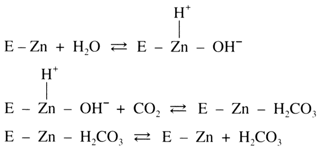
acid (H2CO3) dissociates into bicarbonate (HCO3-) and hydrogen ion (H+ ). In the eye, HCO3- then passes into the posterior chamber along with 
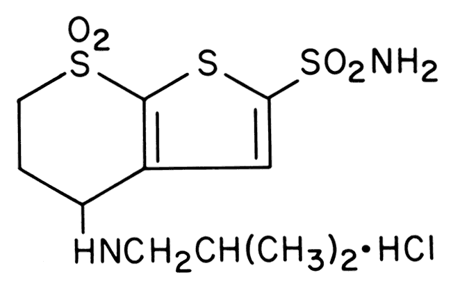
sodium ions (Na+ ). This movement of HCO3- and Na+ draws copious amounts of fluid along with it, thus producing aqueous humor. Although this reaction proceeds in a reversible fashion in the absence of a catalyst, the presence of the enzyme CA provides an enhancement of the reaction rate by more than 100-fold.7

Seven isoenzymes of CA have been isolated in mammals.8,9 Three isoenzymes (CA I, II, and IV) have been isolated in the eye.9,10 CA I and CA II are found within the cellular cytoplasm. CA I has been isolated in corneal endothelial cells, lenticular cells, and capillary endothelial cells in the stroma of ciliary processes and the choroid. CA II is the most widely distributed CA isoenzyme in the eye.9 It is especially abundant in pigmented and nonpigmented epithelial cells of the ciliary processes,9,10 in Müller's cells of the retina, and in a subset of cone photoreceptor cells.9 The distribution of CA IV in the human eye has been determined recently.9 In contrast to CA I and CA II, CA IV is a membrane-associated protein that has not been isolated within the pigmented or nonpigmented epithelial layers of the ciliary body, but has been isolated within the choriocapillaris overlying the ciliary body. It has long been theorized that CA II is the predominant isoenzyme responsible for controlling aqueous humor production. The recent isolation of CA IV within the eye, however, has raised questions as to the certainty of this theory. At the present time, it is unclear whether CA II or CA IV is the predominant regulator of bicarbonate metabolism.11

Aside from ocular tissues, CA has been found in red blood cells,12 the renal tubules,13 gastric mucosa, salivary glands, the pancreas, and the cells responsible for the production of cerebrospinal fluid.14

Systemically, CA serves two important physiologic roles. In red blood cells within tissue capillaries, CO2 is hydrated to form H2CO3 (CO2 + H2O H2CO3). This reaction, when catalyzed by CA, proceeds to the right 13,000 times as fast as the uncatalyzed reaction. As the erythrocyte proceeds from tissue capillaries to pulmonary capillaries, H2CO3 is converted to H2O and CO2 (H2CO3 CO2 + H2O). In pulmonary capillaries, CO2 then diffuses across the red cell membrane into the plasma and then across the alveolocapillary membranes into alveolar gas. Thus, CA plays a critical role in the removal of the body's main metabolic byproduct, CO2.

In the proximal and distal renal tubules of the kidney, the enzyme CA facilitates the production of H+ , which acidifies the urine. In the kidney, the catalytic hydration of CO2 generates H2CO3, which dissociates into H+ and HCO3-. The H+ is excreted into the urine, and the HCO3- diffuses back into the plasma. By inhibiting CA, H+ excretion is greatly decreased, thus producing a mild metabolic acidosis. A second result of the decrease in H+ excretion in the urine is the loss of Na+ in the distal tubule in lieu of H+ . In patients who do not have renal disease, total body levels of Na+ and potassium (K+ ) are initially decreased by up to 10%.15,16 At this point, however, a new equilibrium is reached, as the acidosis forces the reaction to the left (CO2 + H2O ←→ H2CO3), ensuring no further loss of these electrolytes.16,17

Thus, CA inhibition can produce a mixed respiratory and metabolic acidosis.6 This acidosis occurs as a consequence of (1) accumulation of tissue CO2 and reduced concentration of CO2 in expired air; and (2) decreased H+ excretion by the kidneys.18 This acidosis is compensated for by hyperventilation in patients who do not have compromising pulmonary disease, which ensures a sufficient release of CO2.19

Back to Top
MECHANISM OF INTRAOCULAR PRESSURE REDUCTION
The reduction in IOP caused by CAIs is primarily due to local enzyme inhibition,20,21 and in part may be due to systemic acidosis.22–25 Studies on the effects of acetazolamide on nephrectomized animals have suggested that IOP could be reduced in the absence of systemic acidosis.22,26,27 In some clinical evaluations, the degree of IOP reduction after acetazolamide or methazolamide administration in the absence of acidosis is less than that seen in conditions of acidosis.28,29 In other studies, the degree of acidosis did not seem to affect the IOP response.20,30

CAIs reduce IOP by inhibiting aqueous humor formation. This has been demonstrated with the use of indirect measurement techniques22,31 as well as direct fluorophotometric measurement techniques of aqueous humor flow rates.32–34 Fluorophotometric studies have documented reductions in aqueous humor flow rates of 21% to 40% in normal human volunteers and in a few glaucoma patients after acute oral dosing with 250 to 750 mg acetazolamide.32–34 McCannel and associates34 found that acetazolamide reduced aqueous flow rates by 24% below the nocturnal flow rate in the eye of a sleeping patient; timolol produced no such reaction. Aqueous flow measurements performed during the day suggested that acetazolamide and timolol 0.5% are additive after acute dosing.33,34 Outflow facility is unaltered by CAIs.1,31

Back to Top
CLINICAL USES OF CARBONIC ANHYDRASE INHIBITORS
Numerous clinical evaluations of systemically administered CAIs have been performed. Very few have been conducted in masked, randomized fashion, and the number of subjects participating in these studies has been small. The limitations of the study designs allow us to make some generalizations about CAIs, but the conclusions of many of these trials should be evaluated critically. Much of the information we cite about systemically administered CAIs is anecdotal and based on small numbers of patients who are taking additional ocular hypotensive agents.

Three oral CAIs are commercially available: acetazolamide (Diamox), methazolamide, (Neptazane), and dichlorphenamide (Daranide). The longest clinical experience has been with acetazolamide, as this drug was the first to be synthesized and has been used clinically since 1954.1

Two different single-dose dose-response studies35,36 using oral administration of 63, 125, 250, or 500 mg acetazolamide suggested mild enhancement of the IOP effect with increasing dosages. The onset of IOP reduction began 1 hour after dosing, and maximum IOP reductions occurred 2 to 5 hours after dosing and ranged from 30% to 41%. The duration of the IOP effect of single doses of orally administered acetazolamide is 6 to 8 hours. The dose-response characteristics and duration of action of acetazolamide support the common clinical regimen of four times daily dosing with acetazolamide tablets. Patients can be started on a low daily dosage of acetazolamide, such as 62.5 mg or 125 mg four times daily and advanced to 250 mg four times daily as necessary and tolerated. Dosages are generally limited to a total of 1000 mg daily because a higher dosage is associated with more frequent and severe side effects. The dosage in infants and children is 5 to 10 mg/kg administered every 4 to 6 hours.37 The tablets can be crushed and added to foods (e.g., apple sauce) or juices.

An alternative formulation of acetazolamide tablets is acetazolamide 500 mg sustained-release capsules. The onset of IOP reduction is 1 to 2 hours after single-dose capsule administration.36,38 Maximum IOP reductions occur 6 to 18 hours after dosing with durations of up to 23 hours.36,38,39 Twice-daily dosing with acetazolamide sustained-release capsules caused greater reductions in the magnitude of IOP reduction than once-daily dosing.36,39 Twice-daily dosing with 500 mg sustained-release capsules caused IOP reductions of a magnitude similar to that caused by 250 mg acetazolamide tablets administered four times daily.39 The advantages of prescribing sustained-release capsules include the following: (1) patient compliance is enhanced with twice-daily dosing compared with four times daily dosing; and (2) some patients are purported to be able to tolerate the capsules better than the tablets, experiencing fewer side effects.38,40 The cost of the sustained-release capsules is a factor that may need to be considered: the physician may hesitate to prescribe them or the patient may not be willing to purchase them.

Acetazolamide can be administered intravenously (IV) in doses of 250 to 500 mg if acute, rapid IOP reduction is imperative, as the onset of IOP reduction begins within 2 minutes of IV administration.41,42 IV dosing can be repeated in 2 to 4 hours, or an oral CAI can be administered.

Methazolamide was initially evaluated in patients in43 The chemical structure is similar to acetazolamide, but methazolamide has a plasma half-life three times that of the non-sustained-release form of acetazolamide: 14 hours versus 5 hours.44 Fifty-five percent of methazolamide is bound to plasma proteins compared with 93% of acetazolamide, allowing for enhanced diffusion of methazolamide into the tissues. Thus, lower and less frequent doses of methazolamide can be used as opposed to acetazolamide; however, clinical evaluations of methazolamide using doses of up to 450 mg daily have suggested that the drug was not as effective as daily doses of 500 to 2000 mg acetazolamide.29,30,43 The difference in efficacy of these two drugs has been explained in part by the findings of absence of systemic acidosis encountered with low doses of methazolamide44 and milder systemic acidosis with higher doses of methazolamide.43 Methazolamide doses of up to 50 mg three times daily caused less severe metabolic acidosis than acetazolamide in doses as low as 62.5 mg four times daily.44 Systemic acidosis may be one of the explanations for the greater incidence of side effects reported with acetazolamide than with methazolamide.29,30,43 Unlike acetazolamide, methazolamide is neither secreted nor concentrated by the kidney.

Dichlorphenamide is available in 50-mg tablets. It has a side-effect profile similar to acetazolamide because of its potential to induce metabolic acidosis45 and reduce urinary citrate excretion. It is usually prescribed in doses of 50 to 100 mg every 6 to 8 hours.

Anecdotal information suggests that a small number of patients intolerant to one CAI may be able to tolerate one of the other available CAIs.38,43

ORAL CARBONIC ANHYDRASE INHIBITORS AND SYSTEMIC SIDE EFFECTS

Despite the well-documented IOP-lowering effects of oral CAIs, their use is limited by side effects. As mentioned, the side effects of orally administered CAIs can range from mild and annoying to debilitating and life-threatening.

Calcific renal calculi have been documented in patients treated with CAIs. In a retrospective case control study, Kass and colleagues46 demonstrated that the rate of developing one or more kidney stones per year during acetazolamide therapy was 11 times higher in the study group than in the control (untreated) group. Therapeutic doses of orally administered acetazolamide 250 mg four times daily,30,47,48 methazolamide 100 mg three times daily,44 or dichlorphenamide 50 mg four times daily or 100 mg three times daily48 decreased urinary citrate excretion by up to 86% compared with pretreatment values. Significantly less reduction of urinary citrate excretion, however, was induced with oral methazolamide 25 mg three times daily.30,44 The overall incidence of developing renal stones while on methazolamide seems to be lower than with acetazolamide, which may be due to the lower doses of methazolamide that are most commonly used.43,49,50 CAIs induce urolithiasis by reducing urinary excretion of citrate. Citrate chelates urinary calcium salts into a soluble complex and prevents stone formation. In the absence of citrate, calcium is not adequately solubilized and serves as a nidus for stone formation.

A relative hypokalemia and hyponatremia can occur with chronic CAI use.15,19,51,52 These effects are usually self-limited, as is the diuretic effect of CAIs.15,53 The hypokalemia is usually not of clinical significance unless the patient is concurrently using a potassium-depleting medication, such as a hydrochlorothiazide diuretic. Potassium supplementation is generally not indicated in the majority of patients taking CAIs alone.51 Additionally, potassium supplementation does not alleviate the CAI-associated side effects in patients who are not taking concurrent potassium-depleting drugs.54 If the patient is on a potassium-depleting drug, serum potassium levels should be followed and supplementation prescribed as indicated.

A mild mixed respiratory and metabolic acidosis can occur with chronic use of oral CAIs. The acidosis may contribute to the malaise and fatigue often encountered with chronic CAI use.54 Systemic acidosis, as a consequence of CO2 retention, may contribute to acute respiratory failure in patients with severe chronic obstructive pulmonary disease. These patients are unable to increase their minute respiratory volume in order to compensate for the acidosis.45,55–57 Acute systemic acidosis can precipitate sickling in patients with sickle cell trait or disease, and therefore CAIs should be avoided in these patients.58

The likelihood of salicylate toxicity in patients taking high doses of aspirin may be increased with concurrent CAI use because of CAI-related systemic acidosis.59 Salicylate toxicity is related to tissue rather than blood salicylate levels.60 Nonionized salicylic acid passes from the bloodstream into the central nervous system and other cells more rapidly than does ionized salicylate. Systemic acidosis increases the proportion of nonionized drug61 and thus increases the quantity of salicylate entering the central nervous system and other tissues, leading to toxicity.

Liver failure, characterized by confusion, asterixis, and elevated blood ammonia, may be seen in patients with chronic liver diseases. The mechanism by which CAIs cause liver failure remains unclear.

Teratogenicity has been demonstrated in laboratory animals as well as humans.62–65 As a consequence, female patients considering pregnancy should be counseled about the teratogenic effects and switched to another ocular hypotensive regimen before pregnancy.

Blood dyscrasias, including aplastic anemia, agranulocytosis, thrombocytopenia, and neutropenia, have been documented in patients taking CAIs.66–69 Blood dyscrasias may be idiosyncratic, occurring after as little as a single dose of medication, or they may be dose related. The National Registry of Drug-Induced Ocular Side Effects received 79 case reports of suspected hematopoietic toxicity caused by CAIs occurring between 1972 and70 These adverse hematologic reactions occurred in 68% of patients within 6 months of initiating therapy. Thirty-two percent of these cases resulted in death secondary to aplastic anemia. These severe adverse experiences have engendered considerable debate in the literature as to the advisability of routine monitoring of the complete blood counts of patients on CAI therapy. It has been suggested that these patients be monitored every 6 months.69–71 Unfortunately, the majority of the hematologic complications reported occurred before 6 months of therapy. Thus, monitoring every 6 months would not have been soon enough to detect the blood dyscrasias. Of greatest benefit to the individual patient is probably routine careful questioning about anemia, infection, and poor clotting, with follow-up as indicated.72

The following side effects are less serious than those described above, but they tend to be much more frequent and can be particularly annoying to the patient:

  A symptom complex of malaise, fatigue, weight loss, depression, anorexia, and often loss of libido has been reported in up to 48% of patients. The patients in one study54 who complained of this symptom complex were more acidotic than those without such complaints.
  Paresthesias in the proximal and distal extremities have been reported in up to 75% of patients.73 This side effect usually does not necessitate the discontinuation of the drug and tends to diminish with chronic use.
  Maculopapular and urticarial skin eruptions have been reported in patients using methazolamide.74
  Patients may complain of strange tastes occurring during consumption of carbonated beverages. Similar complaints have been elicited when CA has been topically dispensed on the tongue.

TOPICAL CARBONIC ANHYDRASE INHIBITORS

Since the 1950s, numerous investigators75–79 have attempted to formulate a topically active CAI that would have fewer side effects than the currently prescribed systemic formulations. The systemically administered CAIs that are effective ocular hypotensive agents (e.g., acetazolamide, methazolamide, ethoxzolamide, dichlorphenamide) were relatively ineffective when administered topically. Various modifications of these systemically active agents that were evaluated for topical use include trifluoromethazolamide, a derivative of methazolamide that reduced IOP in rabbits75,76 but was unstable in solution and required a long duration of ocular exposure; aminozolamide gel, an analogue of ethoxzolamide that significantly reduced IOP in patients with ocular hypertension77 but had substantial ocular side effects when administered chronically78; and L-650,719, another sulfonamide derivative that reduced IOP in rabbits79 but failed to reduce IOP in a clinical trial of normotensive volunteers.80

In the 1980s, a series of thienothiopyran-2sulfonamide compounds were developed that are effective in reducing IOP when administered topically. These compounds are sulfonamide derivatives,81–83 as is acetazolamide, and they differ from other CAIs previously investigated for topical use in that they are water soluble, are stable in solution, have excellent corneal penetration, and inhibit CA at low concentrations, which can be achieved with topical application.

Three thienothiopyran-2-sulfonamide derivatives have been identified that reduce IOP after topical administration in laboratory and clinical trials: MK-927, which is a racemic mixture (Fig. 1); sezolamide (MK-417), which is the S-enantiomer of MK-927 (Fig. 2); and dorzolamide, a compound structurally similar to MK-927 and sezolamide, which has previously been identified as MK-507 and L-671,152 (Fig. 3).

Fig. 1. Generally accepted catalytic mechanism of carbonic anhydrase.

Fig. 2. The chemical structure of MK-927 and sezolamide. MK-927 is the racemic mixture of the above compound. Sezolamide is the S-enantiomer.

Fig. 3. The chemical structure of dorzolamide.

MK-927

RABBIT STUDIES. MK-927 was the first of these three thienothiopyran-2-sulfonamide derivatives to be identified and extensively evaluated. Studies in normal rabbits demonstrated that the topical application of MK-927 inhibited CA in homogenized iris ciliary body tissues82 and reduced IOP for at least 6 hours.83 IOP was reduced for a longer duration in pigmented rabbits than in albino rabbits,84 possibly because pigment binding of the drug may have caused a depot effect. A dose-response study in albino rabbits demonstrated that 2% MK-927 was at the top of the dose-response curve, producing the longest duration and the largest magnitude of effect on IOP.84 In unilateral alpha-chymotrysinized glaucoma-induced rabbits, MK-927 reduced IOP unilaterally when applied to the normal eyes, but did not affect hypertensive eyes. These data provided support for the local rather than systemic effect of MK-927.

PRIMATE STUDIES. Single-dose and multiple-dose testing of MK-927 was performed in cynomologus monkeys in which ocular hypertension had been created by multiple sessions of high-power argon laser trabeculoplasty. A single-dose dose-response study demonstrated that 2% MK-927 was at the top of the dose-response curve.84 IOP was significantly reduced 1 to 6 hours after drug application, with a mean peak reduction of 9.6 mmHg. Twice-daily application of 2% MK-927 for 5 days to eight glaucomatous monkeys reduced IOP 1 to 8 hours after each dose.85 Maximum reductions in IOP occurred 3 hours after dosing and increased from a mean of 10.1 mmHg on day 1 to 13.2 mmHg on day 5, suggesting enhancement of effect with repetitive dosing.

CLINICAL STUDIES. The first clinical trials (Table 1) in normal volunteers evaluated three doses of 2% MK-927 administered to 10 volunteers and three doses of placebo administered to 2 volunteers.86 IOP was reduced by 30% in the MK-927 treated eyes and by 7% in the fellow eyes, and it was unchanged in the placebo-treated eyes. A similar study in patients with glaucoma or ocular hypertension demonstrated mean IOP reductions of 27% in the 2% MK-927-treated eyes and 11% in the placebo-treated eyes 6 hours after drug instillation.87 A single application of 2% MK-927 to patients with elevated IOP reduced the pressure by 33% in the drug-treated eyes and by 17% in the placebo-treated eyes at 4½ hours after dosing.88


TABLE ONE. Studies of MK-927 and Sezolamide (MK-417) that Demonstrate the Intraocular Pressure Reducing Effect in Patients with Elevated Intraocular Pressure


AuthorsYearDays of StudyStudy Design
Pfeiffer et al8819901MK-927 2%, single dose
Lippa et al8619881MK-927 2%, 3 doses
Bron et al8719891MK-927 2%, 3 doses
Serle et al9019901MK-927 2%, IOP reproducibility
Diestelhorst et al941989 (A)1MK-927 vs MK-417 (1%)
Higginbotham et al921989 (A)14MK-927 1% vs 2% bid vs MK-927 2% qid
Tuulonen et al911989 (A)14MK-927 0.5% vs 1% vs 2% bid
Higginbotham et al8919901MK-927 0.5% vs 1% vs 2%
Serle et al93199242MK-927 2% bid vs timolol 0.5% bid
Bron et al9519911MK-927 2% vs MK-417 1.8%
Lippa et al961990 (A)4MK-417 1.8% tid vs timolol bid
A = abstract   


A single-dose dose-response study89 carried out in patients with ocular hypertension suggested that 2% MK-927 was more effective and had a longer duration of effect than 0.5% or 1% concentrations. In another study,90 eight patients determined to be marked responders (an IOP reduction of the treated eye of at least 6 mmHg corrected for diurnal variation and compared with the contralateral control eye) to 2% MK-927 participated in a four-period crossover study. The effects of 0.125%, 0.5%, and 2% MK-927 in treated eyes were compared with effects in their fellow placebo-treated eyes. The 2% MK-927 substantially reduced IOP 2 to 6 hours after dosing. The 0.125% and 0.5% concentrations reduced IOP only at 4 hours after drug application compared with the fellow placebo-treated eye, which was unaffected. This same study evaluated reproducibility to 2% MK-927. The results indicated considerable variability of response to single-dose drug testing, suggesting that single-dose testing is not sufficient to adequately determine the effectiveness of this therapeutic agent.

A 14-day multiple-dose dose-response study carried out in 76 patients with elevated IOP demonstrated reductions 0 to 12 hours after dosing with the 1% and 2% concentrations.91 A 14-day comparison study of once-daily versus twice-daily application of 2% MK-927 versus once-daily application of 1% MK-927 in 48 patients suggested 1% or 2% MK-927 had minimal effects on IOP 24 hours after once-daily administration.92 Twice-daily administration of 2% MK-927 caused a slightly greater mean reduction of IOP (-2.8 mmHg) 12 hours after dosing. A 6-week safety study evaluating the twice-daily application of 2% MK-927 was carried out in 27 ocular hypertensive patients and 9 additional patients receiving timolol 0.5% as the control.93 Pre-drug morning IOP reductions were up to 15% in the MK-927 treated eyes and 24% in eyes treated with timolol. Outflow facility measured tonographically was unchanged in both groups. Adverse effects were not encountered; side effects were minor and included transient burning or stinging after instillation and a bitter taste.

Sezolamide (MK-417)

Sezolamide, the S-enantiomer of the racemic mixture MK-927, was found to be a more potent inhibitor of the enzyme CA in the ex vivo rabbit iris ciliary body model than the R-enantiomer, which suggested that sezolamide might be more effective clinically than MK-927.82 Single-dose94 and multiple-dose95 clinical comparative studies supported a slightly greater effect of sezolamide compared with MK-927.

A comparison of 1.8% sezolamide administered three times daily with 0.5% timolol administered twice daily in 63 glaucoma patients demonstrated similar IOP reductions except just before the morning dosing, when timolol was more effective.96 A 15-day study of sezolamide added to timolol, compared with placebo added to timolol, demonstrated mild additivity with a duration of effect of less than 12 hours (Greve E, unpublished observations, 1990).

METABOLIC STUDIES OF MK-927 AND SEZOLAMIDE. The possibility of systemic effects of topically applied CAIs was explored in two different studies. After topical administration of three doses of 2% MK-927 to seven healthy subjects, the levels of the racemic mixture of MK-927, sezolamide the S-enantiomer, and the R-enantiomer in plasma, whole blood, and urine were monitored for 14 days.97 The S-enantiomer sezolamide was present in whole blood for a prolonged period of time (300 hours) compared with the R-enantiomer (5.4 hours). Despite the prolonged detection of S-enantiomer in whole blood, the extrapolated steady-state red blood cell level was 7 μM compared with 20-μM concentrations of CA II. The extrapolated blood levels were believed to be probably too low to induce the metabolic effects seen with orally administered CAIs.

The metabolic effects of 2% MK-927 or 1.8% sezolamide were examined in 16 healthy subjects treated four times daily for 14 days.98 Significant changes in blood and urine parameters (e.g., electrolytes, urate, citrate, creatinine, osmolality, acid-base profiles) were not observed, substantiating the suggestion that topical application induces systemic drug levels that are of sufficiently small magnitude to avoid systemic effects.

Dorzolamide (MK-507, L-671,152)

PRECLINICAL STUDIES. Refinement of the chemical structure of the more active S-enantiomer sezolamide led to the development of dorzolamide.99 In ocular hypertensive rabbits, dorzolamide caused IOP reductions of greater magnitude than that produced by MK-927.100 Single- and multiple-dose studies in ocular hypertensive monkeys suggested that dorzolamide was more potent101 and had a longer duration of effect than MK-927. Single-dose administration of 2% dorzolamide or 0.5% timolol in the glaucomatous monkey demonstrated comparable IOP reductions, but timolol had a longer duration of action.102

MECHANISM OF ACTION. An evaluation of the mechanism by which dorzolamide reduces IOP was carried out in eight normal monkeys (Table 2).103 After a single unilateral administration of 2% dorzolamide, aqueous flow rates measured by fluorophotometry were reduced by 38% for 5 hours in the treated eyes when compared with the control eyes. Outflow facility was not altered at 3 hours after drug application. IOP was reduced from 1 to 7 hours after administration in the treated eyes only. This study confirmed the anticipated effect of dorzolamide on aqueous humor formation.


TABLE TWO. Twice Daily (8:30 am and 4:30 pm) Application of 2% Dorzolamide for Five Days to Eight Glaucomatous Monkey Eyes


 Intraocular Pressure
 (Mean mmHg ± SEM)
 0 h2 h8 h
Vehicle treated eyes31.0 ± 2.330.1 ± 2.829.3 ± 2.2
 Drug Treated
Day 131.9 ± 2.523.8 ± 0.9*23.6 ± 1.0*
Day 323.1 ± 0.5*20.4 ± 0.7*20.4 ± 1.2*
Day 522.1 ± 0.5*19.8 ± 0.5*20.0 ± 1.3*

*Drug-treated eye significantly different compared with vehicle-treated eye by paired t-test, p < 0.05.
(Adapted from Wang RF, Serle JB, Podos SM et al: The ocular hypotensive effect of the topical carbonic anhydrase inhibitor L-671,152 in glaucomatous monkeys. Arch Ophthalmol 108:511, 1990)


INITIAL CLINICAL STUDIES. The initial clinical evaluation of dorzolamide performed in 24 healthy volunteers demonstrated excellent local tolerability and IOP reductions of up to 29% 4 hours after the first dose.104

The first study of dorzolamide performed in patients with elevated IOP demonstrated a substantial ocular hypotensive effect.105 Eighteen patients treated unilaterally with one drop of 2% dorzolamide every 12 hours for a total of three doses had IOP reductions of 21.1% 2 hours after dosing on day 2. These and other early studies demonstrated the clinical efficacy of dorzolamide in reducing IOP (Table 3).


TABLE THREE. Preliminary Clinical Evaluations of Dorzolamide


AuthorsYearSubject GroupsResults
Hoffman et al1041990 (A)NormalsReduce IOP, 3 drops
Lippa et al1091990 (A)POAG, OHTReduce IOP, bid, 2 days
Bourgeois et al1081990 (A)POAG, OHTReduce IOP, 6 days

A = year of publication of abstract; IOP = intraocular pressure; OHT = ocular hypotensive; POAG = primary open-angle glaucoma.


CLINICAL COMPARISON BETWEEN DORZOLAMIDE AND SEZOLAMIDE. A 4-day clinical comparison trial of two or three times daily administration of 2% dorzolamide and three times daily administration of 1.8% sezolamide in patients with elevated IOP (Table 4) demonstrated peak mean IOP reductions of 26.2% with dorzolamide and 22.5% with sezolamide.106 This was the pivotal study that suggested that dorzolamide administered three times a day was more effective than sezolamide and could be effective monotherapy for glaucoma in some patients.


TABLE FOUR. A Four-Day Comparison Trial of 2% Dorzolamide (MK-507) and 1.8% Sezolamide (MK-417) in 82 Glaucoma Patients: Intraocular Pressure (Mean [mmHg] ± SD)


   Prestudy Diurnal 
 MK-507 q 8 hMK-507 q 12 hMK-417 q 8 hPlacebo
Hours Post-Dose    
028.4 ± 4.128.2 ± 3.828.8 ± 4.628.2 ± 3.7
227.6 ± 4.626.6 ± 3.528.1 ± 5.427.1 ± 3.8
427.3 ± 4.326.8 ± 3.726.7 ± 4.826.8 ± 4.4
1224.9 ± 4.624.4 ± 3.723.7 ± 4.025.1 ± 4.6
   Day 4 of Treatment 
Hours Post-Dose    
023.3 ± 2.922.8 ± 3.224.3 ± 4.326.9 ± 4.6
220.2 ± 2.720.1 ± 3.921.6 ± 4.724.8 ± 4.6
420.9 ± 3.620.8 ± 3.522.0 ± 4.725.5 ± 4.2
1220.1 ± 3.120.8 ± 3.020.6 ± 3.923.8 ± 3.5

Change in intraocular pressure (IOP) was significantly (p < .05) different from prestudy for all three treatment groups at all times post-dose on day 4. Dosing follows the 0-hour IOP measurement. No statistically significant differences were observed between the dorzolamide and sezolamide treatment groups at any time point.
(Adapted from Lippa EA, Schuman JS, Higginbotham EJ et al: MK-507 versus sezolamide: comparative efficacy of two topically active carbonic anhydrase inhibitors. Ophthalmology 98:308, 1991)


CARBONIC ANHYDRASE ENZYME INHIBITION. The relative potency of dorzolamide, MK-927, and sezolamide in inhibiting the CA II in human red blood cells was measured in vitro.107 The relative 50% inhibitory concentrations were 52 nM for MK-927, 7.7nM for sezolamide, and 2.2nM for dorzolamide. The data supported the clinical finding that dorzolamide is the most potent of these three ocular hypotensive agents.

DOSE-RESPONSE STUDIES. The safety and efficacy of dorzolamide have been evaluated in clinical trials up to 1 year after administration106,108–113 (Table 5). In these various studies, in which dorzolamide was administered as single-agent therapy twice daily or three times daily, IOP reductions ranged from 7% to 21% at trough just prior to morning drug instillation, and from 17% to 27% at peak 2 hours after instillation, compared with baseline IOP. Two formal multiple-dose dose-response studies suggested that 0.7%, 1.4%, or 2% concentrations of dorzolamide caused similar reductions in IOP (14% to 24%).110,111 The 2% concentration of dorzolamide administered two or three times daily was slightly more effective in reducing IOP than the 0.7% or 1.4% concentrations before morning dosing and 12 hours after the initial daily dose. A lower concentration of dorzolamide, 0.2%, administered three times daily was minimally effective in reducing IOP.


TABLE FIVE. Evaluations of Dorzolamide in Patients with Glaucoma or Ocular Hypertension


     IOP Reduction
AuthorsYearStudy DurationNo. Pts.*Dosing RegimenTroughPeak12 h
Bourgeois et al10819906 days492%, bid7.317.512.0
Lippa et al10619914 days822%, bid19.124.414.8
    2%, tid1826.819.3
Lippa et al11019925 days732%, bid1821.812.5
    1.4%, bid13.421.79.5
    0.7%, bid12.921.410.2
    2%, tid21.122.219.2
  7 days 1.4%, tid16.822.114.2
    0.7%, tid17.624.218.9
Kitazawa1091993 (A)52 weeks981%, tid15.621.2-
Lippa et al1111993 (A)6 weeks3320.2%, tid9.212.5-
    0.7%, tid14.117.5-
    2%, tid14.117.2 
Wilkerson et al113199328 days482%, tid13.318.4-
Strahlman et al1121994 (A)1 year5232%, tid16.9||22.9-

*Total number of patients enrolled in the study, not the total number of patients on the regimen indicated, as some patients may have been treated with placebo, other drugs, or other concentrations.
Trough IOP is measured just before am dosing.
Peak IOP is measured 2 hours after the am dosing.
§IOP is measured 12 hours after the am dosing.
||Trough IOP is measured 8 hours after am dosing.
A = year of publication of abstract if otherwise unpublished; IOP = intraocular pressure.


These two multiple-dose dose-response studies suggested that concentrations ranging from 0.7% to 2% may be similarly efficacious in some patients. Whereas treatment twice daily may be sufficient for some patients, therapy three times daily may result in better control of IOP, particularly 10 to 12 hours after the initial daily dosing and just before the morning dose.

SAFETY AND EFFICACY STUDY. Forty-eight patients with IOP greater than 22 mmHg113 participated in a 4-week safety and efficacy study of 2% dorzolamide administered three times daily. The efficacy of dorzolamide was similar to what has been described previously, with IOP reductions of 13.3% at morning trough and 18.4% at peak effect (day 29). CA I and CA II, which are inhibited by sulfonamides, are located in human corneal endothelium.10 This has raised concern that topically applied CAIs may alter corneal endothelial cell function; thus, theoretically dorzolamide could produce adverse corneal side effects. In this 4-week clinical trial, corneal thickness was measured by ultrasound pachymetry, and endothelial cell counts were measured with a specular microscope. Mean corneal thickness was slightly increased in the dorzolamide-treated group compared with the placebo-treated group (0.009 and 0.001 mm, respectively; p < 0.05). This change was not clinically significant. Changes in endothelial cell counts were minimal and similar in the two groups.

CA II is found in human erythrocytes, where it regulates CO2 transport.10 Four weeks after beginning dorzolamide treatment, the activity of CA II in red blood cells was decreased to 21% of baseline, which is below the 99% inhibitory concentration thought necessary to induce physiologic effects. This study reported blood chemistries, complete blood counts, urinalysis, and electrocardiograms to be similar between the dorzolamide- and placebo-treated groups. The results of this investigation suggested that 2% dorzolamide was efficacious, well-tolerated, and could be administered safely three times daily for at least 4 weeks.

ONE-YEAR CLINICAL EVALUATIONS. Two clinical trials have evaluated dosing with dorzolamide three times daily for 1 year. Ninety-eight patients with open-angle glaucoma were enrolled into an open-label evaluation of 1% dorzolamide; 77 patients remained on therapy for 1 year.109 After 52 weeks of therapy, the drug remained effective, reducing mean IOP by 15.6% before the morning dosing and by 21.2% at 2 hours after dosing. Substantial ocular and systemic side effects were not encountered.

A 1-year randomized, double-blinded comparison of 2% dorzolamide, 0.5% timolol, and 0.5% betaxolol (Table 6) included a study group of 523 patients with either open-angle glaucoma or ocular hypertension.112 Dorzolamide was administered three times daily; timolol and betaxolol were administered twice daily. IOP reductions at 1 year were similar between the dorzolamide- and betaxolol-treated patients compared with baseline measurements. Mean percent change in IOP from baseline in the dorzolamide-treated patients was 22.9% at 2 hours after dosing, 18.1% at 5 hours after dosing, and 16.9% at 8 hours after dosing. The betaxolol-treated patients had reductions of 20.8%, 19%, and 15.1% at the same time points. The patients treated with timolol had greater mean percent changes in IOP from baseline than the other two treatment groups: 25.3%, 22.2%, 20.4% at 2, 5, and 8 hours after dosing, respectively. This study suggests that dorzolamide and betaxolol have similar effects on IOP as single-agent therapies, and that both are less efficacious than timolol.


TABLE SIX. One-Year Comparison Study of Dorzolamide tid, Timolol bid, and Betaxolol bid in 523 Glaucoma Patients


Mean Reduction in Intraocular Pressure From Baseline at One Year (%)
 Hour 2Hour 5Hour 8
2% Dorzolamide22.918.116.9
0.5% Timolol25.322.220.4
0.5% Betaxolol20.819.015.1
(Adapted from Strahlman E, Tipping R, Vogel R, et al: A double-masked; randomized 1-year study comparing dorzolamide (Trusopt), timolol, and betaxolol. Arch Ophthalmol 113:1009, 1995)


ATTEMPTS AT ENHANCING THE INTRAOCULAR PRESSURE EFFECT OF DORZOLAMIDE. Modifications of the drug delivery system of dorzolamide and concentrations greater than 2% have been evaluated in an attempt to prolong the duration and enhance the effect of dorzolamide on IOP. In a 4-day trial, the 3% concentration of dorzolamide produced greater local irritation and tearing and IOP reductions of smaller magnitude than noted in previous studies of 2% dorzolamide.114 In another study, twice-daily application of 2% dorzolamide in Gelrite (Kelco, San Diego, CA), a drug delivery system designed to prolong ocular contact time, did not substantially enhance or extend the duration of IOP.115

ADDITIVITY OF DORZOLAMIDE TO TIMOLOL. Both timolol and dorzolamide reduce IOP by suppressing aqueous humor secretion. The additivity of these two aqueous humor suppressants was evaluated in patients with elevated IOP.116 Dorzolamide 2% administered twice daily reduced IOP an additional 13% to 21% in 16 patients already receiving 0.5% timolol twice daily. When placebo was added to a group of timolol-treated patients, no additional IOP reductions occurred. This suggested that dorzolamide may be additive to other drugs that reduce aqueous humor production.

COMPASSIONATE CASE USAGE OF DORZOLAMIDE. The compassionate case use trial was designed to allow patients, for whom dorzolamide may be potentially beneficial, access to the drug before commercial distribution and to assess the efficacy and safety of dorzolamide in patients on maximum tolerated medical therapy, in whom it was preferable to avoid additional surgery or laser therapy. Between December 1992 and April 1994, 25 patients were enrolled at the Mount Sinai Medical Center in New York in this still ongoing study.117 Criteria for enrollment included poorly controlled glaucoma or ocular hypertension on maximum tolerated medical therapy in patients 21 years of age or older. At the time of enrollment, 16 patients were not receiving orally administered CAIs; 9 other patients were taking oral CAIs that were poorly tolerated, in addition to other ocular hypotensive medications.

In the 16 patients who were not receiving an orally administered CAI at the time of enrollment, the IOP after the addition of 2% dorzolamide was reduced compared with study day 1 measurements from week 1 through month 12. Mean IOP on study day 1, before the addition of 2% dorzolamide was 19.6 ± 1.9 mmHg (mean ± SEM). Reductions of IOP were 5.5 ± 1.8 mmHg at week 1, 4.1 ± 2.2 mmHg at month 2, 2.9 ± 2.0 mmHg at month 4, and 2.0 ± 1.1 mmHg at month 12. Only 4 of the 16 patients have completed 12 months of therapy at the time of this writing. One patient was discontinued from the study after 2 months because of insufficient IOP control and underwent successful filtration surgery.

Substitution of 2% dorzolamide for a poorly tolerated orally administered CAI in the nine other patients resulted in additional reductions of IOP from week 1 though month 4 compared with study day 1. IOP on study day 1 was 16.5 ± 1.3 mmHg. IOP reductions were 3.6 ± 0.7 mmHg at week 1 (9 patients), 3.3 ± 1.3 mmHg at week 2 (8 patients) and 2.4 ± 0.9 mmHg at month 4 (8 patients) (Table 7). Side effects were substantially less frequent and less severe than when these patients had been on orally administered CAIs.


TABLE SEVEN. Substitution of 2% Dorzolamide Administered bid or tid for an Oral Carbonic Anhydrase Inhibitor in Glaucoma Patients on Maximum-Tolerated Medical Therapy


 Mean IOP (mmHg) ± SEMIOP Reduction (%)No. Patients
Day 116.5* ± 1.3-9
Week 112.9 ± 0.7229
Week 213.3 ± 1.1208
Month 114.5 ± 1.1129
Month 215.6 ± 1.238
Month 413.8 ± 1.3158

* The study day 1 IOP was measured during treatment with systemically administered carbonic anhydrase inhibitors, before substituting dorzolamide.
IOP significantly different from study day 1, by paired t-test, p < 0.05. IOP = intraocular pressure.


The results of this trial suggested that dorzolamide is effective, well tolerated, and possibly additive to other classes of compounds used to treat glaucoma. Similar IOP results after substitution of dorzolamide for oral CAIs have been reported by Kitazawa and associates.118

SIDE EFFECTS. Mild stinging and burning of the eye and a bitter taste upon ocular drug instillation, both resolving within minutes, have been noted in some patients.106,113 Blood dyscrasias, tingling in the extremities, abdominal discomfort, and diarrhea have not been reported in patients taking dorzolamide, sezolamide, or MK-927. Numerous studies suggest that dorzolamide is well tolerated, and has few systemic side effects. Kitazawa and colleagues118 substituted 0.5% dorzolamide for orally administered CAIs in 31 patients with primary open-angle glaucoma on multiple ocular hypotensive medications. The patients remained on dorzolamide three times daily for 12 weeks. Subjective symptoms that were related to oral CAI administration had resolved within 4 weeks of discontinuation of the oral CAIs.

A radioactive tracer study determined that dorzolamide has a long half-life, approximately 147 days, in red blood cells.113 Despite this long half-life, systemic side effects have not been reported to date in patients receiving the topically active CAI, dorzolamide. Dorzolamide appears to be better tolerated than orally administered CAIs.

Back to Top
SUMMARY
MK-927, sezolamide, and dorzolamide represent an exciting breakthrough in ocular pharmacology. More than 30 years of research have led to the discovery of this series of thienothiopyran-2-sulfonamide derivatives that are effective CAIs when topically administered. Enzyme inhibition studies and clinical trials demonstrate that dorzolamide is the most efficacious of these three compounds.

Dorzolamide is effective in reducing IOP as a single-agent therapy. Thus, in some patients dorzolamide may be used two or three times daily as a first-line drug for the treatment of glaucoma. The utility of this drug as initial therapy is supported by the low incidence of side effects: the drug is well tolerated by patients. Three clinical trials have suggested that dorzolamide is additive to other ocular hypotensive agents. One of these studies demonstrated additivity to beta-adrenergic antagonists. In two of these clinical trials dorzolamide appeared to reduce IOP when added to multiple ocular medications being used to treat glaucoma, and when substituted for orally administered CAIs that are producing side effects. Thus, it may also be useful as a second-line drug to treat glaucoma.

Dorzolamide was approved by the United States Food and Drug Administration for commercial distribution in December 1994. It became available for clinical use in May of 1995. Long-term trials and the clinical use of dorzolamide (trade name Trusopt) will continue to elucidate the issues of ocular side effects, systemic side effects, additivity to other ocular hypotensive drugs, relative efficacy compared with oral CAIs, and possible additivity to various doses of orally administered CAIs.


Supported in part by grant EYO1867 from the National Institutes of Health, Bethesda, MD, and by an unrestricted grant from Research to Prevent Blindness, Inc., New York, NY.
Back to Top
REFERENCES

1. Becker B: Decrease in intraocular pressure in man by a carbonic anhydrase inhibitor, Diamox: a preliminary report. Am J Ophthalmol 37:13, 1954

2. Friedenwald JS: The formation of the intraocular fluid. Am J Ophthalmol 32:9, 1949

3. Kinsey VE: A unified concept of aqueous humor dynamics and the maintenance of intraocular pressure: an elaboration of the secretion-diffusion theory. Arch Ophthalmol 215, 1950

4. Wistrand PJ: Carbonic anhydrase in the anterior uvea of the rabbit. Acta Physiol Scand 24:144, 1951

5. Maren TH: The development of ideas concerning the role of carbonic anhydrase in the secretion of aqueous humor: Relation to the treatment of glaucoma. In Drance SM, Neufeld AH (eds): Glaucoma: Applied Pharmacology in Medical Treatment, pp 325–355. Orlando, FL, Grune & Stratton, 1984

6. Maren TH: Carbonic anhydrase: chemistry, physiology and inhibition. Physiol Rev 47:595, 1967

7. Maren TH: The kinetics of HCO3- synthesis related to fluid secretion, pH control, and CO2 elimination. Ann Rev Physiol 50:695, 1988

8. Kaunisto K, Parkkila S, Tammel T et al: Immunohistochemical localization of carbonic anhydrase isoenzymes in the human male reproductive tract. Histochemistry 94:381, 1990

9. Hageman GS, Zhu XL, Waheed A, Sly WS: Localization of carbonic anhydrase IV in a specific capillary bed of the human eye. Proc Natl Acad Sci USA 88:2716, 1991

10. Wistrand PJ, Schenholm M, Lönnerholm G: Carbonic anhydrase isoenzymes CA I and CA II in the human eye. Invest Ophthalmol Vis Sci 27:419, 1986

11. Maren TH: Current status of membrane-bound carbonic anhydrase. Ann NY Acad Sci 341:246, 1980

12. Wistrand PJ: The importance of carbonic anhydrase B and C for the unloading of CO2 by the human erythrocyte. Acta Physiol Scand 113:417, 1981

13. Maren TH, Wynns GC, Wistrand PJ: Chemical properties of carbonic anhydrase IV, the membrane-bound enzyme. Mol Pharmacol 44:901, 1993

14. Ashby W: Carbonic anhydrase in mammalian tissue. J Biol Chem 151:521, 1943

15. Brechue WF, Stager JM, Lukaski HC: Body water and electrolyte responses to acetazolamide in humans. J Appl Physiol 69:1397, 1990

16. Preisig PA, Toto RD, Alpern RJ: Carbonic anhydrase inhibitors. Renal Physiol 10:136, 1987

17. DuBose TD: Carbonic anhydrase-dependent bicarbonate transport in the kidney. Ann NY Acad Sci 429:528, 1984

18. Chapron DJ, Gomolin IH, Sweeney KR: Acetazolamide blood concentrations are excessive in the elderly: propensity for acidosis and relationship to renal function. J Clin Pharmacol 29:348, 1989

19. Nadell J, Kalinsky H: The effects of the carbonic anhydrase inhibitor “6063” on electrolytes and acid-base balance in two normal subjects and two patients with respiratory acidosis. J Clin Invest 32:622, 1953

20. Benedikt O, Zirm M, Harnoncourt K: Relations between metabolic acidosis and intraocular pressure after inhibition of carbonic anhydrase with acetazolamide. Graefes Arch Klin Ophthalmol 190:247, 1974

21. Zimmerman TJ, Garg LC, Vogh BP et al: The effect of acetazolamide on the movements of anions into the posterior chamber of the dog eye. J Pharmacol Exp Ther 196:510, 1976

22. Becker B: The mechanism of the fall in intraocular pressure induced by the carbonic anhydrase inhibitor, Diamox. Am J Ophthalmol 39:177, 1955

23. Krupin T, Oestrich CJ, Bass J et al: Acidosis, alkalosis, and aqueous humor dynamics in rabbits. Invest Ophthalmol Vis Sci 16:997, 1977

24. Langham ME, Lee PM: Action of Diamox and ammonium chloride on formation of aqueous humor. Br J Ophthalmol 41:65, 1957

25. Wistrand P, Maren TH: The effect of carbonic anhydrase inhibition on intraocular pressure of rabbits with different blood CO2 equilibria. Am J Ophthalmol 50:291, 1960

26. Friedman Z, Krupin T, Becker B: Ocular and systemic effects of acetazolamide in nephrectomized rabbits. Invest Ophthalmol Vis Sci 23:209, 1982

27. Maren TH, Wadsworth BC: Blocking of renal effect of Diamox 2-acetylamino-1,3,4-thiadiazole-5-sulfonamide by metabolic acidosis. Fed Proc 13:383, 1954

28. Bietti G, Virno M, Pecori-Giraldi J et al: Acetazolamide, metabolic acidosis, and intraocular pressure. Am J Ophthalmol 80:360, 1975

29. Stone RA, Zimmerman TJ, Shin DH et al: Low-dose methazolamide and intraocular pressure. Am J Ophthalmol 83:674, 1977

30. Dahlen K, Epstein DL, Grant M et al: A repeated dose-response study of methazolamide in glaucoma. Arch Ophthalmol 96:2214, 1978

31. Becker B: Carbonic anhydrase and the formation of aqueous humor: The Friedenwald Memorial Lecture. Am J Ophthalmol 47:342, 1959

32. Bloom JN, Levene RZ, Thomas G et al: Fluorophotometry and the rate of aqueous flow in man: I. Instrumentation and normal values. Arch Ophthalmol 94:435, 1976

33. Dailey RA, Brubaker RF, Bourne WM: The effects of timolol maleate and acetazolamide on the rate of aqueous formation in normal human subjects. Am J Ophthalmol 93:232, 1982

34. McCannel CA, Heinrich SR, Brubaker RF: Acetazolamide but not timolol lowers aqueous humor flow in sleeping humans. Graefes Arch Clin Exp Ophthalmol 230:518, 1992

35. Friedland BR, Mallonee J, Anderson DR: Short-term dose response characteristics of acetazolamide in man. Arch Ophthalmol 95:1809, 1977

36. Lichter PR, Musch DC, Medzihradsky F et al: Intraocular pressure effects of carbonic anhydrase inhibitors in primary open-angle glaucoma. Am J Ophthalmol 107:11, 1989

37. Kolker AE, Hetherington J: Carbonic anhydrase inhibitors. In Kolker AE, Hetherington J (eds): Becker-Shaffer's Diagnosis and Therapy of the Glaucomas, pp 384–392. St. Louis, CV Mosby, 1983

38. Garner LL, Carl EF, Ferwerda JR: Advantages of sustained-release therapy with acetazolamide in glaucoma. Am J Ophthalmol 55:323, 1963

39. Berson FG, Epstein DL, Grant WM et al: Acetazolamide dosage forms in the treatment of glaucoma. Arch Ophthalmol 98:1051, 1980

40. Joyce PW, Mills ICB, Richardson T et al: Equivalence of conventional and sustained release oral dosage formulations of acetazolamide in primary open angle glaucoma. Br J Clin Pharm 27:597, 1989

41. Lehmann B, Linnér E, Wistrand PJ: The pharmacokinetics of acetazolamide in relation to its use in the treatment of glaucoma and to its effects as an inhibitor of carbonic anhydrases. Adv Biosci 5:197, 1970

42. Linnér E, Wistrand P: The initial drop of the intraocular pressure following intravenous administration of acetazolamide in man. Acta Ophthalmol 37:209, 1959

43. Becker B: Use of methazolamide (Neptazane) in the therapy of glaucoma, comparison with acetazolamide (Diamox). Am J Ophthalmol 49:1307, 1960

44. Maren TH, Haywood JR, Chapman SK et al: The pharmacology of methazolamide in relation to the treatment of glaucoma. Invest Ophthalmol Vis Sci 16:730, 1977

45. Naimark A, Cherniack RM: Effect of dichlorphenamide on gas exchange and CSF acid-base state in chronic respiratory failure. Can Med Assoc J 94:164, 1966

46. Kass MA, Kolker AE, Gordon M et al: Acetazolamide and urolithiasis. Ophthalmology 88:261, 1981

47. Shah A, Constant MA, Becker B: Urinary excretion of citrate in humans following administration of acetazolamide (Diamox). Arch Ophthalmol 59:536, 1958

48. Constant MA, Becker B: The effect of carbonic anhydrase inhibitors on urinary excretion of citrate by humans. Am J Ophthalmol 49:929, 1960

49. Ellis PP: Urinary calculi with methazolamide therapy. Doc Ophthalmol 34:137, 1973

50. Shields MB, Simmons RJ: Urinary calculus during methazolamide therapy. Am J Ophthalmol 81:622, 1976

51. Spaeth GL: Potassium, acetazolamide, and intraocular pressure. Arch Ophthalmol 78:578, 1967

52. Leaf A, Schwartz WB, Relman AS: Oral administration of a potent carbonic anhydrase inhibitor (Diamox): I. Changes in electrolyte and acid-base balance. N Engl J Med 250:759, 1954

53. Hanley T, Platts MM: Observations on the metabolic effects of the carbonic anhydrase inhibitor Diamox: mode and rate of recovery from the drug's action. J Clin Invest 35:20, 1956

54. Epstein DL, Grant WM: Carbonic anhydrase inhibitor side effects: serum chemical analysis. Arch Ophthalmol 95:1378, 1977

55. Block ER, Rostand RA: Carbonic anhydrase inhibition in glaucoma: hazard or benefit for the chronic lunger? Surv Ophthalmol 23:169, 1978

56. Chiesa A, Stretton TB, Massoud AAE et al: The effects of inhibition of carbonic anhydrase with dichlorphenamide on ventilatory control at rest and on exercise in normal subjects. Clin Sci 37:689, 1969

57. Coudon WL, Block AJ: Acute respiratory failure precipitated by a carbonic anhydrase inhibitor. Chest 69:112, 1976

58. Goldberg MF: Sickled erythrocytes, hyphema, and secondary glaucoma: V. The effect of vitamin C on erythrocyte sickling in aqueous humor. Ophthalmic Surg 10:70, 1979

59. Anderson CJ, Kaufman PL, Sturm RJ: Toxicity of combined therapy with carbonic anhydrase inhibitors and aspirin. Am J Ophthalmol 86:516, 1978

60. Hill JB: Salicylate intoxication. N Engl J Med 288:1110, 1973

61. Hill JB: Experimental salicylate poisoning: observations on the effects of altering blood pH on tissue and plasma salicylate concentrations. Pediatrics 47:658, 1971

62. De Vincentiis M, Marmo F: Inhibition of the morphogenesis of the otoliths in the chick embryo in the presence of carbonic anhydrase inhibitors. Experientia 24:818, 1968

63. Worsham F, Beckman EN, Mitchell EH: Sacrococcygeal teratoma in a neonate: association with maternal use of acetazolamide. JAMA 240:251, 1978

64. Layton WM, Hallesy DW: Deformity of forelimb in rats: association with high doses of acetazolamide. Science 149:306, 1965

65. Maren TH: Teratology and carbonic anhydrase inhibition. Arch Ophthalmol 85:1, 1971

66. Rentiers PK, Johnston AC, Buskard N: Severe aplastic anemia as a complication of acetazolamide therapy. Can J Ophthalmol 5:337, 1970

67. Lubeck MJ: Aplastic anemia following acetazolamide therapy. Am J Ophthalmol 69:684, 1970

68. Wisch N, Fischbein FL, Siegel R et al: Aplastic anemia resulting from the use of carbonic anhydrase inhibitors. Am J Ophthalmol 75:130, 1973

69. Werblin TP, Pollack IP, Liss RA: Blood dyscrasias in patients using methazolamide (Neptazane) for glaucoma. Ophthalmology 87:350, 1980

70. Fraunfelder FT, Meyer SM, Bagby GC et al: Hematologic reactions to carbonic anhydrase inhibitors. Am J Ophthalmol 100:79, 1985

71. Ellis PP: Discussion. Ophthalmology 87:354, 1980

72. Mogk LG, Cyrlin MN: Blood dyscrasias and carbonic anhydrase inhibitors. Ophthalmology 95:768, 1988

73. Leopold IH, Eisenberg IJ, Yasuna J: Experiences with Diamox. Am J Ophthalmol 39:885, 1955

74. Gandham SB, Spaeth GL, DiLeonardo M et al: Methazolamide-induced skin eruptions. Arch Ophthalmol 111:370, 1993

75. Maren TH, Jankowska L, Sanyal G et al: The transcorneal permeability of sulfonamide carbonic anhydrase inhibitors and their effect on aqueous humor secretion. Exp Eye Res 36:457, 1983

76. Stein A, Pinke RP, Glabb KT et al: The effect of topically administered carbonic anhydrase inhibitors on aqueous humor dynamics in rabbits. Am J Ophthalmol 95:222, 1983

77. Lewis RA, Schoenwald RD, Barfknecht CF et al: Aminozolamide gel: a trial of topical carbonic anhydrase inhibitor in ocular hypertension. Arch Ophthalmol 104:842, 1986

78. Kalina PH, Shetlar DJ, Lewis RA et al: 6-Amino-2-benzothiazole-sulfonamide: the effect of a topical carbonic anhydrase inhibitor on aqueous humor formation in the normal human eye. Ophthalmology 95:772, 1988

79. Bar-Ilan A, Pessah NI, Maren TH: Ocular hypotensive activity and disposition of the topical carbonic anhydrase inhibitor 6-hydroxy-benzo[b]thiophene-2-sulfonamide, L-650,719, in the rabbit. J Ocul Pharmacol 5:99, 1989

80. Werner EB, Gerber DS, Yoder YJ: Effect of a topical carbonic anhydrase inhibitor, 6-hydroxybenzo[b]thiophene-2-sulfonamide, on intraocular pressure in normotensive subjects. Can J Ophthalmol 22:316, 1987

81. Ponticello GS, Freedman MB, Habecker CN et al: Thienothiopyran-2-sulfonamides: a novel class of water-soluble carbonic anhydrase inhibitors. J Med Chem 30:591, 1987

82. Baldwin JJ, Ponticello GS, Anderson PS et al: Letter to the editor: Thienothiopyran-2-sulfonamides: novel topically active carbonic anhydrase inhibitors for the treatment of glaucoma. J Med Chem 32:2510, 1989

83. Maren TH, Bar-Ilan A, Conroy CW et al: Chemical and pharmacological properties of MK-927, a sulfonamide carbonic anhydrase inhibitor that lowers intraocular pressure by the topical route. Exp Eye Res 50:27, 1990

84. Sugrue MF, Gautheron P, Grove J et al: MK-927: A topically active ocular hypotensive carbonic anhydrase inhibitor. J Ocul Pharmacol 6:9, 1990

85. Wang RF, Serle JB, Podos SM, Sugrue MF: The effect of MK-927, a topical carbonic inhibitor, on IOP in glaucomatous monkeys. Curr Eye Res 9:163, 1990

86. Lippa EA, Von Denffer HA, Hofmann HM et al: Local tolerance and activity of MK-927: a novel topical carbonic anhydrase inhibitor. Arch Ophthalmol 106:1694, 1988

87. Bron AM, Lippa EA, Hofmann HM et al: MK-927: A topically effective carbonic anhydrase inhibitor in patients. Arch Ophthalmol 107:1143, 1989

88. Pfeiffer N, Hennekes R, Lippa EA et al: A single dose of the topical carbonic anhydrase inhibitor MK-927 decreases IOP in patients. Br J Ophthalmol 74:405, 1990

89. Higginbotham EJ, Kass MA, Lippa EA et al: MK-927: A topical carbonic anhydrase inhibitor: dose response and duration of action. Arch Ophthalmol 108:65, 1990

90. Serle JB, Lustgarten JS, Lippa EA et al: MK-927: A topical carbonic anhydrase inhibitor: dose response and reproducibility. Arch Ophthalmol 108:838, 1990

91. Tuulonen A, Hovding G, Gustad L et al: Multiple-dose dose-response curve of the topical carbonic anhydrase inhibitor MK-927 (abstr). Invest Ophthalmol Vis Sci 30(suppl):24, 1989

92. Higginbotham E, Kao SF, Kass M et al: Once-daily and twice-daily treatment with the topical carbonic anhydrase inhibitor MK-927 (abstr). Invest Ophthalmol Vis Sci 30(suppl):23, 1989

93. Serle JB, Lustgarten J, Lippa EA et al: Six week safety study of 2% MK-927 administered twice daily to ocular hypertensive volunteers. J Ocul Pharmacol 8:1, 1992

94. Diestelhorst M, Bechetoille A, Lippa E et al: Comparative potencies of the topical carbonic anhydrase inhibitors MK-417 and MK-927 (abstr). Invest Ophthalmol Vis Sci 30(suppl):23, 1989

95. Bron A, Lippa EA, Gunning F et al: Multiple-dose efficacy comparison of the two topical carbonic anhydrase inhibitors sezolamide and MK-927. Arch Ophthalmol 109:50, 1991

96. Lippa E, Sherwood M, Laibovitz R et al: MK-417 vs. timolol comparative activity (abstr). Invest Ophthalmol Vis Sci 31(suppl):232, 1990

97. Biollaz J, Lippa EA, Buclin T et al: Stereoselective pharmacokinetics of MK-927, a novel topical carbonic anhydrase inhibitor following ocular administration. Eur J Clin Pharmacol 36(suppl):05.20, 1989

98. Buclin T, Biollaz J, Lippa EA et al: Absence of metabolic effects of the topical carbonic anhydrase inhibitors MK-927 and sezolamide during two-week ocular administration to normal subjects. Clin Pharmacol Ther 49:665, 1991

99. Baldwin JJ, Ponticello GS, Murcko et al: Three-dimensional structure of the carbonic anhydrase inhibitor complex derived from human carbonic anhydrase II and the optical isomers of MK-927. Invest Ophthalmol Vis Sci 30(suppl):374, 1989

100. Sugrue MF, Mallorga P, Schwam H et al: A comparison of L-671,152 and MK-927, two topically effective ocular hypotensive carbonic anhydrase inhibitors, in experimental animals. Curr Eye Res 9:607, 1990

101. Wang RF, Serle JB, Podos SM et al: The ocular hypotensive effect of the topical carbonic anhydrase inhibitor L-671,152 in glaucomatous monkeys. Arch Ophthalmol 108:511, 1990

102. Sugrue MF, Funk H, Klotzbuecher C: Comparison of the topical carbonic anhydrase inhibitor L-671,152 and timolol in glaucoma monkeys. Invest Ophthalmol Vis Sci 31(suppl):232, 1990

103. Wang RF, Serle JB, Podos SM et al: MK-507 (L-671,152), a topically active carbonic anhydrase inhibitor, reduces aqueous humor production in monkeys. Arch Ophthalmol 109:1297, 1991

104. Hofmann HM, Feicht B, Brunner-Ferber F et al: MK-507 (L-671,152): lokale Vertäglichkeit und Wirksamkeit eines neven lokalen Karboanhydrasehemmers bei gesunden Probanden. Fortsch Ophthalmol 88:513, 1991

105. Lippa EA, Laibovitz R, Clineschmidt C: L-671,152: A novel, active topical carbonic anhydrase inhibitor in patients (abstr). Singapore, Glaucoma Society International Congress of Ophthalmology, 1990

106. Lippa EA, Schuman JS, Higginbotham EJ et al: MK-507 versus sezolamide: comparative efficacy of two topically active carbonic anhydrase inhibitors. Ophthalmology 98:308, 1991

107. Lippa EA: Topical carbonic anhydrase inhibitors. In Dodgson S (ed): The Carbonic Anhydrases: Cellular Physiology and Molecular Genetics, pp 171–181. New York, Plenum Press, 1991

108. Bourgeois H, Bron A, Lippa E et al: 2% L-671,152: Multiple-dose activity BID. Invest Ophthalmol Vis Sci 31(suppl):233, 1990

109. The MK-507 Clinical Study Group: Long-term glaucoma treatment with MK-507, Dorzolamide, a topical carbonic anhydrase inhibitor. J Glaucoma 4:6, 1995

110. Lippa EA, Carlson LE, Ehinger B et al: Dose response and duration of action of dorzolamide, a topical carbonic anhydrase inhibitor. Arch Ophthalmol 110:495, 1992

111. Lippa EA, Clineschmidt CM, Tipping RW et al: Dorzolamide hydrochloride: six-week, dose-response study of an active topical carbonic anhydrase inhibitor. Invest Ophthalmol Vis Sci 34(suppl):931, 1993

112. Strahlman E, Tipping R, Vogel R et al: A double-masked, randomized 1-year study comparing dorzolamide (Trusopt), timolol, and betaxolol. Arch Ophthalmol 113:1009, 1995

113. Wilkerson M, Cyrlin M, Lippa EA et al: Four-week safety and efficacy study of dorzolamide, a novel active topical carbonic anhydrase inhibitor. Arch Ophthalmol 111:1343, 1993

114. Kass MA, Laibovitz RA, Lippa EA et al: Comparative activity of 3% MK-507, a topical CAI, with betaxolol (abstr). Invest Ophthalmol Vis Sci 32(suppl):989, 1991

115. Gunning FP, Greve EL, Bron AM et al: Two topical carbonic anhydrase inhibitors sezolamide and dorzolamide in Gelrite vehicle: a multiple-dose efficacy study. Graefes Arch Clin Exp Ophthalmol 231:384, 1993

116. Nardin G, Lewis R, Lippa EA et al: Activity of the topical CAI MK-507 bid when added to timolol bid (abstr). Invest Ophthalmol Vis Sci 32(suppl):989, 1991

117. Serle JB, Podos SM, Lippa EA, Klotzbuecher CM: Compassionate case usage of dorzolamide (MK-507) in 16 glaucoma patients (abstr). Invest Ophthalmol Vis Sci 35(suppl):2177, 1994

118. Kitazawa Y, Azuma I, Araie M et al: Topical dorzolamide hydrochloride can be a substitute for oral carbonic anhydrase inhibitors (abstr). Invest Ophthalmol Vis Sci 35(suppl):2177, 1994

Back to Top