Chapter 12
Pathology of the Lens
MARILYN C. KINCAID
Main Menu   Table Of Contents

Search

CONGENITAL ANOMALIES
CAPSULE
EPITHELIUM
CORTEX AND NUCLEUS
SECONDARY CATARACTS
COMPLICATIONS OF CATARACT FORMATION
COMPLICATIONS OF CATARACT SURGERY
REFERENCES

The lens is unique in that it is a closed structure composed of epithelium with its basement membrane on the outside. It is transparent and has no innervation or blood supply.

During the first few weeks of embryonic life, surface ectoderm overlying the optic cup invaginates and pinches off to form a sphere of cells surrounded by basement membrane. The surface ectoderm later forms the eyelid skin, conjunctival epithelium, and corneal epithelium. The posterior cells of the lens vesicle elongate, losing their nuclei and forming the lens fibers, while the anterior cells remain as low cuboidal epithelium a single layer thick. Lens fibers are termed as such because they are long and slender; there is no true connective tissue in the lens.

The lens epithelium keeps replicating throughout life at the lens equator, reflecting its surface ectodermal origin. Because the cells cannot be desquamated, the lens very gradually becomes larger anteroposteriorly throughout life.

Back to Top
CONGENITAL ANOMALIES
Abnormalities of the lens are seen in many ocular and systemic congenital malformations and diseases. Congenital anomalies can also be isolated findings, and can be hereditary or sporadic.

LENS COLOBOMA

The lens has a natural tendency to assume a more spherical shape. This phenomenon accounts for accommodation, when the circular muscle of the ciliary body contracts, allowing the zonules to relax. This tendency probably also explains lens coloboma, in which the lens zonules are missing in the area of a ciliary body coloboma and the lens appears notched in that area. Because there are no zonules in the area of the coloboma, the lens takes on its more natural spherical shape, forming a notch in this area.

CONGENITAL CYSTIC EYE

A congenital cystic eye develops when the optic vesicle fails to invaginate, an event that normally takes place at 4 weeks' gestation. When this happens, the expected contact between the optic cup and the surface ectoderm does not occur. Thus, a congenital cystic eye contains no surface ectodermal component, including the lens.1

ANOPHTHALMIA

Anophthalmia can be primary or secondary, involving central nervous system malformations, or it can be degenerative. In primary and secondary anophthalmia, no optic vesicle is formed and thus the lens is not induced.2 The term degenerative anophthalmia implies the formation and subsequent loss of the optic vesicle. Therefore, in degenerative anophthalmia, the optic vesicle may have existed long enough to induce lens and cornea formation, so remnants of these structures may be found within the orbit.3

The lens may be unformed or resorbed in a number of other conditions. In one bilateral case of congenital absence of the pupils, both lenses were also missing, replaced by fibrous, calcific tissue.4

SYNOPHTHALMIA

Synophthalmia and cyclopia result when the brain does not form two hemispheres. This is called holoprosencephaly, and it leads to failure of the eyes and face to form properly. The eyes are partially fused in synophthalmia and completely fused in cyclopia. In synophthalmia, the more anterior structures are duplicated, and can appear quite normal, but the eye is fused posteriorly and is more disorganized. Cyclopia is extremely rare and results in a single, relatively well-formed eye, since the surface ectodermal structures are properly induced by the optic cup.5 An apparently similar anomaly is the formation of two eyes in a single orbit; however, this is more likely a localized anomaly, in which a fold was formed in the neuroectoderm of the primary optic vesicle.6

MITTENDORF'S DOT

Although not visually significant, Mittendorf's dot, a small remnant of the fetal hyaloid vascular system, may be found on the posterior surface of the lens. Typically it is just inferonasal to the posterior pole.7

MESENCHYMAL DYSGENESIS

Much of the tissue of the anterior segment of the eye is derived from the neural crest.8 In one form of Peters' anomaly, the lens fails to separate from the cornea and to undergo normal inward migration. Clinical findings in such cases are a central corneal leukoma, iris strands attached to the corneal stroma, and anterior cataract. In other forms, the lens separates and forms normally. Peters' anomaly can be an isolated, uniocular finding or part of other ocular and systemic malformations; thus, it appears to be a morphologic abnormality rather than a specific diagnosis. It can be inherited as an autosomal-dominant8 or -recessive trait. Some cases have been associated with chromosomal abnormalities.9 Histologic findings in Peters' anomaly are disorganized central corneal stroma and a gap in Descemet's membrane. There is typically fibrous tissue between that membrane and the adherent lens.10 In one patient with Peters' anomaly who also had persistent hyperplastic primary vitreous, only remnants of the lens capsule remained.11 Even if the lens is not adherent to the cornea, there may be a cortical cataract.8

LENTICONUS

The lens can assume a focally conical shape either anteriorly or posteriorly. Anterior lenticonus apparently occurs only as part of Alport's syndrome,and it is clinically evident as a central nipple-like lenticular protrusion and myopia. The capsule is unusually fragile, and this finding correlates with irregular capsular dehiscences noted on electron microscopic examination. The molecular defect apparently involves a noncollagenous, globular protein domain of type IV collagen.12 Posterior lenticonus can be unilateral or bilateral, and the red reflex looks like an oil droplet. The cause is obscure but may involve either traction or a focal weakness in the capsule. Secondary subcapsular or cortical cataract can occur.13

ZONULAR DEFECTS

Zonules are composed of a glycoprotein with a high amount of cystine; biochemically they are part of an elastic microfibrillar system.14 Lens dislocation and subluxation are components of Marfan's syndrome, homocystinuria, and Weill-Marchesani syndrome.

Marfan's syndrome is a connective tissue disorder characterized by an upward subluxation of the lens. The zonular and capsular fibers have been shown by electron microscopy to be large and granular, with a loss of their normal parallel orientation.15

Homocystinuria is a disorder of homocysteine metabolism. The lenses tend to be dislocated by age 10. The zonular fibers are decreased on the lens surface and recoiled onto the surface of the ciliary body. The nonpigmented ciliary epithelium is atrophic.16 Because the zonular protein has a high sulfur content, disorders of sulfated amino acid metabolism would be expected to cause zonular abnormalities.14

Weill-Marchesani syndrome is a musculoskeletal disorder of unknown cause. The lenses are small and spherical, and affected patients are highly myopic. Lens dislocation tends to occur in a downward direction. The molecular defect is not known.17

Ectopia lentis et pupillae is an autosomalrecessive disorder characterized by lens and pupillary displacement, axial myopia, retinal detachment, cataract, persistent pupillary membranes, iridohyaloid adhesions, and megalocornea. Thecause is not known, and the ocular findings can vary not only among different families, but also between the two eyes of an affected person. Zonules are abnormal and missing, and this disease may also represent an abnormality in their biosynthesis.18

CONGENITAL CATARACT

Congenital cataract is one of the most common significant ocular disorders in infants. Cataracts can occur as an isolated defect, and may be sporadic or familial in any of the Mendelian patterns. Gene loci on chromosomes 1, 2, and 16 have all been implicated in the autosomal-dominant variety. Cataracts can also be autosomal recessive or associated with chromosomal translocations.19

One type of autosomal-dominant cataract is termed zonular pulverulent cataract, because these cataracts are powdery in appearance19 and because they reflect a limited disruption seen as opaque fibers in only one zone of the lens.20 They are usually symmetric but can vary in appearance not only among different members of the same pedigree, but also between the two eyes of an affected person. They can range from being visually insignificant to causing major disability.19

Zonular cataracts are termed as such because they affect only one zone of lens fibers and are nonprogressive, but because the lens continues to form new fibers throughout life, the involved lens fibers gradually are pushed more centrally. Acquired types occur because of trauma or metabolic disorders. On histologic examination, the fibers are seen to have been replaced by disorganized, variably sized globules.20

Galactose cataracts are seen in disorders of galactose metabolism. Galactose, a monosaccharide, is combined with glucose to form the disaccharide lactose. Affected persons therefore cannot metabolize milk products properly. There are three enzymes21 in the metabolic pathway that can be defective; the genes are located on different chromosomes, and defects are inherited as autosomalrecessive traits. The sugar alcohol galactitol accumulates in the lens, resulting in water imbibition and cataract. Early in the course of the disease, this cortical opacity may be reversible.21

Myotonic dystrophy is a muscle disorder characterized by frontal balding and distinctive facies. Affected patients have difficulty relaxing contracted muscles. The characteristic cataract is in the deep subcapsular region and consists of fine, iridescent, bluish particles. Ultrastructurally they correspond to vacuoles with multilaminar membranes. These laminations apparently are responsible for the iridescent color.22

In Lowe's syndrome, there are ocular abnormalities along with mental retardation and renal tubular acidosis.23 Lowe's syndrome is inherited as an X-linked recessive trait, and female carriers may show lens opacities.24 Tripathi and coworkers25 have suggested that the primary lenticular defect resides in the lens cells.

Soemmering's ring cataract appears to be due to defectively formed primary posterior lens fibers that subsequently degenerate, giving rise to a centrally flattened or ring-shaped cataract. There is no demarcation between nuclear and cortical fibers, and lens nuclei are retained. Other lens changes are nonspecific. There can be focal excrescences of the lens capsule,25 which have also been described in Down's syndrome (trisomy 21).26 Down's syndrome patients often have cataracts, but these tend to be nonspecific and to occur in older Down's syndrome patients. In one series, the youngest patient with clinically and histologically evident opacities was 15 years of age.26

Back to Top
CAPSULE
The capsule is, at least in the paracentral region, the thickest basement membrane of the body. Like basement membranes elsewhere, the lens capsule can stretch, so the shape of the lens can be distorted, within limits, without capsular rupture. The intact lens can prolapse through corneal defects, and this is often how the lens is lost when the eye is traumatically ruptured, even if the corneal defect is relatively small.

TRUE EXFOLIATION

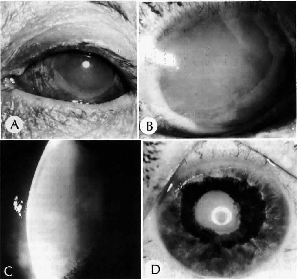
True exfoliation of the lens capsule is a condition in which the anterior aspect of the capsule splits. Clinical findings are a thin, clear, colorless membrane floating in the pupillary axis. It was first described in glass blowers and was believed to be a consequence of the considerable infrared radiation to which they were exposed. It has also been described in blacksmiths and steelworkers as well as in other persons who have had similar infrared exposure.27

It is possible, however, for patients to develop true exfoliation without having an apparent history of infrared radiation exposure. True exfoliation secondary to severe iridocyclitis or occurring as a sequela of trauma has been suggested.28 In some cases, the cause is obscure, but it may be a change associated with aging. In the largest reported series of 11 eyes in seven patients, the youngest patient was 79. All were hyperopic.27

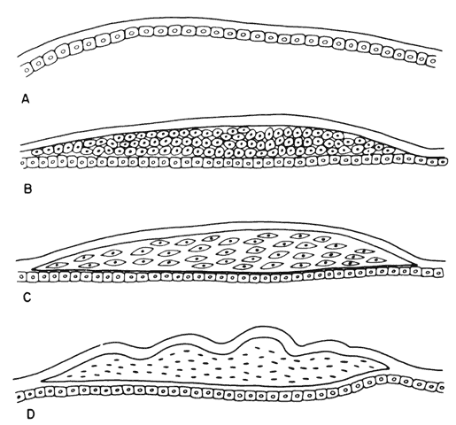
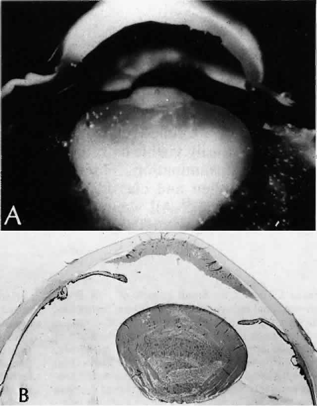
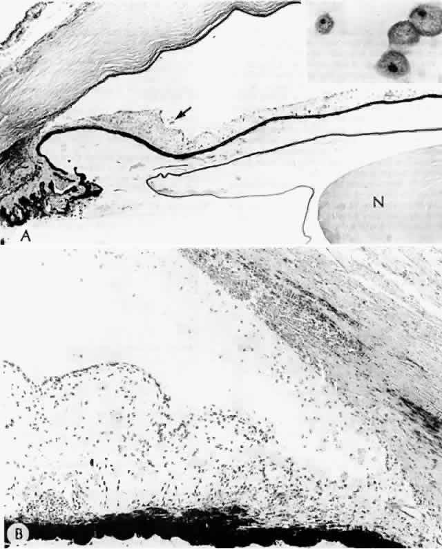
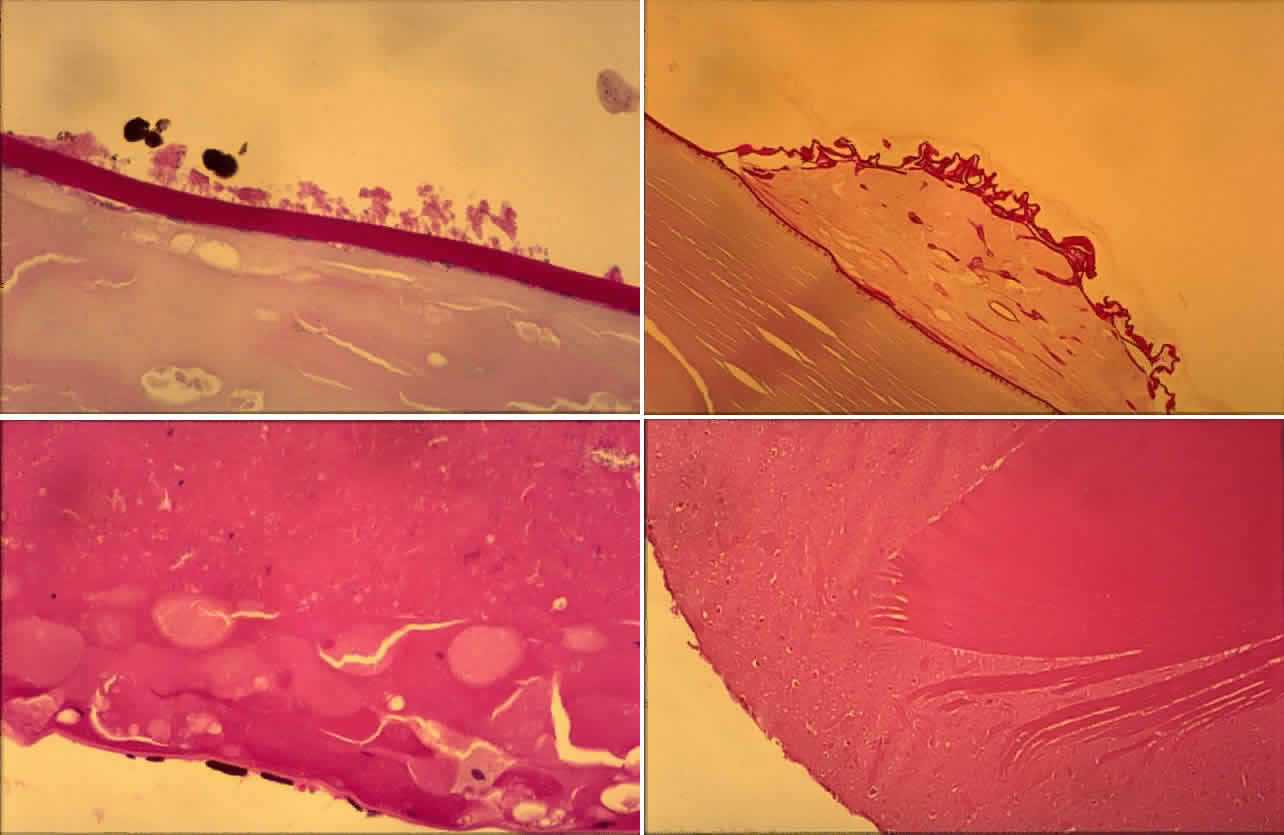
Histologic examination shows an unequal splitting of the anterior capsule (Fig. 1). Electron microscopic examination shows that the lens capsule is laminated and the anterior layer or layers are split free.29 The free portion is quite thin, and it can be rolled or folded over.27

Fig. 1. True exfoliation of the lens capsule. The anterior lens capsule is split and floats away from the rest of the lens (arrow). The subepithelial disruption is artifact. (H & E, magnification × 234; Courtesy of Dr. W.R. Green, Baltimore, MD)

PSEUDOEXFOLIATION SYNDROME

Pseudoexfoliation of the lens is much more common than true exfoliation. The term is actually a misnomer, as it has become clear that the origin of the pseudoexfoliation material is not the lens capsule. It is more prevalent in Scandinavian countries and is associated with secondary open-angle glaucoma.30

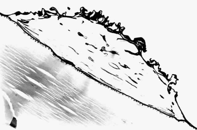
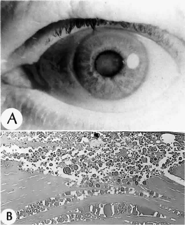
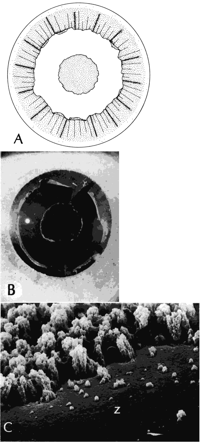
Clinically, in its classic manifestation, pseudoexfoliation syndrome is manifest as a whitish, fluffy deposit on the lens surface and at the pupillary margin. Typically there is a circular clear zone in the midperiphery of the lens, where the iris comes in contact with it (Fig. 2). This syndrome is important to recognize because a type of secondary open-angle glaucoma that can be difficult to control is associated with pseudoexfoliation in more than 20% of eyes.31 Other associated changes include poor dilatation of the iris, melanin dispersion from degeneration of the iris pigment epithelium, and iris stromal atrophy, which are visible clinically as transillumination defects.32

Fig. 2. Pseudoexfoliation syndrome. A and B. Central disc surrounded by a relatively clear zone, surrounded in turn by a peripheral granular area. C. Scanning electron micrograph shows relatively clear zone (Z) surrounded by the central edge of the peripheral granular area. (B, courtesy of Dr. G. Naumann, SEI 73-985; C, courtesy of Dr. R.D. Eagle, Jr;) magnification × 900)

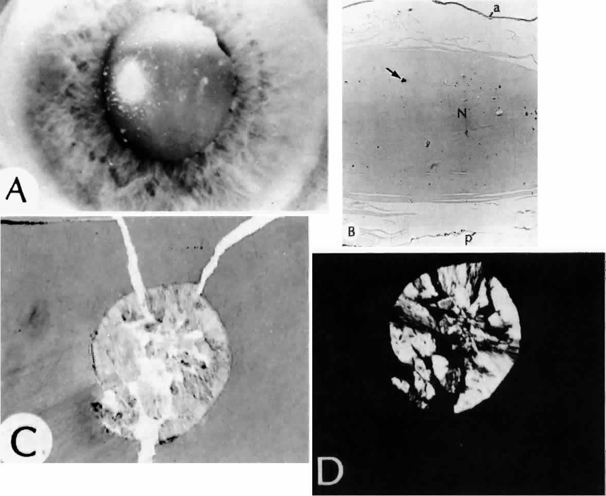
Because of its appearance, it was natural to assume that the exfoliation material was derived from the lens capsule. In some eyes, however, it was observed to persist or to develop long after intracapsular lens extraction. The stimulus for its deposition is likewise unknown. Subsequently, the material has been found in the conjunctiva,33 iris,32 and visceral organs.31 Thus, pseudoexfoliation syndrome appears to be a systemic disease, although there are no clinical symptoms apart from those in the eye. The syndrome has been described as typically unilateral, but it now appears that it is more frequently bilateral but asymmetric. Sometimes, too, eyes with uncontrolled open-angle glaucoma and other signs of exfoliation, such as poorly reactive pupils, but with no evidence of intraocular exfoliation material, can be shown to have the syndrome by conjunctival biopsy.33

Histologic examination shows exfoliation deposits appearing as short strands of eosinophilic material, which are found on the surface of the lens and zonules and within and adjacent to the ciliary body and iris (Color Plate 1A). On electron microscopic examination, these strands are found to be thick, straight, and densely osmiophilic, with associated thin microfibrils.33

COLOR PLATE 1 A. Pseudoexfoliation fibers on the lens capsule. The dark areas are pigment aggregates (PAS, magnification × 146) B. Anterior polar cataract. The lens epithelium has undergone fibrous metaplasia, and the anterior capsule is wrinkled. A nwe anterior capsule has formed with subjacent normal lens epithelium (PAS, × 58). C. Posterior subcapsular cataract with large bladder cells (H & E, × 58). D. The cortex is liquefied and granular, with more solid-appearing nuclear fragments (H & E, × 58).

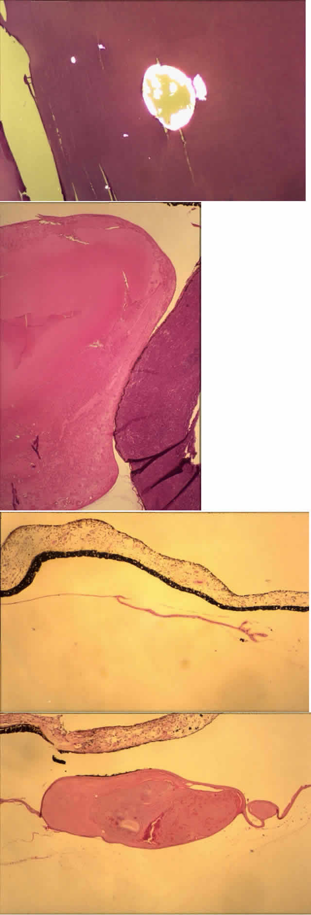
The iris pigment epithelium takes on a characteristic jagged, sawtooth appearance as melanin granules are lost from the cells. Degenerative changes with accumulations of the pseudoexfoliation fibers can be seen in both the sphincter and dilator muscles, probably accounting for the poor reactivity of the pupil.32

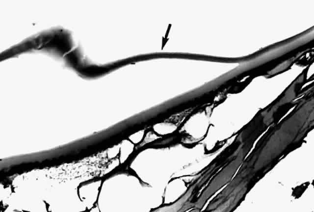
Recently, the pseudoexfoliation syndrome has been identified as a risk factor for lens capsule dislocation and vitreous loss during cataract surgery. On clinical examination, the zonules appear weakened by the exfoliation material. Electron microscopic examination reveals accumulated material along the length of the zonule and also at the origin and insertion of the zonules. Aggregates of exfoliation material interfere with the normal attachment of the zonules and interrupt the length of the zonule. Lysosomal enzymes are present within pseudoexfoliation fiber aggregates, presumably facilitating zonular dissolution.34

The exact nature of the pseudoexfoliation material has not been discerned. Probably it is an abnormal metabolite or component of basement membrane material. Many associated substances have been identified by immunostaining, including proteins of elastic tissue (e.g., elastin, fibrillin, amyloid P) and basement membrane-related components (e.g., chondroitin sulfate).31 Antibody testing has identified specific carbohydrate moieties in pseudoexfoliation material that are also present in various types of adhesion molecules.35 The cause and mechanism of this disease, however, remain elusive.

Back to Top
EPITHELIUM
The lens epithelium is metabolically active, serving to pump out sodium ion and to defend against oxidative injury. In some cataracts studied biochemically, levels of glutathione reductase, an enzyme that protects against oxidative injury, may be deficient.36 There is a tendency for the epithelial cell density to decrease somewhat with age.37

SUBCAPSULAR CATARACT

Anterior subcapsular cataracts can result from a variety of stimuli, including trauma. Anterior polar cataracts, which tend to be congenital, are similar but more localized. Both appear as discrete whitish plaques located just beneath the lens capsule.

Subcapsular cataracts are caused by proliferation of the lens epithelium. Normally a monolayer, the epithelial cells, when stimulated to proliferate, also undergo fibrous metaplasia and generate an irregular fibrous plaque with wrinkling of the lens capsule. The individual cells, however, still secrete basement membrane. On histologic examination, each cell is surrounded by basement membrane material. A new anterior capsule may be secreted, under which is a new layer of lens epithelium (Figs. 3 and 4; Color Plate 1B).

Fig. 3. Anterior subcapsular cataract. Change from a normal lens epithelium (A) through proliferating epithelial cells (B, C) to final subcapsular fibrous plaque and formation of a new continuous basement membrane (lens capsule) (D).

Fig. 4. Anterior polar cataract. The lens epithelium has undergone fibrous metaplasia, and the anterior capsule is wrinkled. A new anterior capsule has formed with subjacent normal lens epithelium. (PAS, magnification × 93)

Posterior subcapsular cataracts are more common. They may be idiopathic, but they also occur after steroid or busulfan use and are histologically similar.38 Clinically they appear as irregular, whitish plaques just anterior to the posterior surface of the lens (Fig. 5A, B, and C). The entire remainder of the lens may be clear, and there may be crystalline-appearing areas within the cataract.

Fig. 5. Posterior subcapsular cataract (PSC). A, B, and C. Different views of a PSC. D. Marked PSC changes consist of posterior cortical bladder cell formation. (A, B, and C, SEI 79-213, 79-214, and 79-215; D, PAS, × 250, 73-28)

The Lens Opacities Classification System II (LOCS II), a clinical grading system based on slit-lamp retroillumination, was developed in an effort to standardize the degree and type of lens opacity for clinical studies. According to this system, posterior subcapsular cataracts are graded from PI through PIV according to the extent of the opacity.39

Posterior subcapsular cataracts also are derived from lens epithelial cells; however, since the cells are not normally present posteriorly, an abnormal migration from the lens equator is a first step in their formation. The migrating cells become more metabolically active as they progress, as evidenced by the presence of cytoplasmic organelles. They secrete fibrillogranular material and basement membrane, sometimes wrinkling the lens capsule. Swollen bladder cells, sometimes called Wedl cells or Elschnig's pearls, are also present (Fig. 5D). These cells are large, with abundant eosinophilic cytoplasm and an inconspicuous eccentric nucleus (Color Plate 1C).40

Back to Top
CORTEX AND NUCLEUS
Cortical cataracts originate as discrete round or elongated opacities involving small groups of fibers in deep cortex. Adjacent fibers may be completely normal morphologically. Later, cortical cataracts become more spoke-like as the opacity advances.

The Lens Opacities Classification System grades cortical cataracts from Ctr (trace) through CIV.39 In one study,41 early changes were correlated by scanning electron microscopy with the clinical grades CI and CII. Small groups of lens fibers showed focal interruption with globule formation, whereas adjacent fibers remained intact.

After these early changes, the degeneration of individual fibers then proceeds either anteriorly, posteriorly, or in both directions of the fibers (Fig. 6). On histologic examination, more advanced cortical cataracts show areas of interrupted cortical fibers with fine granules and larger globules intermixed (Fig. 7, Color Plate 1D).42 Eventually, the entire cortex may be liquefied, allowing the nucleus to float within it; this is termed a mature cataract (Fig. 8).

Fig. 6. Types of changes that lens “fibers” (i.e., cells) undergo.

Fig. 7. Intumescent cataract. A. Swollen lens contains a “milky” cortex and a small anterior subcapsular cataract. B. Swollen cataract shows marked cortical morgagnian degeneration. Note the peripheral anterior synechias and chronic secondary angle closure. (A, macroscopic, SEI 73-117; B, H & E, × 8, SEI 73-15)

Fig. 8. A. Clinical appearance of a mature cataract. B. Morgagnian cortical degeneration comes right up to the anterior capsule so that no clear cortex can be seen clinically (i.e., mature cataract). (A, SEI 73-600; B, H & E, × 40, SEI 73-354)

Retrodots, discrete birefringent opacities found in the deep cortical layers, are common after the age of 50 and may reflect decreased protection from oxidative injury. They are called retrodots because they are much more easily visible by retroillumination than by direct examination. Three types of retrodots were identified and classified clinically by their shape and size.43 All were discrete and varied from 80 to 500 μm, and they occurred in both anterior and posterior cortex. Retrodots are associated with increased nuclear light scatter. Scanning electron microscopy and x-ray dispersion analysis demonstrated that two of the types contained calcium phosphate and the third calcium oxalate. All were well demarcated, suggesting a sequestration from the surrounding normal lens fibers. Their role in cataractogenesis is not understood, but calcium is known to aggregate crystallins.44 The oxalate may be derived from ascorbic acid, which acts as an antioxidant in the lens.43

The increased yellowing and decreased transparency of the lens with age are the hallmarks of nuclear sclerosis. Probably anyone living long enough will develop visually significant nuclear sclerosis. As the lens grows, the central fibers become increasingly compressed. On histologic examination, the nucleus is seen to have become a homogeneous, eosinophilic structure, appearing discrete from the more liquefied cortex.

Nuclear cataracts are graded in the Lens Opacities Classification System according to both color (NCO-NCII) and opalescence (NI-NIV).39 For such a common disease process, it may seem surprising that the mechanism of clouding and color change is not better understood. Only recently have some of the biochemical events become elucidated.

The lens has a very high protein content. Part of the reason for transparency of the lens is the structural arrangement of the proteins. However, as the lens grows throughout life, older lens cells are moved inward; these fibers, having lost their nuclei, are dependent on the peripheral metabolically active cells for maintenance. There is no evident way to repair damaged cells, and with increasing age, the lens nucleus becomes denser and loses transparency. The protein structure of the nuclear fibers becomes altered so that the normally water-soluble proteins become insoluble, apparently by forming high-molecular-weight aggregates with disulfide bonds. These aggregates appear to increase light scatter. With increased cross-linking and changes in membrane permeability, more water is allowed in, promoting opacification.45 Calcium oxalate crystals can be seen clinically as shiny, birefringent deposits, and are identifiable histologically with polarized light (Fig. 9, Color Plate 2A).

Fig. 9. A. Dark nucleus floating within a liquefied, “milky” cortex settled inferiorly because of gravity. B. Hypermature cataract characterized by a wrinkled anterior capsule (a). No cortex is present (it has liquefied and leaked out); only the nucleus (N) is present. Homogeneity of the nucleus marks it as cataractous. A calcium oxalate crystal (arrow) is present in the nucleus; p' = posterior capsule of lens. A calcium oxalate crystal can be seen within the nucleus before (C) and after (D) polarization. (A, courtesy of Dr. G. Naumann, × 100; D, polarized, H & E × 100)

COLOR PLATE 2 A. Calcium oxalate crystal in lens nucleus, as seen with polarized light (H & E, × 146). B. Lens compressed by a ciliochoroidal malignant melanoma (lower right). Changes include cortical liquefaction and nuclear fragmentation (H & E, × 23.4). C. Capsular remnants 6 weeks after extracapsular lens extraction. Only capsule and epithelium remain. D. Capsular remnants years after lens extraction. Lens fibers can proliferate from remaining epithelium (C and D, H & E, × 23.4).

In one study,36 the lens epithelium from some patients with cataracts contained low levels of glutathione reductase, an enzyme that protects against oxidative damage and that requires a flavin cofactor for activity. Since oxidative damage from a variety of sources, including irradiation and certain drugs, has a role in cataract formation, the authors proposed dietary supplementation with riboflavin. It remains to be seen whether such supplementation will help to prevent cataract.

Back to Top
SECONDARY CATARACTS
Cataracts form earlier in patients with diabetes mellitus than in otherwise comparable persons without diabetes. Clinical and histologic examination, however, reveals that diabetic cataracts appear to be no different from ordinary age-related cataracts. A contributing factor to the formation of diabetic cataracts is the aldose reductase pathway. The enzyme aldose reductase reduces glucose and other aldohexoses to sorbitol, the corresponding sugar alcohol. Sorbitol cannot readily cross the lens capsule, but does exert osmotic pressure, so that water from outside of the lens comes in, causing lens swelling.46

Retinitis pigmentosa is the common name for a group of inherited disorders of the retina characterized by constriction of peripheral fields, pigmentary retinopathy, and optic disc pallor. Posterior subcapsular cataracts are characteristic. Histologically and ultrastructurally they are similar to primary age-related posterior subcapsular cataracts.47

Tumors within the eye can press against the lens, altering its shape and causing localized or diffuse cataract. A monocular dense cataract may therefore be evidence of an otherwise unsuspected intraocular mass (Color Plate 2B).

Injury, including direct trauma, also can cause cataract formation. Cataracts caused by direct contusion are cortical and take on a rosette shape, outlining the lens sutures. They can be anterior or posterior. On histologic examination, these lenses show focal vacuolization of cortical lens fibers. Because lens fibers continue to form, the opaque fibers become further separated from the subcapsular area over time, as they do in congenital cataracts.48 There is one report49 of an anterior chamber cyst that formed after penetrating trauma, which when excised proved to be of lenticular origin. A basement-membrane capsule was found to have surrounded fibrous tissue and bladder cells.

Metal deposition, most notably iron and copper, cause cataract formation, both in the context of trauma and as part of ocular or systemic disease. Iron deposition in the epithelial structures of the eye can be the result of an iron-containing foreign body or repeated hemorrhage inside the eye. The retina and retinal pigment epithelium are adversely affected, with early reduction of the B-wave on electroretinography. The lens can be grossly rust-colored, with massive accumulation of iron as demonstrated by light and electron microscopy.50 Pure copper foreign bodies cause panophthalmitis, but copper alloys may be tolerated for longer periods. As with iron toxicity, the electroretinographic A-wave and B-wave are reduced. Copper is deposited in basement membranes, including Descemet's membrane and the lens capsule, giving rise to a characteristic yellow-green discoloration.51 Wilson's disease, a defect in copper metabolism,52 and secondary causes of hypercupremia51 can cause a similar copper uptake in the lens capsule. In Wilson's disease, the cataract resembles a sunflower, having a central ring anteriorly and peripheral tapering extensions.52 Electron-dense copper particles are seen at all levels of the capsule,51 both anteriorly and posteriorly.52

Radiation, including ionizing radiation and microwaves, causes posterior subcapsular cataracts. Recently, the incidence of subcapsular cataract, as well as nuclear sclerosis and cortical cataract, was studied after charged particle irradiation for uveal melanoma. As might be anticipated, posterior subcapsular cataract formation was dose dependent. Other lenticular opacities were age related.53

Laser energy can also cause cataracts. Inadvertent argon laser energy causes focal white opacities in the anterior cortical region immediately after administration. Subsequently, these lesions remain unchanged.42 Histologically and ultrastructurally, they appear spindle shaped, in deep anterior cortex, and consisted of clear spaces with cellular debris. The lesions are discrete, with normal lens fibers adjacent to them.54

Lightning55 and electrical shock injury56 are known to cause cataracts. These are subcapsular or cortical in location, and form over a period of several weeks. The pathogenesis is obscure. It is possible that the electrical injury causes coagulation of lens protein directly, but in that case the cataract would be expected to be manifest immediately. Another theory is a tetanic contraction of the ciliary muscle, mechanically displacing the lens fibers.56

Maternal alcohol abuse can lead to a constellation of abnormalities termed fetal alcohol syndrome. This syndrome is an important cause of mental retardation and central nervous system abnormalities. Ocular abnormalities are frequent, especially optic nerve hypoplasia.57 Anterior segment abnormalities, including cataract and Peters' anomaly, are sometimes present.58

A number of pharmaceuticals are associated with cataract formation, most notably systemic or topical steroids. These cataracts are typically posterior subcapsular.59 Busulfan, used for treating leukemia, has also been associated with posterior subcapsular cataract.38,60 Amiodarone, an antiarrhythmic drug, causes whorl-like figures in the cornea that resemble those seen in Fabry's disease. In one study,61 the cataracts of a patient who had had cataracts before having taken amiodarone were removed and examined. The lenses showed dense inclusions of concentric membranous lamellae within lens epithelium, similar to those of the conjunctival and corneal epithelium seen in this patient and in others who have taken amiodarone.

Back to Top
COMPLICATIONS OF CATARACT FORMATION
Apart from visual loss, the main indication for cataract extraction, the major complication of cataract formation is some form of glaucoma.

The aging lens, even without a clinically significant cataract, increases in its anteroposterior dimension throughout life, since fibers cannot be desquamated. Because of osmotic changes, cataractous lenses can be even larger anteroposteriorly, approaching a spherical shape, thus forming an intumescent cataract. Such an eye may develop a shallow anterior chamber and an increased risk of pupil block as the lens enlarges. This is called phacomorphic glaucoma because it is the shape, or morphology, of the lens that causes the glaucoma.

With age, the cortical lens fibers disintegrate and liquefy. The lens proteins are able to leak across a clinically and histologically intact lens capsule. As the proteins escape, macrophages ingest them. As the macrophages accumulate, however, they clog the trabecular meshwork, interfering with aqueous outflow. This is called phacolytic glaucoma (Figs. 10 and 11).62 In the clinical setting, specular microscopy shows these macrophages to be adherent to the posterior corneal surface.63

Fig. 10. Phacolytic glaucoma. A. Patient presented with symptoms of acute closed-angle glaucoma. Chalky material is present in the anterior chamber, and the angle is open. B and C. Another patient with symptoms of acute closed-angle glaucoma. D. Neglected case of phacolytic glaucoma shortly before enucleation. (A, courtesy of Dr. T.R. Thorp, SEI 79-220; B, C, and D, courtesy of Dr. G. Naumann, SEI 79-221, 79-222, and 79-223)

Fig. 11. Phacolytic glaucoma. A. Hypermature cataract. Most of the cortex leaked through the intact capsule. (N, nuclear cataract). Lens-filled macrophages are shown in high magnification (inset) (arrow). B. Another case shows lens-filled macrophages in the anterior chamber, on the iris surface, in the iris stroma, and in the anterior chamber angle (note angle recession). (A, main figure, PAS, × 70, SEI 73-65; inset, H & E × 220, SEI 73-3; B, H & E, × 220).

Along with the macrophages containing lens material, free lens protein, macrophages with pigment, and erythrocytes have been found in the aqueous fluid. The macrophages and free lens material obstruct the trabecular meshwork, as demonstrated ultrastructurally.64

Phacoanaphylactic endophthalmitis is a misleading name for what is actually a cell-mediated hypersensitivity reaction: there is no anaphylactic, or IgE-mediated, reaction. In contrast to phacolytic glaucoma, the capsule is ruptured as a result of a trauma, including surgery. Massive leakage of lens proteins incites a granulomatous inflammatory response. Rupture of the lens capsule, however, may not be a sufficient cause, as suggested by observation after controlled needling of cataracts.65 It is an extremely rare complication of planned extracapsular surgery.

Back to Top
COMPLICATIONS OF CATARACT SURGERY
Extracapsular lens extraction with placement of a plastic intraocular lens is the most common ophthalmic surgical procedure currently performed.

An important function of the lens capsular remnant is to support and stabilize the lens implant, which is inserted either into it or in front of it. Anterior capsulotomy, especially if the removed portion of capsule is large, may disrupt a significant number of zonules, thereby weakening support of the capsular remnant. The diameter of the central zonule-free area of anterior capsule decreases with age, from 8 mm in 20-year-old persons to 6 mm in 70-year-old persons.66 In a recent study of 50 anterior capsulotomy specimens, the average diameter was 5 to 6 mm, and the specimens were about the same size whether excised via a multiple puncture technique or a continuous-curve capsulotomy.36

Equatorial and posterior zonular disinsertion may occur during the removal of the cortex and nucleus, as the instruments are maneuvered within the capsule. One postmortem study showed large disinsertions in 5 of the 20 eyes studied, although in none was there a significant dislocation of the lens implant.67

A major cause of postoperative disability is opacification of the posterior capsule. Just as the epithelium in the intact lens can undergo fibrous metaplasia, it can also do so after extracapsular lens extraction, causing clinical opacity. Histologic examination reveals wrinkling of the posterior capsule by the proliferating cells, which show myofibroblastic differentiation. It has been proposed that a large anterior capsulotomy would delay this opacification,68 but it would not be possible to remove all lens epithelial cells because they normally extend past the equator. The epithelium can also form bladder cells, especially at the margin of contact between the posterior capsule and the anterior capsule margin. The cells can extend across the pupillary space as well as along the anterior surface of the lens capsule remnant.69 The ability of the lens epithelium to proliferate onto the capsule also has been documented in tissue culture.70

Varying amounts of cortical material may be left behind; the lens capsular bag may also “refill” as lens epithelial cells proliferate, creating a secondary Soemmering ring configuration (Color Plates 2C and D).71 Fibrous material can also encase intraocular lenses, especially after multiple operative procedures or intraocular inflammation incites a diffuse fibrous proliferation. It has long been speculated that some of the fibrous membrane is of lens epithelial origin, and recently this has been documented immunohistochemically.72

Rarely, heterologous material can enter the capsule during surgery or shortly afterward. Endocapsular hematoma, or blood within the capsule, has been reported as a postoperative complication.73 Sequestered endophthalmitis is a cause of postoperative low-grade inflammation. Histologically, bacteria of low virulence, especially Propionibacterium acnes, have been documented histologically within the lens capsule.74 Subsequent rupture of the capsule by the YAG laser has led to more fulminant endophthalmitis.75


The author and editors wish to express their appreciation to Myron Yanoff, MD, and Ben S. Fine, MD, authors of the original chapter. Some of the material and many of the illustrations have been retained in this revision.
Back to Top
REFERENCES

1. Pasquale LR, Romayananda N, Kubacki J et al: Congenital cystic eye with multiple ocular and intracranial anomalies. Arch Ophthalmol 109:985, 1991

2. Sassani JW, Yanoff M: Anophthalmos in an infant with multiple congenital anomalies. Am J Ophthalmol 83:43, 1977

3. Marcus DM, Shore JW, Albert DM: Anophthalmica in the focal dermal hypoplasia syndrome. Arch Ophthalmol 108:96, 1990

4. Maden A, Buyukgebiz B, Gunenc U et al: Bilateral congenital absence of pupillary aperture. Am J Ophthalmol 112:608, 1991

5. Torczynski E, Jacobiec FA, Johnston MC et al: Synophthalmia and cyclopia: a histopathologic, radiographic, and organogenetic analysis. Doc Ophthalmol 44:311, 1977

6. Stefani FH, Hausmann N, Lund O-E: Unilateral diplophthalmos. Am J Ophthalmol 112:581, 1991

7. Hamming NA, Apple DJ, Gieser DK et al: Ultrastructure of the hyaloid vasculature in primates. Invest Ophthalmol Vis Sci 16:408, 1977

8. Hittner HM, Kretzer FL, Antoszyk JH et al: Variable expressivity of autosomal dominant anterior segment mesenchymal dysgenesis in six generations. Am J Ophthalmol 93:57, 1982

9. Traboulsi EI, Maumenee IH: Peters' anomaly and associated congenital malformations. Arch Ophthalmol 110:1739, 1992

10. Stone DL, Kenyon KR, Green WR et al: Congenital central corneal leukoma (Peters' anomaly). Am J Ophthalmol 81:173, 1976

11. Kivlin JD, Fineman RM, Crandall AS et al: Peters' anomaly as a consequence of genetic and nongenetic syndromes. Arch Ophthalmol 104:61, 1986

12. Streeten BW, Robinson MR, Wallace R et al: Lens capsule abnormalities in Alport's syndrome. Arch Ophthalmol 105:1693, 1987

13. Khalil M, Saheb N: Posterior lenticonus. Ophthalmology 91:1429, 1984

14. Streeten B: The nature of the ocular zonule. Trans Am Ophthalmol Soc 80:823, 1982

15. Farnsworth PN, Burke P, Dotto ME et al: Ultrastructural abnormalities in a Marfan's syndrome lens. Arch Ophthalmol 95:1601, 1977

16. Ramsey MS, Yanoff M, Fine BS: The ocular histopathology of homocystinuria. Am J Ophthalmol 74:377, 1972

17. Jensen AD, Cross HE, Paton D: Ocular complications in the Weill-Marchesani syndrome. Am J Ophthalmol 77:261, 1974

18. Goldberg MF: Clinical manifestations of ectopia lentis et pupillae in 16 patients. Ophthalmology 95:1080, 1988

19. Scott MH, Hejtmancik JF, Wozencraft LA et al: Autosomal dominant congenital cataract. Ophthalmology 101:866, 1994

20. de Gottrau P, U, Dörfler S et al: Congenital zonular cataract: clinicopathologic correlation with electron microscopy and review of the literature. Arch Ophthalmol 111:235, 1993

21. Stambolian D: Galactose and cataract. Surv Ophthalmol 32:333, 1988

22. Dark AJ, Streeten BW: Ultrastructural study of cataract in myotonia dystrophica. Am J Ophthalmol 84:666, 1977

23. Ginsberg J, Bove KE, Fogelson MH: Pathological features of the eye in the oculocerebrorenal (Lowe) syndrome. J Pediatr Ophthalmol Strabismus 18(4):16, 1981

24. Cibis GW, Waeltermann JM, Whitcraft CT et al: Lenticular opacities in carriers of Lowe's syndrome. Ophthalmology 93:1041, 1986

25. Tripathi RC, Cibis GW, Tipathi BJ: Pathogenesis of cataracts in patients with Lowe's syndrome. Ophthalmology 93:1046, 1986

26. Robb RM, Marchevsky A: Pathology of the lens in Down's syndrome. Arch Ophthalmol 96:1039, 1978

27. Cashwell LFJ, Holleman IL, Weaver RG et al: Idiopathic true exfoliation of the lens capsule. Ophthalmology 96:348, 1989

28. Brodrick JD, Tate GW Jr: Capsular delamination (true exfoliation) of the lens: report of a case. Arch Ophthalmol 97:1693, 1979

29. Fiore PM, Shingleton BJ: Senile lens exfoliation. JAMA 264:2755, 1990

30. Chen V, Blumenthal M: Exfoliation syndrome after cataract extraction. Ophthalmology 99:445, 1992

31. Streeten BW, Li Z-Y, Wallace RN et al: Pseudoexfoliative fibrillopathy in visceral organs of a patient with pseudoexfoliation syndrome. Arch Ophthalmol 110:1757, 1992

32. Asano N, U, Naumann GOH: A histopathologic study of iris changes in pseudoexfoliation syndrome. Ophthalmology 102:1279, 1995

33. Prince AM, Streeten BW, Ritch R et al: Preclinical diagnosis of pseudoexfoliation syndrome. Arch Ophthalmol 105:1076, 1987

34. U, Naumann GOH: A histopathologic study of zonular instability in pseudoexfoliation syndrome. Am J Ophthalmol 118:730, 1994

35. Uusitalo M, Kiveldä T, Tarkkanen A: Immunoreactivity of exfoliation material for the cell adhesion-related HNK-1 carbohydrate epitope. Arch Ophthalmol 111:1419, 1993

36. Straatsma BR, Lightfoot DO, Barke RM et al: Lens capsule and epithelium in age-related cataract. Am J Ophthalmol 112:283, 1991

37. Konofsky K, Naumann GOH, Guggenmoos-Holzmann I: Cell density and sex chromatin in lens epithelium of human cataracts: quantitative studies in flat preparation. Ophthalmology 94:875, 1987

38. Eshaghian J, Rafferty NS, Goossens W: Human cataracta complicata: clinicopathologic correlation. Ophthalmology 88:155, 1981

39. Chylack LT Jr, Leske MC, McCarthy D et al: Lens opacities classification system II (LOCS II). Arch Ophthalmol 107:991, 1989

40. Eshaghian J, Streeten BW: Human posterior subcapsular cataract: an ultrastructural study of the posteriorly migrating cells. Arch Ophthalmol 98:134, 1980

41. Vrensen G, Willekens B: Biomicroscopy and scanning electron microscopy of early opacities in the aging human lens. Invest Ophthalmol Vis Sci 31:1582, 1990

42. McCanna P, Chandra SR, Stevens TS et al: Argon laser-induced cataract as a complication of retinal photocoagulation. Arch Ophthalmol 100:1071, 1982

43. Bron AJ, Brown NAP: Perinuclear lens retrodots: a role for ascorbate in cataractogenesis. Br J Ophthalmol 71:86, 1987

44. Vrensen GFJM, Willekens B, De Jong PTVM et al: Heterogeneity in ultrastructure and elemental composition of perinuclear lens retrodots. Invest Ophthalmol Vis Sci 35:199, 1994

45. Spector A: The search for a solution to senile cataracts: Proctor lecture. Invest Ophthalmol Vis Sci 25:130, 1984

46. Kinoshita JH: Aldose reductase in the diabetic eye: XLIII Edward Jackson Memorial Lecture. Am J Ophthalmol 102:685, 1986

47. Eshaghian J, Rafferty NS, Goossens W: Ultrastructure of human cataract in retinitis pigmentosa. Arch Ophthalmol 98:2227, 1980

48. Asano N, U, Dörfler S et al: Ultrastructure of contusion cataract. Ophthalmology 113:210, 1995

49. Salisbury JA, Foulks GN, Klintworth GK: Lens capsular cyst. Am J Ophthalmol 90:229, 1980

50. Talamo JH, Topping TM, Maumenee AE et al: Ultrastructural studies of cornea, iris and lens in a case of siderosis bulbi. Ophthalmology 92:1675, 1985

51. Martin NF, Kincaid MC, Stark WJ et al: Ocular copper deposition associated with pulmonary carcinoma, IgG monoclonal gammopathy and hypercupremia: a clinicopathologic study. Ophthalmology 90:110, 1983

52. Tso MOM, Fine BS, Thorpe HE: Kayser-Fleischer ring and associated cataract in Wilson's disease. Am J Ophthalmol 79:479, 1975

53. Gragoudas ES, Egan KM, Walsh SM et al: Lens changes after proton beam irradiation for uveal melanoma. Am J Ophthalmol 119:157, 1995

54. Shapiro A, Tso MOM, Goldberg MF: Argon laser-induced cataract: a clinicopathologic study. Arch Ophthalmol 102:579, 1984

55. Lagrèze W-DA, Bömer TG, Aiello LP: Lightning-induced ocular injury. Arch Ophthalmol 113:1076, 1995

56. Johnson EV, Kline LB, Skalka HW: Electrical cataracts: a case report and review of the literature. Ophthalmic Surg 18:283, 1987

57. Strömland K: Ocular involvement in the fetal alcohol syndrome. Surv Ophthalmol 31:277, 1986

58. Chan T, Bowell R, O'Keefe M et al: Ocular manifestations in fetal alcohol syndrome. Br J Ophthalmol 75:524, 1991

59. Yablonski ME, Burde RM, Kolker AE et al: Cataracts induced by topical dexamethasone in diabetics. Arch Ophthalmol 96:474, 1978

60. Podos SM, Canellos GP: Lens changes in chronic granulocytic leukemia: possible relationship to chemotherapy. Am J Ophthalmol 68:500, 1969

61. D'Amico DJ, Kenyon KR, Ruskin JN: Amiodarone keratopathy: drug-induced lipid storage disease. Arch Ophthalmol 99:257, 1981

62. Flocks M, Littwin CS, Zimmerman LE: Phacolytic glaucoma. Arch Ophthalmol 54:37, 1955

63. Brooks AMV, Grant G, Gillies WE: Comparison of specular microscopy and examination of aspirate in phacolytic glaucoma. Ophthalmology 97:85, 1990

64. Ueno H, Tamai A, Iyota K et al: Electron microscopic observation of the cells floating in the anterior chamber in a case of phacolytic glaucoma. Jpn J Ophthalmol 33:103, 1989

65. Yanoff M, Scheie HG: Cytology of human lens aspirate: its relationship to phacolytic glaucoma and phacoanaphylactic endophthalmitis. Arch Ophthalmol 80:166, 1968

66. Stark WJ, Streeten B: The anterior capsulotomy of extracapsular cataract extraction. Ophthalmic Surg 15:911, 1984

67. Wilson DJ, Jaeger MJ, Green WR: Effects of extracapsular cataract extraction on the lens zonules. Ophthalmology 94:467, 1987

68. McDonnell PJ, Zarbin MA, Green WR: Posterior capsule opacification in pseudophakic eyes. Ophthalmology 90:1548, 1983

69. Kappelhof JP, Vrensen GFJM, de Jong PTVM et al: An ultrastructural study of Elshnig's pearls in the pseudophakic eye. Am J Ophthalmol 101:58, 1986

70. Ayaki M, Ohara K, Ibaraki N et al: The outgrowth of lens epithelial cells onto the anterior capsule after intraocular lens implantation. Am J Ophthalmol 115:668, 1993

71. Assia El, Legler UFC, Apple DJ: The capsular bag after short- and long-term fixation of intraocular lenses. Ophthalmology 102:1151, 1995

72. Pavilack MA, Foster CS, Kowal VO et al: Peripseudophakic membrane: pathologic features. Arch Ophthalmol 111:240, 1993

73. Hagan JC III, Gaasterland DE: Endocapsular hematoma: description and treatment of a unique form of postoperative hemorrhage. Arch Ophthalmol 109:514, 1991

74. Meisler DM, Mandelbaum S: Propionibacterium-associated endophthalmitis after extracapsular cataract extraction: review of reported cases. Ophthalmology 96:54, 1989

75. Tetz MR, Apple DJ, Price FW Jr et al: A newly described complication of neodymium-YAG laser capsulotomy: exacerbation of an intraocular infection. Arch Ophthalmol 105:1324, 1987

Back to Top