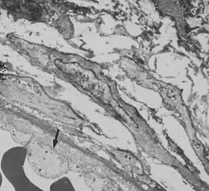
Fig. 8.
Electron micrograph from eye with pars planitis (see also
Fig. 9
) showing collagen deposition and high endothelial venules (arrow).