|
|
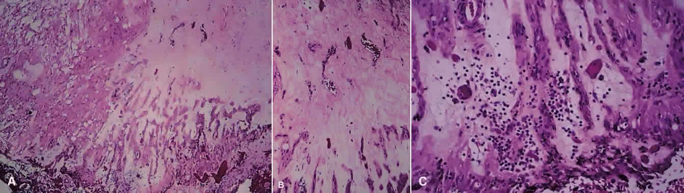
| Fig. 9. A. Photomicrograph of “snowbank” in patient with pars planitis showing a fibroglial mass with proliferated epithelial elements (hematoxylin and eosin; × 31). B. Higher magnification showing epithelial elements and glial proliferation (hematoxylin and eosin; × 80). C. Chronic inflammatory cell infiltrate in pars planitis (hematoxylin and eosin; × 200). |