Chapter 97
Vaccinia
R. MARK BULLER and JAY S. PEPOSE
Main Menu   Table Of Contents

Search

INTRODUCTION
VIROLOGY
PREDISPOSING FACTORS
EPIDEMIOLOGY
PATHOGENESIS
ANIMAL MODELS
LABORATORY DIAGNOSIS
REFERENCES

INTRODUCTION
Poxviruses infect the eye during endogenous viremia or after exogenous inoculation from another site. Historically, in unvaccinated individuals, smallpox and human monkeypox were associated with (corneal) blindness at a frequency of 0.45% to 0.9% and 1.7%, respectively.1,2 With the eradication of smallpox in 1979, and the paucity of human monkeypox outside of Central Africa, this complication is rarely seen. The majority of poxvirus-associated ocular disease is caused by molluscum contagiosum virus after a natural infection3 and by vaccinia virus as a complication of the smallpox vaccine.

Ocular vaccinial infections result from autoinoculation from a smallpox vaccination site before scab formation or from contact with a recently vaccinated individual. Ocular lesions are characterized as pustular blepharitis, preseptal cellulitis, conjunctivitis, epithelial keratitis, and stromal keratitis sometimes accompanied by iritis.4 These complications are currently lower than the historic average because of enhanced clinical care associated with smallpox vaccination—especially the use of occlusive bandages over the vaccination site until lesion resolution. In the event of a bioterrorist release of variola virus and the need to vaccinate many citizens quickly, it is conceivable that ocular vaccinial complications could revisit or exceed previous rates if vaccination precautions are not implemented.

Back to Top
VIROLOGY

CLASSIFICATION

The earliest classification of poxviruses was based on disease symptoms and gross pathological manifestations. Later criteria included morphological characterization of virions and staining patterns of cytoplasmic inclusion bodies in infected cells. The poxvirus family is divided into two subfamilies: Entomopoxvirinae (poxviruses of insects) and Chordopoxvirinae (poxviruses of vertebrates). The Chordopoxvirinae are further classified into genera through studies of cross-protection in animals and cross-neutralization in tissue culture. Species of the same genera cross-protect or cross-neutralize each other and share similar biological properties. Current taxonomic classification is based on viral genomic DNA sequences. Vaccinia virus is the prototypic member of the orthopoxvirus genus along with closely related cowpox, ectromelia, monkeypox, and variola (smallpox) viruses.

VIRION MORPHOLOGY

Poxviruses are the largest of all animal viruses and can just be visualized by light microscopy. High-resolution electron microscopy shows virions to be oval or brick-shaped structures 200 to 400 nm in length, with axial ratios of 1.2 to 1.7 (Fig. 1A). Images of electron microscopic thin sections show the surface membrane to enclose a biconcave or cylindrical core that contains genomic DNA.

Fig. 1 Morphology and structure of an orthopoxvirus virion. A: An electron micrograph of a thin section of orthopoxvirus infected cells. The structure of the virion reveals a condensed nucleoprotein organization of DNA. The core assumes a dumbbell shape surrounded by a core wall invaginated by large lateral bodies, which are in turn enclosed within a membrane to form intracellular mature virus (IMV). The circular and partially circular shapes are intermediate stages in virus assembly (immature particles). Magnification 120,000×. B: A schematic form of the virion.

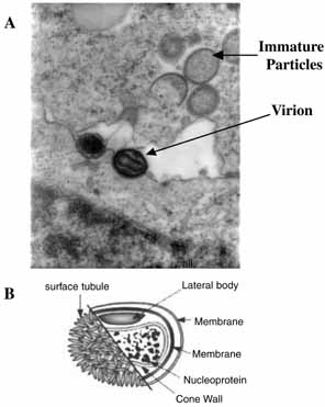
The vaccinial genome comprises a linear molecule of double-stranded DNA with covalently closed termini. Terminal hairpins constitute two isomeric, imperfectly paired, “flip-flop” DNA forms consisting of inverted complementary sequences. Variably sized, tandem repeat sequence arrays are present near the ends. Each virion contains one DNA molecule associated with proteins and organized in a nucleoprotein core. One or two lateral bodies are present in the concave region between the core wall and a single membrane, which contains a number of virus-encoded proteins. This virion form is known as the intracellular mature virus (Fig. 1B).

REPLICATION CYCLE

The intracellular replication cycle of vaccinia virus involves a sequence of events (Fig. 2).5 The vaccinia virion containing early RNA transcription machinery attaches to and fuses with the plasma membrane. Within 15 minutes the virion transcription machinery is activated (uncoating I). Early genes are expressed that code for a variety of functions that modify the host cell for optimal virus replication, attenuate the host response to infection, and mediate viral synthetic processes. After further uncoating (uncoating II), between 1.5 to 6 hours postinfection, the viral genome is replicated via concatamers.

Fig. 2 The cytoplasmic vaccinia virus replication cycle. IMV, intracellular mature virus; IEV, intracellular enveloped virus; CEV, cell-associated enveloped virus; EEV, extracellular enveloped virus.

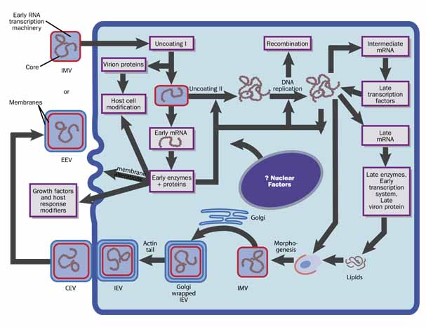
From progeny DNA templates, late transcription factors are expressed from intermediate genes, and late gene RNA is synthesized. Late genes encode the early transcription system, enzymes, and structural proteins necessary for virion assembly. By 4 hours postinfection, virion morphogenesis commences with the formation of membrane structures in the intermediate compartment and the packaging of resolved unit-length genomic DNA. The intracellular mature virus (IMV) has one membrane derived from the intermediate compartment. Some IMVs acquire an additional double layer of intracellular membrane derived from the trans Golgi network that contains unique virus proteins, the intracellular enveloped virus (IEV).

IEVs are transported to the periphery of the cell where fusion with the plasma membrane ultimately results in release of extracellular enveloped virus (EEV) or, if attached to the exterior surface of the plasma membrane, as cell-associated enveloped virus (CEV). While IMV and CEV/EEV are infectious, the external antigens on the two virion forms are different. Thus, each virion type probably binds to different cellular receptors and are likely uncoated by different mechanisms. EEVs are thought to be the most important form involved in cell-to-cell spread and systemic disease.

Back to Top
PREDISPOSING FACTORS
Exposure can occur by accidental autoinoculation of the eye or eyelid with virus from the inoculation site or from close physical contact with a vaccinee shedding virus before eschar detachment.6 The majority of ocular manifestations of smallpox vaccination in immunocompetent patients are self-limiting. Selective cases may require treatment with topical trifluridine, topical corticosteroids, and vaccinia immune globulin (VIG). More severe ocular and periocular manifestations reflect a higher viral inoculum, increased local viral replication, and host immune response.

RISK FACTORS

Several factors predispose to serious systemic complications that can follow vaccination or contact with a vaccinated person. In patients with impaired T-cell function, the primary vaccination site may not heal and the infection can spread both locally and by viremia. This condition, known as vaccinia necrosum (i.e., progressive vaccinia), produces necrotic lesions, most commonly in individuals who have HIV infection, cancer, or congenital T-cell deficiency, or who are on immunosuppressive chemotherapy.7 Cutaneous manifestations of HIV infection or other skin conditions, such as seborrheic dermatitis, impetigo, scabies, burns, and pemphigoid foliaceus, may also predispose to secondary contact acquisition of vaccinia. Smallpox vaccination is contraindicated in subjects with eczema or a history of eczema or atopic dermatitis, once they are susceptible to developing eczema vaccinatum. Eczema vaccinatum manifests with fever, lymphadenopathy, and generalized (often confluent) lesions in areas other than the vaccination site, frequently the face and limbs.

PREVENTIVE PRACTICES

Because autoinoculation is the most frequent mode for developing ocular vaccinia, the vaccination site should be covered until the scab detaches (which can be up to 21 days).8 A semi-permeable polyurethane dressing may be useful.9 Vaccinees should avoid touching the site when bathing. Patients with inflammatory eye disease requiring steroid treatment might be at increased risk of inadvertent inoculation as a result of touching or rubbing the eye.10

Health care workers who vaccinate or care for recently vaccinated patients should adhere to routine blood-borne pathogen precautions, including handwashing and disinfection of contaminated equipment with 70% alcohol or 10% bleach. Health care workers who are pregnant, have active atopic dermatitis, are immunocompromised, or take immunosuppressant drugs should not evaluate or care for recent vaccinees or those with vaccine-related adverse events.

Back to Top
EPIDEMIOLOGY
Between 1963 and 1968, 348 patients were identified with severe ocular vaccinia through the American Red Cross vaccinia immunoglobulin (VIG) distribution system.11 In the 1968 US smallpox vaccine survey, the frequency of ocular vaccinia was approximately 1 per 40,000 primary vaccinations.6 In the 2003 Vaccine Adverse Event Reporting System, ocular vaccinia occurred in approximately 1 per 10,000 vaccinations.12

Nearly three-fourths of ocular vaccinia cases occur after a primary vaccination, as compared to revaccinees or unvaccinated persons exposed to recent vaccinees. In revaccinees, complications from accidental autoinoculation onto the eye or through contact with a recent vaccinee are generally limited to blepharoconjunctivitis. Half of the patients are younger than age 4 years, with a slight preponderance of females.

Corneal involvement occurs in 6% of cases, although some studies have reported keratitis in 20% to 37%.13–16 Long-term sequelae, such as punctal stenosis, cicatricial lid changes, and madarosis are more common in cases with corneal manifestations than in ocular cases without keratitis. Reexamination of cases of vaccinial keratitis after 5 years reveals minor or no corneal changes, limited to light scarring, ghost vessels, and subepithelial opacification.11

Ninety percent of ocular vaccinia cases are diagnosed 1 to 9 days after primary vaccination, consistent with direct infection of the eye from the initial vaccine inoculum. The eye becomes infected with virus released 1 to 4 days after vaccination, when the integrity of the skin over the primary lesion site breaks down. The short time interval for development of ocular vaccinia argues against a consequence of viremia.

Back to Top
PATHOGENESIS
Biopsies of the primary vaccination site have characterized the histological changes that occur throughout the development and resolution of the vaccinial lesion, but little is known concerning the systemic pathological changes. Similarly, little is known concerning the pathogenesis of adverse ocular complications to vaccination. Based on studies with vaccinia virus infections of primary and established cell cultures and on knowledge of the annotated sequence of the vaccinia genome, the molecular pathogenesis of ocular vaccinial infections is emerging.17,18 Vaccinia virus has been evaluated as a possible viral vector for gene transfer into ocular cells.19,20

VIRAL PATHOGENESIS

Epidemiological studies suggest that ocular vaccinia virus infections are associated mainly with primary vaccinations. Histopathological changes are likely a direct result of virus-mediated necrosis and apoptosis of infected cells. Vaccinia virus has a very broad tissue tropism and infects most cell types that have been examined; however, replication in certain cell lineages such as human dendritic cells may be abortive.21 Abortive infection can lead to cell death and thus may also contribute to clinical signs.

Vaccinia virus infections of epidermal and fibroblast lineage cells usually lead to the production of progeny virions. Neighboring cells are infected via the EEV and CEV (Fig. 2). Because the onset of ocular vaccinia occurs rapidly after primary vaccination, the inflammatory response to infection likely does not contribute to acute pathological changes. On the contrary, the immune response to infection is inhibited by vaccinia virus expression of a number of secreted proteins that bind to, and inhibit the function of, key immune response regulators (complement interleukins, such as IL-18 and IL-1B, and interferon). Virus-encoded proteins also block the activation of NFκB, RNase L, and the double-stranded RNA-dependent protein kinase pathways induced by exposure to interferon.

CLINICAL PATHOGENESIS

Manifestations of ocular vaccinia depend on the location of the site where the virus initially infects epidermal cells via an abrasion.

Dermatoblepharitis

Infection of the cornified skin of the eyelid develops in a manner similar to the site of the primary vaccination. In a primary vaccination, the earliest changes are cytoplasmic and perinuclear vacuolation in the epithelium, accompanied by swelling, coagulation necrosis, intercellular edema, and vesicle formation. Within 48 hours after vaccination, a cup-shaped vesicle appears, with the stratum corneum and dermis forming the roof and the floor of the lesion, respectively.1 The lesion increases in size with time. Progression through the pustular phase is marked with mononuclear and polymorphonuclear cellular infiltrates and the formation of a crust of dense, homogeneous, deeply staining reticulum by 14 days postvaccination. Preauricular lymphadenopathy frequently accompanies eyelid and conjunctival lesions.

Conjunctivitis

Infection of the conjunctiva manifests as vesicles, often near the limbus. Like eruptions on the skin, they frequently evolve through a pustular phase and can be accompanied by conjunctival injection, hemorrhage, chemosis, mucopurulent discharge, and either a papillary or follicular reaction. Membranes or pseudomembranes can be present, with symblepharon formation as late sequelae.

Epithelial Keratitis

The pathological development of lesions on the cornea is different from those of the eyelid skin because of the lack of a cornified surface that would contain the developing vesicle. Experimental vaccinia virus infection of the rabbit cornea showed that infection begins deep in the corneal epithelium where free viral particles and virus-infected cells are observed.22 Similar findings were made after infection of mouse and human skin with ectromelia and molluscum contagiosum viruses, respectively. Poxviruses apparently need a cell that is less differentiated and more like the pluripotent keratinocyte stem cell for initial infection.23,24 Corneal epithelial lesions progressively enlarge for 3 to 5 days. Progeny virus from the initially infected cells infect neighboring cells until the bed of the lesion rests on the stroma. Infected cells erode, producing punctate epithelial keratitis, dendritic epithelial keratitis, and geographic epithelial keratitis.

Stromal Keratitis and Keratouveitis

With time polymorphonuclear cellular infiltrates are associated with the lesions, which usually heal by 10 days postinfection. Vaccinial keratitis can evolve to nummular anterior stromal infiltrates, disciform keratitis with granulomatous keratic precipitates, and interstitial keratitis.

Back to Top
ANIMAL MODELS
Experimental vaccinial keratitis has been studied in rabbits and monkeys. Vaccinia is generally introduced into the cornea by scarification. The keratitis is easily and reliably produced; its activity can be assessed by slit-lamp biomicroscopy and fluorescein staining. The epithelium can be removed for culture, and the ocular surface is accessible for topical application of therapeutics.25 These models have been useful in evaluation of a number of antiviral agents, including topical trifluridine, vidarabine, idoxuridine, and interferon.25–28 A study of the effect of human VIG on rabbit vaccinial keratitis showed VIG to be of no benefit and, in high doses, may have contributed to larger corneal scars.29
Back to Top
LABORATORY DIAGNOSIS
Because most cases of ocular vaccinia are associated with recent smallpox vaccination or exposure to a recent vaccinee, laboratory confirmation of the clinical diagnosis may not be needed. In cases in which the history may not be obtainable or the patient is unaware of inadvertent exposure, ancillary laboratory testing may be helpful.

Smears of mucopurulent discharge contain numerous polymorphonuclear cells. Direct scraping of lesions may reveal eosinophilic cytoplasmic inclusions called Guarnieri bodies. Vaccinia virus can be propagated from swabs of lesions plated onto a wide variety of tissue-culture cells, including HeLa, rabbit or monkey kidney, MRC-5, and human embryonic kidney cells.30 Restriction endonuclease analysis of infected cell DNA extracts can be used for confirmation purposes.

Electron microscopy, polymerase chain reaction gene amplification, and viral culture are currently undergoing multicenter validation studies.4 After regulatory approval, testing will be made available in the United States through the Laboratory Response Network, a system of private and public health laboratories that can be accessed through consultation with state and local health departments.

Back to Top
REFERENCES

1. Fenner F, Henderson DA, Arita I: Smallpox and its Eradication. Geneva: World Health Organization , 1988

2. Jezek Z, Fenner F: Human Monkeypox. Basel: Karger, 1988

3. Charteris DG, Bonshek RE, Tullo AB: Ophthalmic molluscum contagiosum: clinical and immunopathological features. Br J Ophthalmol 79:476–481, 1995

4. Pepose JS, Margolis TP, LaRussa P, Pavan-Langston D: Ocular complications of smallpox vaccination. Am J Ophthalmol 136:343–352, 2003

5. Moss B: The viruses and their replication, 4th ed. In Knipe DM, Howley PM, eds. Fields' Virology. Philadelphia: Lippincott, Williams & Wilkins, 2001:2849–2883

6. Lane JM, Ruben FL, Neff JM: Complications of smallpox vaccination, 1968: results of ten statewide surveys. J Inf Dis 122:303, 1970

7. Centers for Disease Control and Prevention: Smallpox vaccination and adverse reactions: guidance for clinicians. MMWR Dispatch 52:1, 2003

8. Semba RD: The ocular complications of smallpox and smallpox immunization. Arch Ophthalmol 121:715–719, 2003

9. Fulginiti VA, Papier A, Lane JM, Neff JM, Henderson DA: Smallpox vaccination: a review, part II. Adverse events. Clin Infect Dis 37:251–271, 2003

10. Wharton M, Strikas RA, Harpaz R, et al: Recommendations for using smallpox vaccine in a pre-event vaccination program. Supplemental recommendations of the Advisory Committee on Immunization Practices (ACIP) and the Healthcare Infection Control Practices Advisory Committee (HICPAC). MMWR Recomm Rep 52:1–16, 2003

11. Ruben FL, Lane JM: Ocular vaccinia. An epidemiologic analysis of 348 cases. Arch Ophthalmol 84:45–48, 1970

12. Update: adverse events following civilian smallpox vaccination–United States, 2003. MMWR Morb Mortal Wkly Rep 52:819–820, 2003

13. Sedan J, Ourgaud AG, Guillot P: Les accidents oculaires d'origine vaccinale observés dans le Department des Bouches-du-Rhone au cours de l'epidemie variolique de l'hiver. Ann Oculist 186:34, 1953

14. Ellis PP, Winograd LA: Current concepts of ocular vaccinia. Trans Pac Coast Ophthalmol Soc 44:141, 1963

15. Jones BR, Al-Hussaini MK: Therapeutic considerations in ocular vaccinia. Trans Ophthalmol Soc UK 83:61, 1963

16. Duke-Elder S: The saga of a century. Am J Ophthalmol 59:163, 1965

17. Buller RM, Palumbo GJ: Poxvirus pathogenesis. Microbiol Rev 55:80–122, 1991

18. Goebel SJ, Johnson GP, Perkus ME, Davis SW, Winslow JP, Paoletti E: The complete DNA sequence of vaccinia virus. Virology 179:247–266, 517–563, 1990

19. Banin E, Obolensky A, Piontek E, et al: Gene delivery by viral vectors in primary cultures of lacrimal gland tissue. Invest Ophthalmol Vis Sci 44:1529–1533, 2003

20. Chowers I, Banin E, Hemo Y, et al: Gene transfer by viral vectors into blood vessels in a rat model of retinopathy of prematurity. Br J Ophthalmol 85:991–995, 2001

21. Drillien R, Spehner D, Bohbot A, Hanau D: Vaccinia virus-related events and phenotypic changes after infection of dendritic cells derived from human monocytes. Virology 268:471–481, 2000

22. Matas BR, Dawson CR, Togni B: Vaccinia-infected rabbit cornea: a transmission electron microscopic study. Invest Ophthalmol 12:782–786, 1973

23. Buller RM, Burnett J, Chen W, Kreider J: Replication of molluscum contagiosum virus. Virology 213:655–659, 1995

24. Chen N, Buller RM, Wall EM, Upton C: Analysis of host response modifier ORFs of ectromelia virus, the causative agent of mousepox. Virus Res 66:155–173, 2000

25. Kaufman HE, Nesburn AB, Maloney ED: Cure of vaccinia infection by 5-iodo-2'-deoxyuridine. Virology 18:567–569, 1962

26. Hyndiuk RA, Okumoto M, Damiano RA, Valenton M, Smolin G: Treatment of vaccinial keratitis with vidarabine. Arch Ophthalmol 94:1363–1364, 1976

27. Hyndiuk RA, Seideman S, Leibsohn JM: Treatment of vaccinial keratitis with trifluorothymidine. Arch Ophthalmol 94:1785–1786, 1976

28. Neumann-Haefelin D, Sundmacher R, Sauter B, Karges HE, Manthey KF: Effect of human leukocyte interferon on vaccinia- and herpes virus-infected cell cultures and monkey corneas. Infect Immun 12:148–155, 1975

29. Fulginiti VA, Winograd LA, Jackson M, Ellis P: Therapy of experimental vaccinal keratitis: effect of idoxuridine and VIG. Arch Ophthalmol 74:539–544, 1965

30. Lee SF, Buller R, Chansue E, et al: Vaccinia keratouveitis manifesting as a masquerade syndrome. Am J Ophthalmol 117:480–487, 1994

Back to Top