Chapter 95
Periocular Involvement of Papillomavirus and Molluscum Contagiosum Virus
DAVID S. BARDENSTEIN and JONATHAN H. LASS
Main Menu   Table Of Contents

Search

HUMAN PAPILLOMAVIRUS
MOLLUSCUM CONTAGIOSUM VIRUS
SUMMARY
REFERENCES

Both human papillomavirus (HPV) and molluscum contagiosum virus (MCV) cause squamoproliferative lesions of periocular tissues, and the incidence of both has become much higher given the current rise in sexually transmitted diseases and severe immunosuppression. More sensitive molecular biologic and genetic techniques have revealed that the role of HPV in periocular disease is much greater than was thought previously. HPV is also now known to encompass entities previously unsuspected to have a viral etiology. MCV has been known to cause molluscum contagiosum, but the new techniques have advanced the study of its biology. Periocular involvement by these two viruses is far less common than in other sites, and most of the genetic and molecular biologic data are derived from study of these other sites. Since optimal diagnosis and treatment depend on a full understanding of the problem at hand, this chapter will discuss not only periocular lesions, but also current concepts of the biology of these viruses derived from other sites.
Back to Top
HUMAN PAPILLOMAVIRUS
HPV is a small double-stranded DNA virus. Until recently it was classified as a papovavirus, but it is now considered a distinct family1 for two reasons: (1) molecular biology and genetics studies revealed that it has different-sized DNA, with different sequences and larger capsids2; and (2) unlike papovaviruses, papillomaviruses are unable to propagate in culture (a significant functional difference).3

The papillomavirus virion consists of an outer protein capsid, 55 nm in diameter, enclosing the DNA. The capsid is made of 72 capsomeres that form an icosahedron. The capsid is composed of two groups of proteins: a major protein (molecular weight [MW] 54 kd) and a minor protein (MW 76 kd). The major protein is antigenically similar between animals and humans. The minor protein appears to be type-specific. Papillomavirus coats differ from those of other DNA viruses in that they do not have an outer lipid envelope. They are resistant to inactivation by freezing, ether, and desiccation, but sensitive to high temperature, aldehyde fixatives, and detergents.

Classification of papillomaviruses is based on the host organism and further typed and subtyped in accordance with their degree of genomic homology. A virus is named on the basis of its natural animal host, and host specificity is high. Using solution hybridization, viral isolates demonstrating less than 50% homology are considered new types, isolates demonstrating more than 50% are considered subtypes, and those with almost 100% homology are considered variants. It is important to realize that 50% hybridization corresponds to almost 90% homology of DNA sequence.

Antigenic serotyping of papillomaviruses as is done with adenoviruses is not possible because of insufficient antigen and the failure of the virus to replicate in vitro. To date, more than 70 types of HPV have been identified, and these have been divided into the following three groups:

  Genital-mucosal lesions
  Nongenital lesions
  Epidermodysplasia verruciformis4

The types most often seen in the periocular tissues are from the first group.

Another method of grouping HPV subtypes is on the basis of clinical behavior, as follows:

  High-risk subtype: Those associated with invasive malignant tumors (e.g., HPV-16, HPV-18)
  Low-risk subtype: Those associated with benign tumors (e.g., HPV-6, HPV-11).

Simultaneous infection with multiple HPV subtypes has been reported.

The HPV genome is a closed circle of double-stranded DNA, 7900 base pairs in length. The genome consists of nine open reading frames (ORFs) and an upper regulatory region (URR), all located on one strand of the DNA.5 The nine ORFs encode nine proteins, which are divided into seven early or “E” proteins and two late or “L” proteins, which are capsid proteins. El ORF encodes proteins that are important in modulating DNA replication.6,7 E2 ORF encodes proteins that regulate viral gene transcription.8–10 E4 ORF encodes a protein that makes up a large portion of wart tissue but whose function is unknown.11 E5 ORF encodes a small protein found in the cell membrane that can transform cultured murine cells.12 E6 ORF encodes a protein involved in cellular transformation and control of the number of viral DNA copies. E7 ORF encodes a protein involved in transcriptional transactivation and cellular immortalization.

PATHOGENESIS AND TUMORIGENESIS

HPV infection is thought to occur when virus is inoculated into viable epithelium. Loss of integrity of epithelium is thought to enhance susceptibility to infection.13 The viral DNA infects basal cells of the epithelium. There it remains extrachromosomal and replicates along with the host cell DNA.14 Viral DNA episomes are carried upward through the differentiating epithelium. In the malpighian cell layer, transcription of viral DNA occurs. Viral DNA replication occurs in the granular cell layer, and hundreds of viral DNA replicas may be made. The viral capsid proteins are then synthesized and the virions assembled in the host cell nucleus as it progresses to the stratum corneum.

Specific questions regarding HPV tumorigenesis arise: Why do certain types cause certain pathology? Are there group differences between high- and low-risk types?

It appears that HPV DNA in benign lesions from low-risk types rarely inserts in the host genome but exists as an episome, whereas in severe dysplasia and carcinoma associated with high-risk HPV types, the HPV DNA incorporates into the host genome.15 Moreover, neoplasia is not simply the result of viral DNA insertion or its site of insertion, but of insertion of the viral DNA into the host genome with concurrent disruption of the E2 ORF. Disruption of the E2 ORF allows for increased production of E6 and E7 proteins, which appear to exert important effects on the tumor suppressor proteins p53 and the product of the retinoblastoma (Rb) gene.

Tumor suppressor proteins typically have regulatory functions that affect normal cell growth. Homozygous absence of these proteins allows loss of normal cell regulation and tumorigenesis, as in the example of the Rb gene where absence of a normal copy of the Rb gene leads to clinical retinoblastoma. E6 binds and accelerates the degradation of p53 protein, which is active in limiting cell reproduction.16 E6 protein from high-risk HPV-16 and HPV-18 is more effective at inactivating p53 than that from low-risk types. High-risk E6 binds E6-associated protein, which is necessary for the degradation of p53; low-risk E6 binds p53 but does not lead to degradation.17,18 High-risk HPV DNA also encodes truncated E6* proteins, whose function is not yet known.15

E7 protein binds the product of the Rb and p107 genes, both of which are negative growth regulators.19,20 Although originally thought to require E6 to cause immortalization of cells, E7 can immortalize cells by itself when in the presence of a strong promoter.21,22 However, it does appear that E6 and E7 act synergistically in the tumorigenic process.

In studies of HPV infection of cultured cells, so-called cytopathic effects—loss of contact inhibition, alteration of cell shape, loss of growth factor dependence, and immortalization—have been described. Three of the early genes, E5, E6, and E7, are believed responsible for these properties. The amounts and forms of these proteins produced vary with different HPV subtypes and often correlate with their clinical behavior.

ROLE OF HUMAN PAPILLOMAVIRUS IN PERIOCULAR DISEASE

The relationship between periocular squamous epithelial proliferations and viral infection was historically addressed by the categorization of these lesions as viral (infectious) and noninfectious.23 Viral papillomas were believed to occur more often in young patients and to be essentially benign in nature. Noninfectious papillomas were thought to be related to ultraviolet exposure, more commonly found in adults, and capable of undergoing malignant transformation. The role of HPV was explored because of similarities in the histology of HPV-induced cervical disease.

The earliest work by Lass and associates24 using immunohistochemical techniques directed at antigens of the viral capsid demonstrated HPV involvement in 2 of 21 conjunctival papillomas examined. In a subsequent immunohistochemical study, McDonnell and colleagues25 detected HPV-common antigen in 46% of 50 papillomas and 8% of 61 dysplastic or carcinomatous lesions of conjunctival epithelium. Subsequent application of more sensitive DNA in situ hybridization techniques showed HPV-11 DNA in one case in which HPV antigen was detected.26 Other cases of papilloma with detectable HPV antigen and DNA were reported.27–29 Using in situ DNA hybridization, HPV 6 or HPV 11 was detected in 65% of 23 papillomas but in none of 28 dysplastic lesions.30 In a study using polymerase chain reaction (PCR)-aided analysis, six of six tissue specimens with conjunctival or corneal dysplasia were found to have HPV-16 DNA, but not HPV-18 DNA.31 Lauer and co-workers used PCR-aided analysis to detect the E6 region of HPV DNA in four of five conjunctival intraepithelial neoplasia lesions.32 HPV-16 was found in all four lesions, and coinfection with HPV-18 was found in two of these. One study from an experienced laboratory did not detect HPV-6, -11, -16, or -18 in specimens of squamous cell carcinoma of the conjunctiva with the use of either in situ hybridization or PCR-aided analysis, and the authors speculated that another HPV type might be involved.33 In a larger series, HPV-16 DNA was found using PCR-aided analysis in 88% of 42 tissue specimens and in 83% of six conjunctival swabs from patients with dysplastic disease.34,35 A small series of bilateral squamous conjunctival tumors associated with HPV-16 also has been reported.36

Of the eyes in the McDonnell PCR DNA studies35,36 in which conjunctival swab specimens were found adequate for analysis, the clinically uninvolved eye of one patient showed evidence of HPV DNA and HPV-16 DNA that persisted years after “successful” excision of the lesion in the affected eye. This finding demonstrates the following: (1) an unsuspected subclinical HPV infection can exist; and (2) induction of clinical disease might be multifactorial.30,34 In the McDonnell studies,35,36 corneal swabs from women with cervical HPV-16 disease contained HPV-16 DNA in 76.5% of patients, none of whom had clinical ocular disease.

Data on HPV detection in other squamoproliferative conditions have been limited. Identification of HPV in pterygia has been controversial. One study using immunohistochemical analysis of HPV antigen found HPV in 56% of pterygia patients and in 25% of controls.37 Other studies have used pterygia as negative control tissue in which HPV DNA was not detected.30,34 HPV-16 DNA has been detected in recurrent squamous cell carcinoma of the eyelid.38 HPV DNA also has been detected in epithelial tumors of the lacrimal sac.39 Three of three papillomas contained HPV-11 DNA. One of three carcinomas studied contained HPV-18 DNA. In another study,40 a case of inverted papilloma was positive for HPV antigen and polytypic DNA probe, whereas specific probes for HPV-6 and HPV-11 were negative.

In summary, HPV-6, -11, -16, and -18 have been identified in periocular squamous epithelial lesions. Similar to findings in the anogenital region, HPV-6 and HPV-11 are associated with benign lesions and HPV-16 and HPV-18 with dysplastic or malignant lesions. Infection can be found in the following cases: (1) in the clinically normal eyes of patients with infection of the other eye and after successful excision of lesions; and (2) in the clinically uninvolved eyes of patients with cervical infection. This may reflect the multifactorial nature of this disease. The rates of HPV detection in all types of ocular disease approach those in anogenital disease when PCR-aided analysis is used.

EPIDEMIOLOGY

The analysis of HPV-related lesions of the periocular tissues is complicated by a variety of factors, including the advent of new technologies, demographic considerations, occult infections, immune deficiency, and risk factors such as solar exposure and smoking. As mentioned above, some types of lesions now known to harbor HPV in at least some cases were previously not thought to be virus induced. Thus both the spectrum and prevalence of HPV-related periocular disease have been underestimated. Demography is an important issue because these lesions are relatively uncommon, and each has a distinct clinical and pathologic presentation, affecting very different age groups and both genders. Until extensive studies are done regarding the prevalence of HPV infection in these diseases, a comprehensive discussion of ocular HPV demographics will not be possible. Demographic factors can be considered both by studying the demographics of HPV infection in other sites where data are available and by analyzing demographic characteristics based on the clinical type of lesion.

General Demographics

The only reported systematic demographic study of ocular disease (thought to have an HPV-related etiology) has evaluated the occurrence of histologically proved conjunctival dysplasia and carcinoma during a 10-year period in Brisbane, Australia.41 An average incidence of 1.9 cases per 100,000 persons per year was calculated. The male to female ratio was 2.3:1.

WOMEN. The most comprehensive available demographic data have been reported on anogenital lesions, specifically those of the female genital tract. The incidence of new anogenital condylomatous lesions or dysplasias in the United States is 1 million per year.42 Since infection can exist without clinical disease, however, the incidence of infection is probably much higher. This is evident from the data on anogenital HPV infections. Among female patients aged 14 to 23 years old, 32% had cervical swabs that contained HPV DNA.43 In two thirds of these specimens, HPV-16, HPV-18, or HPV-31, types frequently associated with cervical carcinoma, were detected. Among 505 Norwegian women requesting first-trimester termination of conception, 6.1% had detectable HPV in cervical swabs.44 In a study of older women (65 and older), 3.5% had HPV DNA detected in cervical swabs.45

OCCULT INFECTION AND IMMUNE DEFICIENCY. Among men, occult HPV infection has been found in up to 40% of genitourinary clinic patients with the use of cytology and DNA hybridization studies.46 Homosexual men can have up to a 65% prevalence of detectable HPV in their anal mucosa.47 The fact that occult infection with HPV-16 was found in the oral mucosa of 43% of 62 asymptomatic subjects without significant known risk factors is perhaps an even more significant finding.48

Immune deficiency enhances the manifestation of HPV clinical disease. Warts were seen in 43% of unselected renal transplant patients.49 An increased incidence of skin lesions also was seen in a group of AIDS patients.50

OTHER RISK FACTORS. Other factors associated with squamoproliferative diseases include solar exposure and smoking. A case control study in the ophthalmic literature claimed there was no effect of solar exposure on squamous epithelial disease.51 A larger study in a renal transplant population, however, showed that warts and carcinomas were more prevalent in this group compared to control subjects. When patients were separated on the basis of high versus low solar exposure, all carcinomas and more than twice the incidence of warts were found in the group with high solar exposure.49

Ocular Transmission

Ocular transmissibility of HPV is inferred from the fact that sexual partners of patients with genital HPV have an increased rate of infection and that neonates of mothers with genital HPV may become ocularly infected at birth.52,53 An infant of a mother with condylomata acuminata was found to have conjunctival and respiratory papillomas that tested positive for HPV-6 DNA.54

DEMOGRAPHICS. Demographic data are available on the clinical periocular lesions likely to be related to HPV infection or transformation. These include eyelid warts, eyelid papillomas, conjunctival papillomas, conjunctival dysplasia and carcinoma, lacrimal sac papillomas, and pterygia.

Nonocular cutaneous HPV lesions most commonly occur in patients between the ages of 12 to 16 years old, but demographic data on clinically diagnosed eyelid papillomas are not available. In the slightly older sexually active population, anogenital lesions are becoming more common. Conjunctival papillomas comprised 12% of epibulbar lesions; 7% of these occurred in patients who were less than 20 years old.55 In a later series of 302 epibulbar lesions in children less than 17 years old, conjunctival papillomas constituted 7% of lesions.56 Conjunctival papillomas are more often multiple in children. Despite the large number of patients with AIDS who show multiple molluscum contagiosum of the eyelids and an increase in nonocular papillomas, a similar increase in HPVassociated lesions in the conjunctiva or eyelids has not been noted in this group.

Conjunctival intraepithelial neoplasia and squamous cell carcinoma are most common in late adulthood, with a 3:1 to 20:1 male to female ratio. Patients with squamous cell carcinoma are an average of 10 years older (mean = 68.7 years; range = 41 to 96 years) than patients with dysplasia (mean = 58 years; range = 8 to 90 years).57,58 Conjunctival intraepithelial neoplasia and squamous cell carcinoma are thought to be associated with solar exposure, although collecting accurate direct quantitative data of ocular sun exposure has proved difficult.

CLINICAL PRESENTATION AND DIAGNOSIS

Comprehensive discussions of the presentation and diagnosis of each lesion presumed to be HPV-induced are beyond the scope of this chapter and have been reviewed elsewhere.59,60 Rather, a brief discussion of each lesion type will be presented.

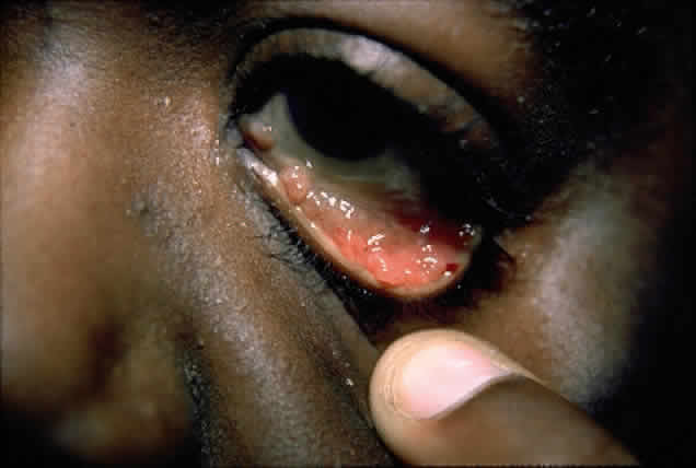
In the conjunctiva, HPV can induce papillomatous lesions (papillomas and inverted papillomas) and epithelial dysplastic lesions consisting of conjunctival intraepithelial neoplasia and squamous cell carcinoma. Conjunctival papillomas were traditionally divided into infectious, limbal, and inverted types; however, current data suggest that all types have a viral etiology. All sites of the conjunctiva can be affected, and involvement of the contiguous epithelium of the lacrimal canaliculus, lacrimal sac, and nasolacrimal duct has been noted to occur.38,61,62 Conjunctival papillomas can be pedunculated or sessile and are typically shiny, flesh-colored, and papillomatous (Fig. 1). Each papillary frond has a vascular core that is easily seen through the nonkeratinized epithelium. The papillomatosis is responsible for the typical appearance of evenly spaced vessels throughout the lesion. If keratinized, these lesions may appear whitish and dull. Papillomas can be either unilateral or bilateral, and they can be either solitary or multifocal (Fig. 2). Inverted papillomas are the least common type of papilloma in all sites, but they constitute a larger portion of lacrimal sac tumors than other periocular tumors.63,64

Fig. 1. Solitary conjunctival papilloma in fornix of immunocompetent man.

Fig. 2. Confluent conjunctival papillomata in patient with multiple recurrent lesions and chronic immunosuppression after multiple renal transplants.

The diagnosis is usually made on the basis of the history and clinical appearance. In questionable cases, excisional biopsy should be performed. Conjunctival papillomatous lesions must be differentiated from squamous cell carcinomas, which may occur as a papillomatous variant indistinguishable from both typical papillomas and other squamoproliferative lesions.

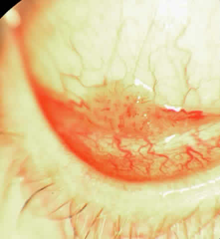
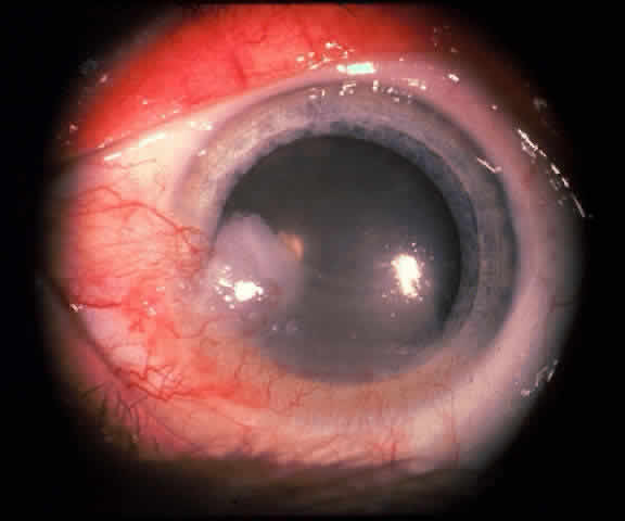
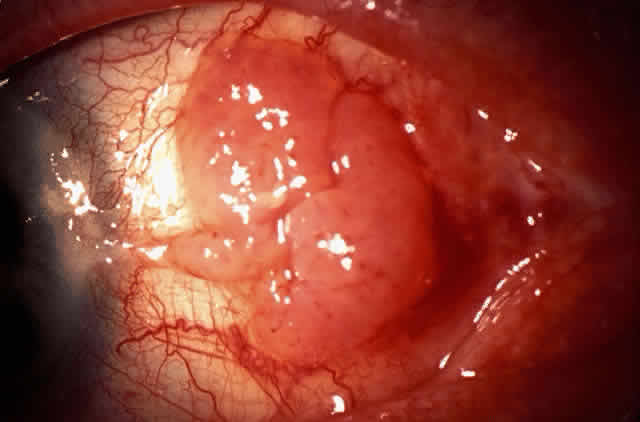
The epithelial dysplastic lesions—conjunctival intraepithelial neoplasia and squamous cell carcinoma—almost always begin at the limbus, most commonly in the interpalpebral zone.57,58 The initial appearance is a gelatinous, gray lesion with some thickening (Fig. 3). There may be increased vascularization. The epithelial changes may extend onto the cornea, causing a hazy, gray appearance. Tumor vascularity lags behind the lead margin of the epithelial change. As the lesions progress from dysplastic to carcinomatous, they become thicker, more vascularized and may show keratinization, giving a leukoplakic appearance (Fig. 4). Lesions confined to the epithelium move freely over the sclera, whereas invasive lesions are fixed to underlying tissue. Occasionally squamous cell carcinomas appear identical to classic papillomas.

Fig. 3. Multiple eyelid papillomas in an immunocompetent woman.

Fig. 4. Eyelid papilloma, histology. Low-power view of frond-like architecture with acanthotic epithelium with hyperkeratosis and fibrovascular cores. (Original magnification × 25)

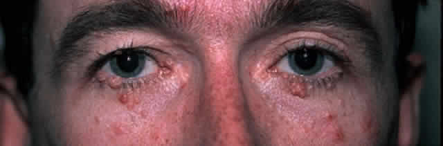
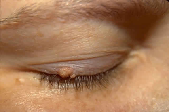
The diagnosis is at best suspected clinically but histologic confirmation allows a distinction between epithelial dysplasia and entities such as pterygium, keratosis, melanoma, nevus, and other lesions. Pterygia are almost always in the horizontal meridian. A lesion centered away from the limbus and one in other than a horizontal meridian should be suspect for a dysplastic lesion. Handling and processing these specimens to ensure that an accurate diagnosis can be made requires the active participation of a pathologist with experience evaluating this type of specimen. Eyelid papillomas have the typical appearance of skin warts with a flesh colored, papillomatous surface (Fig. 5). Hyperkeratotic scaling is common. Dysplastic and squamous cell carcinomas of the eyelids typically show an elevated flesh colored lesion with hyperkeratosis and abnormal vasculature. Eyelid lesions must be differentiated from seborrheic keratosis, acrochordons, nevi, and other squamoproliferative lesions. Lacrimal sac papillomas and carcinomas are very uncommon tumors.63 Patients present with epiphora and a medial canthus mass, usually above the medial canthal tendon. Bloody epiphora is suggestive of malignancy. Diagnosis is made with the use of imaging and biopsy.

Fig. 5. Conjunctival papilloma, histology. High-power view of acanthotic epithelium surrounding fibrovascular cores. Koilocytosis is absent. (Original magnification × 200)

PATHOLOGY

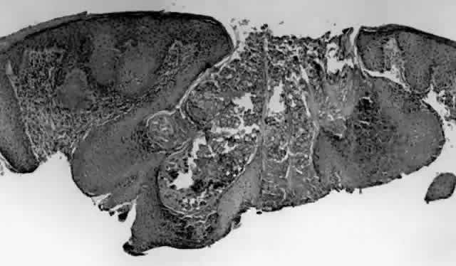
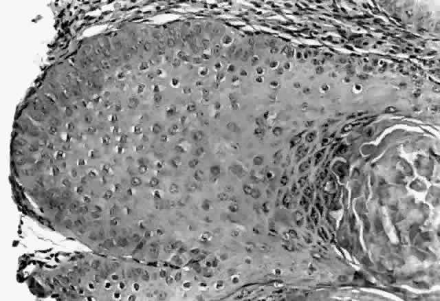
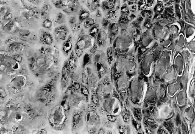
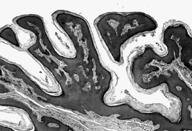
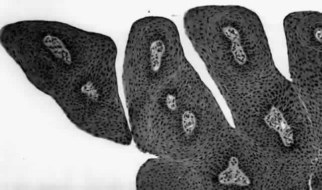
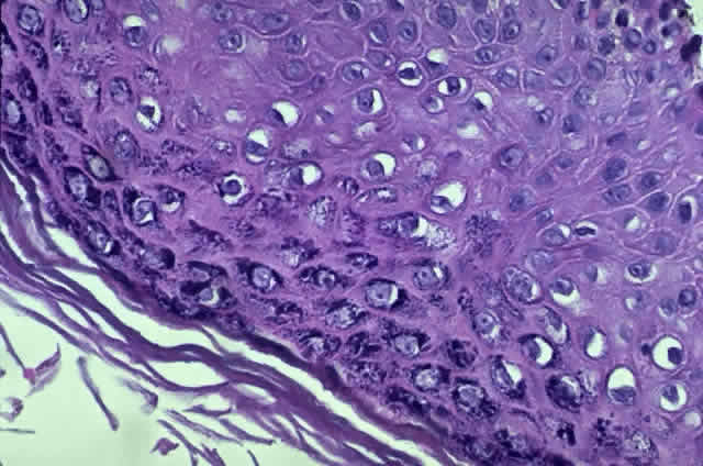
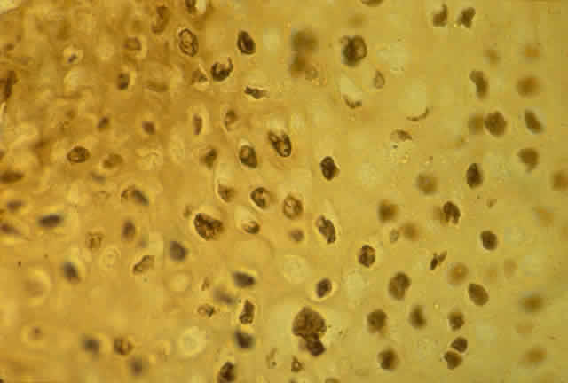
HPV-induced conjunctival squamous papillomas demonstrate papillomatosis, acanthosis, and koilocytosis (Fig. 6). Because they are mucous membrane lesions, those of the conjunctiva and lacrimal sac show no granular cell layer but will show keratinization if they are large or exposed (Fig. 7). In our experience the frequency of koilocytes is variable, even in lesions almost certain to be viral (Fig. 8). Scattered inflammatory cells may be present in the cores or epithelium. Virus can be detected immunohistochemically in the nuclei of the cells (Fig. 9).

Fig. 6. Eyelid papilloma, histology. High-power view of acanthotic epithelium and koilocytes. (Original magnification × 400)

Fig. 7. Immunohistochemical detection of human papillomavirus antigen. (Original magnification × 400)

Fig. 8. Conjunctival intraepithelial neoplasia. Gray, gelatinous mass is centered at limbus and has prominent vessels. A veil of intraepithelial disease extends laterally across the inferior third of the cornea.

Fig. 9. Squamous cell carcinoma of conjunctiva.

Early dysplastic lesions (conjunctival intraepithelial neoplasia) show thickened epithelium with extension of basal-like cells beyond the basal layer, but cells continue to mature into flat squamous cells. Mitoses are also seen higher in the epithelium. As the degree of dysplasia worsens, the more superficial epithelium is replaced at higher levels by basal-type cells, mitoses can be seen further from the basal layer, and atypia of cells becomes more pronounced. With squamous cell carcinoma in situ, there is full-thickness dysplasia, cellular atypia is variable, and mitoses can be seen at all layers of the epithelium. Invasive squamous cell carcinomas are usually well differentiated. Pleomorphism, hyperchromaticity, dyskeratosis, and horn pearls are seen. Epithelial cell clusters can be seen invading below the basement membrane. Inflammatory cells are common in the substantia propria.

Squamous papillomas of the eyelid are exophytic lesions with multiple fronds, each showing acanthotic epithelium around a fibrovascular core. There is typically hyperkeratosis and koilocytosis. Inflammatory cells may be present.

The histopathology of lacrimal sac epithelial tumors is typical of papillomas or squamous cell carcinoma. Koilocytosis may be present.

Historically, viral involvement was indicated by the presence of koilocytosis. Later, immunohistochemical analysis of HPV-common antigens allowed the first demonstration of viral components in lesional cells (see Fig. 8). Newer, more specific DNA-based techniques allowed typing of viruses. In situ hybridization, which bridged DNA and immunohistochemical techniques, was somewhat more sensitive than either technique alone. The development of PCR amplification allowed detection of a tiny amount of original source DNA and, with proper controls, powerful statements could be made about specific virus involvement. Because DNA is relatively stable compared to many antigens, DNA techniques can be used on archival material or on specimens fixed with aldehydes and embedded in paraffin.

TREATMENT AND PROGNOSIS

The treatment of periocular HPV-induced lesions is divided on the basis of anatomic considerations. In contrast to the epithelium of the eyelid skin and genital tract, where tissue destruction can be better tolerated, the ocular surface has a delicate balance of tear film and epithelial and stromal tissues, and therefore treatment must be gentler to afford preservation of vision. Certain HPV treatment modalities thus are not applicable to periocular lesions. Additionally, most treatment data were generated before the full spectrum of HPV involvement in periocular epithelial disease was suspected and before the biology of HPV infection was understood, and thus the theoretic rationale behind these original approaches is of uncertain value.

Excision or local destruction of involved tissue has been the mainstay of HPV-induced lesion treatment. When the distinction between infectious or viral papillomas and other presumably noninfectious lesions was made on clinical grounds, excisional surgery was deemed adequate for noninfectious lesions, whereas infectious lesions required some attempt to “sterilize” the base of the lesion. In view of our current understanding, such distinctions are probably of little value. Modalities that have been used to achieve sterilization include cryotherapy, cautery, chemical agents, keratolysis, and radiation.57,58,65,66

Special surgical modalities such as the argon and carbon dioxide laser have been used as to destroy periocular HPV lesions. While initially reported to be more effective than routine surgery in cervical lesions, further studies showed no difference between the two.67,68 In addition, the plume of vaporized tissue has been shown to contain HPV DNA, which could theoretically cause spread of disease, specifically of the respiratory tract, to operating room personnel and the surgeon.69,70 Such cases have not been reported, and one study has suggested that the HPV material recovered from the plume is not clinically infectious.71 This issue is still not fully resolved, and the fact that many patients have simultaneous infection with HIV and other viruses warrants care before selecting vaporizing modalities and employment of proper plume-management techniques.

Chemotherapeutic efforts have involved antimetabolites. Topical 5-fluorouracil has appeared effective in treating lesions of the female genital tract. The sensitivity of the ocular surface, however, is an important consideration; topical 5% 5-fluorouracil applied to the rabbit cornea was found to retard epithelial healing.72 This agent and the related agent mitomycin C might have the potential to retard epithelial proliferation without affecting the virus specifically. These agents can cause serious complications, including severe corneal scarring and melting of the ocular connective tissue coat. Recent studies have claimed no significant side effects associated with topical 0.01% mitomycin C solution when used for pterygium or conjunctival intraepithelial neoplasia.73,74

Immunotherapeutic efforts have employed dinitrochlorobenzene and interferon. Isolated case reports using multiple topical applications of dinitrochlorobenzene after systemic sensitization have been successful.75–77 Intralesional injection was used to augment the effect. The method has not been studied in any series or compared to alternate methods, leaving its efficacy uncertain. Interferon alfa has been used in the treatment of recurrent conjunctival papillomas following reports of initial success in other sites.78 Patients in the initial report of five cases underwent surgical excision of the tumor, followed by daily intramuscular injections of 5 million units interferon alfa-n1 for 1 month.79 This interferon was extracted from cultured lymphoblastoid cells. Administration was then decreased to two to three injections weekly for 6 months. Subsequently, some patients had administration stopped, while others continued on more prolonged therapy. All patients experienced reduction in the rate of tumor recurrence and extent of disease; however, three patients experienced recurrence, in two of whom the severity of the disease was judged to be less after interferon treatment. An additional case of interferon treatment of papillomas has also been reported.80

Other factors must be considered regarding the use of interferon. Systemic side effects can be significant, including fever, chills, weight loss, leukopenia, thrombocytopenia, and alterations in renal and hepatic function.81,82 Side effects have occurred with recombinant interferon alfa-2b as well as with extracted human interferon.83 Intralesional injections of cervical lesions still resulted in side effects in 83% of patients. Long-term studies of laryngeal papillomatosis and cervical disease have shown no long-term benefit compared to other modalities.84 In addition, interferons are extremely expensive, and the treatment regimen is very demanding. Currently, condyloma acuminata of the anogenital region is the only indication for interferon use in HPV disease that is approved by the Food and Drug Administration.

Although treatment of HPV-induced disease has been challenging, other agents might provide benefit. Retinoic acid (vitamin A) has been shown to regulate squamous epithelial differentiation. It inhibits growth of HPV-18-positive immortalized cervical carcinoma cells and downregulates production of proteins E6 and E7 in HPV-16-infected human keratinocytes.85,86 Because HPV replication is dependent on the state of differentiation of the epithelial cells, retinoic acid might alter HPV replication. Retinoic acid can also prevent malignant transformation. Retinoids have been initially effective in treating warts in immunosuppressed patients, but lesions have recurred after cessation of therapy. Topical retinoic acid has been investigated for the treatment of keratoconjunctivitis sicca and appears to be noninjurious to the ocular surface.87

Treatment of conjunctival papillomas remains enigmatic, with recurrence rates approaching 40% or more. Ironically, the dysplastic HPV-induced lesions, conjunctival intraepithelial neoplasia, and squamous cell carcinoma have higher rates of successful treatment. Recurrence of conjunctival intraepithelial neoplasia or squamous cell carcinoma is less than 5% when excision and cryotherapy are used together, compared to 33% when either modality is used alone.66 It is hoped that a better understanding of the pathogenesis of these lesions will allow for a more efficacious treatment.

Skin carcinoma and dysplasia are effectively cured with excision and frozen-section control of the margins. Metastasis occurs less than 1% of the time.

The number of lacrimal sac lesions treated has been relatively few, and management consists primarily of wide local surgical excision. Adjunct radiation therapy can be effective in some cases.

Back to Top
MOLLUSCUM CONTAGIOSUM VIRUS
Molluscum contagiosum virus (MCV) induces benign squamous epithelial proliferations characterized by a typical clinical and histopathologic picture. Historically, MCV occurred as a directly transmitted lesion seen in adults and children. As in the case of HPV, increases in both the incidence of sexually transmitted diseases and the incidence and severity of immunosuppression have led to an increased incidence and occurrence of MCV in new populations.88,89 Powerful genetic and molecular biologic techniques have led to an increased understanding of the biology of MCV that causes the lesions. With MCV, the data obtained from the study of periocular lesions are limited compared to those obtained from the remainder of the squamous epithelium of the body. Most of the mechanistic and basic science data presented are from studies of extraperiocular lesions.

VIRUS STRUCTURE

The MCV ranges in size from 100 × 200 × 300 nm to 150 × 350 × 350 nm. Scanning electron microscopy has demonstrated spherical, ellipsoid, rectangular, and immature smaller spheroidal forms.90 Ultrastructurally, the virion has a rectangular electron-dense core contained between the inner and outer membranes.91 The core contains the virus genome and enzymes required for infection and replication. MCV shows similar nucleic acid content and sedimentation behavior to vaccinia virions.92

The virus genome is linear double-stranded DNA, 180 to 200 kilobases in length, which places it in the midst of the range for poxviruses (130 to 280 kilobases).93–95 At each terminus are inverted repeats with blocks of tandem direct repetitions that are believed to be important for replication.96

VIRUS SUBTYPES

Analysis of viral isolates, using DNA restriction endonuclease digests, has identified three genetic subtypes known as MCV I, MCV II, and MCV III.97 The sizes of these subtype genomes are 190, 200, and 196 kilobases, respectively. Initially, two subtypes were described, but further study showed that MCV II contained a distinct subtype now classified as MCV III.94,95,97,98 Restriction fragment analysis using the enzymes BamHI, Clal, and Hind III shows that restriction fragments of these three subtypes produce very different hybridization patterns, with digests of the whole genome strongly demonstrating that the subtypes are not the result of recombination of the other subtypes. At the same time, while distinct, the three subtypes show strong nucleotide similarity. The genomes are also colinear, with only two documented exceptions.95,97

MOLECULAR EPIDEMIOLOGY

Molecular genetic data on MCV subtype are available for more than 200 lesions.99 The ratio of MCV I to MCV II to MCV III is 226:32:3. In familial cases, lesions found on different family members were found to have the same subtype, and different lesions on a single patient contained the same subtype even when the lesions occurred at different times, with only one exception.100,101 In contrast to papillomavirus, mixed MCV infections have not been identified. Gender and age do not affect subtype.98,101,102 Unlike HSV, in which certain subtypes display a strong affinity for specific body sites, such preferences are absent with MCV. Immunodeficiency does not favor infection by specific subtypes.

EPIDEMIOLOGY

MCV infection occurs from the Arctic (Alaska) to the tropics (New Guinea).103,104 Transmission occurs by sexual activity and other direct contact. Other risk factors include warmth, humidity, and poor hygiene. Sexual transmission of MCV has increased 400% between 1971 and 1983 in England.88,89 Incidence data about periocular MCV are not available.

With respect to age, molluscum contagiosum was once a disease of children, having a peak incidence of 10 to 12 years. An increase in the incidence of sexually transmitted diseases has elevated the age of affected patients. Sporadic cases have been reported in association with various diseases involving immune compromise (e.g., sarcoid, atopic dermatitis, leukemia) as well as with iatrogenic immunosuppression with prednisone and methotrexate, but AIDS patients comprise the largest and most rapidly growing group of immunosuppressed MCV patients.105–113 Estimates of incidence run as high as 5% to 18%. MCV infection correlates with low CD4+ T-cell counts, and the incidence may increase to 33% in patients with CD4+ cell counts of less than 100 cells/mm3.110,111,113

EXPERIMENTAL STUDIES

MCV has not yet been cultured successfully, and transmission appears to be specific to the human species.

CLINICAL DISEASE

Molluscum contagiosum is a benign epithelial neoplasm that most commonly affects the skin and less commonly the mucous membranes. The typical lesion, whether early or mature, is a skincolored papule with a smooth convex contour. A small central umbilication on the uppermost surface is typical in the mature lesion but may be absent, especially when the lesion is seen on the eyelids. Typical papules range from 3 to 5 mm. So-called giant molluscum lesions occasionally occur, more often in severely immunosuppressed individuals110,111,113,114 (Fig. 10). Elsewhere in the body, inflammation and eczematous reactions can be seen around the lesions, although this is rare in periocular lesions. Molluscum contagiosum lesions display a varied distribution depending on the clinical scenario. In immunocompetent children, lesions are usually found on the face, trunk, and extremities. They may be individual or in small crops. Involvement of the anogenital region may be present and are suggestive of possible sexual activity and child abuse. In immunocompetent adults, the disease is usually sexually transmitted, so lesions are found in the anogenital region and on the abdomen. In immunocompromised individuals, such as those with AIDS, the lesions are often widespread and involve the face, eyelids, neck, and trunk. Lesions are often numerous, sometimes reaching the hundreds. Multiple lesions may form a plaque.

Fig. 10. Multiple molluscum contagiosum lesions in a patient with AIDS. Note typical umbilicated lesions as well as “giant” molluscum near lid margins.

Periocular molluscum contagiosum consists of typical lesions on the eyelids in more than 99% of cases. Conjunctival mollusca are rare.115–117 Only one series of any size reporting on periocular molluscum contagiosum has been published to date.118 Most other reports have anecdotally described isolated cases. In this series of 35 patients, only two were immunosuppressed, so the series describes the characteristics of MCV infection in the “natural” state. The upper lids were involved slightly more often than the lower lids. Bilateral involvement was seen in only two cases. Conjunctivitis, characteristically follicular (resulting from dispersal of material from the lesion), occurred in 54% of cases. Corneal involvement consisting of punctate epitheliopathy was seen in 37% of all patients; 80% of those with corneal findings also had conjunctivitis. Subepithelial opacities and pannus were seen rarely. No conjunctival mollusca were found in this series.

Clinical differential diagnosis includes basal cell carcinoma, papilloma, chalazion, keratoacanthoma, wart, and blepharitis. In a study correlating clinical diagnosis with pathologic diagnosis, the clinical diagnosis was correctly made in only 20% of cases of nonocular molluscum contagiosum. In AIDS patients, cryptococcosis and histoplasmosis have simulated molluscum contagiosum.119,120

The incubation period for MCV ranges from 7 to 50 days. The natural history of untreated lesions in immunocompetent patients is usually spontaneous resolution within months. Lesions in immunocompromised patients persist indefinitely.

PATHOLOGY

Molluscum contagiosum is a papule with lobular downgrowth of epithelium into the dermis and a central “crater” on the surface (Fig. 11). The epithelium and infected cells demonstrate hyperplasia. Basal cells divide more rapidly (6.1 day turnover decreasing to 3.4 days) and infected cells show more rapid migration through the epithelium as well as cessation of host DNA transcription.121,122 Each lobule demonstrates a progression from base to top. The malpighian cells contain eosinophilic elementary bodies (Fig. 12). These coalesce to form a hyaline molluscum body (HendersonPaterson body).123 The cytoplasmic molluscum bodies increase in size until they distort and displace the nucleus, sometimes becoming larger than the original host cell (Fig. 13). At the top of the lobule in the granular cell layer, the Henderson-Paterson inclusion body changes from eosinophilic to basophilic. When the overlying stratum corneum loses its integrity, the crater is formed (see Fig. 10). Microdissection revealed124 and scanning electron microscopy confirmed125 the presence of a sac surrounding the viral inclusion body within each cell, which has been speculated to provide an immunologically sheltered site for viral reproduction.

Fig. 11. Molluscum contagiosum, histology. Low-power view of lobular epithelial downgrowth and central umbilicated crater. (Original magnification × 50)

Fig. 12. Molluscum contagiosum, histology. Evolution of molluscum bodies from base (left) to center of lobule (right). (Original magnification × 200)

Fig. 13. Molluscum contagiosum, histology. High-power view of transition zone. Molluscum bodies are apparent (right). Note the dark, displaced nuclei. (Original magnification × 400)

Although only one third of cells in a molluscum contagiosum lesion show evidence of viral infection, signs of viral infection have been found in cells as far as 1 cm from these lesions in HIV patients, a finding explaining the high rate of new lesions.126

Inflammation around intact lesions is said to be uncommon. A recent study using immunohistochemical analysis, however, showed T-cells in 75% of lesions and macrophages in more than 50%.118 Inflammatory cells were at the periphery of the lesion and did not invade it, although there was strong antigenic cross-reactivity between the molluscum bodies and CD3 antigen. Ruptured lesions may demonstrate a mixed inflammatory response. Involuting lesions demonstrate a mononuclear inflammatory response.

DIAGNOSIS

The diagnosis of periocular molluscum contagiosum is typically clinically straightforward, based on the history and examination. In the Manchester series, however, only 60% of molluscum contagiosum lesions were diagnosed at the time of first visit. A mean of 3.1 subsequent visits was required to make the diagnosis.118 Biopsy is advised for lesions with an atypical appearance or clinical setting. Diagnosis can be made using cytologic smear.127,128 MCV DNA can be detected with the use of in situ DNA hybridization and dot-blot hybridization.129–131

TREATMENT

Although molluscum contagiosum lesions are benign and self-limited in most patients, therapy is of value to prevent transmission, autoinoculation of other sites, and secondary conjunctivitis as well as to improve cosmesis. Specific considerations in treating periocular lesions include the following:

  Avoidance of injury to the underlying ocular surface
  Limitation of scarring to the delicately functioning eyelid
  Avoidance of discomfort
  Avoidance of risk of disease transmission through unnecessary use of sharp instruments.

Treatment can be selected based on the ability of the patient to cooperate, the number of lesions, the risk of associated disease transmission (e.g., HIV, hepatitis) and the likelihood of development of new lesions. In patients with a limited number of lesions, who are cooperative and with little likelihood of developing new lesions, most modalities work extremely well.

Molluscum contagiosum is sensitive to a wide range of therapeutic modalities: mechanical, thermal, chemical, and pharmacologic—alone and in combination.132–136 Mechanical techniques such as expression of the contents of the papule by pressure or curettage are successful. Thermal methods such as electrocautery, laser, thermal cautery, and cryotherapy are also highly effective. Either injected local anesthetics or a topical eutectic mixture of local anesthetic (EMLA) can be used.137 Hyperfocal cryotherapy using nitrous oxide and fine probes, while taking advantage of the ability to elevate the skin of the eyelid off the underlying tissue, has been an extremely effective form of cryotherapy that avoids the need to place protective devices on the eye, inadvertent epilation, and scarring.138 In combination with the use of topical anesthesia with eutectic mixture of local anesthetics, this technique has advantages in AIDS patients with multiple lesions because it minimizes the risk of disease transmission and causes insignificant scarring, an important factor given the likelihood that new lesions will develop in these patients.

Effective topical agents include cantharidin, tri-retinoin, trichloroacetic acid, silver nitrate, tincture of iodine, and podophyllin, although these are not usually used to treat periocular lesions. Successful systemic agents for the treatment of molluscum contagiosum include griseofulvin and methisazone.

PROGNOSIS

Although almost any modality can be used successfully to treat molluscum contagiosum (in immunocompetent individuals), this is not true in severely immunosuppressed patients with multiple lesions. In many of these patients, these lesions are considered refractory to treatment.139,140 Development of new lesions is also common. It has been difficult to sort out “recurrence” from development of new lesions. Therefore, patients with immunosuppression and multiple lesions should be advised of the natural history of the condition, and the treatment selected should be effective while causing minimal permanent damage.

Back to Top
SUMMARY
Although knowledge of the clinical setting enhances the clinician's ability to make the diagnosis of disease caused by HPV and MCV, it is at least as important that the diagnosis of these infections bring to the diagnostician's mind the possibility of other entities whose diagnosis is of critical importance to the patient and others. The finding of widespread infection should raise suspicion of sexually transmitted disease, immunosuppression, and sexual abuse in children.
Back to Top
REFERENCES

1. Howley PM, Schlegel R: The human papillomaviruses: An overview. Am J Med 85(suppl 2A):155, 1988

2. Chang F: Role of papillomaviruses. J Clin Pathol 43:269, 1990

3. Cobb MW: Human papillomavirus infection. J Am Acad Dermatol 22:547, 1990

4. deVilliers EM: Human pathogenic papillomavirus types: An update. Curr Top Microbiol Immunol 186:101, 1994

5. Smotkin: Virology of human papillomavirus. Clin Obstet Gynecol 32:117, 1989

6. Berg L, Lusky BL, Stenlund A, Botchan MR: Repression of bovine papilloma virus replication is mediated by a virally encoded trans-acting factor. Cell 46:753, 1986

7. Roberts JM, Weintraub H: Negative control of DNA replication in composite SV40-Bovine papilloma virus plasmids. Cell 46:741, 1986

8. Androphy EJ, Lowy DR, Schiller JT: Bovine papillomavirus E2 transactivating gene product binds to specific sites in papillomavirus DNA. Nature 325:70, 1987

9. Moskaluk C, Bastia D: The E2 “gene” of bovine papillomavirus encodes an enhancer binding protein. Proc Natl Acad Sci USA 84:1215, 1987

10. Lambert PF, Spalholz BA, Howley PM: A transcriptional repressor encoded by BPV-1 shares a common carboxy-terminal domain with the E2 transactivator. Cell 50:69, 1987

11. Doorbar J, Campbell D, Grand RJA et al: Identification of the human papilloma virus-la E4 gene products. EMBO J 5:355, 1986

12. Schlegel R, Wade-Glass M, Rabson MS et al: The E5 transforming gene of bovine papillomavirus encodes a small hydrophobic polypeptide. Science 233:464, 1986

13. Gentles JC: Foot infection in swimming baths. Br Med J 2:260, 1973

14. Lowy DR, Androphy EJ: Warts. In Fitzpatrick TB, Eisen AZ, Wolff K (eds): Dermatology in General Medicine, pp 2611–2621. New York, McGraw-Hill, 1994

15. Kao GF, Kao WH: Malignant transformation of keratinocytes by human papillomaviruses. J Cutan Pathol 21:193, 1994

16. Scheffner M, Werness BA, Huibregtse JM et al: The E6 oncoprotein encoded by human papillomavirus types 16 and 18 promotes the degradation of p53. Cell 63:1129, 1990

17. Crook T, Tidy J, Vousden K: Degradation of p53 can be targeted by HPV E6 sequences distinct from those required for p53 binding and trans-activation. Cell 67:547, 1991

18. Huibregtse JM, Scheffner M, Howley PM: A cellular protein mediates association of p53 with the E6 oncoprotein of human papillomavirus types 16 and 18. EMBO J 11:2425, 1992

19. Huang PS, Patrick DR, Edwards G et al: Protein domains governing interactions between E2F, the retinoblastoma gene product and human papillomavirus type 16 E7 protein. Mol Cell Biol 13:953, 1993

20. Wu EW, Clemens KE, Heck DV et al: The human papillomavirus E7 oncoprotein and the cellular transcription factor E2F bind to separate sites on the retinoblastoma tumor suppressor protein. J Virol 67:2402, 1993

21. Munger K, Werness BA, Dyson N et al: Complex formation of human papillomavirus E7 proteins with the retinoblastoma tumor suppressor gene product. EMBO J 8:4099, 1989

22. Halbert CB, Demers GW, Galloway DA: The E7 gene of human papillomavirus type 16 is sufficient for immortalization of human epithelial cells. J Virol 65:473, 1991

23. Spencer WH, Zimmerman LE: Conjunctiva. In Spencer WH (ed): Ophthalmic Pathology, pp 178–180. Philadelphia, WB Saunders, 1985

24. Lass JH, Jenson AB, Papale JJ et al: Papillomavirus in human conjunctival papillomas. Am J Ophthalmol 95:364, 1983

25. McDonnell JM, McDonnell PJ, Mounts P et al: Demonstration of papillomavirus capsid antigen in human conjunctival neoplasia. Arch Ophthalmol 104:1801, 1986

26. Lass JH, Grove AS, Papale JJ et al: Detection of human papillomavirus DNA sequences in conjunctival papilloma. Am J Ophthalmol 96:670, 1983

27. Pfister H, Fuchs PG, Volcker HE: Human papillomavirus DNA in conjunctival papilloma. Graefes Arch Clin Exp Ophthalmol 223:164, 1985

28. Mantyjarvi J, Syrjanen S, Kaipiainen S et al: Detection of human papillomavirus type 11 DNA in a conjunctival squamous cell papilloma by in situ hybridization with biotinylated probes. Acta Ophthalmol (Copenh) 67(4):425, 1989

29. Mincione GP, Taddei GL, Wolovsky M et al: Detection of human papillomavirus (HPV) DNA type 6/11 in a conjunctival papilloma by in situ hybridization with biotinylated probes. Pathologica 84(1092):483, 1992

30. McDonnell PJ, McDonnell JM, Kessis T et al: Detection of human papillomavirus type 6/11 DNA in conjunctival papillomas by in situ hybridization with radioactive probes. Hum Pathol 18:1115, 1987

31. McDonnell JM, Mayr AJ, Martin WJ: DNA of human papillomavirus type 16 in dysplastic and malignant lesions of the conjunctiva and cornea. N Engl J Med 320:1442, 1989

32. Lauer SA, Malter JS, Meier JR: Human papillomavirus type 18 in conjunctival intraepithelial neoplasia. Am J Ophthalmol 110:23, 1990

33. Tuppurainen K, Raninen A, Kosunen O et al: Squamous cell carcinoma of the conjunctiva: Failure to demonstrate HPV DNA by in situ hybridization and polymerase chain reaction. Acta Ophthal 70:248, 1992

34. McDonnell JM, McDonnell PJ, Sun YY: Human papillomavirus DNA in tissues and ocular surface swabs of patients with conjunctival epithelial neoplasia. Invest Ophthalmol Vis Sci 33:184, 1992

35. McDonnell JM, Wagner D, Ng ST et al: Human papillomavirus type 16 DNA in ocular and cervical swabs of women with genital tract condylomata. Am J Ophthalmol 112:61, 1991

36. Odrich MG, Jakobiec FA, Lancaster WD et al: A spectrum of bilateral squamous conjunctival tumors associated with human papillomavirus type 16. Ophthalmology 98:628, 1991

37. Varinli S, Varinli I, Koksal Erkisi M, Doran F: Human papillomavirus in pterygium. Cent Afr J Med 40:24, 1994

38. McDonnell JM, McDonnell PJ, Stout WC, Martin WJ: Human papillomavirus DNA in a recurrent squamous carcinoma of the eyelid. Arch Ophthalmol 107:1631, 1989

39. Madreperla SA, Green WR, Daniel R, Shah KV: Human papillomavirus in primary epithelial tumors of the lacrimal sac. Ophthalmology 100:569, 1993

40. Nakamura Y, Mashima Y, Kameyama K: Letter to the Editor: Human papilloma virus DNA detected in case of inverted squamous papilloma of the lacrimal sac. Br J Ophthalmol 79:392, 1995

41. Lee GA, Hirst LW: Incidence of ocular surface epithelial dysplasia in metropolitan Brisbane: A 10-year study. Arch Ophthalmol 110:525, 1992

42. Institute of Allergy and Infectious Diseases: Fact Sheet on Sexually Transmitted Diseases. Washington, DC, Institute of Allergy and Infectious Diseases, 1989

43. Fisher M, Rosenfeld WD, Burk RD: Cervicovaginal human papillomavirus infection in suburban adolescents and young adults. J Pediatr 119:821, 1991

44. Csango PA, Skuland J, Nilsen A et al: Papillomavirus infection among abortion applicants and patients at a sexually transmitted disease clinic. Sex Trans Dis 19:149, 1992

45. Mandelblatt J, Richart R, Thomas L et al: Is human papillomavirus associated with cervical neoplasia in the elderly? Gynecol Oncol 46:6, 1992

46. Mandal D, Haye KR, Ray TK et al: Prevalence of occult human papillomavirus infection, determined by cytology and DNA hybridization in heterosexual men attending a genitourinary medicine clinic. Int J STD AIDS 2:351, 1991

47. Caussy D, Goedert JJ, Palefsky J: Interaction of human immunodeficiency and papillomaviruses: Association with anal epithelial abnormality in homosexual men. Int J Cancer 46:214, 1990

48. Jalal H, Sanders CA, Prime SS et al: Detection of human papilloma virus type 16 DNA in oral squames from normal young adults. J Oral Pathol Med 21:465, 1992

49. Rudlinger R, Smith IW, Bunney MH et al: Human papillomavirus infections in a group of renal transplant patients. Br J Dermatol 115:681, 1986

50. Valle SL: Dermatologic findings related to human immunodeficiency virus infection in high risk individuals. J Am Acad Dermatol 17:951, 1987

51. Napora C, Cohen EJ, Genvert GI et al: Factors associated with conjunctival intraepithelial neoplasia: A case control study. Ophthalmic Surg 21:27, 1990

52. Bergman A, Nalick R: Prevalence of human papillomavirus infection in men: Comparison of the partners of infected and uninfected women. J Reprod Med 37:710, 1992

53. Fredericks BD, Balkin A, Daniel HW et al: Transmission of human papillomaviruses from mother to child. Aust NZ J Obstet Gynecol 33:30, 1993

54. Naghashfar Z, McDonnell PJ, McDonnell JM et al: Genital tract papillomavirus type 6 in recurrent conjunctival papilloma. Arch Ophthalmol 104:1814, 1986

55. Ash JE: Epibulbar tumors. Am J Ophthalmol 33:1203, 1950

56. Nicholson DH, Green WR: Tumors of the eye, lids and orbits in children. In Harley RH (ed): Pediatric Ophthalmology, pp 923–1067. Philadelphia, WB Saunders, 1975

57. Erie JC, Campbell RJ, Liesegang TJ: Conjunctival and corneal intraepithelial and invasive neoplasia. Ophthalmology 93:176, 1986

58. Waring GO, Roth AM, Elkins MB: Clinical and pathologic description of 17 cases of corneal intraepithelial neoplasia. Am J Ophthalmol 97:547, 1984

59. Char DH: Clinical Ocular Oncology. New York, Churchill Livingstone, 1989

60. Elner VM, Bardenstein DS, Lichter AM: Tumors of the eye and orbit. In Abeloff, Armitage, Lichter, Niederhuber (eds): Clinical Oncology, pp 913–959. New York, Churchill Livingstone, 1995

61. Williams R, Ilsar M, Welham RAN: Lacrimal canalicular papillomatosis. Br J Ophthalmol 69:464, 1985

62. Migliori ME, Putterman AM: Recurrent conjunctival papilloma causing nasolacrimal duct obstruction. Am J Ophthalmol 110:17, 1990

63. Ryan SJ, Font RL: Primary epithelial neoplasms of the lacrimal sac. Am J Ophthalmol 76:73, 1973

64. Streeten BW, Carrillo R, Jamison R et al: Inverted papilloma of the conjunctiva. Am J Ophthalmol 88:1062, 1979

65. Harkey ME, Metz HS: Cryotherapy of conjunctival papillomata. Am J Ophthalmol 66:872, 1968

66. Fraunfelder FT, Wingfield D: Management of intraepithelial conjunctival tumors and squamous cell carcinomas. Am J Ophthalmol 95:359, 1983

67. Schachat A, Illiff WJ, Kashima HK: Carbon dioxide laser therapy of recurrent squamous papilloma of the conjunctiva. Ophthalmic Surg 13:916, 1982

68. Bosniak SL, Novick NL, Sachs ME: Treatment of recurrent squamous papillomata of the conjunctiva by carbon dioxide laser vaporization. Ophthalmology 93:1078, 1986

69. Garden JM, O'Banion MK, Shelmitz LS et al: Papillomavirus in the vapor of carbon dioxide laser-treated verrucae. JAMA 259:1199, 1988

70. Ferenczy A, Bergeron C, Richart RM: Human papillomavirus DNA in CO2 laser-generated plume of smoke and its consequences to the surgeon. Obstet Gynecol 75:114, 1990

71. Sawchuk WS, Weber PJ, Lowy DR, Dzubow LM: Infectious papillomavirus in the vapor of warts treated with carbon dioxide laser or electrocoagulation: Detection and protection. J Am Acad Dermatol 21:41, 1989

72. Anod H, Ido T, Kaway Y et al: Inhibition of corneal epithelial wound healing: A comparative study of mitomycin C and 5-fluorouracil. Ophthalmology 99:1809, 1992

73. Frucht-Pery J, Ilsar M: The use of low dose mitomycin C for prevention of recurrent pterygium. Ophthalmology 101:759, 1994

74. Frucht-Pery J, Rozenmann Y: Mitomycin C therapy for corneal intraepithelial neoplasia. Am J Ophthalmol 117:164, 1994

75. Ferry AP, Meltzer MA, Taub RN: Immunotherapy with dinitrochlorobenzene for recurrent squamous cell tumor of conjunctiva. Trans Am Ophthalmol Soc 74:154, 1976

76. Petrelli R, Cotlier E, Robins et al: Dinitrochlorobenzene immunotherapy of recurrent squamous papilloma of the conjunctiva. Ophthalmology 88:1221, 1981

77. Burns RP, Wankum G, Giangiocoma J, Anderson PC: Dinitrochlorobenzene and debulking therapy of conjunctival papilloma. J Pediatr Ophthalmol Strabismus 20:221, 1983

78. Haglund S, Lundquist PG, Cantell K et al: Interferon therapy in juvenile laryngeal papillomatosis. Arch Otolaryngol 107:327, 1981

79. Lass JH, Foster CS, Grove AS et al: Interferon-alpha therapy of recurrent conjunctival papillomas. Am J Ophthalmol 103:294, 1987

80. Chiemchaisri Y, Wasi C, Tuchinda C et al: The regression of recurrent conjunctival papillomas by lymphoblastoid interferon treatment. J Med Assoc Thai 73(7):406, 1990

81. Crockett DM, McCabe BF, Lusk RP et al: Side effects and toxicity of interferon in the treatment of recurrent respiratory papillomatosis. Ann Otol Rhinol Laryngol 96:601, 1987

82. Renault PF, Hoofnagle JH, Park Y et al: Psychiatric complications of long term interferon alpha therapy. Arch Intern Med 147:1577, 1987

83. Stellato G: Intralesional recombinant alpha 2B interferon in the treatment of human papillomavirus-associated cervical intraepithelial neoplasia. Sex Trans Dis 19:124, 1992

84. Healy BG, Gelber RD, Trowbridge AL et al: Treatment of recurrent respiratory papillomatosis with human leukocyte interferon: Results of a multicenter randomized clinical trial. N Engl J Med 391:401, 1988

85. Bartsch D, Boye B, Baust C et al: Retinoic acid-mediated repression of human papillomavirus 18 transcription and different ligand regulation of the retinoic acid receptor beta gene in non-tumorigenic and tumorigenic HeLa hybrid cells. EMBO J 11:2283, 1992

86. Khan MA, Jenkins GR, Tollesion WH et al: Retinoic acid inhibition of human papillomavirus type 16-mediated transformation of human keratinocytes. Cancer Res 53:905, 1993

87. Gilbard JP, Huang AJW, Belldegrun R et al: Open label crossover study of vitamin A ointment (all-trans retinoic acid) as a treatment for keratoconjunctivitis sicca. Ophthalmology 96:244, 1989

88. Department of Health and Social Security: Sexually transmitted diseases: Extract from the annual report of the Chief Medical Officer to the Department of Health and Social Security for the year 1971. Br J Vener Dis 49:89, 1973

89. Department of Health and Social Security: Sexually transmitted diseases: Extract from the annual report of the Chief Medical Officer to the Department of Health and Social Security for the year 1983. Genitourin Med 61:204, 1985

90. Mihara M: Three-dimensional ultrastructural study of molluscum contagiosum in the skin using scanning-electron microscopy. Br J Dermatol 125:557, 1991

91. Postelthwaite R: Molluscum contagiosum: A review. Arch Environ Health 21:432, 1970

92. Pirie GD, Bishop PM, Burke DC et al: Some properties of purified molluscum contagiosum virus. J Gen Virol 13:311, 1971

93. Parr RP, Burnett JW, Garon CF: Structural characteristics of the molluscum contagiosum virus. Virology 81:247, 1977

94. Darai G, Reisner H, Scholz J et al: Analysis of the genome of molluscum contagiosum virus by restriction endonuclease analysis and molecular cloning. J Med Virol 18:29, 1986

95. Porter CD, Archard LC: Characterization and physical mapping of molluscum contagiosum virus DNA and location of a sequence capable of encoding a conserved domain of epidermal growth factor. J Gen Virol 68:673, 1987

96. Wittek R, Moss B: Tandem repeats within the inverted terminal repetition of vaccinia virus DNA. Cell 21:277, 1980

97. Porter CD: Molluscum contagiosum virus: Genome structure and mechanism of tumorigenesis. Ph.D. Thesis, University of London, UK, 1989

98. Scholz J, Rosen-Wolff A, Bugert J et al: Epidemiology of molluscum contagiosum using genetic analysis of the viral DNA. J Med Virol 27:87, 1988

99. Porter CW, Blake NW, Cream JJ et al: Molluscum contagiosum virus. In Wright D, Archard L (eds): Molecular and Cell Biology of Sexually Transmitted Diseases. London, Chapman and Hall, 1992

100. Porter CD, Muhlemann MF, Cream JJ, Archard LC: Molluscum contagiosum: Characterization of viral DNA and clinical features. Epidemiol Infect 99:563, 1987

101. Scholz J, Rosen-Wolff A, Bugert J et al: Molecular epidemiology of molluscum contagiosum. J Infect Dis 158:898, 1988

102. Porter CD, Blake NW, Archard LC et al: Molluscum contagiosum virus types in genital and non-genital lesions. Br J Dermatol 120:37, 1989

103. Overfield TM, Brody JA: An epidemiologic study of molluscum contagiosum in Anchorage, Alaska. J Pediatr 69:640, 1966

104. Sturt RJ, Muller HK, Francis GD: Molluscum contagiosum in villages of the West Sepik District of New Guinea. Med J Aust 2:751, 1971

105. Ganpule M, Garretts M: Molluscum contagiosum and sarcoidosis: Report of a case. Br J Dermatol 85:587, 1971

106. Cotton DWK, Cooper C, Barrett DF et al: Severe atypical molluscum contagiosum infection in an immunocompromised host. Br J Dermatol 116:871, 1987

107. Rosenberg EW, Yusk JW: Molluscum contagiosum: Eruption following treatment with prednisone and methotrexate. Arch Dermatol 101:439, 1970

108. Hellier FF: Profuse molluscum contagiosum of the face induced by corticosteroids. Br J Dermatol 85:398, 1971

109. Hughes WT, Parham DM: Molluscum contagiosum in children with cancer or acquired immunodeficiency syndrome. Pediatr Infect Dis J 10:152, 1991

110. Katzman M, Elmets CA, Lederman MM: Molluscum contagiosum and the acquired immunodeficiency syndrome. Ann Intern Med 102:413, 1985

111. Koopman RJJ, Van Merrienboer FCJ, Vreden SGS et al: Molluscum contagiosum: A marker for advanced HIV infection. Br J Dermatol 126:528, 1992

112. Goodman DS, Teplitz ED, Wishner A et al: Prevalence of cutaneous disease in patients with acquired immunodeficiency syndrome (AIDS) or AIDS-related complex. J Am Acad Dermatol 17:210, 1987

113. Schwartz JJ, Myskowski PL: Molluscum contagiosum in patients with human immunodeficiency virus infection: A review of twenty-seven patients. J Am Acad Dermatol 27:583, 1992

114. Linberg JV, Blaylock WK: Giant molluscum contagiosum following splenectomy. Arch Ophthalmol 108:1076, 1990

115. Sinha TR, Choudhary OP, Sinha PV: Molluscum contagiosum (in conjunctiva). Indian J Ophthalmol 32(Mar-Apr): 115, 1984

116. Charles NC, Friedberg DN: Epibulbar molluscum contagiosum in acquired immune deficiency syndrome: Case report and review of the literature. Ophthalmology 99:1123, 1992

117. Merisier H, Cochereau I, Hoang-Xuan T et al: Multiple molluscum contagiosum lesions of the limbus in a patient with HIV infection. Br J Ophthalmol 79:476, 1995

118. Charteris DG, Bonshek RE, Tullo AB: Ophthalmic molluscum contagiosum: Clinical and immunopathological features. Br J Ophthalmol 79:476, 1995

119. Rico MJ, Penneys NS: Cutaneous cryptococcosis resembling molluscum contagiosum in a patient with AIDS. Arch Dermatol 121:901, 1985

120. DeChavin MF, Revuz J, Deniau M: Histoplasmose a histoplasma duboisi: Lesions cutanees simulant des molluscum contagiosum. Ann Dermatol Venereol 113:715, 1983

121. Epstein WL, Conant MA, Krasnobrod H: Molluscum contagiosum: Normal and virus infected cell kinetics. J Invest Dermatol 46:91, 1966

122. Epstein WL, Fukuyama K: Maturation of molluscum contagiosum virus (MCV) in vivo: Quantitative electron microscopic autoradiography. J Invest Dermatol 60:73, 1973

123. Torres A: The molluscum body: The Henderson-Paterson body with Lipschutz granules. Am J Dermatol 8:260, 1986

124. van Rooyen CE: The micromanipulation and microdissection of the molluscum contagiosum body. J Pathol 46:425, 1983

125. Shelley WB, Burmeister V: Demonstration of a unique viral structure, the molluscum viral colony sac. Br J Dermatol 115:557, 1986

126. Smith KJ, Skelton HG, Yeager J et al: Molluscum contagiosum: Ultrastructural evidence for its presence in skin adjacent to clinical lesions in patients infected with human immunodeficiency virus type 1. Arch Dermatol 128:223, 1992

127. Oppedal BR: Molluscum contagiosum diagnosed in a cytologic smear. Acta Cytol 29:501, 1985

128. Shelley WB, Burmeister V: Office diagnosis of molluscum contagiosum by light microscopic demonstration of virions. Cutis 36:465, 1985

129. Thompson CH, Biggs IM, de Zwart-Steffe RT: Detection of molluscum contagiosum virus DNA by in situ hybridization. Pathology 22:181, 1990

130. Forghani B, Oshiro LS, Chan CS et al: Direct detection of molluscum contagiosum virus in clinical specimens by in situ hybridization using biotinylated probe. Mol Cell Probes 6:67, 1992

131. Hurst JW, Forghani B, Chan DS et al: Direct detection of molluscum contagiosum virus in clinical specimens by dot blot hybridization. J Clin Microbiol 29:1959, 1991

132. Syed TA, Lundin S, Ahmad M: Topical 0.3% and 0.5% podophyllotoxin cream for self-treatment of molluscum contagiosum in males: A placebo-controlled, double-blind study. Dermatology 189:65, 1994

133. Garrett SJ, Robinson JK, Roenigk HH Jr: Trichloroacetic acid peel of molluscum contagiosum in immunocompromised patients. Dermatol Surg Oncol 18:855, 1992

134. Ohkuma J: Molluscum contagiosum treated with iodine solution and salicylic acid plaster. Int J Dermatol 29:443, 1990

135. Gonnering RS, Kronish JW: Treatment of periorbital molluscum contagiosum by incision and curettage. Ophthalmic Surg 19:325, 1988

136. Gottlieb SL, Myskowski PL: Molluscum contagiosum. Int J Dermatol 33(7):453, 1994

137. de Waard-van der Spek FB, Oranje AP, Lillieborg S et al: Treatment of molluscum contagiosum using a lidocaine/prilocaine cream (EMLA) for analgesia. J Am Acad Dermatol 23(4 Pt 1):685, 1990

138. Bardenstein DS, Elmets C: Hyperfocal cryotherapy for multiple molluscum contagiosum in patients with AIDS. Ophthalmology 102:1031, 1995

139. Katzman M, Carey JT, Elmets CA et al: Molluscum contagiosum and the acquired immunodeficiency syndrome: Clinical and immunological details of two cases. Br J Dermatol 116:131, 1987

140. Robinson MR, Udell IJ, Garber PF et al: Molluscum contagiosum of the eyelids in patients with acquired immune deficiency syndrome. Ophthalmology 99:1745, 1992

Back to Top