Chapter 92
Enterovirus and Coxsackievirus
EIICHI UCHIO
Main Menu   Table Of Contents

Search

ACUTE HEMORRHAGIC CONJUNCTIVITIS
VIROLOGY
VIROLOGIC DIAGNOSIS
CONTROL
PATHOGENICITY ANDANIMAL MODELS
EVOLUTION ANDANTIGENIC RELATIONSHIPS
REFERENCES

Human enteroviruses make up one genus in the family of Picornaviridae, which also contains the genera Rhinovirus, Hepatovirus, Cardiovirus, and Aphthovirus. The members of the human Enterovirus genus include the polioviruses, the coxsackievirus groups A and B, the echoviruses, and the enteroviruses 68 through 71 (Table 1). Human enteroviruses inhabit the alimentary tract, and most can infect the central nervous system. The first enterovirus, a group A coxsackievirus, was recovered from suckling mice that had been inoculated with a cell-free filtered stool from children suffering from paralysis in Coxsackie, New York in 1948.1 The first group B coxsackievirus was also isolated in 1948.2

Enterovirus 70 (EV70) and an antigenic variant of coxsackievirus A24 (CA24v) are the two major pathogenic agents inducing acute hemorrhagic conjunctivitis (AHC). The ocular symptoms caused by EV70 and CA24v are clinically indistinguishable and constitute the AHC syndrome. Therefore, AHC should be used as the term representing an enteroviral eye disease characterized by the AHC syndrome. AHC in this chapter includes conditions caused not only by EV70 and CA24v but also by echoviruses 7 and 11 whose epidemic occurred in Sweden in 1977; AHC caused by adenovirus type 11 is excluded.

Back to Top
ACUTE HEMORRHAGIC CONJUNCTIVITIS

EPIDEMIOLOGY

Explosive epidemics of AHC were initially reported in West Africa and Indonesia in 1969 with subsequent pandemic spread.3 A causative agent of AHC was first recovered by Kono and coworkers4 from conjunctival scrapings of patients with AHC. The isolate was identified as a new type of enterovirus when the first outbreak of AHC in Japan occurred, during the worldwide pandemic of 1969 to 1972. The first pandemic of AHC was caused by EV70 in Africa, Southeast Asia (including Singapore and Hong Kong), Japan, and India in 1969 to 1971, with several million cases. In 1970, a year before EV70 was found, Yin-Murphy5 reported a large outbreak of epidemic conjunctivitis in Singapore, where she isolated a new enterovirus from the conjunctival scrapings of patients. The virus was identified as the causative agent of the disease, which was called Singapore epidemic conjunctivitis by Lin and Yin-Murphy.6 The virus was later renamed CA24v.7

Epidemics of this disease recurred during the last quarter of the 20th century. Both viruses cause the same disease, according to viral isolation, serologic investigation, and the reverse transcription-polymerase chain reaction (RT-PCR) method. AHC spread throughout Southeast Asia and first occurred outside Asia in American Samoa in 1986, where nearly half the population was affected.8 After a sporadic outbreak in French Polynesia in 1982, EV70 was introduced into the Western hemisphere, including the United States.9 CA24v was responsible for the epidemics of 197510 and 1985.11,12

There is a noticeable difference in the geographic distribution of these two viruses. EV70 has widely spread to various parts of the world from its original focus in West Africa, and it has so far caused two major pandemics in 1969 to 1972 and 1980 to 1982. AHC caused by CA24v first appeared in Singapore and Malaysia in 1970, and since then, AHC epidemics have been intermittently observed only in Southeast and East Asia and India.

CLINICAL CHARACTERISTICS

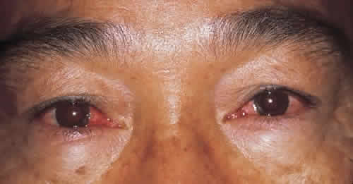
The onset is sudden and alarming. After beginning with one eye, the other often becomes inflamed in a matter of hours. Most patients develop signs in the second eye within 1 to 3 days (Fig. 1). Pain accompanies the other symptoms.

Fig. 1. Bilateral case of acute hemorrhagic conjunctivitis caused by enterovirus 70.

The most common symptoms are a profuse and predominantly watery discharge, a foreign body sensation, burning, photophobia, swelling of the eyelids, and pain in the upper eyelid that is aggravated on bending over. Although patients often complained of coryza, fewer than 5% showed systemic manifestations, such as fever or malaise. Ocular symptoms are often usually severe enough, however, to disrupt routine activities for several days.

Signs of AHC caused by EV70 and CA24v are similar, and it is impossible to determine the causative virus from clinical characteristics. Edematous swelling of the lid occurs in 72% to 100% of patients, although severity varies. Swelling usually subsides within 3 or 4 days. Preauricular lymph nodes become enlarged and palpable in 65% of patients, and approximately 80% are tender.13 The upper conjunctivae become inflamed from a mild to a severe degree, with injection, infiltration, and follicle formation. In moderately inflamed eyes, the fornix is reddened and cloudy, obscuring blood vessels.

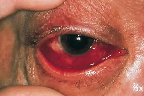
The most characteristic finding is marked hemorrhagic involvement of the bulbar conjunctival and subconjunctival layers. Punctate hemorrhages often appear within a few hours of onset of symptoms and rapidly coalesce to form larger hemorrhagic patches (Fig. 2). These patches tend to form ridges concentric with the corneoscleral limbus and occasionally become confluent (Fig. 3). Although 40% of patients have with unilateral involvement, more than 90% have bilateral disease when examined the next day.

Fig. 2. Diffuse patchy conjunctival hemorrhages in acute hemorrhagic conjunctivitis. Hemorrhagic patches diffusely distribute in the bulbar conjunctiva in patients 3 days after the onset of the condition.

Fig. 3. Typical conjunctival hemorrhage in the late phase of acute hemorrhagic conjunctivitis. Blot conjunctival hemorrhages form a ridge at the corneoscleral limbus 6 days after the onset.

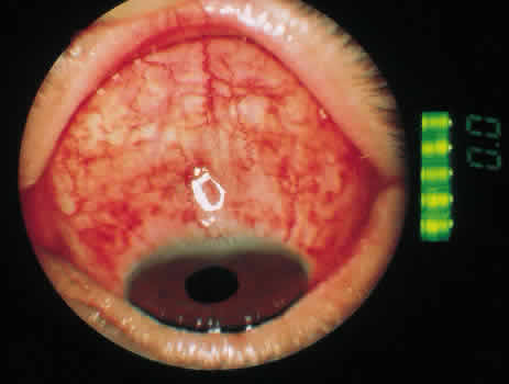
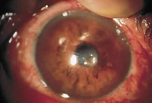
Corneal complications are generally rare. The incidence of epithelial keratitis ranges from 0% to 30% (Fig. 4). Extraocular symptoms and signs are usually mild and resolve without sequelae.

Fig. 4. Corneal complication of acute hemorrhagic conjunctivitis. Moderate epithelial keratitis and conjunctival hyperemia are observed.

Most symptoms resolve by the fourth day of illness, as do most physical findings. The most persistent signs are conjunctival hemorrhages and follicles, which may aid physicians in making the diagnosis of AHC when patients present during the convalescent phase of disease.

Back to Top
VIROLOGY

VIRAL MORPHOLOGY AND PHYSICOCHEMICAL PROPERTIES

The virions of EV70 and CA24v are spherical and have diameters of 30 ± 1 nm, showing a typical enterovirus morphology. Virions of these viruses have a buoyant density of 1.34 g/cm3 in CsCl2, and their sedimentation coefficient is 160S in sucrose.14,15 The virion consists of a single-stranded RNA genome and four structural proteins, similar to other enteroviruses.16

The sedimentation coefficient of the genome RNA is 34S, and its molecular weight is 2.5 × 10⁶ daltons.14,15 The single-stranded genomic RNA has positive-strand sense, and it codes in a single open reading frame for the four capsid proteins and additionally for functional proteins with, for example, RNA polymerase and protease activities. The genome RNA contains 7,391 nucleotides.

The capsid consists of 60 protomers, each of which contains four nonglycosylated virus proteins. The capsid proteins are virus protein (VP) 1 (molecular weight, 35 kilodaltons), VP2 (28 kilodaltons), VP3 (27 kilodaltons), and VP4 (9 kilodaltons). VP4 is myristoylated at its N-terminus. Structure analysis by x-ray crystallography and biochemical accessibility studies have shown that the capsid proteins VP1, VP2, and VP3 are at the capsid surface, whereas VP4 is covered inside the capsid shell and has contact to the viral RNA (Fig. 5). VP1, VP2, and VP3 together make up a pseudoequivalent packing arrangement in the capsid. VP1 and VP3 form a depression or canyon (approximately 2.5 nm deep and up to 3 nm wide) that is oriented around the fivefold symmetry axis of the capsid. It is proposed that the canyon is the recognition site for the virus-specific receptor; this canyon hypothesis has also been described for the related picornavirus, human rhinovirus 14.17

Fig. 5. Organization of the enterovirus genome. The boxes represent the coding region of the single-strand genomic RNA of enterovirus. A small hydrophobic protein is covalently linked to the terminal uracil of the 5-NTR and called virus protein genome linked (VPg). Region P1 codes for the capsid proteins VP4, VP2, VP1, and VP3. Regions P2 and P3 code for functional proteins.

BIOLOGICAL PROPERTIES

Portal of Entry

For most enteroviruses, the primary site of infection is the alimentary tract, where the viruses replicate and produce systemic infection through viremia. For EV70 and CA24v, however, the primary infection site seems to be the conjunctiva, and there has been no definite evidence to suggest active replication of these viruses in the gastrointestinal tract. In the occasional case, these viruses are recovered from the feces of patients with AHC. Although one cannot completely exclude the possibility that EV70 and CA24v replicated in the intestine, in such cases, it is more reasonable to assume that these viruses grew in the conjunctiva, passed from the nasopharynx into the alimentary tract, and were then incidentally recovered from feces. This assumption is also supported by in vitro findings that EV70 replicates better at 32°C to 34°C than at 37°C and usually does not replicate at 39°C,18,19 suggesting that the virus cannot readily grow in the alimentary tract. This temperature-sensitive growth of EV70 suggests that these viruses have acquired human pathogenicity as a result of their adaptation to the lower temperature of the ocular surface through evolution. However, in contrast, as for CA24v, no difference was observed in the isolation rate in HeLa cell culture at 33°C and those incubated at 37°C.20,21 Studies to investigate molecular events leading to a temperature-sensitive defect in EV70 replication showed that viral RNA synthesis was blocked at the nonpermissive temperature (39°C).22,23 The temperature-sensitive defect of EV70 replication is most likely the result of lack of uridylation of VPg at the restricted temperature.24

Virus-specific Receptors

Most human enteroviruses, with a few exceptions, grow only in primary or established cell cultures derived from human and monkey tissues. EV70 is one of those that replicate in nonprimate cell cultures. Replication of EV70 occurs in nonprimate cell cultures at 33°C, and this virus can adsorb and replicate in cells from the mouse (L), hamster (BHK21), rabbit (RK13), pig (IB-RS-2), and cow (BK1).25 In contrast, a CA24v can replicate and cause cytopathic effect only in primate cell cultures,26 like poliovirus and many other enterovi-ruses. Although a specific viral receptor of CA24v has not been determined, an additional membrane protein, decay accelerating factor (CD55), is the receptor for echoviruses 6, 7, 12, and 21,27 some of which are considered to cause AHC, and for EV70.28

Viral Reproduction

The early phase of the enterovirus reproduction cycle can be divided into adsorption, penetration, and uncoating. Viral entry into the host cell (penetration) occurs by way of receptor-mediated endocytosis followed by a pH-dependent release of viral RNA from the virus capsid. The internal capsid protein VP4 is released from the virus, and the N-terminus of the capsid VP1 becomes accessible. After the virus has been uncoated, synthesis of viral protein and RNA are initiated by using the viral parental positive-strand RNA as a template. One reproduction cycle of enterovirus lasts 6 to 8 hours, at which time as many as 10,000 virus particles have been synthesized in a single cell.

Back to Top
VIROLOGIC DIAGNOSIS

ENTEROVIRUS 70

To establish the etiologic diagnosis of AHC caused by EV70, viral isolation and serologic tests are required. The selection of sensitive batches of cells or of sensitive sublines is important. To effectively isolate EV70, more sensitive cell types should be selected among primary cell cultures (e.g., human embryonic kidney, human embryonic lung, or monkey kidney cells) and diploid (e.g., human embryonic lung fibroblast or WI-38) or heteroploid (e.g., HeLa or HEp-2) cells. Suitable specimens should be inoculated onto at least two different cell types sensitive to EV70 propagation.

Although commonly used diagnostic tests for enterovirus infection are based on virus isolation in cell culture, followed by identification of serotypes with neutralizing antisera, or on serologic tests, no EV70 strain has been isolated by the cell culture method since 1988.29 Changes in the characteristics of the virus may explain this phenomenon. Takeda and coworkers30 have shown with oligonucleotide fingerprinting that EV70 isolates from cases of AHC in 1981 were genetically distinct from isolates observed during the 1970-1971 pandemic. Immunofluorescence,31 enzyme-linked immunosorbent assay,32 and electron microscopy33 have been applied to detect epidemic EV70 that could not be isolated by cell culture.

Recently, the sequences of the full-length genome of EV70 standard strain were determined.34 Uchio and coworkers35 have previously reported that out of culture-negative specimens from conjunctival swabs of patients among the population of an AHC epidemic in Okinawa, Japan in 1994, several specimens were positive by RT-PCR (Fig. 6). Three sets of primers (VP1, VP2, and VP3 regions) were designed based on the nucleotide sequence of the standard strain of EV70, J670/7120. That no PCR products obtained from specimens from patients with symptoms of more than 3 days' duration gave positive results indicates that the virus might be present in the eye for only a brief duration, and timely collection of specimens may be important.36 This method has been applied for phylogenetic analysis of EV70 using specimens from patients among a community outbreak of AHC in Israel.37

Fig. 6. Polymerase chain reaction analysis of clinical conjunctival swab specimens deemed enterovirus tissue-culture negative using VP1 region primer (top right and left) and VP2 region primer (bottom right and left). Ethidium bromide-stained plates are shown. Lanes 1–27, clinical samples; lane 28, enterovirus 70 prototype virus (J670/71); lane 29, CA24v prototype virus (EH24/70); lane 30, negative control. mk represents the 1-kilobase pair ladder marker. The molecular weights of the polymerase chain reaction products are indicated to the side of the figures. Lanes 1, 5, 9, 10, 12, 14, 15, 17, 20, 21, and 24 show a positive signal in both primers, but lane 8 shows a positive signal only in the VP2 primer.

COXSACKIEVIRUS 24

Identification of CA24v can be carried out by the conventional neutralization test in test tubes or more economically in microtitration plates.33 The micrometabolic inhibition test in microtitration plates is also reliable and convenient.38 The CA24v specific antiserum should be used for identification by the neutralization test because the virus is known to be antigenically distinct from the prototype (Joseph strain) and other variants of CA24v.7

TYPING

Although the neutralization test is generally reliable for enterovirus typing, it is labor-intensive and time-consuming and may fail to identify the serotype of clinical isolates because of antigenic drift, recombination, or the presence of virus mixtures in the specimen being tested. The technique of viral protein fingerprinting has recently been used for the typing and characterization of clinical enterovirus isolates.39 The method is specific and 97% accurate, but the radiolabeled proteins generate radioactive waste, and the method requires the use of specialized instrumentation for data acquisition and analysis. Obserste and coworkers40 have developed a molecular typing system based on RT-PCR and nucleotide sequencing of the 3' half of the genomic region encoding VP1. The serotype of an unknown may be inferred by comparison of the partial VP1 sequence to those in a database containing VP1 sequences for the prototypes strains of all 66 human enterovirus serotypes. The antigenic and molecular typing results agreed for all isolates. This method can greatly reduce the time required to type an enterovirus isolate and can be used to type isolates that are difficult or impossible to type with standard immunologic reagents, such as recent isolates of EV70.

Back to Top
CONTROL

DISINFECTANTS

Enterovirus 70 is readily killed by various germicides, such as phenol, cresol, formaldehyde, and sublimate, under the conditions by which other enteroviruses are not or are only partially inactivated. However, neither EV70 nor any of the other enteroviruses were inactivated by the common preservatives: benzalkonium chloride, benzethonium chloride, chlorhexidine gluconate, and hexachlorophen at levels of practical use.41

Iodine is widely used in eye clinics. The solution containing free iodine at the concentration of 0.0025% to 0.01% iodine is generally used for disinfection of hands and instruments. Kono and Yoshii42 studied the virucidal activity of the Pliacid-Nutra flow system containing iodine and found it effective for EV70. Previous investigation showed that among six enteroviruses (polio 1, echo 7, coxsackie A1, coxsackie B5, EV70, and EV71), EV70 was most sensitive to methanol, ethanol, isopropanol, and n-propanol.43

ANTIVIRAL AGENTS

Antiviral chemotherapy cannot yet be used for the treatment of enterovirus infection of humans. Some antiviral compounds have been proven to be potent only in vitro. Guanidine and some benzimidazoles specifically inhibit the viral RNA polymerization. Hydrophobic compounds intercalating in the viral capsid protein VP1 stabilize the virus, resulting in inhibition of viral uncoating.44 Arildone is a broad-spectrum antiviral agent that has been shown to selectively inhibit replication of picornaviruses. The mechanism of action of arildone is consistent with inhibition of the uncoating process.45 Arildone inhibited the infectivity of AHC viruses, EV70, and CA24v in tissue culture. Arildone did not inhibit interferon production or interferon activity.

Back to Top
PATHOGENICITY ANDANIMAL MODELS

ENTEROVIRUS 70

Although it was reported that EV70 isolates grew well in the primary cultured epithelial cells derived from mouse, rabbit, and monkey conjunctival and corneal tissue, no laboratory animals have so far been found to develop noticeable conjunctivitis.26 Unlike coxsackievirus, enterovirus isolates are nonpathogenic to suckling mice and adult mice.4 However, EV70 showed a distinct neurovirulence when inoculated into the central nervous system of cynomolgus monkeys.46 Monkeys inoculated with EV70 simultaneously in the spinal cord and thalamus developed paraplegia or monoplegia of the lower limbs. This finding gave virologic evidence to the later recognized clinicoepidemiologic observation that EV70 infection is occasionally complicated by poliolike motor paralysis, which usually appears several weeks after the onset of conjunctivitis.47

COXSACKIEVIRUS 24

Although suckling mice are the most popular isolation system for the coxsackievirus A group, they are not sensitive as tissue cell cultures for the isolation of CA24v. The pathogenicity of the virus for mice can be enhanced by passing the virus through the intracerebral route into consecutive batches of suckling mice.48 Higgins and coworkers15 observed stunting of growth and paralysis in some newborn mice inoculated subcutaneously and intracerebrally with the Singapore and Hong Kong strains of CA24v. They also observed that the Hong Kong strain produced a higher morbidity and mortality rate than did the Singapore strain and that the virus was found in the brain and torso of paralyzed mice. Loss of striation and fragmentation of fibers in the voluntary muscles occur with infiltration of mononuclear cells, polymorphonuclear cells, and eosinophils typical of coxsackievirus A infection.49

Back to Top
EVOLUTION ANDANTIGENIC RELATIONSHIPS
Enterovirus 70 and coxsackievirus 24 have continued their intermittent participation in AHC epidemics since 1971. It seemed strange that two different enteroviruses causing the newly recognized AHC syndrome emerged suddenly at almost the same time. Thus, there arose a question whether these viruses might have been derived from a common ancestor and might genetically be closely related.

The phylogenetic tree of EV70 indicates that the virus originated from one focus around 1967 and was transmitted among humans since then.50 West Africa is considered to be the most likely place where EV70 was born. A hot and humid coastal monsoonal climate and a dense population are favorable factors for the spread of EV70. The phylogenetic tree of EV70 showed an evolutionary pattern in divergent fashion; the virus branched into many lineages during the pandemic period that were transmitted independently of one another, resulting in the appearance of vastly polymorphic viruses in the nucleotide sequence.

According to the evolutionary tree of CA24v, the strains during the early epidemic in Singapore and Hong Kong were closely related.51 In contrast, all late isolates from various areas including Singapore, Pakistan, Taiwan, and Japan were distinct from the strains of early epidemics. The tree indicated that all strains isolated in 1985 and 1986 were the descendants of the virus that prevailed between 1978 and 1980. After branching, the viruses transmitted independently in certain areas, and after 1985, one of them spread to Pakistan, one to Singapore, and another to Taiwan and Japan. The tree indicated that all isolates were derived from a common ancestor that appeared around 1965 leading to the hypothesis that AHC due to CA24v originated from a Java focus. It is conceivable that one of the strains of CA24v had undergone nucleotide substitutions and pathogenic change and came to preferentially grow in human conjunctiva. Through this new pathogenicity, the virus has spread and caused major epidemics of AHC in humans.

From these reports, the two enteroviruses causing the AHC syndrome, EV70 and CA24v, are entirely different, not only in antigenic properties but also in viral genomes. This was shown by cross-neutralization tests, complement-fixation tests using antisera against purified viral antigens, and RNA-RNA hybridization experiments in which 32P-labeled ssRNA (-) extracted from infected cells was hybridized with ssRNA (+) of homologous or heterologous virions. EV70 is considered as a new distinct type of human enterovirus, and its origin remains unknown. The first emergence of this virus has been estimated to be around 1967 by molecular epidemiology using oligonucleotide mapping.52 Conversely, EH24/70, the representative strain of CA24v, shares nucleotide sequence with the prototype strain of Joseph and other variant strains of coxsackievirus A24 to various degrees. Thus, these two closely related virus seem to arose, one from Africa and the other from Asia, at approximately the same time.

Back to Top
REFERENCES

1. Dalldorf G, Sickles GM: An unidentified, filterable agent isolated from the feces of children with paralysis. Science 108:61–63, 1948.

2. Melnick JL, Shaw EW, Cumen EC: A virus isolated from patients diagnosed as nonparalytic poliomyelitis or aseptic meningitis. Proc Soc Exp Biol Med 71:344–349, 1949.

3. Kono R: Apollo 11 disease or acute haemorrhagic conjunctivitis: a pandemic of a new enterovirus infection of the eyes. Am J Epidemiol 101:383–390, 1975.

4. Kono R, Sasagawa A, Ishii K et al: Pandemic of new type of conjunctivitis. Lancet i:1191–1194, 1972.

5. Yin-Murphy M: An epidemic of picornavirus conjunctivitis in Singapore. Southeast Asin J Trop Med Pub Hlth 3:303–309, 1972.

6. Lin KH, Yin-Murphy M: Epidemic conjunctivitis in Singapore in 1970 and 1971. Singapore Med J 14:86–89, 1973.

7. Mirkovic RR, Schmidt NJ, Yin-Murphy M et al: Enterovirus etiology of the 1970 Singapore epidemic of acute conjunctivitis. Intervirology 4:119–127, 1974.

8. Onorato IM, Morens DM, Schonberger LB et al: Acute haemorrhagic conjunctivitis caused by enterovirus 70: an epidemic in American Samoa. Am J Trop Med Hyg 34:984–991, 1985.

9. Asbell PA, de la Pena W, Harms D et al: Acute haemorrhagic conjunctivitis in central America: first enterovirus epidemic in the western hemisphere. Ann Ophthalmol 17:205–210, 1985.

10. Yin-Murphy M, Lim KH, Ho YM: A coxsackievirus A24 epidemic of acute conjunctivitis. Southeast Asian J Trop Med Publ Health 7:1–5, 1976.

11. Yin-Murphy M, Baharuddin-Ishak, Phoon MC et al: A recent epidemic of coxsackievirus type A24 acute haemorrhagic conjunctivitis in Singapore. Br J Ophthalmol 70:869–873, 1986.

12. Miyamura K, Yamashita K, Takeda N et al: The first epidemic of acute haemorrhagic conjunctivitis due to a coxsackievirus A24 variant in Okinawa, Japan, in 1985-1986. Jpn J Med Sci Biol 41:159–174, 1988.

13. Mitsui Y, Kajima M, Matsumura K et al: Haemorrhagic conjunctiitis, a new type of epidemic viral keratoconjunctivitis. Jpn J Ophthalmol 16:33–40, 1972.

14. Yamazaki S, Natori K, Kono R: Purification and biophysical properties of acute haemorrhagic conjunctivitis virus. J Virol 14:1357–1360, 1974.

15. Higgins PG, Scott RJ, Davies PM et al: A comparative study of viruses associated with acute haemorrhagic conjunctivitis. J Clin Pathol 27:292–296, 1974.

16. Esposito JJ, Obifeski JF: Enterovirus type 70 virion and intercellular proteins. J Virol 18:1160–1162, 1976.

17. Rossman MG, Arnold E, Erickson JW et al: Structure of a human common cold virus and functional relationship to other picornaviruses. Nature 317:145–153, 1985.

18. Miyamura K, Yamazaki S, Tajiri E et al: Growth characteristics of acute hemorrhagic conjunctivitis (AHC) virus in monkey kidney cells. I. Effect of temperature on viral growth. Intervirology 4:279–286, 1974.

19. Miyamura K, Sasagawa A, Tajiri E et al: Growth characteristics of acute hemorrhagic conjunctivitis (AHC) virus in monkey kidney cells. II. Temperature sensitivity of the isolates obtained at various epidemic areas. Intervirology 7:192–200, 1976.

20. Lin KH, Yin-Murphy M: An epidemic of conjunctivitis in Singapore in 1970. Singapore Med J 12:247–249, 1971.

21. Yin-Murphy M: An epidemic of picornavirus conjunctivitis in Singapore. Southeast Asian J Trop Med Pub Hlth 3:303–309, 1972.

22. Takeda N, Miyamura K, Kono R et al: Characterization of a temperature-sensitive defect of enterovirus type 70. J Virol 44:98–106, 1982.

23. Miyamura K, Takeda N, Yamazaki S: Characterization of temperature-sensitive defect of enterovirus 70: effect of elevated temperature on in vitro transcription. J Virol 51:192–198, 1984.

24. Takeda N, Kuhn RJ, Yang CF et al: Initiation of poliovirus plus-strand RNA synthesis in a membrane complex of infected HeLa cells. J Virol 60:43–53, 1986.

25. Yoshii T, Natori K, Kono R: Replication of enterovirus 70 in non-primate cell cultures. J Gen Virol 36:370–384 1977.

26. Langford MP, Stanton GC: Replication of acute hemorrhagic conjunctivitis viruses in conjunctival-corneal cell cultures of mice, rabbits and monkeys. Invest Ophthalmol Vis Sci 19:1477–1481, 1980.

27. Bergelson JM, Chan M, Solomon KR et al: Decay accelerating factor (CD55), a glycosylphosphatidylinositol-anchored complement regulatory protein, is a receptor for several echoviruses. Proc Natl Acad Sci USA 91:6245–6249, 1994.

28. Karnauchow TM, Tolson DL Harrison BA et al: The HeLa cell receptor for enterovirus 70 is decay accelerating factor (CD55). J Virol 70:5143–5152, 1996.

29. Ramia S, Arif M: Isolation of enterovirus 70 (EV70) from patients with acute haemorrhagic conjunctivitis in two areas of Saudi Arabia. Trans Roy Soc Trop Med Hyg 84:139–140, 1990.

30. Takeda N, Miyamura K, Ogino T et al: Evolution of enterovirus type 70: Oligonucleotide mapping analysis of RNA genome. Virology 134:375–388, 1984.

31. Pal SR, Szucs GY, Melnick JL: Rapid immunofluorescence diagnosis of acute haemorrhagic conjunctivitis caused by enterovirus 70. Intervirology 20:19–22, 1983.

32. Anderson LJ, Hatch MH, Flemister MR et al: Detection of enterovirus 70 with monoclonal antibodies. J Clin Microbiol 20:405–408, 1984.

33. Yin-Murphy M, Rahim NA, Phoon MC et al: Early and rapid diagnosis of acute haemorrhagic conjunctivitis with tear specimens. Bull World Health Organ 63:705–709, 1985.

34. Ryan MD, Jenkins O, Hughes PJ et al: The complete nucleotide sequence of enterovirus type 70: relationships with other members of the Picornaviridae. J Gen Virol 71:2291–2299, 1990.

35. Uchio E, Yamazaki K, Aoki K et al: Detection of enterovirus 70 by polymerase chain reaction in acute haemorrhagic conjunctivitis. Am J Ophthalmol 122:273–275, 1996.

36. Kishore J, Nanjunath N, Bareja U et al: Study of an outbreak of epidemic conjunctivitis in Delhi in 1986. Indian J Pathol Microbiol 32:266–269, 1989.

37. Shulman LM, Manor Y, Azar R et al: Identification of a new strain offastidious enterovirus 70 as the causative agent of an outbreak of haemorrhagic conjunctivitis. J Clin Microbiol 35:2145–2149, 1997.

38. Yin-Murphy M: Simple tests for the diagnosis of picornavirus epidemic conjunctivitis (acute haemorrhagic conjunctivitis). Bull World Health Organ 54:675–679, 1976.

39. Holland DT, Sene J, Peter CR et al: Differentiation and characterization of enteroviruses by computer-assisted viral protein fingerprinting. J Clin Microbiol 36:1588–1594, 1998.

40. Oberste MS, Maher K, Kilpatrick DR et al: Typing of human enteroviruses by partial sequencing of VP1. J Clin Microbiol 37:1288–1293, 1999.

41. Yamazaki S, Miyamura K: General characteristics of enterovirus 70. In: Uchida Y, Ishii K, Miyamura K, Yamazaki S, eds. Acute hemorrhagic conjunctivitis. Basel, Karger, 1989:345–358.

42. Kono R, Yoshii T: Antiviral effect of a disinfectant (Pliacid/Nutraflow system) for soft contact lenses. J Jpn Contact Lens Soc 23:150–154, 1981.

43. Noda N, Watanabe M, Yamada F: Virucidal activity of alcohols: virucidal efficiency of alcohols against viruses is liquid phase. J Jpn Ass Infect Dis 55:355–366, 1981.

44. Eggers HJ. Assay systems: testing of antiviral drugs in cell culture (in vitro). In: De Claercq E, Walker RT, eds. Antiviral drug development. New York, Plenum Press, 1988:139–148.

45. McSharry JJ, Caliguiri LA, Eggers HJ: Inhibition of uncoating of poliovirus by arildone, a new antiviral drug. Virology 97:307–315, 1979.

46. Kono R, Uchida N, Sasagawa A et al: Neurovirulence of acute haemorrhagic conjunctivitis virus in monkeys. Lancet ii:61–63, 1973.

47. Wadia NH, Irani PF, Katrak SM: Neurological complications of new conjunctivitis. Lancet ii:970–971, 1972.

48. Christopher S, Theogaraj S, Godbole S et al: An epidemic of acute haemorrhagic conjunctivitis due to coxsackievirus A24. J Infect Dis 146:16–19, 1982.

49. Gogate SS, Gupta NO, Anantanarayan TR et al: Laboratory investigations of an epidemic of acute haemorrhagic conjuntivitis in Bombay and Poona in 1975. J Indian Med Ass 70:34–37, 1978.

50. Takeda N, Tanimura M, Miyamura K: Molecular evolution of major capsid protein VP1 of enterovirus 70. J Virol 68:854–862, 1994.

51. Supanaranond K, Takeda N, Yamazaki S: The complete nucleotide sequence of a variant of coxsackievirus A24, an agent causing acute hemorrhagic conjunctivitis. Virus Genes 6:149–158, 1992.

52. Miyamura K, Tanimura M, Takeda N et al: Evolution of enterovirus 70 in nature: all isolates were recently derived from a common ancestor. Arch Virol 89:1–14, 1986.

Back to Top