Chapter 91
The Epstein-Barr Virus in Ocular Disease
STEPHEN C. PFLUGFELDER and JAMES CHODOSH
Main Menu   Table Of Contents

Search

EPSTEIN-BARR VIRUS INFECTION IN HUMANS
DIAGNOSIS OF EBV INFECTION
MOLECULAR GENETICS OF THE EPSTEIN-BARR VIRUS LIFE CYCLE
EBV OCULAR DISEASE
NEURO-OPHTHALMIC, RETINAL, AND UVEAL DISEASES
THERAPY OF OCULAR EPSTEIN-BARR VIRUS INFECTION
REFERENCES

Epstein-Barr virus (EBV) is a human herpesvirus belonging to the subfamily Gammaherpesvirinae. EBV is a ubiquitous virus which infects the majority of humans by early adulthood.1 Epstein and associates reported the initial observation of EBV in cell lines established from biopsies of Burkitt's lymphomas, a neoplasia which occurs predominantly in children in certain tropical regions of Africa.2 EBV was subsequently found by Henle and colleagues to be the agent responsible for infectious mononucleosis (IM), a common febrile illness.3 In addition to being associated with Burkitt's lymphoma, EBV has been detected in other human neoplasias including nasopharyngeal carcinoma,4 thymic carcinoma,5 Hodgkin's disease,6 oral hairy leukoplakia,7 and B-cell lymphoproliferation in allograft recipients receiving immunosuppressive therapy.8,9 The association of EBV with these neoplasias has led to the speculation that EBV is involved in the pathogenesis of these diseases. There is increasing evidence that EBV is capable of infecting ocular and adnexal tissues and causing ocular disease. The current concepts regarding the molecular genetics of the EBV life cycle, diagnosis of EBV infection, and the role of EBV in ocular disease is reviewed in this chapter.
Back to Top
EPSTEIN-BARR VIRUS INFECTION IN HUMANS
Primary EBV infection may manifest as IM, a disease characterized by fever, severe fatigue, pharyngitis, lymphadenopathy, hepatosplenomegaly, and occasionally hepatitis or lymphoproliferative disease, or more commonly as a mild upper respiratory tract infection which is frequently unrecognized as a primary EBV infection.1,10

The fate of EBV following infection of human cells depends on the particular type of cell infected and the pattern of gene transcription used by the virus.11 There are two major consequences of EBV infection of B lymphocytes depending on whether the active or passive latent transcriptional programs are activated. If the active latent transcription program is utilized, B cells are growthtransformed into lymphoblasts.12 EBV-infected lymphoblasts, many of which are in mitosis, can be readily detected in the blood and lymphoid tissues of patients with acute IM.13 A potent T-cell-mediated immunologic response is rapidly mounted against these virus-infected B cells, and they are cleared from the blood within several weeks. The proliferative potential of EBV is limited in vivo by HLA class I (and occasionally class II) EBV-specific cytotoxic T lymphocytes (CTLs) for the remainder of an EBV-infected individual's lifetime.14,15 Systematic surveys identifying the immunodominant EBV epitopes suggest that most EBV-specific CTL responses are directed toward antigens expressed during active latent infection, with EBV nuclear antigens 3, 4, and 6 (EBNA-3, -4, and -6) being the most frequently identified. CTLs recognizing EBNA-2, EBNA-5, LMP-1 and LMP-2 have also been demonstrated, but at a much lower frequency and with fewer HLA class I restrictions.16 Suppression of cellular immunity in EBV-seropositive individuals (e.g., high-dose immunosuppressive therapy in transplant recipients) results in increased virus shedding in the saliva,17,18 and can lead to lymphoproliferative disease as a result of B lymphocytes expressing active latent genes that escape T-cell-mediated destruction.8,19,20

Use of the other transcriptional program in EBV-infected B cells results in “passive” latent infection. It is typically used by the virus after infecting small resting B cells, and gives rise to mRNA that encodes only EBNA-1, a nonimmunogenic protein that fails to induce an EBV-specific CTL response.16 Thus cells expressing only EBNA-1 escape T-cell-mediated destruction, and this passive latent state may represent an evolutionary strategy by the virus for lifelong persistence in immunocompetent hosts. In contrast to B cells, EBV infection of epithelial cells is a more complicated issue. Depending on the type of epithelial cell infected and the state of cellular differentiation, either a latent persistent infection may ensue or a lytic infection may occur with release of infectious viral particles.21–23

Similar to other herpesviruses, EBV usually persists in infected hosts following primary infection in a latent, nonpathogenic state.23,24 A number of sites of EBV persistence have been identified. Thus far, all of these sites are components of the mucosal associated lymphoid tissue (MALT) and include the major and minor salivary glands,25,26 oropharyngeal epithelia,21 epithelia in the male and female genital tracts,27,28 lacrimal gland,29 and corneal and conjunctival epithelia.30,31 At the present time, there are limited data regarding the cell type(s) in which persistent mucosal EBV infection occurs, and the repertoire of EBV proteins expressed in these latently infected cells.23,24 Because EBV DNA17 and infectious virus32 can be detected in saliva specimens of a small percentage (15% to 20%) of EBV-seropositive individuals, it appears that EBV may occasionally undergo spontaneous lytic transformation with production of infectious virus by latently infected cells in oropharyngeal MALT.

Back to Top
DIAGNOSIS OF EBV INFECTION
The standard for diagnosis of IM at the present time is serology. A characteristic and specific temporal pattern of appearance of serum antibodies to EBV is observed in patients with IM.10,33 By the time clinical symptoms of IM have developed, both IgM and IgG antibodies to viral capsid antigens (VCA) have reached their peak. After recovery from IM, viral capsid IgM antibodies disappear; however, anti-VCA IgG persists at low levels throughout life. Antibodies to early antigens which are indicative of viral replication, also rise during the acute phase of the disease and subsequently decrease to low or nondetectable levels in most individuals after the disease resolves. Antibodies to EBV nuclear antigens (EBNAs) appear weeks to months after the onset of IM, persist for life, and provide serologic evidence of past EBV infection.10,33,34

Additional laboratory tests may be helpful in confirming a diagnosis of IM. By the second week, patients typically have a relative or absolute lymphocytosis, and an increased percentage of atypical lymphocytes (10% to 25%).10,33 Detection of serum heterophil antibodies of the Paul-Bunnell type (Monospot test) is specific for IM due to EBV.10 These IgM antibodies, which agglutinate sheep and horse red blood cells (RBCs) and lyse beef RBCs, usually appear by the end of the first week of the infection.10 At least 10% of young adults with a typical serologic picture of acute EBV infection do not produce heterophil antibodies.35

Chronic elevations of EBV serum antibodies have been observed in a number of neoplasic and immunologic diseases. Henle and associates have reported that these serologic findings may reflect an enhanced viral carrier state manifesting as increased shedding of EBV into the saliva.36 Specific patterns of persistently elevated antibodies to EBV antigens have been found in the following EBV-associated diseases: Burkitt's lymphoma (VCA IgG, early antigen-restricted component, or EA-R), nasopharyngeal carcinoma (VCA IgG and IgA, early antigen-diffuse component, or EA-D), recurrent parotitis in children (VCA IgG and IgA, EA-R), and chronic fatigue syndrome (VCA IgG and early antigens).37,38 Elevated EBV VCA IgG and anti-early antigen titers may also be observed in diseases with altered immunoregulation including sarcoidosis, systemic lupus erythematosis, rheumatoid arthritis, Sjögren's syndrome, HIV infection, and immunosuppressed allograft recipients.36,39,40 The majority of reports describing an etiologic role for EBV in ocular inflammatory diseases have relied on diagnosis of EBV infection by serologic tests indicating acute or chronic infection. To conclusively prove that EBV is the causative agent for ocular diseases requires either culture of virus from or demonstration of viral proteins or nucleic acids in involved tissues.

Diagnosis of EBV infection can be made by immortalization of B lymphocytes after exposure to EBV-containing clinical specimens.1 Umbilical cord lymphocytes are the most commonly used source of B lymphocytes for EBV cell culture. Unfortunately, these cells are not always readily available, and detection of EBV-infected lymphoblasts in culture typically requires 4 to 6 weeks after inoculation.

Molecular biology techniques can also be used for detecting virus-specific proteins (antigens) and nucleic acids (DNA, RNA) in human fluid and tissue specimens. Antigens can be detected in pathologic specimens using monoclonal antibodies against EBV-specific proteins. A number of monoclonal antibodies reactive to EBV latent and lytic antigens have been developed. These antibodies have been used to detect EBV antigens in desquamated oropharyngeal epithelial cells from patients with IM,21 as well as in EBV-induced neoplasia.8,9

EBV nucleic acid sequences can be detected in specimens by hybridization with labeled nucleic acid probes specific to EBV.1 Hybridization can be performed as an in situ technique on sections cut from biopsied tissue, or on membranes onto which nucleic acids extracted from fluid or tissue specimens have been blotted.25 These techniques often lack the sensitivity for detecting the small copy number of viral genomes which may be present in the minute quantities of tissue or fluids obtained from ocular diagnostic procedures.41 The polymerase chain reaction (PCR) is a recently described molecular technique which can amplify genomic sequences from an infectious agent over one million times in several hours.42 The proven advantages of this technique for diagnosis of viral infection are its specificity for amplification of genomic sequences of a particular virus, its rapidity, and its sensitivity.42 This technique has recently been found to be sensitive enough to amplify EBV genomic sequences in tears of primary Sjögren's syndrome patients43 and cytomegalovirus (CMV) sequences in aqueous humor specimens from patients with CMV retinitis.41 This technique hasgreat potential for evaluating the role of EBV in ocular inflammatory diseases.

Back to Top
MOLECULAR GENETICS OF THE EPSTEIN-BARR VIRUS LIFE CYCLE
EBV has many morphologic similarities to other herpesviruses including: (1) a protein sheet that is surrounded by DNA, (2) a nucleocapsid, (3) a protein sheet that separates the nucleocapsid and the envelope, and (4) an outer envelope composed of glycoprotein spikes, predominantly the gp350/220 protein.1 During the life cycle of herpesviruses such as EBV, a sequential pattern of gene transcription occurs as the virus progresses from the latent to the lytic phase of its life cycle.44

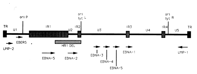
The entire EBV genome has been cloned and sequenced.45–47 Within virus particles, the EBV genome is linear and contains terminal repeats on either end of the genome (putatively thought to be important in circularization of the virus and/or integration of the EBV genome into cellular DNA), four internal repeat (IR1-IR4) regions, and five unique (U1-U5) sequence domains (Fig. 1). Approximately 100 open reading frames have been identified within the EBV genome that may potentially be transcribed and translated into proteins that are essential for the maintenance of the different phases of the EBV life cycle.44 Molecular biology techniques have allowed for the manipulation of many of these EBV genetic sequences. As a result, these in vitro studies have provided insight into the potential function of many of the EBV genes and how infection may be controlled.

Fig. 1. Diagram of the EBV genome indicating the terminal (TR) and internal (IR) repeat regions, unique (U) regions, and the location of latent genes, several of which (EBNA-2 and LMP) may be essential for growth transformation of B lymphocytes. Arrows indicate length and direction of transcription of latency associated mRNAs. The origin of DNA replication in latent (ori P) or lytically (ori lyt, right and left) infected cells is noted.

Although it is known that adsorption of EBV to a permissive cell occurs by interaction of the EBV gp350/220 envelope glycoprotein with the EBV/complement receptor, CD 21, on the surface of the cell,48 it is not clear how the envelope or nucleocapsid is dissolved or how the EBV DNA penetrates the cell nucleus. It is known that once the EBV genome enters the nucleus, it circularizes and that the cellular phenotype is a key factor in determining the pattern of EBV gene transcription and the consequences of virus infection on the cell.49

EXPRESSION OF EBV GENES DURING THE LATENT STAGES OF INFECTION

Two types of latent EBV infection have been identified: (1) active latent infection with transcription of EBV oncoproteins that transform (or immortalize) B cells into lymphoblasts,12 and (2) passive latent infection that is typically utilized by the virus after infecting small resting B cells and during which transcription of only one gene (EBNA-1) has been identified.

A limited number of EBV genes have been reported to be expressed during the active latent (or growth transformation) phase of EBV infection (see Fig. 1).44 These include (1) six EBV nuclear antigens (EBNA-1, -2, -3, -4, -5, -6); (2) the latent membrane proteins (LMP-1 and LMP-2); and (3) two small, nonpolyadenylated RNAs (designated EBER-1 and EBER-2).11 mRNAs encoding each of the EBNA proteins are generated by individual splicing of long rightward transcripts initiated at one of two promoters, one located in the Bam HI C region within the U1 region of the genome, and the other in the IR1 region of the genome. The LMP transcripts are expressed from separate promoters which run in opposite directions, but share the same bidirectional control region.

The EBNA-2 protein appears to be important in the transformation of B lymphocytes into immortalized cells because mutant EBV strains lacking this gene (such as the HR-1 strain with the deletion noted in Fig. 1) are incapable of immortalizing B lymphocytes.44 One of the mechanisms by which EBNA-2 may modulate growth transformation is through its ability to upregulate latent membrane protein-1 (LMP-1) transcription.50

LMP-1 is one of the most abundantly transcribed genes in EBV infected lymphoblastoid cell lines (LCLs).51 LMP-1 has recently been reported to be essential for B-lymphocyte growth transformation. EBV strains with deletion mutations in the LMP-1 gene were found to be incapable of immortalizing B lymphocytes.52 LMP-1 has been shown to be responsible for many of the phenotypic changes observed in EBV-infected B-lymphocytes including upregulation of expression of B-cell activation markers (CD21 and CD23), the cell membrane adhesion molecule ICAM-1, and the bcl-2 protooncogene.53 Increased expression of adhesion molecules facilitates cell clumping which may enhance B-cell growth and proliferation via paracrine growth factors. T cells also adhere to B cells expressing these adhesion molecules, and such T-cell adherence may represent the initial step in the host immune response to EBV infection that ultimately leads to elimination of EBV infected lymphoblasts in vivo.54

EBNA-1 is the only protein transcribed in the passive latent state. EBNA-1 has been reported to play an important role in maintaining EBV latency.44 EBNA-1 has been shown to bind to specific DNA sequences located at three sites within the EBV genome. One of these sites is located within the oriP domain of the EBV genome (see Fig. 1). Binding of EBNA-1 to the oriP sequences maintains EBV DNA as a closed circular episomal molecule, allows replication of EBV episomal DNA, facilitates transcription of latent cycle genes, and prevents transcription of lytic cycle genes.12 Expression of EBV latent associated genes apparently does not necessarily ensure maintenance of the latent state, because a small percentage (approximately 1%) of latently infected B cells in vitro spontaneously enter the lytic phase of the life cycle and produce infectious virus.1

EBER-1 and EBER-2 are the most abundantly transcribed latent genes with approximately 10)7 copies of each per cell.55 EBER-1 and EBER-2 are small (158 and 178 base pairs, respectively), nonpolyadenylated RNAs that are not translated into proteins. These small RNA molecules are thought to be important in RNA splicing which is an integral function in maintaining latency because EBNA mRNAs are extensively spliced.56 Their abundant copy number per cell makes EBERs excellent targets for in situ hybridization techniques that demonstrate cellular sites of EBV latent infection.

EXPRESSION OF EBV GENES DURING THE LYTIC STAGES OF INFECTION

In general, most normal B cells that are permissive for latent EBV infection cannot support EBV replication. Epithelial cells have been found to support lytic EBV infection in humans11; however, at the present time an EBV-permissive epithelial cell line for in vitro studies has not been identified.16 Because of the lack of an EBV-permissive cell line, the expression of EBV genomic sequences during the lytic phase of the virus infection has been investigated predominantly in latently infected B lymphocytes induced into the lytic phase using phorbol esters, the most reliable and reproducible inducers of viral replication.57 The most widely used cell line for these experiments, Raji, is derived from a Burkitt's lymphoma and contains a deletion which does not allow for expression of genes associated with the late phases of lytic infection although most immediate-early and early genes may be expressed.58 Superinfecting Raji cells with the P3HR-1 EBV strain results in expression of most lytic EBV genes in superinfected cells.59,60

The majority of genes expressed in the immediate-early phase of EBV infection are the latency-associated genes described above.44 It has been reported that there are several abundant EBV mRNAs expressed approximately 4 hours after induction into the lytic phase that may be key trans-activators of early lytic gene transcription.61 One immediate-early gene in particular, BZLF1 (often termed ZEBRA), is a “switch” gene that has been shown to play an important role in the switch from the EBV latent phase to the lytic phase.62

There are at least 30 EBV early genes that are defined by their constant synthesis (even when inhibitors of DNA synthesis are present) once a cell is induced into the lytic cycle by superinfection.44 The genomic locations of many of the genes detected during the early phase of EBV replication have been mapped. Although the function of many of these genes is currently unknown, the functions of some of the early EBV proteins have been identified by comparing the genomic location and sequence of these early genes to their counterparts in other herpesviruses. Most of the early proteins identified thus far are involved in DNA replication and include DNA polymerase, thymidine kinase, DNA binding proteins, alkaline reductase, and ribonucleotide reductase.44

EBV genes expressed during the late phase of the lytic cycle predominantly encode EBV structuralproteins and proteins that allow for the transport of the virus out of the cell. Several late genes have been mapped within the EBV genome although not all of the RNA transcripts for these genes have been detected in vitro. Most of the late EBV proteins that have been identified are heavily glycosylated and are associated with the outer envelope of the virus particle. One example is gp350/220, an abundantly expressed glycoprotein that is found in the cell membrane as well as the outer envelope of the virus particle.63 The gp350/220 protein is the predominant envelope protein recognized by neutralizing antibodies.64

Back to Top
EBV OCULAR DISEASE

CONJUNCTIVA AND CORNEA

There is increasing clinical and laboratory evidence suggesting that EBV is capable of infectingsusceptible cells in the ocular surface epithelia. Expression of CD21, the putative EBV receptor, has been detected in conjunctival and corneal epithelial cells.65 EBV genomic sequences have been detected in 10% of normal corneal epithelial specimens.30 Using highly sensitive reverse transcriptase PCR, EBNA-1 mRNA sequences, but not active latent or lytic cycle associated gene sequences, were detected in 95% of human conjunctival epithelial specimens obtained by impression cytology.31 Taken together, these results suggest that the ocular surface epithelia are sites of EBV persistence following primary infection.

Numerous cases of conjunctivitis occuring in patients with IM syndrome were reported prior to the availability of specific tests to confirm a clinical diagnosis of IM.66 More recently, conjunctival involvement has been reported in patients with serologically confirmed IM. In 1981, Meisler and associates reported a case of a unilateral conjunctival inflammatory mass and enlarged preauricular lymph node in an 11-year-old boy with acute IM.67 The conjunctival lesion was biopsied, and an intense lymphocytic infiltrate with occasional multinucleated giant cells was observed in histologic sections. Wilhelmus reported a case of unilateral keratoconjunctivitis in a 16-year-old girl with acute IM.68 This patient had a follicular conjunctivitis and preauricular lymph node. Virus was cultured from the tears and conjunctiva of this patient. Matoba and associates also noted conjunctival inflammation consisting of mild hyperemia occasionally accompanied by a follicular tarsal conjunctival response in a series of patients with IM.69 Gardner and co-workers recently reported a case of a bulbar conjunctival nodule associated with unilateral enlarged preauricular and submandibular lymph nodes in a 38-year-old patient with acute IM. Mature lymphocytes and plasma cells were noted in histologic section, and scattered cells in the lesion stained positively for EBV active latent cycle antigens (LMP-1 and EBNA-2).70

The first reported association between IM and keratitis came from Payrau and Hoel in 1958.71 The authors described a patient with IM, follicular conjunctivitis, and in a single quadrant of one cornea, interstitial infiltrative keratitis. However, theputative association between IM and corneal stromal inflammation in their case is in doubt, because they also found a corneal foreign body within the area of keratitis.

In 1980, Pinnolis and colleagues described a 16-year-old boy with classical heterophil antibody-positive (IM) who, upon discontinuation of oral corticosteroids, developed bilateral “nummular interstitial keratitis” without stromal vascularization.72 The corneal opacities were bilateral and symmetric unlike herpes simplex (HSV) and varicella-zoster virus-induced disease, and deeper within the corneal stroma than the subepithelial infiltrates of adenovirus keratitis. Acute and convalescent serologies by indirect immunofluorescence for specific antibody against EBV capsid antigen showed a greater than fourfold rise consistent with acute infection, while antibody titers by complement fixation for HSV, adenovirus, mumps, cytomegalovirus, influenza virus, and respiratory syncytial virus remained low or undetectable throughout the illness.

In 1986, Matoba and associates reported 7 patients with purported EBV stromal keratitis, but only 3 of the patients had clinical symptoms of IM or evidence of recent seroconversion.69 In one of the patients, heterophil antibody-positive IM preceded by 1 week the onset of chronic multifocal keratitis. When the authors first examined the patient in referral 9 months later, they found multiple, discrete, anterior stromal opacities in the patient's left eye. In another patient, bilateral, patchy, deep peripheral infiltrative keratitis and a fourfold decrease in antibody against EBV viral capsid antigen were documented at the time of referral 5 months after an “infectious mononucleosis-like illness” associated with bilateral red eyes and photophobia. In another patient, the authors state that IM preceded subepithelial infiltrative keratitis in the left eye by 1 month. When examined 8 months after onset of the keratitis, the patient's left cornea showed features of multifocal anterior stromal and deep peripheral stromal keratitis. Although the other 4 patients in this series did not show evidence of recent EBV seroconversion, their keratitis appeared similar to that of the patients with documented IM. On clinical grounds alone, Matoba and associates postulated that the development of EBV keratitis did not depend on recent EBV infection, but could occur in the chronic carrier state. To summarize the authors' findings in the combined group of 7 patients, the interstitial keratitis appeared in 4 patients as unilateral, multifocal, discrete, sharply demarcated, anterior stromal opacities, 0.1 to 2 mm in diameter, with either a blotchy pleomorphic (Fig. 2) or granular ringlike appearance (Fig. 3), in two patients as bilateral, multifocal, full-thickness or deep stromal peripheral infiltrates reminescent of luetic keratitis (Fig. 4), and in 1 patient with features of both. No patient tested had showed serologic evidence of acute systemic HSV or adenovirus infection. Both patients with keratitis restricted to the peripheral cornea had bilateral disease but lacked serologic evidence for syphilis (nonreactive MHA-TP). Five patients had mild or moderate corneal stromal vascularization. Two patients showed corneal epithelial granularity overlying the stromal opacities.

Fig. 2. Blotchy pleomorphic multifocal anterior stromal corneal infiltrates in a 25-year-old woman. (Matoba AY, Wilhelmus KR, Jones DB: Epstein-Barr viral stromal keratitis. Ophthalmology 93:746, 1986)

Fig. 3. Sharply demarcated ring opacities of the anterior corneal stroma in an 11-year-old boy. (Matoba AY, Wilhelmus KR, Jones DB: Epstein-Barr viral stromal keratitis. Ophthalmology 93:746, 1986)

Fig. 4. Multifocal deep peripheral corneal stromal infiltrates in a 17-year-old man. (Matoba AY, Wilhelmus KR, Jones DB: Epstein-Barr viral stromal keratitis. Ophthalmology 93:746, 1986)

In a second report, Matoba and Jones described two additional patients with subepithelial corneal infiltrates similar to those seen in adenoviral epidemic keratoconjunctivitis.73 One patient showed a greater than fourfold rise in antibody against EBV nuclear antigen. Serum antibodies to adenovirus were not detected in either patient.

In 1990, Pflugfelder and associates reported a 66-year-old woman with bilateral, pleomorphic, ring-shaped, anterior stromal opacities which developed 3 months after initial onset of concurrent bilateral, dendritic epithelial keratitis (Fig. 5).74 Dendritic epithelium removed by impression cytology at the second recurrence of epithelial disease bound monoclonal antibody to EBV early antigen-diffuse and contained EBV genomic sequences as shown by PCR. Corneal epithelial cultures for HSV were negative on two occasions. EBV serology test did not indicate acute infection, and thus confirmed the earlier impression of Matoba and associates, that EBV keratitis may follow viral reactivation from the chronic carrier state.

Fig. 5. Peripheral corneal dendrite (arrows) in a 66-year-old woman who later developed ring opacities of the anterior corneal stroma.

Recently, Palay and associates described a 21-month-old boy with bilateral, anterior stromal, nummular opacities which progressed in 1 month to confluent peripheral infiltrates associated with stromal vascularization.75 Paired acute and convalescent serum showed a greater than fourfold rise in antibody against EBV early antigen. Serologic tests for syphilis, HSV, and varicella-zoster virus were negative.

Among the 12 reported cases of EBV interstitial keratitis, there appears to be three distinct morphologic patterns of the corneal lesions. Subepithelial infiltrates (type I) most closely resemble those of adenovirus epidemic keratoconjunctivitis. Anterior-to-midstromal opacities (type II) occur in two forms: small, granular, circular or ring-shaped opacities with minimal associated inflammation, or larger, blotchy, pleomorphic infiltrates with active inflammation. Full-thickness or deep stromal keratitis (type III) is pleomorphic and blotchy, predominantly involves the deep peripheral cornea, and may mimic luetic interstitial keratitis, or when unilateral, HSV stromal keratitis.

-The clinical course in two of the reported cases suggests that one form of keratitis may progress toanother, and that deep or full-thickness peripheral infiltrates with vascularization may represent a later stage of the disease. One of the cases reported by Matoba and associates developed multifocal subepithelial opacities (type I) 1 month after acute IM.69 Eight months later, the now chronic keratitis had progressed to anterior stromal, pleomorphic, course, granular infiltrates (type II) which became confluent adjacent to the limbus (type III). Similarly, the young patient reported by Palay and associates75 demonstrated at initial presentation blotchy, anterior stromal, “nummular” infiltrates (type II), but 2 months later showed confluent peripheral opacities at all levels of the stroma (type III) associated with intrastromal vascularization.

The pathogenesis of EBV-associated keratitis has not been established; however, the case reported by Pflugfelder and associates74 suggests that epithelial infection by EBV may lead to stromal keratitis. EBV was demonstrated in the dendrite by PCR and EBV-specific monoclonal antibody staining. The epithelial keratitis began 4 days after a chemical facial peel. The chemoexfoliant included phorbol ester, which can induce EBV replication in latently infected B lymphocytes and epithelia and has been implicated as a cofactor in the development of nasopharyngeal carcinoma. Pflugfelder and associates suggested that the keratitis was due to phorbol ester-induced reactivation of EBV latent within corneal epithelium. Although the detection of EBV genome in this case may be due to persistently infected cells in the corneal epithelium, the immunohistochemical evidence of EBV early antigen within the dendritic epithelium indicates a replicative EBV infection was occurring and implicates EBV as the cause of the keratitis. Interestingly, among the patients with EBV stromal keratitis described by Matoba and associates,69 two patients were noted to have punctate epithelial granularity adjacent to stromal opacities. It is unknown whether these epithelial changes occurred as a result of recent EBV replication within the corneal epithelium or were secondary to underlying stromal inflammation.

EBV may infect the corneal epithelium and produce a dendritic epithelial keratitis without subsequent development of stromal keratitis. Wilhelmus reported a 16-year-old girl with heterophil antibody-positive IM who developed a unilateral follicular conjunctivitis and dendritic epithelial keratitis without stromal keratitis.68 EBV was cultured from conjunctival and tear specimens. Although the epithelial keratitis resembled that caused by HSV, no serum antibody to HSV types I or II was detected, nor could HSV be cultured from the ocular specimens. The examining physician later became ill with fever, vesicular glossitis, lymphopenia, splenomegaly, and heterophil antibody positivity suggesting possible transmission of EBV via the patient's tears.

Tsai and associates recently reported the association of elevated EBV VCA antibodies in patients with iridocorneal endothelial (ICE) syndrome.76 Because lymphocytic infiltration of the endothelium has been observed histologically in corneal buttons obtained from patients with ICE syndrome,77 Tsai and co-workers postulated that the corneal endothelial disease in patients with the ICE syndromes may be due to EBV infection of the endothelium.76 However, at the present time, EBV has not been found in the endothelium of corneal buttons obtained at the time of corneal transplantation for ICE syndrome to confirm these serologic observations.

SJ<auO>GREN'S SYNDROME

Direct and indirect evidence has been reported that EBV plays a pathogenic role in the lacrimal gland pathology of primary Sjögren's syndrome. There are multiple case reports of primary Sjögren's syndrome developing immediately after serologically confirmed IM.78 In 1990, Pflugfelder and associates reported that primary Sjögren's syndrome patients have significant elevations of serum antibodies to EBV viral capsid and early antigens compared to patients with non- Sjögren's syndrome aqueous tear deficiency and normal controls.79 These results suggested that primary Sjögren's syndrome patients have chronic persistent EBV infection that is a risk factor for their disease. Subsequently, Pflugfelder and associates reported the results of studies evaluating peripheral blood mononuclear (PBMN) cells, lacrimal gland biopsies, and tear specimens from EBV-seropositive controls and primary Sjögren's syndrome for the presence of EBV genomes using PCR. EBV DNA sequences were amplified by PCR in 50% of Sjögren's syndrome PBMN cell specimens and 80% of the Sjögren's syndrome lacrimal gland and tear specimens.80 In contrast, EBV genomic sequences were detected in 32% of normal human lacrimal glands, but in none of the PBMN cell specimens from normal controls. Tsubota and collaborators reported the results of similar studies evaluating lacrimal and salivary gland biopsies from normal controls and primary Sjögren's syndrome patients for the presence of EBV genomes by PCR.81 They detected the presence of EBV genomes in 100% of lacrimal gland biopsies from Sjögren's syndrome patients, and in only 40% of lacrimal gland biopsies from normal controls. They also detected the presence of the EBV genome in the majority of salivary gland biopsies from Sjögren's syndrome patients; however, quantitative analysis of the number of EBV genomes indicated there was a 10-fold greater number of EBV genomes in lacrimal gland than in salivary gland biopsies from primary Sjögren's syndrome patients. Taken together, these studies indicate that EBV may persist in a small percentage of normal lacrimal glands, and that EBV genomes are found in the majority of lacrimal glands from primary Sjögren's syndrome patients, suggesting that EBV may be a risk factor for the pathogenesis of the lacrimal gland disease of Sjögren's syndrome.

Reported studies using PCR to detect EBV genomes in normal and Sjögren's syndrome lacrimal gland biopsies did not indicate the infected cell types within the lacrimal gland, nor did they determine if the amplified EBV DNA sequences were from latent EBV genomes or replicating virus. The results of studies reported by Pflugfelder and associates82,83 suggest that EBV may persist in the normal human lacrimal gland in a latent nonpathologic state. The cellular site and state of genome expression in normal human lacrimal glands persistently infected with EBV appears to be similar to that reported to occur in normal salivary glands (Fig. 6).25 In contrast, the results of studies using in situ DNA hybridization and immunohistochemical techniques to evaluate Sjögren's syndrome lacrimal glands for EBV infection indicate that there may be a much more extensive infection of ductal epithelia than observed in normal lacrimal glands, as well as infection of mononuclear cells in areas of B-cell lymphoproliferation. EBV antigens were detected in both lymphocytes and epithelial cells in Sjögren's syndrome lacrimal glands; however, the pattern of antigen expression differs in these two cell types. EBV antigens associated with immortalization of B cells, LMP-1, and EBNA-2 were detected in mononuclear cells in areas of B-cell lymphoproliferation. B cells in Sjögren's syndrome lacrimal glands expressing EBV latent infection cycle antigens also expressed ICAM-1, CD-23 and CD-21, the typical repertoire of antigens upregulated by EBV following immortalization of B cells. Based on these findings, it appears that EBV infection of B lymphocytes in Sjögren's syndrome lacrimal glands may be responsible for the B-cell lymphoproliferation observed in these glands. In contrast, epithelial cells located in areas of lymphoproliferation in Sjögren's syndrome lacrimal gland strongly expressed early (EA-R) and late (VCA) EBV lytic-cycle antigens. These findings suggest that a lytic EBV infection may occur in epithelial cells in Sjögren's syndrome lacrimal glands. Because EBV genomes have been detected in the majority (80%) of tear specimens obtained from primary Sjögren's syndrome patients, it is possible that EBV-infected ductal epithelium may be the source of the virus shed into the tears. Similar to other EBV-associated neoplasias, a lymphoepithelial pathology is frequently observed in Sjögren's syndrome lacrimal gland biopsies.83 The lymphoepithelial pathology in Sjögren's syndrome lacrimal glands differs from nasopharyngeal carcinoma in that lymphoproliferation surrounding epithelium in Sjögren's syndrome lacrimal glands consists predominantly of B lymphocytes, whereas T cells typically surround epithelia in nasopharyngeal carcinoma.84 In lacrimal gland lobules with mild inflammation occurring in patients with Sjögren's syndrome, the B-cell lymphoproliferation is observed surrounding ducts in the center of the lobule and normal-appearing acini may still be present in the peripheral lobule. In more severely affected glands, the lymphoproliferation replaces all secretory acini and the ducts in areas of B-cell lymphoproliferation have an abnormal morphology and pattern of cytokeratin expression.83

Fig. 6. A. Normal lacrimal gland biopsy showing hybridization signals for EBV DNA in intralobular duct epithelia indicated by black arrow (X 100 original magnification). B. Magnified photomicrograph of EBV-positive ductal epithelia in upper figure (X 400 original magnification).

PCR genotype analysis indicates that the majority of EBV-positive Sjögren's syndrome lacrimal glands are infected with type I EBV. Type I EBV strains efficiently transform B lymphocytes into continuous cell lines and the detection of this strain of virus in Sjögren's syndrome lacrimal gland is consistent with the B-cell lymphoproliferation observed in these lacrimal glands. This contrasts with normal lacrimal glands from which Type I EBV strains were not detected; these glands were infected exclusively by EBV strains with EBNA-2 deletions typical of nontransforming Type II EBV. Although the sample size in these studies is small, and additional studies are needed to confirm our results, the difference in virus strain between normal and Sjögren's syndrome lacrimal glands may be important in the pathogenesis of the lacrimal gland destruction in Sjögren's syndrome.

The predominant EBV-specific CTLs in humans are human leukocyte antigen (HLA) Class I restricted (CD8) T cells.85 EBV-specific CTLs have been reported to efficiently lyse HLA-restricted B cells infected with type I strains and poorly recognize cells infected with Type II EBV strains.86,87 CD8 T cells are the predominant population surrounding acini and proximal ducts in normal lacrimal glands.88 One potential role of CD8 cells in the lacrimal gland may be to recognize and destroy cells within the lacrimal gland infected with type I EBV. These cells could include EBV-infected ductal epithelium or B cells which continuously traffic into the gland. Lacrimal gland cells infected with type II EBV strains may be able to elude recognition by resident CD8 CTLs. This hypothesis may explain the fact that only EBNA-2 deleted type II EBV DNA was found in normal lacrimal glands.

The higher frequency of EBV infection in the blood and lacrimal glands of Sjögren's syndrome patients may result from the inability of CTLs from Sjögren's syndrome patients to recognize and destroy cells infected with certain strains of type I EBV. Misko and associates studied paternal EBV-specific CTL activity against EBV-infected lymphoblastoid cell lines established by infecting peripheral blood B cells obtained from five different children in a family with either the B95-8 or the BL 74 EBV strains.89 The parental HLA type was A1, 11; B51, 8; DR3, 7. Parental EBV-specific CTLs lysed haploidentical EBV cell lines infected with the B95-8 strain expressing the HLA A11, B51, DR7 paternal haplotype, but failed to lyse haploidentical cell lines infected with the B95-8 strain expressing the HLA A1, B8, DR3 paternal haplotype. Cell lines expressing either of the paternal HLA haplotypes infected with the BL74 strains were efficiently lysed by paternal CTLs. The authors found that failure to lyse HLA B8-restricted cell lines infected with the B95-8 strain was not due to T-cell dysfunction, and they concluded that the failure to lyse was probably due to an inability of the HLA B8 antigen to present the immunodominant B95-8 epitope to HLA Class I restricted CTLs. B95-8 cell lines coated with the BL74 immunodominant peptide were efficiently lysed by paternal CTLs. Interestingly, the HLA B8, DR3, DW52A, DQW2 haplotype is strongly associated with primary Sjögren's syndrome (relative risk of 8).90 As suggested by Misko and associates, the HLA B8 haplotype association in Sjögren's syndrome patients may be one of the principal risk factors for their abnormal EBV infection.89 Alternatively, the EBV-induced lacrimal gland B-cell lymphoproliferation in Sjögren's syndrome may be related to other cellular immune derangements previously reported to occur in Sjögren's syndrome patients with severe dry eyes.

Back to Top
NEURO-OPHTHALMIC, RETINAL, AND UVEAL DISEASES
Neuro-ophthalmologic and ocular posterior segment diseases have been reported in patients with clinical or serologic evidence of IM. In 1952, Tanner reviewed several cases of unilateral or bilateral optic neuritis, occasionally accompanied by retinal edema, occurring in patients with clinical diagnosis of IM that was confirmed serologically in some cases by detection of heterophil antibodies.66 A case of severe optic neuritis in a patient with serologically confirmed IM was reported by Jones and associates.91 Ophthalmoplegia due to single or multiple palsies of cranial nerves III, IV, and VI, as well as Bell's palsy, has been observed in patients with IM.92,93

Retinitis has been observed in two patients with clinical and serologic diagnosis of IM. This manifested as a deep punctate retinitis in one patient,94 and full-thickness retinitis in the other.95 Both of these patients also had detectable serum antibodies to Toxoplasma gondii, and both received systemic treatment for this infectious agent. Grossniklaus and associates reported a case of bilateral retinal necrosis in an 8-year-old boy with X-linked lymphoproliferative disease, a disease resulting from a deletion of Xq 23–25, a region of the X chromosome containing a gene orchestrating the cellular immune response to EBV.96 EBV DNA was detected in paraffin section from one of the eyes of this patient; however, imunohistochemistry failed to demonstrate which cell type in the necrotic retina was infected with EBV.

Mild nongranulomatous anterior uveitis has been observed in several patients with IM.66 More recently, Wong and associates reported two cases of bilateral anterior uveitis in a patient with acute IM.97 One of these patients had bilateral papilledema, and another patient had peripheral corneal edema with multiple, confluent, geographic patches of white keratic precipitates underlying the stromal edema. In the same article, these authors also reported bilateral panuveitis in a patient with markedly elevated EBV serum antibodies consistent with a chronic infection.97 In 1987, Teideman reported a syndrome of multifocal choroiditis, pigment epithelial disturbance, and vitritis in patients with persistent VCA IgM and early antigen titers.98

The clinical and laboratory data suggest that EBV may be directly or indirectly involved in the pathogenesis of a variety of ocular diseases. In some cases, the association between EBV and certain ocular diseases has only recently been recognized, and additional research will be required to establish the exact pathogenic role of EBV.

Back to Top
THERAPY OF OCULAR EPSTEIN-BARR VIRUS INFECTION
The efficacy of antiviral therapy for the ocular manifestations of systemic EBV infection has not been established. This is due in part to the rare occurence and self-limiting clinical course of most of these conditions. Similar to HSV, EBV has been found to have a thymidine kinase (TK) gene,44 andreplication of the EBV genome in infected cells in vitro is inhibited by the antiviral agent acyclovir [9-2(2-hydroxyethoxymethyl)guanine], a nucleoside analog which inhibits viral DNA polymerase after being phosphorylated by TK.99 Acyclovir has been reported to have beneficial effects in the treatment of clinical EBV infections. A significant decrease in salivary shedding of infectious EBV was noted in IM patients treated with intravenous acyclovir,100 and EBV-associated epithelial keratitis has been successfully treated by topical68 and systemic acyclovir.74

It is likely that some of the ocular inflammatory conditions observed in patients with acute EBV infection, such as stromal keratitis and uveitis, are due to inflammation incited by the virus rather than active viral replication. Corticosteroid therapy either alone, or in combination with acyclovir, have been reported to be effective in these conditions,97,101 and may be considered for severe ocular inflammation.

If a causative role for EBV in the lacrimal gland B-cell lymphoproliferation of Sjögren's syndrome is firmly established in the future, then therapeutic strategies aimed at inhibiting the growth transforming effects of latent EBV in infected cells may have beneficial effects on the ocular manifestations of Sjögren's syndrome. Systemic acyclovir therapy has been reported to be ineffective for the treatment of EBV-associated B-cell lymphoproliferative disease in immunocompromised transplant recipients.102 In contrast, both alpha interferon and interleukin-2-stimulated cytotoxic T cells have been reported to have efficacy for the treatment of EBV-associated B-cell lymphoproliferation.103–105

Back to Top
REFERENCES

1. Miller G: Epstein-Barr Virus: Biology, pathogenesis, and medical aspects. In Fields BN, Knipe DM et al (eds): Virology, 2nd ed, pp 1921-1939. New York, Raven Press, 1990

2. Epstein MA, Barr YM, Achong BG: Virus particles in cultured lymphoblasts from Burkitt's lymphoma. Lancet i:702, 1964

3. Henle G, Henle W, Diehl V: Relation of Burkitt's tumor associated herpes-type virus to infectious mononucleosis. Proc Natl Acad Sci USA 59:94, 1968

4. Wolf H, Zur Hausen H, Becker V: EB viral genome in epithelial nasopharyngeal carcinoma cells. Nature New Biol 244:245, 1973

5. Leyvraz S, Henle W, Chahinian PA et al: Association of Epstein-Barr virus with thymic carcinoma. N Engl J Med 312:1296, 1985

6. Weiss LM, Movahed AM, Warnke RA, Sklar J: Detection of Epstein-Barr virus genomes in Reed-Sternberg cells of Hodgkin's disease. N Engl J Med 320:502, 1989

7. Thomas JA, Felix DH, Wray D et al: Epstein-Barr virus gene expression and epithelial differentiation in oral hairy leukoplakia. Am J Pathol 139:1369, 1991

8. Thomas JA, Hotchin NA, Allday MJ et al: Immunohistology of Epstein-Barr virus-associated antigens in B cell disorders from immunocompromised individuals. Transplantation 49:944, 1990

9. Young L, Alfieri C, Hennessy K et al: Expression of Epstein-Barr virus transformation-associated genes in tissues of patients with EBV lymphoproliferative disease. N Engl J Med 321:1080, 1989

10. Henle W, Henle G, Horowitz C: Infectious mononucleosis and Epstein-Barr virus associated malignancies. In Lennette EH, Schmidt NJ (eds): Diagnostic Procedures for Viral, Rickettsial, and Chlamydial Infections, 5th ed, pp 441–470. Washington, DC, American Public Health Association, 1979

11. Editorial: Epstein-Barr virus silver anniversary. Lancet ii:1171, 1989

12. Sugden B: An intricate route to immortality. Cell 57:5, 1989

13. Rocchi G, De Felici A, Ragona G, Heinz A: Quantitative evaluation of Epstein-Barr-virus-infected mononuclear peripheral blood leukocytes in infectious mononucleosis. N Engl J Med 296:132, 1977

14. Wallace LE, Rickinson AB, Rowe H, Epstein MA: Epstein-Barr virus-specific cytotoxic T-cell clones restricted through a single HLA antigen. Nature 297:413, 1982

15. Misko I, Pope R, Hutter T et al: HLA-DR antigen associated restriction of EBV specific cytotoxic T-cell colonies. Int J Cancer 33:239, 1984

16. Masucci MG, Gavioli R, de Campos-Lima PO et al: Transformation-associated Epstein-Barr virus antigens as targets for immune attack. Ann NY Acad Sci 690:86, 1993

17. Sixbey JW, Shirley P, Chesney PJ et al: Detection of a second widespread strain of Epstein-Barr virus. Lancet ii:761, 1989

18. Yao Q, Rickinson A, Gaston J, Epstein M: In vitro analysis of the Epstein-Barr virus: Host balance in long-term renal allograft receipients. Int J Cancer 35:43, 1985

19. Ambinber RF: Human lymphotrophic viruses associated with lymphoid malignancy: Epstein-Barr and HTLV-1. Hematol Oncol Clin North Am 4:821, 1990

20. Swinnen L, Costanzo-Nordin M, Fischer S et al: Increased incidence of lymphoproliferative disorder after immunosuppression with monoclonal antibody OKT3 in cardiac-transplant recipients. N Engl J Med 323:1723, 1990

21. Sixbey JW, Nedrud JG, Raab-Traub N et al: Epstein-Barr virus replication in oropharyngeal epithelial cells. N Engl J Med 310:1225, 1984

22. Sixbey JW: Epstein-Barr virus and epithelial cells. Adv Viral Oncol 8:187, 1989

23. Allday MJ, Crawford DH: Role of epithelium in EBV persistence and pathogenesis of B-cell tumours. Lancet i:855, 1988

24. Klein G: Viral latency and transformation: The strategy of Epstein-Barr virus. Cell 58:5, 1989

25. Wolf H, Haus M, Wilmes E: Persistence of Epstein-Barr virus in the parotid gland. J Virol 51:795, 1984

26. Saito I, Compton T, Fox RI: Detection of Epstein-Barr virus DNA by polymerase chain reaction in blood and tissue biopsies from patients with Sjögren's syndrome. J Exp Med 169:2191, 1989

27. Sixbey JW, Lemon SM, Pagano JS: A second site for Epstein-Barr virus shedding: The uterine cervix. Lancet 2:1122, 1986

28. Israele V, Shirley P, Sixbey JW: Excretion of the Epstein-Barr virus from the genital tract of men. J Infect Dis 163:1341, 1991

29. Crouse CA, Pflugfelder S, Cleary T et al: Detection of Epstein-Barr virus genomes in normal human lacrimal glands. J Clin Microbiol 28:1026, 1990

30. Crouse CA, Pflugfelder SC, Pereira I et al: Sjögren's syndrome: Detection of herpes viral genomes in normal and diseased corneal epithelium. Curr Eye Res 9:569, 1990

31. Jones DT, Monroy D, Ji Z et al: Sjögren's syndrome: Cytokine and Epstein-Barr viral gene expression within the conjunctival epithelium. Invest Ophthalmol Vis Sci 35:3493, 1994

32. Miller G, Neiderman JC, Andrews LL: Prolonged oropharyngeal excretion of Epstein-Barr virus after infectious mononucleosis. N Engl J Med 288:229, 1973

33. Fleisher GR, Collins M, Fager S: Limitations of available tests for diagnosis of infectious mononucleosis. J Clin Microbiol 17:619, 1983

34. Rowe M, Finke J, Szigeti R, Klein G: Characterization of the serological response in man to latent membrane protein and the six nuclear antigens encoded by Epstein-Barr virus. J Gen Virol 69:1217, 1988

35. Evans A, Neiderman J, McCollum R: Seroepidemiologic studies of infectious mononucleosis with EB virus. N Engl J Med 279:1121, 1968

36. Henle W, Henle G: Epstein-Barr virus-specific serology in immunologically compromised individuals. Cancer Res 41:4222, 1981

37. Akaboshi I, Izumi T, Jamamoto J et al: Unique pattern of Epstein-Barr virus specific antibodies in recurrent parotitis. Lancet 2:1049, 1983

38. Straus SE, Tostato G, Armstrong G et al: Persisting illness and fatigue in adults with evidence of Epstein-Barr virus infection. Ann Intern Med 102:7, 1985

39. Pflugfelder SC, Tseng SCG, Pepose JS et al: Epstein-Barr virus infection and immunologic dysfunction in patients with aqueous tear deficiency. Ophthalmology 97:313, 1990

40. Merlin TL: Chronic mononucleosis: Pitfalls in laboratory diagnosis. Hum Pathol 17:2, 1986

41. Fox G, Crouse C, Chaung E et al: Detection of herpesvirus DNA in vitreous and aqueous specimens by polymerase chain reaction. Arch Ophthalmol 109:266, 1991

42. Saiki RK, Scharf S, Faloona F et al: Enzymatic amplification of ß-globin genomic sequences and restriction site analysis for diagnosis of sickle cell anemia. Science 230:1350, 1985

43. Pflugfelder S, Crouse C, Pereira I, Atherton S: Amplification of Epstein-Barr virus genomic sequences in blood cells, lacrimal glands, and tears from primary Sjögren's syndrome patients. Ophthalmology 97:976, 1990

44. Kieff E, Liebowitz D: Epstein-Barr virus and its replication. In Fields BN (ed): Virology, 2nd ed, pp 1896–1997. New York, Raven Press, 1990

45. Dambaugh T, Beisel C, Hummel M et al: EBV DNA VII: Molecular cloning and detailed mapping of EBV (B95-8) DNA. Proc Natl Acad Sci USA 77:2999, 1980

46. Raab-Traub N, Dambaugh T, Kieff E: DNA of EBV VIII: B95-8, the previous prototype, is an unusual deletion derivative. Cell 22:257, 1980

47. Baer R, Bankier A, Biggin M et al: DNA sequence and expression of the B95-8 Epstein-Barr virus genome. Nature 310:207, 1984

48. Tanner J, Weis J, Fearon D et al: Epstein-Barr virus gp 350/220 binding to the B lymphocyte C3d receptors mediates adsorption, capping, and endocytosis. Cell 50:203, 1987

49. Dales S, Chadornet Y: Early events in the interaction of adenovirus with Hela cells. IV. Association with microtubules and the nuclear pore complex during vectorial movement of the inoculum. Virology 56:465, 1973

50. Fahraeus R, Jansson A, Ricksten A et al: Epstein-Barr virus-encoded nuclear antigen 2 activates the viral latent membrane protein promoter by modulating the activity of a negative regulatory element. Proc Natl Acad Sci USA 87:7390, 1990

51. Fennewald S, van Santen V, Keiff E: The nucleotide sequence of a messenger RNA transcribed in latent growth transforming virus infection indicates that it may encode a membrane protein. J Virol 51:411, 1984

52. Kaye KM, Izumi KM, Kieff E: Epstein-Barr virus latent membrane protein 1 is essential for B-lymphocyte growth transformation. Proc Natl Acad Sci USA 90:9150, 1993

53. Wang D, Liebowitz D, Wang F et al: Epstein-Barr virus latent infection membrane protein alters the human B lymphocyte phenotype: Deletion of the amino terminus abolishes activity. J Virol 62:4173, 1988

54. Springer T: Adhesion receptors of the immune system. Nature 346:425, 1990

55. Howe JG, Steitz JA: Localization of Epstein-Barr virus-encoded small RNAs by in situ hybridization. Proc Natl Acad Sci. USA 83:9006, 1986

56. Howe JG, Shu MD: Epstein-Barr virus small RNA (EBER) genes: Unique transcription units that combine RNA polymerase II and III promoter elements. Cell 57:825, 1989

57. Hudewentz J, zur Hausen H: Effect of the diterpene ester TPA on Epstein-Barr virus antigen and DNA synthesis in producer and non-producer cell lines. Virology 100:175, 1980

58. Hatfull G, Barrell BG: Sequence analysis of Raji Epstein-Barr virus DNA. Virology 164:334, 1988

59. Miller G, Rabson M, Heston L: Differences between laboratory strains of Epstein-Barr virus based on immortalization, abortive infection, and interference. Proc Natl Acad Sci USA 71:4006, 1974

60. Rooney C, Taylor N, Countryman J et al: Genome rearrangements activate the Epstein-Barr virus gene whose product disrupts latency. Proc Natl Acad Sci USA 85:9801, 1988

61. Takaki K, Polack A, Bornkamm GW: Expression of a nuclear and cytoplasmic Epstein-Barr virus early antigen after DNA transfer: Cooperation of two distinct parts of the genome for expression of the cytoplasmic antigen. Proc Natl Acad Sci USA 81:4178, 1984

62. Farrell JP, Roew DT, Rooney CM, Keumarides T: Epstein-Barr virus BZLF1 transactivator specifically binds to a consensus AP1 site and is related to c-fos. EMBO J 8:127, 1989

63. Beisel C, Tanner J, Matsuo T et al: Two major outer envelope glycoproteins of Epstein-Barr virus are encoded by the same gene. J Virol 54:665, 1985

64. Ulaeto D, Wallace L, Morgan A et al: In vitro T-cell responses to an Epstein-Barr virus vaccine: Human CD4+ T-cell clones specific for the major envelope glycoprotein gp350. Eur J Immunol 18:1689, 1988

65. Levine J, Pflugfelder SC, Yen M et al: Detection of the complement (CD21)/EBV receptor in human lacrimal gland and ocular surface epithelia. Reg Immunol 3:164, 1990-1991

66. Tanner OR: Ocular manifestations of infectious mononucleosis. Arch Ophthalmol 51:229, 1952

67. Meisler DM, Bosworth DE, Krachmer JH: Ocular infectious mononucleosis manifested as Parinaud's oculoglandular syndrome. Am J Ophthalmol 92:722, 1981

68. Wilhelmus KR: Ocular involvement in infectious mononucleosis. Am J Ophthalmol 91:117, 1981

69. Matoba AY, Wilhelmus KR, Jones DB: Epstein-Barr viral stromal keratitis. Ophthalmology 93:746, 1986

70. Gardner BP, Margolis TP, Mondino BJ: Conjunctival lymphocytic nodule associated with Epstein-Barr virus. Am J Ophthalmol 112:567, 1991

71. Payrau P, Hoel J: Mononucleose Infectieuse et Keratite. Bull Soc Ophtalmol Fr p 381, 1958

72. Pinnolis M, McCulley JP, Urman JD: Nummular keratitis associated with infectious mononucleosis. Am J Ophthalmol 89:791, 1980

73. Matoba AY, Jones DB: Corneal subepithelial infiltrates associated with systemic Epstein-Barr viral infection. Ophthalmology 94:1669, 1987

74. Pflugfelder S, Huang A, Crouse C: Epstein-Barr keratitis after a chemical facial peel. Am J Ophthalmol 110:572, 1990

75. Palay DA, Litoff D, Krachmer JH: Stromal keratitis associated with Epstein-Barr virus infection in a young child. Arch Ophthalmol 111:1323, 1993

76. Tsai CS, Ritch R, Straus SE et al: Antibodies to Epstein-Barr virus in iridocorneal endothelial syndrome. Arch Ophthalmol 108:1572, 1990

77. Alvarado JA, Murphy CG, Juster RP, Hetherington J: Pathogenesis of Chandler's syndrome, essential iris atrophy and the Cogan-Reese syndrome. II. Estimated age at disease onset. Invest Ophthalmol Vis Sci 27:873, 1986

78. Pflugfelder SC, Crous CA, Atherton SS: Epstein-Barr virus and the lacrimal gland pathology of Sjögren's syndrome. In Sullivan DA (ed): Lacrimal Gland, Tear Film and Dry Eye Syndromes, pp 641–646. New York, Plenum Press, 1994

79. Pflugfelder SC, Tseng SCG, Pepose JS et al: Chronic Epstein-Barr viral infection and immunologic dysfunction in patients with aqueous tear deficiency. Ophthalmology 97:313, 1990

80. Pflugfelder SC, Crouse CA, Pereira I, Atherton SS: Amplification of Epstein-Barr virus genomic sequences in blood cells, lacrimal glands, and tears from primary Sjögren's syndrome patients. Ophthalmology 97:976, 1990

81. Tsubota K, Fujishima H, Toda I et al: Increased level of Epstein-Barr virus DNA in lacrimal and salivary glands of patients with Sjögren's syndrome (ARVO Abstracts). Invest Ophthalmol Vis Sci 32:807, 1991

82. Pflugfelder SC, Crouse CA, Monroy D et al: Epstein-Barr virus and the lacrimal gland pathology of Sjögren's syndrome. Am J Pathol 143:49, 1993

83. Pflugfelder SC: Lacrimal gland epithelial and immunopathology. In Homma M, Sugai S, Tojo T et al (eds): Sjögren's Syndrome--State of the Art, pp 9–12. Amsterdam, Kugler, 1994

84. Pepose JS, Akata RF, Pflugfelder SC, Voight W: Mononuclear cell phenotypes and immunoglobulin gene rearrangements in lacrimal gland biopsies from patients with Sjögren's syndrome. Ophthalmology 97:1599, 1990

85. Moss DJ, Misko IS, Sculley TB et al: Immune regulation of Epstein-Barr virus (EBV); EBV nuclear antigen as a target for EBV-specific T-cell lysis. Springer Semin Immunopathol 13:147, 1991

86. Murray RJ, Young LS, Calander A et al: Different patterns of Epstein-Barr virus gene expression and of cytotoxic T-cell recognition in B-cell lines infected with transforming (B95-8) or non-transforming (P3HR1) virus strains. J Virol 62:894, 1988

87. Moss DJ, Misko IS, Burrows SR et al: Cytotoxic T-cell clones discriminate between A- and B-type Epstein-Barr virus transformants. Nature 331:719, 1988

88. Wieczorek R, Jakobiec FA, Sacks EH, Knowles DM: The immunoarchitecture of the normal human lacrimal gland: Relevancy for understanding pathologic conditions. Ophthalmology 95:100, 1988

89. Misko IS, Schmidt C, Honeyman M et al: Failure of Epstein-Barr virus-specific cytotoxic T-lymphocytes to lyse B-cells transformed with the B95-A strain is mapped to epitope that associates with the HLA B8 antigen. Clin Exp Immunol 87:65, 1992

90. Arnett FC, Bias WB, Reveille JD: Genetic studies in Sjögren's syndrome and systemic lupus erythematosus. J Autoimmun 2:403, 1989

91. Jones J, Gardner W, Newman T: Severe optic neuritis in infectious mononucleosis. Ann Emerg Med 17:361, 1988

92. Davie CJ, Ceballos R, Little SC: Infectious mononucleosis with fatal neuronitis. Arch Neurol 9:71, 1963

93. Grose C, Henle W, Henle G, Feorine PM: Primary Epstein-Barr virus infections in acute neurologic diseases. N Engl J Med 292:392, 1975

94. Raymond LA, Wilson CA, Linnemann CC et al: Punctate outer retinitis in acute Epstein-Barr virus infection. Am J Ophthalmol 104:424, 1987

95. Kelly SP, Rosenthal AR, Nicholson KG, Woodard CG: Retinochoroiditis in acute Epstein-Barr virus infection. Br J Ophthalmol 73:1002, 1989

96. Grossniklaus HE, Aaberg TM, Purnell EW et al: Retinal necrosis in X-linked lymphoproliferative disease. Ophthalmology 101:705, 1994

97. Wong KW, D'Amico DJ, Hedges TR et al: Ocular involvement associated with chronic Epstein-Barr virus disease. Arch Ophthalmol 105:788, 1987

98. Tiedeman JS: Epstein-Barr viral antibodies in multifocal choroiditis and panuveitis. Am J Ophthalmol 103:659, 1987

99. Colby BM, Shaw JM, Elion GB: Effect of acyclovir [9-(2-hydroxy-ethoxymethyl)guanine] on Epstein-Barr virus DNA replication. J Virol 34:560, 1980

100. Andersson J, Ernberg I: Management of Epstein-Barr Infections. Am J Med 85 (suppl 2A):107, 1988

101. Matoba AY: Ocular disease associated with Epstein-Barr virus infection. Surv Ophthalmol 35:145, 1990

102. Hanto DW, Najarian JS: Advances in the diagnosis and treatment of EBV-associated lymphoproliferative disease in immunocompromised hosts. J Surg Oncol 30:215, 1985

103. Kundu SK, Menezes J: Interleukin-2 induced killer cell activity against Epstein-Barr virus immortalized human B-cells. Immunol Lett 20:299, 1989

104. Shapiro RS, Chauvenet A, McGuire W et al: Treatment of B-cell lymphoproliferative disorders with intravenous interferon alpha and gamma globulin. N Engl J Med 318:1334, 1988

105. Lieberman J, Buchsbaum RJ: Editorial: Using T cells to treat B-cell cancers. N Engl J Med 330:1231, 1994

Back to Top