Chapter 70
Spirochetes
STEVEN J. NORRIS
Main Menu   Table Of Contents

Search

TREPONEMA PALLIDUM SUBSPECIES PALLIDUM
OTHER TREPONEMA SPECIES AND SUBSPECIES
BORRELIA BURGDORFERI
OTHER BORRELIA SPECIES
LEPTOSPIRA SPECIES
REFERENCES

Spirochetes are spiral or wave-shaped bacteria that represent a separate phylum among the eubacteria. In addition to their overall morphologic similarity, spirochetes possess outer and cytoplasmic membranes, a thin peptidoglycan layer, and flagella originating at each end and located entirely within the periplasmic space. This structure gives rise to the unique corkscrew motility characteristic of spirochetes and central to their invasive properties and pathogenesis.

The spirochetes pathogenic to humans include members of the genera Treponema, Borrelia, and Leptospira. The principal diseases associated with each genus are syphilis, Lyme borreliosis, and leptospirosis, respectively (Table 1). All are highly invasive organisms that readily disseminate from the initial site of infection, spread hematogenously, invade multiple organs, and produce chronic infections with prominent immunopathologic reactions.


TABLE 1. Spirochetes Pathogenic to Humans


OrganismDiseaseOther Natural HostsDistributionOcular Manifestations (Representative)
Treponema pallidum subsp. pallidumVenereal syphilisNoneWorldwideInterstitial keratitis, iridocyclitis, retinitis, retinal vasculitis, or optic atrophy
Treponema pallidum subsp. endemicumEndemic syphilis (bejel, dichucwa)NoneArid areas, Africa, Middle EastIridocyclitis, chorioretinitis, or optic atrophy
Treponema pallidum subsp. pertenueYaws (frambesia, pian)NoneTropical areas, Africa, South America, Caribbean, IndonesiaIridocyclitis, chorioretinitis, or optic atrophy
Treponema carateumPinta (carate, cute)NoneSemiarid warm areas, Central and South AmericaIritis, chorioretinitis
Borrelia burgdorferiLyme borreliosis (tick-transmitted)Ixodes ticks, rodents, other mammals and birdsNorth America, Europe, AsiaEarly (Stage 1) – conjunctivitis or episcleritis;
Borrelia garinii  Europe, AsiaLate (Stages 2 and 3) – interstitial keratitis, uveitis (iridocyclitis, iritis, pars planitis, vitreitis, panophthalmitis), neuroretinitis, choroiditis, papilledema, or rare ocular motility disorders
Borrelia afzelii  Europe, Asia 
Borrelia hermsii, other Borrelia speciesEndemic relapsing fever (tick-transmitted)Ornithodoros ticks, rodents, other mammalsWorldwide (sporadic)Iridocyclitis, choroiditis, or optic neuritis
Borrelia recurrentisEpidemic relapsing fever (louse-transmitted)Body liceRegions of Africa, AsiaNot reported
Leptospira interrogans, other Leptospira speciesLeptospirosisRats, mice, dogs, horses, cattle, many other mammalsWorldwide (sporadic); epidemics associated with flooding in Latin America, Africa, AsiaConjunctival hemorrhage, interstitial keratitis, pars planitis, posterior uveitis, or optic atrophy


None of these bacteria can be easily or rapidly cultured. Diagnosis is typically clinical and is based on a combination of history, physical manifestations, microscopic detection, serologic confirmation, or polymerase chain reaction (PCR) detection of organisms. Eye disease is associated with chronic spirochetal infection, with various inflammatory and degenerative manifestations.

Back to Top
TREPONEMA PALLIDUM SUBSPECIES PALLIDUM
Before the availability of penicillin therapy in the 1940s, syphilis was a common and devastating disease. During the 20th century, the annual reported incidence of syphilis in the United States peaked in 1946.4 With the availability of effective treatment, the reported cases in the United States plummeted in the 1950s but again increased to a high of 135,043 cases in 1990. Since that time, the number has decreased to historically low levels (e.g., 32,871 reported cases in 2002). A recent, slight increase in the reported rates was restricted to the male population and was largely attributable to transmission among males seeking males (Fig. 1).3 Based on the relatively low incidence of syphilis in the United States during the past decade, the U.S. Centers for Disease Control and Prevention (CDC) and the National Institutes of Health launched a national plan to eliminate syphilis. However, the disease still occurs in epidemic proportions in many areas of the world, including sub-Saharan Africa, Southeast Asia, Russia, and other countries of the former Soviet Union. The World Health Organization estimated that there were 12 million new cases of syphilis per year worldwide in 1995.5

Fig. 1 Annual reported rates (per 100,000) of primary and secondary syphilis in males and females in the United States and male-to-female rate ratios, 1981–2002. Figure provided by the Centers for Disease Control and Prevention.3

PATHOPHYSIOLOGY OF VENEREAL SYPHILIS

Transmission of Treponema pallidum subspecies pallidum results from direct contact with early syphilitic lesions; entry is thought to occur through microscopic fissures in the epidermis or mucous membranes. T. pallidum proliferates at the initial site of infection, resulting in well-circumscribed, indurated, painless lesions called chancres that often become ulcerated or encrusted. This primary stage of syphilis occurs within 10 to 90 days postinfection and typically will resolve within a few weeks. The organism disseminates during this period, and the patient enters into a period of latent infection, in which there are no signs of infection other than serologic reactivity.

Secondary syphilis occurs in nearly one third of untreated cases, appearing within 6 weeks to 6 months postinfection. Manifestations consist of multiple macular or papular skin lesions, often accompanied by mucous membrane lesions, patchy hair loss, “nickel-and-dime” lesions on the palms of the hands and soles of the feet, fever, malaise, and lymphadenopathy. Primary and secondary skin and mucosal lesions contain large numbers of T. pallidum and are highly infectious. Syphilitic patients are considered to be infectious during the first year of infection.

A period of latent, asymptomatic infection occurs in all untreated individuals and may last the lifetime of the patient. Approximately two-thirds of syphilis patients in the United States are diagnosed by serologic reactivity during the early (<1 year postinfection) or late (≥1 year) latent stages.

Tertiary (also called late) manifestations of syphilis typically occur after years to decades in approximately one third of untreated patients and consist of gummatous, cardiovascular, and neurosyphilis forms. Gummas are granuloma-like accumulations of mononuclear cells that can occur in any organ, causing damage through the displacement of normal tissue. Cardiovascular syphilis may present as aortic aneurysm or cardiac valve abnormalities. Neurosyphilis can actually occur at either early or late stages of syphilis and may result in meningovascular inflammation, spinal cord demyelination and deterioration of dorsal root ganglia (tabes dorsalis), or frank infection of the brain (paresis).

Congenital syphilis occurs following transmission of T. pallidum across the placenta during the second or third trimester of pregnancy. The range of manifestations includes stillbirth, fulminant, multiorgan infection at birth, and asymptomatic infection at birth followed by the constellation of symptoms that constitute late congenital syphilis.

OCULAR MANIFESTATIONS

Interstitial keratitis and uveitis in congenital syphilis patients were first reported by Jonathan Hutchinson in 1858.32 Since that time, ophthalmic manifestations have been recognized as common complications of both sexually transmitted (acquired) and congenital syphilis. Any region of the eye can be affected, and presentations include interstitial keratitis, granulomatous and non-granulomatous iridocyclitis, panuveitis, retinitis, retinal vasculitis, and optic atrophy.6,86 Uveitis in nearly all forms may occur at either the secondary or late stages, months to years after the initial infection. In the preantibiotic era, the majority of interstitial keratitis cases were caused by untreated late congenital syphilis, occurring at 5 to 20 years of age. Acquired syphilis also caused uveitis in up to 5% of patients.

With the wide availability of penicillin in the mid 1940s, the incidence of syphilis and its impact on ocular disease decreased dramatically in countries where health care and antibiotic therapy were readily available. Any reduction in incidence may apply only to countries and regions with health care access; occurrence of all forms of syphilis (including congenital disease) in some areas of sub-Saharan Africa and Asia often reflect preantibiotic era rates. Recent studies in the United States indicate that syphilis is still a cause of corneal scarring with ghost vessels but is rarely associated with active keratitis.86 In a referral center in New York City,11 8% of uveitis patients had reactive treponemal antibody tests, but other potential causes could be ruled out in only 4% of patients.

Ulcerative early lesions of syphilis are thought to increase the transmission rate of human immunodeficiency virus (HIV). Conversely, HIV infection may affect the manifestations of syphilis,31,50 including ocular lesions. For example, dense vitreitis was identified in three patients infected with HIV.37 In addition, coinfection with HIV, particularly in the late symptomatic phase, may result in deficient antibody responses and lack of reactivity in nontreponemal and treponemal serologic tests for syphilis. For these reasons, it is recommended that patients suspected of having syphilis also be tested for HIV antibody reactivity. Another consideration is that corticosteroid treatment may alter the ocular manifestations of syphilis or serologic reactivity.

LABORATORY DIAGNOSIS

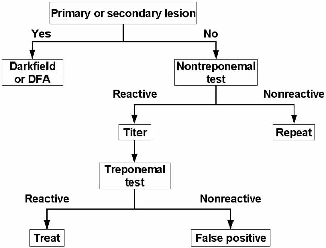
No ocular manifestations are pathognomonic for syphilis, so diagnosis is largely dependent on a combination of history, general clinical signs, detection of T. pallidum (when early lesions are present), and serologic analysis.51 A general scheme for the diagnosis of syphilis is depicted in Figure 2.

Fig. 2 Suggested paradigm for the diagnosis of syphilis. Nontreponemal tests are exemplified by the RPR Circle Card Test and the VDRL Test, whereas commonly used treponemal tests include the particle agglutination T. pallidum (PA-TP) test and the fluorescent treponemal antibody absorbed (FTA-ABS) test. Evaluation of lesion exudates for presence of T. pallidum can be performed by darkfield microscopy or direct fluorescent antibody (DFA) staining. Adapted from reference 16.

T. pallidum in patient specimens can be detected by darkfield microscopy, immunofluorescent staining, polymerase chain reaction (PCR), or animal inoculation. Characteristic primary or secondary lesions combined with the demonstration of T. pallidum in lesion exudates or biopsy samples by darkfield microscopy or immunostaining techniques is considered to be a definitive diagnosis.51 However, detection of T. pallidum by these means is not always available. In addition, spirochetes are present in exceedingly low numbers past the primary and secondary stages and are then difficult to detect by any method.

Immunofluorescent identification usually involves direct fluorescent antibody (DFA-TP) staining of lesion exudates or tissue using fluorescein-labeled anti-T. pallidum polyclonal or monoclonal antibodies.38,51 Immunohistochemical techniques have also been developed.29 T. pallidum has been detected in aqueous humor using darkfield microscopy or immunofluorescence of concentrated samples,68,87 but the numbers are exceedingly small and artifacts can yield false-positive interpretations.

PCR has been used to detect T. pallidum DNA in blood, tissue, and cerebrospinal fluid56,74 but is not routinely available. Inoculation of a rabbit was used to isolate the type strain T. pallidum subspecies pallidum Nichols from a patient's cerebrospinal fluid in 1906, and animal inoculation is still the only procedure available for the isolation of T. pallidum strains. It is conceivable that these sensitive methods could be used to detect or isolate T. pallidum from ocular fluids or biopsies.

Diagnosis of syphilis is highly dependent on serologic tests, including the so-called nontreponemal and treponemal antibody assays.8,16,51 Nontreponemal tests detect reaginic antibodies that react with cardiolipin combined with lecithin; these antibodies arise for unknown reasons during treponemal infections. The Rapid Plasma Reagin (RPR) circle card test is the most commonly used nontreponemal assay, although the Venereal Disease Research Laboratory (VDRL) test is the only assay available for testing the reactivity of cerebrospinal fluid. Treponemal assays detect anti-T. pallidum antibodies using whole T. pallidum, sonicates, or recombinant antigens. The T. pallidum particle agglutination assay (TP-PA) replaced the microhemagglutination T. pallidum (MHA-TP) test and is widely used today, but the fluorescent treponemal antibody-absorbed (FTA-ABS) test and enzyme immunoassays are also available. The nontreponemal tests are of value because they can be quantitated by titration, and typically the reciprocal titer decreases after treatment. The treponemal tests have the advantage of being specific for treponemal antigens, but reactivity is usually retained after treatment. Both types of tests may yield false-positive reactions in autoimmune disease, which should be kept in mind when attempting to distinguish autoimmune from syphilitic disease.

TREATMENT

Treatment should be consistent with the stage of disease, HIV status, and other factors.2 A single dose of benzathine penicillin (2.4 million units intramuscularly [IM]) is currently recommended for primary, secondary, and early latent syphilis, whereas three equivalent dosages at weekly intervals are recommended for late latent, latent of unknown duration, or tertiary syphilis.2 Aqueous crystalline penicillin G is recommended for neurosyphilis (3–4 million units intravenous [IV] every 4 hours for 10–14 days) and for congenital syphilis in infants (100,000–150,000 units/kg per day for 10 days). Re-covery of viable T. pallidum from CSF has been reported in neurosyphilis patients treated with benzathine penicillin, consistent with its poor penetration of blood–brain barrier; recurrence of manifestations in HIV-positive individuals also occurred.44 For this reason, treatment of patients with ocular syphilis with a regimen of aqueous penicillin G consistent with current neurosyphilis treatment guidelines should be considered.

ANIMAL MODELS

The earliest report of an animal model of ocular syphilis occurred before the discovery of T. pallidum. In 1881, Haensell30 described the occurrence of keratitis and iritis after inoculation of a syphilitic lesion exudate into the anterior chamber of a rabbit eye. In 1906, 1 year after T. pallidum was first identified by Schaudinn and Hoffman,65 Bertarelli14 also demonstrated the occurrence of corneal lesions in rabbits after T. pallidum inoculation. As part of their extensive characterization of experimental syphilis in the rabbit, Brown and Pearce17 described the eye manifestations observed after infection. In the 1960s, Wells and Smith84 and Elsas et al.26 were able to demonstrate ocular manifestations after inoculation of owl monkeys or squirrel monkeys with T. pallidum via the cornea, vitreous, cisterna magna, carotid artery, skin, and intravenous routes. The pathologic findings varied but included pupillary inequality, non-granulomatous iridocyclitis, retinal hemorrhage, multiple lens opacities, and flare. In addition, T. pallidum could be found in the aqueous humor using either darkfield microscopy or immunofluorescent staining up to 1 year after inoculation. Although five of the six monkeys studied developed serologic reactivity in the VDRL, FTA-ABS, and T. pallidum immobilization (TPI) tests, the reactivity was often weak (e.g., VDRL titers never exceeded 1:4). It is unclear whether this low reactivity was caused by poor antibody responses in these animals or by the inadequacy of the serologic tests for serum samples obtained from monkey species.

Concerns have long existed that corneal transplants could serve as a potential source of T. pallidum infection. Corneas from donors with reactive RPR tests were excluded from the transplant pool, resulting in a substantial reduction in the number of corneas available for transplant. In an extensive study by Dr. Elliot Randolph published in 1949,59 the presence of infectious organisms in corneas from rabbits infected with T. pallidum for 3 or more months was analyzed to provide information on the potential for transmission by corneal transplantation. None of the recipient rabbits was infected after injection of corneal extracts from 40 infected ``donor'' rabbits. In more recent studies,45 rabbits were infected by intratesticular inoculation, and 10 days later the corneas were removed, minced, and extracted. When injected into naïve rabbits, these extracts produced lesions if not washed with saline before extraction, indicating the presence of T. pallidum. However, with saline rinsing, no lesions were obtained, consistent with the presence of viable treponemes in blood or ocular fluids but not in the corneal tissue itself. In addition, incubation of T. pallidum in OptiSol® cornea storage medium was found to render the organisms noninfectious within 24 hours. These data were used in combination with other information to remove RPR reactivity as a criterion for the exclusion of corneas for transplantation.

Back to Top
OTHER TREPONEMA SPECIES AND SUBSPECIES
The species Treponema pallidum contains several closely related subspecies that each correspond to a particular human disease: subspecies pallidum (syphilis), subspecies endemicum (endemic syphilis), and subspecies pertenue (yaws) (Table 1). Treponema carateum is the causative agent of pinta and remains a separate species because of the lack of genetic information. Distribution of the sexually transmitted disease syphilis is worldwide, whereas the other treponematoses are currently restricted to tropical and desert regions of South and Central America, Africa, and Asia. These organisms are obligate pathogens of humans and are not found elsewhere in nature. They are among a handful of pathogenic bacteria that have not been cultivated continuously in vitro. Other Treponema species can be cultured from subgingival plaque (e.g., Treponema denticola) or moist skin surfaces (Treponema phagedenis). Oral treponemes are associated with periodontal disease, whereas T. phagedenis and other skin saprophytes are nonpathogenic; however, neither group is known to cause eye infections.

PATHOPHYSIOLOGY OF NONVENEREAL TREPONEMATOSES

Nonvenereal treponematoses are typically transmitted by non-sexual contact with active lesions during childhood or adolescence. The degree of systemic manifestations of endemic syphilis tends to be somewhat less than that of venereal syphilis; yaws and pinta each exhibit progressively more localized skin lesions and less systemic involvement.7,63 Congenital disease occurs rarely in endemic syphilis and is not thought to occur in yaws and pinta.

OCULAR MANIFESTATIONS

Relatively few studies address the occurrence of ocular lesions in endemic syphilis, yaws, and pinta. Tabbara et al.76 examined late endemic syphilis patients in nomadic Bedouins in Saudi Arabia in the 1980s and found uveitis, chorioretinal scars, and optic atrophy. An extensive study of yaws patients in Venezuela69,70 found a high percentage of cases of abnormal pupils (32%), abnormal fundi (26%), or optic atrophy (20%). Evidence of chorioretinitis and abnormal pupils was also observed in some pinta patients.69

Spirochetes were identified by darkfield microscopy or immunofluorescence in the aqueous humour of some yaws patients and in one pinta patient, but positive results were also obtained in 10% of presumed-normal controls. Many of these presumed-positive results were described as ``nonfluorescent spiral forms,'' meaning that they did not bind anti-T. pallidum antibodies; interestingly, photomicrographs provided for several cases (especially cases 175 and 178) bear a strong resemblance to Leptospira, which also has a high incidence in this region.69 Taken together, the data available indicate that ocular manifestations can occur in patients with endemic (nonvenereal) treponematoses but that care must be taken to distinguish these diseases from other infections (or co-infections).

Back to Top
BORRELIA BURGDORFERI
Borrelia species are epizootic, arthropod-transmitted pathogens that cause Lyme borreliosis or relapsing fever in humans. Lyme borreliosis (also called Lyme disease) is the most common arthropod-transmitted bacterial infection in the United States, with 23,763 cases reported in 2002. It is caused primarily by Borrelia burgdorferi in North America, whereas B. burgdorferi, Borrelia garinii, and Borrelia afzelii all cause human disease in Europe and Asia.

The predominant arthropod vectors of the disease are hard-bodied Ixodes ticks, including Ixodes scapularis in North America and Ixodes ricinus in Europe and Asia. Lyme Borrelia are maintained through an infectious cycle between small mammals (or birds) and the Ixodes ticks; large mammals, most notably deer, are required for feeding and ovulation of the adult ticks. Humans can be infected by nymphal or adult ticks, and human-to-human transmission does not occur. Although small numbers of cases occur in almost every state of the United States, most reported cases are concentrated in the northeastern and mid-Atlantic states, with another focus in Wisconsin and Minnesota.

PATHOPHYSIOLOGY OF LYME BORRELIOSIS

The clinical course has been subdivided into three stages, with the understanding that these stages commonly overlap.71 Stage 1 is the initial, localized infection and typically consists of erythema migrans, a well-demarcated expanding erythematous rash with central pallor that develops at the site of the tick bite. Regional lymphadenopathy and flu-like constitutional symptoms, including fever and chills, myalgias, arthralgias, malaise, and nausea are also often present. Stage 2 represents disseminated infection, occurring weeks to months after the initial presentation. Migrating monoarthritis of the knee and other large joints, neuroborreliosis (including cranial neuropathy, aseptic meningitis, headache, encephalitis, or peripheral neuritis), cardiac atrioventricular block, and ophthalmic manifestations can occur during this stage.

The third or chronic stage of Lyme borreliosis is characterized by oligoarthritis, chronic neurologic manifestations, and a hypopigmented skin lesion called acroderma chronicum atrophicans (ACA). Arthritis is the most common stage 3 symptom of B. burgdorferi infection and, hence, is seen commonly in North America. In contrast, neurologic symptoms predominate the chronic stage of B. garinii infection, and B. afzelii gives rise to ACA; thus, these manifestations are found more commonly in Europe and Asia.83

Other tick-transmitted diseases, including babesiosis, human monocytic ehrlichiosis, and human granulocytic ehrlichiosis (caused by Anaplasma phagocytophilum), occur in areas endemic for Lyme borreliosis and may cause co-infections.

OCULAR MANIFESTATIONS

As in syphilis, Lyme borreliosis can affect any segment of the eye.10,13,48,92 Mild follicular conjunctivitis, subconjunctival hemorrhages, photophobia, and periorbital edema may coincide with erythema migrans during the early stage of the disease. The occurrence of conjunctivitis is reported to be 10% during this stage, but this value may be underestimated. During stage 2, diplopia may result from cranial neuropathy, although involvement of cranial nerve VII (with resulting Bell's palsy) is much more common than paresis of cranial nerve VI, III, or IV. Blurred vision also occurs at this stage and may be associated with papilledema, optic atrophy, optic or retrobulbar neuritis, or pseudotumor cerebri. These may coincide with aseptic meningitis and associated symptoms, which are present in approximately 15% of untreated Lyme borreliosis patients 4 weeks after the appearance of erythema migrans.

The most severe manifestations typically occur months to years after the initial infection and may include pars planitis, iritis, vitreitis, keratitis, and a wide variety of forms of posterior uveitis.10,92 Although the first reported case of ocular Lyme borreliosis resulted in unilateral blindness,72 eye involvement typically is mild to moderate, responsive to therapy, and not associated with loss of visual acuity.

LABORATORY DIAGNOSIS

The diagnosis of Lyme disease remains primarily clinical. The recognition of the erythema migrans lesion and other associated symptoms, a history of tick bite or potential exposure, and residence in (or travel to) an endemic area are the most important diagnostic clues. Verification is usually performed by detection of antibodies against Borrelia antigens, although culture, immunologic staining of tissue specimens, or PCR can also be used.18

The currently recommended method for serodiagnosis is a two-tiered approach.1,88 Either serum or CSF can be examined. An enzyme immunoassay using a whole-cell lysate of Lyme disease Borrelia or recombinant antigens is used as a screening test. If a positive result is obtained, the enzyme-linked immunosorbent assay (ELISA) is followed by Western blot analysis; both whole-cell extracts and mixtures of recombinant proteins have been used as the antigens in immunoblots. Serologic tests are nonreactive during the erythema migrans stage in approximately one half of the patients, but seroconversion may occur after therapy. Sensitivity approaches 100% at later stages. Specificity of whole–cell-based immunoassays is limited by the occurrence of cross-reactive antibodies, particularly in patients with autoimmune disease or syphilis; therefore, serologic testing is recommended only when there is a strong suspicion (>20%) of Lyme borreliosis. Recently, a rapid "first-tie" test using chimeric recombinant proteins with epitopes from multiple B. burgdorferi antigens has been developed.28 In addition, immunoassays using recombinant protein constructs39 or the C6 synthetic peptide42 of the Borrelia antigenic variation protein VlsE have been shown to be highly sensitive and specific.9,67

Direct detection of Borrelia by culture or PCR is another means of confirmation but is not recommended for general diagnostic use because of low sensitivity.18 In one study, culture of skin punch biopsy samples from the advancing edge of erythema migrans lesions in Barbour-Steiner-Kelly (BSK) medium yielded positive results in 60% of cases.89 Blood cultures are positive in one fourth of early Lyme disease cases; this sensitivity was only achieved by using serum as opposed to whole blood and by using high sample volumes.91 Spirochetes with characteristics of Lyme disease Borrelia were cultured from tissue removed during sector iridectomy and prepupillary membranectomy in a case of recurrent Lyme uveitis after treatment with systemic doxycycline; however, organisms were detected only after 16 subcultures.57

PCR has also been examined as a possible means of laboratory confirmation of Lyme borreliosis.18,22,23,66 Skin and synovial fluid yield the highest rates of PCR positivity (68% and 73%, respectively), whereas CSF and plasma have lower rates (18% and 29%).22 Mikkilä et al.47 reported positive PCR results in 7 of 20 cases of ocular Lyme borreliosis. Spirochetes resembling B. burgdorferi were detected histologically in vitreous debris by Dieterle silver staining of a surgical specimen in a case of panophthalmitis.72 However, microscopic detection of Lyme Borrelia in tissue specimens is difficult because of low numbers and possible artifacts; interpretation is aided by the use of immunofluorescence or immunohistochemical techniques. Overall, culture, PCR, and microscopic detection are currently research techniques and are not generally available for routine confirmation of Lyme borreliosis.

TREATMENT

The preferred treatment of uncomplicated stage 1 disease, arthritis, or cranial nerve palsies consists of oral regimens of either amoxicillin or doxycycline, with cefuroxime as an alternative.83,90 Ocular symptoms associated with stage 1 disease (e.g., conjunctivitis and photophobia) resolve with oral therapy. The recommended therapy for central nervous system Lyme borreliosis is parenteral treatment with ceftriaxone (preferred), cefotaxime, or penicillin G for 14 to 28 days.83,90 Although standardized regimens for ophthalmic disease have not been developed, eye involvement should be provided treatment consistent with that of CNS disease to optimize ocular penetration.10,13,34,75,92

The benefit of systemic corticosteroid treatment as an adjunct therapy is unclear, but such treatment without antibiotic therapy is contraindicated. A Jarisch-Herxheimer reaction (hypotension, chills, fever, arthralgia, and myalgia) occurs following treatment in approximately 14% of patients with Lyme borreliosis and may result in a worsening of optic manifestations. This effect can be counteracted by treatment with nonsteroidal antiinflammatory agents or corticosteroids.73

ANIMAL MODELS

Infection of the eyes and conjunctivitis has been reported to occur consistently in Syrian hamsters inoculated with B. burgdorferi.24,33 Conjunctivitis has also been noted in experimentally infected hamsters and rhesus monkeys.55 However, animal models of ocular Lyme borreliosis have not been studied in detail.

Back to Top
OTHER BORRELIA SPECIES
Several Borrelia species, including B. hermsii, B. turicatae, B. crocidurae, and B. duttoni, give rise to sporadic, epizootic cases of relapsing fever and are transmitted by soft-bodied ticks of the genus Ornithodoros. In contrast, Borrelia recurrentis gives rise to epidemic relapsing fever and is borne by body lice. Both are characterized by recurring episodes of fever (lasting 2 to 5 days) separated by afebrile periods of 7 to 9 days. Although epidemic relapsing fever (also called trench fever) caused many deaths during World War I, both forms of the disease are relatively rare today. Relapsing fever is primarily a spirochetemia. Up to 108 organisms per mL of blood are present during the periods of fever, and spirochetes can be detected easily at these times by Wright staining of peripheral blood smears.

Eye involvement is common in tick-borne relapsing fever and can result in a rapid and severe loss of visual acuity and permanent vision deficits.19 Most ocular disease reported is caused by infection with B. duttonii, B. hispanica, and B. turicatae. Manifestations include iridocyclitis, choroiditis, and optic neuritis. In one study in East Africa, the incidence of optic neuritis or uveitis was 11%. Ocular manifestations of louse-borne relapsing fever have not been reported. Treatment with β-lactam antibiotics or tetracycline derivatives is highly effective but may result in a Jarisch-Herxheimer reaction.

A number of animal models of tick-borne relapsing fever are available, the most widely used of which is mouse infection.19 Changes in the variable small protein (Vsp) antigenic variation proteins of B. turicatae have been shown to affect pathogenesis in mice, in some cases resulting in a neurotropic clonotype that causes measurable neurologic deficits.20

Back to Top
LEPTOSPIRA SPECIES
Leptospirosis is an epizootic illness of global distribution caused by members of the genus Leptospira.15,40,41 Leptospira are slender, tightly wound, highly motile spirochetes with hooked ends. Unlike Treponema and Borrelia, they are obligate aerobes, are easily cultured, and can survive for long periods in water or soil. The 4.7-megabase genome of Leptospira interrogans was sequenced recently and reveals a greatly increased biosynthetic capacity relative to other pathogenic spirochetes.62 As a result of recent genotypic classification, this genus has been speciated into 12 named and five unnamed genospecies.15,40 The pathogenic Leptospira are subdivided into more than 200 different serovars based on serologic reactivity using the microagglutination test (as described later). The serovars are based on variation in the lipopolysaccharide (LPS) side chains; interestingly, this phenotypic classification does not always follow genotypic characteristics.

Leptospira commonly infect a variety of both wild and domestic animals, including rodents, bats, mongooses, possums, dogs, horses, and cattle.15 Animals become chronically infected, and Leptospira colonize the renal tubules and are shed in large numbers in the urine. Infection of humans occurs as the result of exposure of broken skin, mucous membranes, or conjunctiva to urine or tissue of infected animals, or to water contaminated by urine.

Leptospirosis is generally related to occupational or recreational exposure or residence in rural or underdeveloped urban areas. Outbreaks frequently are associated with flooding, in which effluent from soil contaminates untreated water supplies.61,64 Urban epidemics occur in areas of poor sanitation as the result of rat infestation or contamination of water supplies.37,78,80 An outbreak of leptospirosis among 98 of 834 triathlon athletes after an event in Springfield, Illinois underlines the global distribution of pathogenic Leptospira.49 Insufficient reporting or epidemiologic analysis is available to estimate the incidence of leptospiral infection, but it may be the most common spirochetal pathogen in humans as well as domestic and wild animals.

PATHOPHYSIOLOGY OF LEPTOSPIROSIS

The infection in humans and animals follows a similar course. The organism multiplies rapidly and disseminates by the hematogenous route, resulting in spirochetemia and invasion of virtually every tissue. During this acute phase, manifestations include headache, fever, malaise, myalgia, and conjunctival chemosis (sometimes accompanied by subconjunctival hemorrhage).60 In 10% to 15% of untreated cases, a severe spirochetemia accompanied by endothelial damage and hemorrhage results in Weil's disease, which has a fatality rate ranging from 10% to 50% in different reports.

The acute phase is followed by a period of asymptomatic infection lasting 2 to 20 months. The immune phase then develops. Immunologic reactions lead to damage to the liver, kidneys, eyes, and other organs. Damage to endothelial cells and resulting vasculitis, hemorrhage, ischemia, and tissue necrosis appear to be the major pathogenic mechanisms. The expression of LPS, which is not found in Treponema or Borrelia, is likely to be involved in endothelial damage, fever, malaise, and hypotension. Jaundice, anuria, altered sensorium, and rhabdomyolysis are indicators of poor prognosis.60

OCULAR MANIFESTATIONS

Conjunctival suffusion and hemorrhage are frequent manifestations in patients hospitalized for leptospirosis, with rates ranging from 28.5% to 99% in different studies.15 During the late stages of leptospirosis, uveitis is a common occurrence that can potentially lead to blindness. Panuveitis is characterized by rapid onset, severe manifestations, and relapses. Nongranulomatous uveitis occurs more frequently than the granulomatous form, and hypopyon and moderate to severe vitreitis (with membranous opacities) are also common presentations. Intermittent perivasculitis can be distinguished from Behçet disease by the relative infrequency of vascular occlusion. Anterior forms of leptospiral uveitis tend to be less severe and gradual in onset. Cataracts and glaucoma are also associated with leptospirosis.

LABORATORY DIAGNOSIS

The diagnosis of early leptospirosis is complicated by its varied and indistinct symptomology that could be mistaken as influenza, hepatitis, and many other infections.36,40,41 The latent and late forms may be even more challenging because of the lack of recognition of early disease, prolonged asymptomatic periods (e.g., 2 months to 2 years), and varied forms of late presentation. Careful assessment of potential exposure to contaminated water or soil, close contact with animals, and travel to high-incidence areas is needed. Exclusion of other potential causes and consistency of the ocular and systemic manifestations with leptospiral infection are of course also important.

Culture of Leptospira is possible and leads to a definitive diagnosis; however, it requires the specialized EMJH medium, which contains long-chain fatty acids required for growth. In addition, culture can only be performed from blood or other samples during the fulminant early stage of infection, and outgrowth may require several weeks. Immunohistochemical methods for detecting Leptospira in tissue specimens have been developed.85

The microscopic agglutination test (MAT) is considered the gold standard of serologic analysis but is a technically cumbersome procedure. A panel of Leptospira serovars (typically 12 to 16) that occur commonly in the geographic region are cultured, washed, and frozen. The cells are combined with increasing dilutions of the patient's serum along with appropriate controls and examined microscopically to determine the endpoint where more than 50% of the organisms are agglutinated. A four-fold increase in titer during the acute phase or a titer >1:100 in chronic disease with one or more of the serovars tested is considered to be diagnostic. False-negative results can be obtained if the infecting serovar is missing from the test panel, and false-positive results may occur because of persistent antibodies from previous leptospiral infections. Because of the complexity of the MAT, a number of new immunoassay techniques, including ELISA, macroagglutination assays, and a rapid leptospira dipstick test (Lepto-DST) are in use or undergoing evaluation.40

PCR is of particular value in ocular disease. In one study in south India,21 80% of aqueous humor samples from patients with four or more signs suggestive of leptospiral uveitis (e.g., panuveitis, anterior chamber cells, flare, and vasculitis) were positive for leptospiral DNA. Only 5% of patients with uveitis or cataracts inconsistent with leptospirosis were PCR-positive. Very similar results were obtained for the detection of leptospiral uveitis in horses by PCR,27 in which leptospiral DNA was detected in 70% of leptospirosis cases and only 6% of control cases. Less than one third of PCR-positive cases were culture-positive.

TREATMENT

No standardized treatment guidelines have been developed for leptospirosis, and few controlled studies exist that clarify the value of antimicrobial therapy. While all experts would agree that severe leptospirosis should be treated, some controversy exists regarding the treatment of early leptospirosis. Levett41 indicated that patients with early leptospirosis and only mild fever and flu-like symptoms need only supportive therapy, in that the course of disease is usually self-limiting. Conversely, other authors would argue that availability of an effective, inexpensive, antimicrobial regimen for early mild leptospirosis would prevent the potential occurrence of Weil disease or other severe sequelae in a significant number of patients.79 A practical, and important, side of this issue is that the availability of medical care may be extremely limited in many of the indigent populations in which leptospirosis is common.

In a recent randomized, open-label, nonplacebo study, Panaphut et al.52 determined that ceftriaxone and aqueous penicillin reduced the duration of fever in severe leptospirosis cases to 3 days; however, the mortality rate was still 50% in both treatment groups. In another study, Kobayashi36 found that β-lactam antibiotics are bactericidal in vitro against actively growing L. interrogans but are considerably less effective against organisms in the stationary phase. This result may explain why penicillin generally has a limited or varied effect on the course of late or severe disease.25,35,36,82

Streptomycin is particularly effective in treating Leptospira infections,36 presumably because of its bactericidal effect on organisms in both logarithmic growth and stationary phases. Doxycycline therapy (100 mg twice per day for 7 days) of icteric leptospirosis reduced the disease severity and the period of illness by 2 days.46 Studies in hamsters indicate that doxycycline is capable of clearing experimental L. interrogans serovar icterohaemorrhagiae infection, whereas penicillin was only partially effective and ofloxacin was noneffective.77 Such analyses may be useful in optimizing therapies for clinical trials in humans.

ANIMAL MODELS

Leptospirosis is a zoonotic infection in which humans are accidental hosts. Leptospira species can infect a wide variety of small and large mammals, ranging from mice, rats, and bats to neotropical opossums, dogs, horses, and cattle.15 It thus represents a substantial problem in both livestock and pet veterinary practices. Different Leptospira species and serovars preferentially infect certain mammalian hosts, contributing to the variation in distribution and epidemiology of Leptospira variants in geographic regions. These strains in turn have different patterns of pathogenesis in humans. Studies in horses27,43,53,54,81 and hamsters12 have been particularly helpful in understanding the pathobiology of ocular and kidney infections, respectively.

Back to Top
REFERENCES

1. Centers for Disease Control and Prevention: Conditions under public health surveillance. Morbid Mortal Weekly Rep 46:34, 1997

2. Centers for Disease Control and Prevention: Summary of notable diseases–United States 2000. Morbid Mortal Weekly Rep 49:1, 2002

3. Centers for Disease Control: Primary and secondary syphilis–United States. Morbid Mortal Weekly Rep 52:1117, 2003

4. Centers for Disease Control: Summary of notable diseases, United States. Morbid Mortal Weekly Rep 43:3, 1994

5. WHO/64: Sexually transmitted diseases: three hundred and thirty-three million new curable cases in 1995. Geneva: World Health Organization, 1999

6. Aldave AJ, King JA, Cunningham ET Jr : Ocular syphilis. Curr Opin Ophthalmol 12(6):433–41, 2001

7. Antal GM, Lukehart SA, Meheus AZ: The endemic treponematoses. Microbes Infect 4(1):83–94, 2002

8. Augenbraun M, Rolfs R, Johnson R, Joesoef R, Pope V: Treponemal specific tests for the serodiagnosis of syphilis. Syphilis and HIV Study Group. Sex Transm Dis 25(10):549–552, 1998

9. Bacon RM, Biggerstaff BJ, Schriefer ME, Gilmore RD Jr. , Philipp MT, Steere AC, Wormser GP, Marques AR, Johnson BJ: Serodiagnosis of Lyme disease by kinetic enzyme-linked immunosorbent assay using recombinant VlsE1 or peptide antigens of Borrelia burgdorferi compared with 2-tiered testing using whole-cell lysates. J Infect Dis 187(8):1187–1199, 2003

10. Balcer LJ, Winterkorn JM, Galetta SL: Neuro-ophthalmic manifestations of Lyme disease. J Neuroophthalmol 17(2):108–121, 1997

11. Barile GR, Flynn TE: Syphilis exposure in patients with uveitis. Ophthalmology 104(10):1605–1609, 1997

12. Barnett JK, Barnett D, Bolin CA, Summers TA, Wagar EA, Cheville NF, Hartskeerl RA, Haake DA: Expression and distribution of leptospiral outer membrane components during renal infection of hamsters. Infect Immun 67(2):853–861, 1999

13. Bergloff J, Gasser R, Feigl B: Ophthalmic manifestations in Lyme borreliosis. A review. J Neuroophthalmol 14(1):15–20, 1994

14. Bertarelli E: Über die transmission der syphilis auf das kaninchen. Centralbl Bakt 41:320, 1906

15. Bharti AR, Nally JE, Ricaldi JN, Matthias MA, Diaz MM, Lovett MA, Levett PN, Gilman RH, Willig MR, Gotuzzo E, Vinetz JM: Leptospirosis: a zoonotic disease of global importance. Lancet Infect Dis 3(12):757–771, 2003

16. Brown SL, Hansen SL, Langone JJ: Role of serology in the diagnosis of Lyme disease. JAMA 282(1):62–66, 1999

17. Brown WH, Pearce L: Experimental syphilis in the rabbit: VI. affection of the eyes. J Exp Med 34:167, 1921

18. Bunikis J, Barbour AG: Laboratory testing for suspected Lyme disease. Med Clin North Am 86(2):311–340, 2002

19. Cadavid D, Barbour AG: Neuroborreliosis during relapsing fever: review of the clinical manifestations, pathology, and treatment of infections in humans and experimental animals. Clin Infect Dis 26(1):151–164, 1998

20. Cadavid D, Thomas DD, Crawley R, Barbour AG: Variability of a bacterial surface protein and disease expression in a possible mouse model of systemic Lyme borreliosis. J Exp Med 179(2):631–642, 1994

21. Chu KM, Rathinam R, Namperumalsamy P, Dean D: Identification of Leptospira species in the pathogenesis of uveitis and determination of clinical ocular characteristics in south India. J Infect Dis 177(5):1314–1321, 1998

22. Dumler JS: Molecular diagnosis of Lyme disease: review and meta-analysis. Mol Diagn 6(1):1–11, 2001

23. Dumler JS: Molecular methods for ehrlichiosis and Lyme disease. Clin Lab Med 23(4):867–884, vi, 2003

24. Duray PH, Johnson RC: The histopathology of experimentally infected hamsters with the Lyme disease spirochete, Borrelia burgdorferi. Proc Soc Exp Biol Med 181(2):263–269, 1986

25. Edwards CN, Nicholson GD, Hassell TA, Everard CO, Callender J: Penicillin therapy in icteric leptospirosis. Am J Trop Med Hyg 39(4):388–390, 1988

26. Elsas FJ, Smith JL, Israel CW, Gager WE: Late syphilis in the primate. Br J Vener Dis 44(4):267–273, 1968

27. Faber NA, Crawford M, LeFebvre RB, Buyukmihci NC, Madigan JE, Willits NH: Detection of Leptospira spp. in the aqueous humor of horses with naturally acquired recurrent uveitis. J Clin Microbiol 38(7):2731–2733, 2000

28. Gomes-Solecki MJ, Wormser GP, Persing DH, Berger BW, Glass JD, Yang X, Dattwyler RJ: A first-tier rapid assay for the serodiagnosis of Borrelia burgdorferi infection. Arch Intern Med 161(16):2015–2020, 2001

29. Guarner J, Southwick K, Greer P, Bartlett J, Santander A, Blanco S, Pope V, Levine W, Zaki S: Testing umbilical cords for funisitis due to Treponema pallidum infection, Bolivia. Emerg Infect Dis 6(5):487–492, 2000

30. Haensell P: Vorläufiger Mittheilung über Versuche von Impfsyphlis der Iris und Cornea des Kanichanauges. Arch Oophthalmol Berlin 27:93, 1881

31. Hook EW 3rd : Syphilis and HIV infection. J Infect Dis 160(3):530–534, 1989

32. Hutchinson J: On the different form of inflammation of the eye consequent on inhereted syphilis. Royal London Ophthalm Hosp Rep 1:191; 226:1859; 2:54;1860; 3:258 (Reprinted in Med Classics 5:107, 1940, 1858

33. Johnson RC, Marek N, Kodner C: Infection of Syrian hamsters with Lyme disease spirochetes. J Clin Microbiol 20(6):1099–1101, 1984

34. Karma A, Seppala I, Mikkila H, Kaakkola S, Viljanen M, Tarkkanen A: Diagnosis and clinical characteristics of ocular Lyme borreliosis. Am J Ophthalmol 119(2):127–135, 1995

35. Katz AR, Ansdell VE, Effler PV, Middleton CR, Sasaki DM: Assessment of the clinical presentation and treatment of 353 cases of laboratory-confirmed leptospirosis in Hawaii, 1974–1998. Clin Infect Dis 33(11):1834–1841, 2001

36. Kobayashi Y: Clinical observation and treatment of leptospirosis. J Infect Chemother 7(2):59–68, 2001

37. Kuo IC, Kapusta MA, Rao NA: Vitritis as the primary manifestation of ocular syphilis in patients with HIV infection. Am J Ophthalmol 125(3):306–311, 1998

38. Larsen SA, Pope V, Johnson RE, Kennedy EJ Jr : A Manual of Tests for Syphilis, 9th ed. Washington, DC: American Public Health Association, 1998:1

39. Lawrenz MB, Hardham JM, Owens RT, Nowakowski J, Steere AC, Wormser GP, Norris SJ: Human antibody responses to VlsE antigenic variation protein of Borrelia burgdorferi. J Clin Microbiol 37(12):3997–4004, 1999

40. Levett PN: Leptospira and Leptonema. In Murray PR, ed. Manual of Clinical Microbiology, 8th edition. Vol. 1. Washington, DC: ASM Press, 2003:929

41. Levett PN: Leptospirosis. Clin Microbiol Rev 14(2):296–326, 2001

42. Liang FT, Steere AC, Marques AR, Johnson BJ, Miller JN, Philipp MT: Sensitive and specific serodiagnosis of Lyme disease by enzyme-linked immunosorbent assay with a peptide based on an immunodominant conserved region of Borrelia burgdorferi vlsE. J Clin Microbiol 37(12):3990–3996, 1999

43. Lucchesi PM, Parma AE, Arroyo GH: Serovar distribution of a DNA sequence involved in the antigenic relationship between Leptospira and equine cornea. BMC Microbiol 2(1):3, 2002

44. Lukehart SA, Hook EW 3rd : Baker-Zander SA, Collier AC, Critchlow CW, Handsfield HH: Invasion of the central nervous system by Treponema pallidum: implications for diagnosis and treatment. Ann Intern Med 109(11):855–862, 1988

45. Macsai MS, Norris SJ: OptiSol corneal storage medium and transmission of Treponema pallidum. Cornea 14(6):595–600, 1995

46. McClain JB, Ballou WR, Harrison SM, Steinweg DL: Doxycycline therapy for leptospirosis. Ann Intern Med 100(5):696–698, 1984

47. Mikkilä H, Karma A, Viljanen M, Seppala I: The laboratory diagnosis of ocular Lyme borreliosis. Graefes Arch Clin Exp Ophthalmol 237(3):225–30, 1999

48. Mikkilä HO, Seppala IJ, Viljanen MK, Peltomaa MP, Karma A: The expanding clinical spectrum of ocular lyme borreliosis. Ophthalmology 107(3):581–587, 2000

49. Morgan J, Bornstein SL, Karpati AM, Bruce M, Bolin CA, Austin CC, Woods CW, Lingappa J, Langkop C, Davis B, Graham DR, Proctor M, Ashford DA, Bajani M, Bragg SL, Shutt K, Perkins BA, Tappero JW: Outbreak of leptospirosis among triathlon participants and community residents in Springfield, Illinois, 1998. Clin Infect Dis 34(12):1593–1599, 2002

50. Musher DM: Syphilis, neurosyphilis, penicillin, and AIDS. J Infect Dis 163(6):1201–1206, 1991

51. Norris SJ, Pope V, Johnson RE, Larsen SA: Treponema and other human host-associated spirochetes. In Murray PR, ed. Manual of Clinical Microbiology, 8th ed. Washington, DC: ASM Press, 2003

52. Panaphut T, Domrongkitchaiporn S, Vibhagool A, Thinkamrop B, Susaengrat W: Ceftriaxone compared with sodium penicillin g for treatment of severe leptospirosis. Clin Infect Dis 36(12):1507–1513, 2003

53. Parma AE, Cerone SI, Sansinanea SA: Biochemical analysis by SDS-PAGE and western blotting of the antigenic relationship between Leptospira and equine ocular tissues. Vet Immunol Immunopathol 33(1–2):179–185, 1992

54. Parma AE, Cerone SI, Sansinanea SA, Ghezzi M: C3 fixed in vivo to cornea from horses inoculated with Leptospira interrogans. Vet Immunol Immunopathol 34(1–2):181–187, 1992

55. Philipp MT, Aydintug MK, Bohm RP Jr. , Cogswell FB, Dennis VA, Lanners HN, Lowrie RC Jr. , Roberts ED, Conway MD, Karacorlu M, et al: Early and early disseminated phases of Lyme disease in the rhesus monkey: a model for infection in humans. Infect Immun 61(7):3047–3059, 1993

56. Pillay A, Liu H, Chen CY, Holloway B, Sturm AW, Steiner B, Morse SA: Molecular subtyping of Treponema pallidum subspecies pallidum. Sex Transm Dis 25(8):408–414, 1998

57. Preac-Mursic V, Pfister HW, Spiegel H, Burk R, Wilske B, Reinhardt S, Bohmer R: First isolation of Borrelia burgdorferi from an iris biopsy. J Clin Neuroophthalmol 13(3):155–161; discussion 162, 1993

58. Preston KE, Kacica MA, Limberger RJ, Archinal WA, Venezia RA: The resistance and integrase genes of pACM1, a conjugative multiple-resistance plasmid, from Klebsiella oxytoca. Plasmid 37(2):105–118, 1997

59. Randolph ME: An experimental study of the possibility of transmitting syphilis by a corneal graft. Trans Am Ophthalmol Soc 47:683, 1949

60. Rathinam SR: Ocular leptospirosis. Curr Opin Ophthalmol 13(6):381–386, 2002

61. Rathinam SR, Rathnam S, Selvaraj S, Dean D, Nozik RA, Namperumalsamy P: Uveitis associated with an epidemic outbreak of leptospirosis. Am J Ophthalmol 124(1):71–79, 1997

62. Ren SX, Fu G, Jiang XG, Zeng R, Miao YG, Xu H, Zhang YX, Xiong H, Lu G, Lu LF, Jiang HQ, Jia J, Tu YF, Jiang JX, Gu WY, Zhang YQ, Cai Z, Sheng HH, Yin HF, Zhang Y, Zhu GF, Wan M, Huang HL, Qian Z, Wang SY, Ma W, Yao ZJ, Shen Y, Qiang BQ, Xia QC, Guo XK, Danchin A, Saint Girons I, Somerville RL, Wen YM, Shi MH, Chen Z, Xu JG, Zhao GP: Unique physiological and pathogenic features of Leptospira interrogans revealed by whole-genome sequencing. Nature 422(6934):888–893, 2003

63. Roman GC, Roman LN: Occurrence of congenital, cardiovascular, visceral, neurologic, and neuro-ophthalmologic complications in late yaws: a theme for future research. Rev Infect Dis 8(5):760–770, 1986

64. Sanders EJ, Rigau-Perez JG, Smits HL, Deseda CC, Vorndam VA, Aye T, Spiegel RA, Weyant RS, Bragg SL: Increase of leptospirosis in dengue-negative patients after a hurricane in Puerto Rico in 1996 [correction of 1966]. Am J Trop Med Hyg 61(3):399–404, 1999

65. Schaudinn F, Hoffman E: Vorläufiger bericht uber das vorkommen fur spirochaeten in syphilitischen krankheitsprodukten und be papillomen. Arb Gesundh Amt Berlin 22:528, 1905

66. Schmidt BL: PCR in laboratory diagnosis of human Borrelia burgdorferi infections. Clin Microbiol Rev 10(1):185–201, 1997

67. Schulte-Spechtel U, Lehnert G, Liegl G, Fingerle V, Heimerl C, Johnson BJ, Wilske B: Significant improvement of the recombinant Borrelia-specific immunoglobulin G immunoblot test by addition of VlsE and a DbpA homologue derived from Borrelia garinii for diagnosis of early neuroborreliosis. J Clin Microbiol 41(3):1299–1303, 2003

68. Smith JL: Testing for congenital syphilis in interstitial keratitis. Am J Ophthalmol 72(4):816–820, 1971

69. Smith JL, David NJ, Indgin S, Israel CW, Levine BM, Justice J Jr. , McCrary JA 3rd : Medina R, Paez P, Santana E, Sarkar M, Schatz NJ, Spitzer ML, Spitzer WO, Walter EK: Neuro-ophthalmological study of late yaws and pinta. II. The Caracas project. Br J Vener Dis 47(4):226–251, 1971

70. Smith JL, Israel CW: A neuro-ophthalmologic study of late yaws and pinta. Trans Am Ophthalmol Soc 68:292–300, 1970

71. Steere AC: Lyme disease. N Engl J Med 345(2):115–125, 2001

72. Steere AC, Duray PH, Kauffmann DJ, Wormser GP: Unilateral blindness caused by infection with the Lyme disease spirochete, Borrelia burgdorferi. Ann Intern Med 103(3):382–384, 1985

73. Strominger MB, Slamovits TL, Herskovitz S, Lipton RB: Transient worsening of optic neuropathy as a sequela of the Jarisch-Herxheimer reaction in the treatment of Lyme disease. J Neuroophthalmol 14(2):77–80, 1994

74. Sutton MY, Liu H, Steiner B, Pillay A, Mickey T, Finelli L, Morse S, Markowitz LE, St Louis ME: Molecular subtyping of Treponema pallidum in an Arizona County with increasing syphilis morbidity: use of specimens from ulcers and blood. J Infect Dis 183(11):1601–1606, 2001

75. Suttorp-Schulten MS, Kuiper H, Kijlstra A, van Dam AP, Rothova A: Long-term effects of ceftriaxone treatment on intraocular Lyme borreliosis. Am J Ophthalmol 116(5):571–575, 1993

76. Tabbara KF, al Kaff AS, Fadel T: Ocular manifestations of endemic syphilis (bejel). Ophthalmology 96(7):1087–1091, 1989

77. Truccolo J, Charavay F, Merien F, Perolat P: Quantitative PCR assay to evaluate ampicillin, ofloxacin, and doxycycline for treatment of experimental leptospirosis. Antimicrob Agents Chemother 46(3):848–853, 2002

78. Vinetz JM: Leptospirosis. Curr Opin Infect Dis 14(5):527–538, 2001

79. Vinetz JM: A mountain out of a molehill: do we treat acute leptospirosis, and if so, with what? Clin Infect Dis 36(12):1514–1515, 2003

80. Vinetz JM, Glass GE, Flexner CE, Mueller P, Kaslow DC: Sporadic urban leptospirosis. Ann Intern Med 125(10):794–798, 1996

81. Wada S, Yoshinari M, Katayama Y, Anzai T, Wada R, Akuzawa M: Nonulcerative keratouveitis as a manifestation of Leptospiral infection in a horse. Vet Ophthalmol 6(3):191–195, 2003

82. Watt G, Padre LP, Tuazon ML, Calubaquib C, Santiago E, Ranoa CP, Laughlin LW: Placebo-controlled trial of intravenous penicillin for severe and late leptospirosis. Lancet 1(8583):433–435, 1988

83. Weber K: Aspects of Lyme borreliosis in Europe. Eur J Clin Microbiol Infect Dis 20(1):6–13, 2001

84. Wells JA, Smith JL: The fluorescent antibody tissue stain in experimental ocular syphilis. Arch Ophthalmol 77(4):530–535, 1967

85. Wild CJ, Greenlee JJ, Bolin CA, Barnett JK, Haake DA, Cheville NE: An improved immunohistochemical diagnostic technique for canine leptospirosis using antileptospiral antibodies on renal tissue. J Vet Diagn Invest 14(1):20–24, 2002

86. Wilhelmus KR: Syphilitic Interstitial Keratitis. In Krachmer JH, Mannis MJ, Holland EJ, eds. Cornea. Vol. II. St. Louis: Mosby, 1997:1275–1306

87. Wilkinson AE: Study of late ocular syphilis. Demonstration of treponemes in aqueous humour and cerebrospinal fluid. I. Methods of demonstration of treponemes. Trans Ophthalmol Soc U K 88:251–256, 1969

88. Wilske B: Microbiological diagnosis in Lyme borreliosis. Int J Med Microbiol 291(Suppl 33):114–119, 2002

89. Wormser GP, Forseter G, Cooper D, Nowakowski J, Nadelman RB, Horowitz H, Schwartz I, Bowen SL, Campbell GL, Goldberg NS: Use of a novel technique of cutaneous lavage for diagnosis of Lyme disease associated with erythema migrans. JAMA 268(10):1311–1313, 1992

90. Wormser GP, Nadelman RB, Dattwyler RJ, Dennis DT, Shapiro ED, Steere AC, Rush TJ, Rahn DW, Coyle PK, Persing DH, Fish D, Luft BJ: Practice guidelines for the treatment of Lyme disease. The Infectious Diseases Society of America. Clin Infect Dis 31(Suppl 1):1–14, 2000

91. Wormser GP, Nowakowski J, Nadelman RB, Bittker S, Cooper D, Pavia C: Improving the yield of blood cultures for patients with early Lyme disease. J Clin Microbiol 36(1): 296–298, 1998

92. Zaidman GW: The ocular manifestations of Lyme disease. Int Ophthalmol Clin 37(2):13–28, 1997

Back to Top