Chapter 54
Principles of Ocular Mycology
JOHN BRINSER
Main Menu   Table Of Contents

Search

FUNGAL CHARACTERISTICS
OCULAR FUNGAL INFECTIONS
CULTURE MEDIA
DIRECT MICROSCOPIC EXAMINATION
IDENTIFICATION CHARACTERISTICS
FUNGI OF OCULAR IMPORTANCE
YEASTS AND YEASTLIKE FUNGI
AEROBIC ACTINOMYCETES
ANTIFUNGAL SUSCEPTIBILITY TESTING
REFERENCES

The Kingdom Fungi is composed of five classes of fungi that are medically important: the Zygomycetes; the Ascomycetes; the Basidiomycetes; the Deutermycetes [Fungi Imperfecti]; and the Oomycetes (Table 1). Zygomycetes have a saclike cell, a sporangium, in which the entire internal contents are cleaved into spores. Some Zygomycetes form vegetative hyphae similar to roots called rhizoids and vegetative hyphae similar to runners called stolons. The stolons are useful in identifying several genera by their location to the origin of the sporangiophore and the location of the rhizoids. Ascomycetes are characterized by the development of asci, saclike cells that usually contain eight ascospores. Asci may form within a specialized fruiting body termed an ascocarp. The Basidiomycetes have vegetative cells that are dikaryotic (n + n). This dikaryotic state is maintained by the use of clamp connection and dolipore septa that prevent the nuclei moving from cell to cell. Deutermycetes [Fungi Imperfecti] include yeasts and filamentous fungi in which the perfect state or telomorph are not known. The Deutermycetes can be separated into three subclasses: the Blastomycetes comprised of yeast and two subclasses of filamentous fungi based upon the presence or absence of fruiting structures, namely, Coelomycetes and Hypomycetes. The majority of the filamentous fungi isolated in the clinical microbiology laboratory belong to the subclass Hypomycetes. Members of the Class Oomycetes produce biflagellated oospores within a large cell, the oogonium, which has been fertilized by a smaller cell, the anthidium. Oomycetes do not grow on ordinary mycologic media and are not isolated in clinical microbiology laboratories. Filamentous fungi that remain sterile despite attempts to induce the formation of conidia or spore producing structures are placed in a group called the Mycelia-Sterilia. Reasons for the lack of formation of reproductive structures may be due to nutritional needs, appropriate environment, or the lack of a compatible mating strain.


TABLE ONE. Simplified Taxonomic Scheme of Medically Important Fungi


Class and OrderDisease Produced
Class: ZygomycetesZygomycosis
Order: Mucorales 
Genera: Rhizopus, Mucor, Rhizomucor, Absidia, Cumminghamella, Saksenaea 
Order: EntomorphthoralesSubcutaneous zygomycosis
Genera: Basidiobolus, Conidiobolus 
Class: Ascomycetes 
Order: EndomycetalesNumerous mycoses
Genera: Saccharomyces, Pichia (telemorphs of Candida species) 
Order: Onygenales 
Genera: Arthroderma and Nannizzia (telemorphs of Trichophyton and Microsporum species)Dermatophytoses
Ajellomyces (telemorphs of Histoplasma and Blastomyces species)Systemic mycoses
Order: EurotialesAspergillosis
Genera: Emericella, Eurotium, Sartorya, Eupenicillium, Penicilliopsis, Talaromyces (telemorphs of Aspergillus and Penicillium species) 
Class: Basidiomycetes 
Order: AgaricalesMushroom poisoning
Genera: Amanita, Agaricus 
Order: UstilagenalesCryptococcosis
Genera: Filobasidiella (telemorph of Cryptococcus neoformans) 
Class: DeuteromycetesNumerous mycoses
Subclass: Blastomycetes 
Order: Cryptococcales 
Genera: Candida, Cryptococcus, Trichosporon, Malassezia 
Subclass: Hyphomycetes 
Order: Moniliales 
Family: Moniliaceae 
Genera: Epidermophyton, Coccidioides, Paracoccidioides, Sporothrix, Aspergillus, FusariumNumerous mycoses
Family: DematiaceaeChromoblastomycosis, phaeohyphomycosis, mycetoma
Genera: Alternaria, Culvularia, Bipolaris, Phialophora, Fonsecaea, Exophiala, Wangiella 
Order: Sphaeopsidales 
Genera: PhomaPhaeohyphomycosis
Class: Oomycetes 
Genera: PythiumPythiosis


Approximately 20 genera of fungi are capable of causing systemic infections in humans, 20 genera cause cutaneous infections, and 12 genera are capable of causing severe localized subcutaneous disease.1 However, there is an ever-increasing number of opportunistic fungal pathogens that may produce disease in the debilitated or immunocompromised host and in the nonimmunocompromised host.

Human fungal infections occur when an individual is exposed to the fungus in its natural environment by either direct inhalation of the spores, by ingestion of the spores, or by trauma.

When an opportunistic fungal pathogen is isolated from a normally sterile site and is capable of growing at 35 °C, it must be considered a possible pathogen.

Back to Top
FUNGAL CHARACTERISTICS
Fungi are a heterogeneous, heterotrophic, and ubiquitous group of eukaryotic organisms that require complex organic compounds for growth. They are capable of living as either saprophytes, parasites, or as symbiots. Characteristics that separate them from bacteria include having a nucleus with a nucleolus, mitochondria, 80S ribosomes, centrioles, and a flagellum which has a 9 + 2 fibril configuration when the fungus is motile. Fungal cell walls are composed of chitin, chitosan, glucan, mannan, and occasionally cellulose. When a preformed organic compound is supplied as a carbon source, they are capable of synthesizing proteins and most of the amino acids and vitmains necessary for their growth. The capability of a fungus to produce enzymes governs its ability to utilize substrates. Excess food that is produced can be stored as either glycogen or as oil. The temperature range of fungal growth is between 0 to 35 °C, with an optimum range between 20 to 30 °C. Fungi, unlike bacteria, prefer growth in an acid environment with an optimum pH of 6.

Fungi exist primarily in either a filamentous or yeast form. Dimorphism occurs when certain fungiexist either as a filamentous fungus at 25 °C or as a yeast at 35 °C. Filamentous fungi are characterized by the development of tubular structures termed hyphae which branch and intertwine to form the mycelium. The hyphal walls are parallel because they grow by linear elongation. Hyphae may be either septated or nonseptated. The crosswalls in septated hyphae may be close together or widely spaced depending on the characteristic of the genus. The septations may be either complete or incomplete. An incomplete septum has a central pore which allows the protoplasm to extend from one cell to the next.

Certain yeasts are capable of producing pseudohyphae which can be confused with true hyphae. True hyphae can be distinguished from pseudohyphae because true hyphal cell walls are parallel to each other without constrictions of the cell wall at the location of each septum and the septations are straight and easily seen. When fungi are exposed to an unfavorable environment, specialized hyphal structures such as chlamydospores, vesicles, or sclerotia may be produced.

Reproduction in fungi occurs either asexually, sexually, or by a combination of both methods. Sexual reproduction involves the union of nuclei or gametes. Asexual reproduction occurs by either fragmentation, fission, budding, or by the formation of spores. In fragmentation, the hyphae break into individual cells called arthrospores. Fission occurs when the yeast cell splits into two daughter cells. Budding is found in the majority of yeasts and occurs when the parent cell produces an outgrowth termed a daughter cell. The daughter cell increases in size and then detaches, forming a second organism. If multiple daughter cells are formed, they may exist in various stages of development.

The most common type of asexual reproduction occurs with the formation of spores. These spores are consistent in their size, shape, color, arrangement, and number of cells in each spore; therefore, these characteristics are very important in the identification of the organism. Certain fungi are capable of producing more than one type of spore, for example the macroconidia and microconidia of Fusarium species and the dermatophytes.

Back to Top
OCULAR FUNGAL INFECTIONS
Fungi are part of the normal eyelid flora in up to 17% of the normal population. The species that are present are representative of the geographic area of residence and the occupation of the individual. Individuals who work outside, such as farmers and construction workers, have a higher incidence of fungi growing on their eyelid margins than does someone who works in an air-conditioned office building. Fungi, especially yeasts, are capable of growing in eye makeup; therefore, women may have an increased incidence of yeast growing on the eyelids.

Fungal infections of the eyelids are caused primarily by yeasts and the dermatophytes, rarely by the dimorphic fungi and may be accompanied by the loss of eyelashes.

Conjunctival fungal infections present either as an oculoglandular syndrome or as an inflammatory mass. The oculoglandular syndrome can be caused by Blastomyces dermatitidis, Paracoccidioides brasiliensis or Sporothrix schenckii. Rhinosporidium seeberi produces a cystic pedunculated mass in the conjunctiva.

The incidence of fungal keratitis, worldwide, has increased because of a greater awareness of fungi causing corneal ulcers and the incorporation of fungal media into the routine workup for all corneal ulcers. In addition, there is a true increase in the number of fungal corneal ulcers because of an increase in outdoor activities and outdoor occupations, the enhanced survival of patients with altered host defenses, and the use of topical corticosteroids.

There are two distinct disease entities involved in fungal keratitis. The first is caused by filamentous fungi, normally present in soil and vegetative matter, and occurs primarily in healthy males. These ulcers are caused by direct inoculation of the fungus following trauma to the cornea. The second entity is caused by yeast and yeastlike fungi and occurs in patients who have pre-existing corneal disease, have had corneal surgery, or are on long-term immunosuppressive drugs including corticosteroids. The exception to this second entity is those corneal ulcers that occur following trauma while applying eye makeup.

Fungal endophthalmitis can be either exogenous or endogenous in origin. Exogenous fungal endophthalmitis occurs in healthy individuals after a penetrating injury to the globe in which the fungus is deposited into either the anterior chamber, the vitreous, or both. The fungus is usually filamentous in type and is present in the soil or vegetativematter and accompanies the foreign body into the eye. Progression of the infection is dependent on size of the inoculum at the time of injury, the growth rate of the fungus, and the status of the host's immunologic system.

Endogenous fungal endophthalmitis occurs primarily in patients who are in a compromised state of health. Risk factors include the use of antibiotics, corticosteroids and cytotoxic agents, the use of indwelling intravenous catheters and hyperalimentation, increased survival of patients with debilitating diseases, and the use of intravenous narcotic drugs. The spread of the fungus to the eye characteristically occurs following a fungemia, usually due to a yeast. Growth of the yeast occurs slowly and, unlike bacteria, the organisms do not diffuse rapidly through the vitreous. Yeast and yeastlike fungi grow by budding or extending pseudohyphal and hyphal elements into the vitreous, forming colonies within the vitreous body (“fluff balls”). The most common agents are Candida species (primarily C. albicans), Cryptococcus species, and Pneumocytis carinii. The presence of budding yeast cells or pseudohyphal elements in a urine microscopic study performed on a patient with suspected endophthalmitis and a recent history of intravenous catheter use is highly suggestive of an endophthalmitis due to a yeast. The major filamentous fungus seen in endogenous endophthalmitis is Aspergillus species, which is primarily seen in patients with a history of intravenous drug use.

Back to Top
CULTURE MEDIA
Media necessary for the primary isolation of ocular fungal pathogens include blood agar containing 10% sheep blood, Sabouraud dextrose agar, brain heart infusion agar, and SABHI (a combination of Sabouraud dextrose agar and brain heart infusion agar).2 There are two different types of Sabouraud dextrose agar available for use in the clinical mycology laboratory. The first has a pH 5.6, contains 4% dextrose and is used for the isolation and identification of dermatophytes. The second (Emmons modification) has a pH of 6.5, contains 2% dextrose, and is used for the isolation of opportunistic and dimorphic fungi from clinical specimens. If the Emmons modification is available, it is the medium of choice. Media containing cycloheximide (Mycosel [BioQuest, Cockeysville, MD] and Mycobiotic agar [Difco, Detroit, MI]) should not be used because cycloheximide inhibits the growth of mycelium-producing fungi which constitute the majority of ocular pathogens. In its place, either Sabouraud dextrose agar containing 100 μg/mL of gentamicin or Snyder medium (BioQuest, Cockeysville, MD) should be used to isolate fungi from ocular specimens contaminated with bacteria. Broths that can be used include Sabouraud dextrose broth, brain heart infusion broth, and Czapek's-Dox broth. However, the majority of media used for the isolation of bacteria are capable of supporting fungal growth, such as blood agar, chocolate agar, mannitol salts agar, eosin methylene blue agar, and thioglycolate medium.

Two additional media which can be used in ocular mycology as a primary and as a differential medium are potato dextrose agar and Czapek's agar. Potato dextrose agar is used to increase sporulation and pigment production in fungi. Its major use in ocular mycology is for the production of macroconidia necessary for the identification of Fusarium species. Czapek's agar is used for the primary isolation and pigment production necessary for the identification of Aspergillus species.

The majority of microbiology laboratories utilize screw-cap tube media for the isolation of fungi. Ideally, culture media in petri dishes should be used rather than tube media because they provide a larger area for inoculation, especially for “C” streaks. They also provide better aeration for growth and afford easier preparation of microscopic slides for identification when growth appears. The major drawback to using petri dishes is that the media will dry out during the extended incubation period. To reduce this problem, petri dishes should be poured with at least 40 mL of agar per 100 × 15 mm petri dish and the relative humidity of the incubator should be 40% to 50%. This can be accomplished by placing a pan of water in the bottom of the incubator. The petri dishes can be sealed with Parafilm (American National Can Company) to further prevent drying.

Fungal cultures should be incubated at 25 to 30 °C for 4 to 6 weeks and examined three times a week for growth. If infection by a dimorphic fungus is suspected (e.g., Histoplasma capsulatum), the cultures should be held for 8 weeks. The petri dishes should be opened only in a biologic safety cabinet.

Back to Top
DIRECT MICROSCOPIC EXAMINATION
Routine microbiologic stains for fungi include the Gram stain, Giemsa stain, and the fluorochrome stains: acridine orange and calcofluor white.2,3 The cell walls and septations of fungi do not stain with either Gram or Giemsa stain. With Gram stain, the internal contents of the filamentous fungal hyphal elements stain either gram positive, gram negative, gram variable, or do not stain and remain hyaline. Yeast and pseudohyphal elements usually stain gram positive. With the Giemsa stain, the internal contents of yeast and filamentous fungi stain dark blue, while the cell walls and septations remain clear or colorless. Potassium hydroxide (KOH)(20%) with 40% dimethyl sulfoxide (DMSO) is used to examine skin scrapings for the presence of fungi. The KOH clears the epithelial cells so that the hyphae can be more easily seen. Because corneal epithelial cells are already clear, the KOH preparation should be reserved for specimens from the ocular adnexa.

The KOH preparation has been replaced by the use of direct fluorescent staining such as the acridine orange and the calcofluor white stains. Specimens of the cornea, aqueous and vitreous fluids can be examined using either a drop of acridine orange stain or calcofluor white stain. The main drawback to the use of fluorochrome stains is that they require the use of a fluorescent microscope; however, because the stained organisms are viewed against a black background, they are easier to detect. The three most common stains are the acridine orange stain, the calcofluor white stain, and fluorescein-conjugated lectins.

The acridine orange stain [AO], buffered at a pH of 3.8 to 4, stains RNA reddish orange and DNA light green. Bacteria stain reddish orange, filamentous fungi stain bright green, and yeast stain reddish orange cytoplasm with a green nucleus. The background is either black or a light green. Erythrocytes and pigment granules do not stain, which makes the AO stain useful for examining anterior chamber and vitreous fluids.

Calcofluor white binds to both chitin and cellulose resulting in yeast, filamentous fungi, and cysts of Acanthamoeba staining bright green.

Lectins are plant glycoproteins that bind to specific carbohydrates of fungi, mycobacteria, and Acanthamoeba species. Identification is achieved by using a standard panel and comparing the fluorescence pattern of the unknown.

HISTOPATHOLOGICAL STAINS

The hematoxylin and eosin (H & E) stain is useful for demonstrating the type of tissue reaction that is present, but it is not an acceptable stain for demonstrating yeast and fungi. They either stain poorly or are difficult to distinguish from the tissue. The exception to this is the excellent staining of the hyphae of Zygomycetes.

The periodic acid-Schiff (PAS) stain is specific for fungal polysaccharides and can be used for both direct staining and for tissue sections. The fungal polysaccharides are oxidized to aldehyde groups at the 1,2-glycol positions by periodic acid. These aldehyde groups react with Schiff's leucofuchsin reagent to produce a deep reddish color.

Gomori's methenamine silver (GMS) stain is specific for fungal cells which stain brown-black against a light green background. Chromic acid oxidizes the fungal polysaccharides to aldehyde groups which then reacts with the methenamine-silver nitrate to form a brown-black complex. The GMS stained slide can be counterstained with hematoxylin and eosin rather than the light green counterstain to allow visualization of the tissue reaction.

The major drawback to using these stains as a primary means of establishing a fungal etiology in keratitis and endophthalmitis is that they require technical methods that may not be available at night and on the weekends. Slides can be prepared and held until they can be stained by the histology laboratory personnel.

Back to Top
IDENTIFICATION CHARACTERISTICS
Unlike bacteria which are identified using biochemical testing, fungi are identified by using the gross colonial morphology and, more importantly, the microscopic morphology.4–6 Exceptions to this are the yeasts and the nutritional testing of the dermatophytes. Use of the microscopic morphologic characteristics is the most definitive method to identify fungi because these characteristics are stable. Microscopic identification is based on the spore shape, the method of spore production, and the arrangement of the spores. The size and color of the hyphae provide additional information. There are times when spore production on primary isolation is absent or very sparse and sporulation media such as potato dextrose agar or cornmeal agar must be used to induce the fungus to sporulate. It is important when attempting to identify an unknown isolate that the medium used be the same as was used to create the identification scheme.

It is imperative that all filamentous fungal cultures be handled in a laminar flow biologic safety hood (Class II). Yeast cultures should be handled with the same precautions as those used for bacterial cultures. When a hood is not used, not only is there a health risk for the laboratory personnel, but there is a risk of airborne contamination in the laboratory.

The gross morphology of a fungal colony includes its general topography, texture, surface pigmentation, and pigmentation on the reverse of the colony.

The growth rate of fungi is variable depending on the size of the inoculum and the temperature of incubation. Dimorphic fungi undergo a slow growth rate of 1 to 4 weeks before colonies are visible. Zygomycetes produce visible colonies within 1 to 3 days, whereas the remaining fungi produce visible colonies between 3 to 14 days. Although the growth rate cannot be used to identify fungi by itself, it is a useful adjunct for identification.

The general topography of a fungal colony is a variable characteristic and like the growth rate, cannot be used by itself to identify fungi. There is both a natural variation among fungal isolates and a variation of a single isolate growing on different media. The topography of a colony may be flat, raised, verrucose, or cerebriform. It may have radial grooves and the edges of the colony may be entire or scalloped. There can be any combination of these characteristics. The texture of the colony can be either yeastlike, glabrous, powdery, granular, velvety, or cottony. The more granular the texture, the more abundant are the spores. The texture is influenced by the type of medium used, the incubation temperature, and the age of the colony.

There are two types of pigmentation produced by fungi. The first is surface pigmentation which is due to the pigmentation occurring in the spores. The second is pigmentation of the reverse side of the colony which is caused by pigmentation of the vegetative hyphae or by the production of soluble pigments, or both.

There are three major techniques for preparing fungi for microscopic examination: the wet mount, the Scotch tape preparation, and the microslide culture method.5 Because the basis of fungal identification is the shape and arrangement of the spores, it is important that these structures remain intact. The portion of the fungal colony where the specimen should be taken from is three fourths of the distance between the center and the edge of the colony. The mounting medium for these preparations should be either lactophenol cotton blue stain, lactofuchsin stain, or saline. Lactophenol cotton blue is composed of lactic acid which preserves the fungus, phenol which kills the fungus, and cotton blue which stains the fungal structures. Lactofuchsin is composed of lactic acid which preserves the fungus and fuchsin which stains the fungal structures. This stain does not kill the fungus being examined because it does not contain phenol.

The wet mount is very easy to perform but because the spore arrangement is disrupted it is not a good method to identify filamentous fungi. It is suitable for the microscopic examination of yeast. A heavy-gauge wire, bent at a 90-degree angle, is used to cut out a small area of the colony. This area is removed and placed in a drop of lactophenol cotton blue stain on a clean glass slide. A coverslip is placed on the mount and gently pressed to disperse the growth and remove the air bubbles. Microscopic examination then follows using both low and high magnification.

The Scotch tape preparation is the most convenient method to study spore formation in the filamentous fungi (Table 2). This preparation allows for the easy identification of fungi because the spores and the spore-forming elements are intact. If the tape is not firmly pressed against the surface of the colony, the specimen may be inadequate for identification. In cases where only spores are seen (i.e., Aspergillus species or Penicillium species) another preparation is made either at the same area as the first preparation, or at the periphery of the colony.


TABLE TWO. Scotch Tape Preparation

  Perform All Work Within a Biologic Safety Cabinet

  1. Label microscopic glass slide with the specimen number and the media from which the slide preparation will be taken.
  2. Place one drop of mounting medium (lactophenol cotton blue or lactofuchsin) in the center of the glass slide.
  3. Pull a 1 to 2 inch long piece of transparent scotch tape from the tape dispenser. Grasp the cut end of the tape with a forceps and cut the tape 1/2 to 1 inch long from the tip of the forceps. Discard the piece held with your fingers (contains epithelial cells which interferes with the interpretation).
  4. Touch the adhesive side of the scotch tape to the surface of the colony approximately three fourths of the way between the center and the periphery of the colony. Use a second forceps to press the tape against the colony surface.
  5. Place the free end of the scotch tape in the drop of mounting medium and using the second pair of forceps, press the tape onto the slide.
  6. Place a drop of the mounting medium on the top of the tape and gently place a coverslip on the preparation. Avoid formation of an air bubble. If necessary, using the forceps, gently press the coverslip to remove any air bubbles that are formed.
  7. Observe microscopically for the characteristic shape and arrangement of the spores.


In the few cases in which definitive identification cannot be made by using the Scotch tape preparation, the microslide culture can be used (Table 3). When sufficient growth has occurred, the coverslip is removed and placed on a clean microscopic slide containing a drop of mounting medium.


TABLE THREE. Slide Cultures

  Perform All Work Within a Biologic Safety Cabinet

  1. Medium-Pablum cereal agar
    1. Add 25 g of pablum mixed cereal (Mead Johnson [Division of Bristol-Myers], Bellevue, Ontario, Canada) and 5 g of agar to 250 mL of distilled water.
    2. Autoclave at 121 ºC for 20 minutes. Pour 15- to 20-mL amounts into 100×15 mm petri dishes and allow to solidify. Store at 4 ºC to 6 ºC.

  2. Place a piece of filter paper in the bottom of a sterile petri dish. Place three or four wooden applicator sticks in the bottom of the petri dish to hold the finished coverslips.
  3. Sterilize a supply of coverslips (22×40 mm) by either autoclaving them or soaking them in 70% ethanol and then flaming them before use.
  4. Using a sterilized spatula or firm inoculating needle, cut a 5×5 mm block of pablum cereal agar and place it in the center of the coverslip.
  5. Using an inoculating needle, remove fungal fragments from the colony and inoculate two or three areas of the agar block.
  6. Place a second sterile coverslip on the top of the inoculated agar block and press gently on the top coverslip to flatten the agar block to a height of 2 to 3 mm.
  7. Place the finished coverslips on the wooden applicator sticks in the petri dish. Wet the filter paper, but do not have standing water in the dish.
  8. Incubate for 7 to 14 days at 25 ºC and examine periodically using a dissecting microscope until sporulation is observed.
  9. Using forceps, remove the top coverslip. If the agar block adheres to the coverslip, use a sterile dissecting needle to transfer the block into the petri dish.
  10. Holding the coverslip over the petri dish, apply 1 to 2 drops of a wetting solution (95% ethanol) to the area of growth.
  11. Gently lower the coverslip, growth side down, onto the drop of mounting media (lactophenol cotton blue or lactofuschin) on a labeled glass slide. Align the coverslip carefully to avoid disrupting the preparation.
  12. Repeat Steps 9 through 11 to mount the lower coverslip in the mounting media.
  13. Observe microscopically for the characteristic sporulation of the filamentous fungi. Modes of conidiogenesis and other methods of sporulation will be clearly visible for study.


The microslide culture procedure must be done in a Class II biologic cabinet. Never examine the slide with the agar block in place. The main advantage to the microslide culture is that it allows for the direct examination of fungus. The main disadvantage is that it is an expensive, tedious, and time-consuming method which should be reserved for those fungi that cannot be identified using the Scotch tape preparation. Never make slide cultures of the slow-growing dimorphic fungi Histoplasma capsulatum, Blastomyces dermatitidis, Paracoccidioides brasiliensis, Coccidioides immitis, or Sporothix schenckii.

Back to Top
FUNGI OF OCULAR IMPORTANCE
The majority of the filamentous fungi isolated in the ocular microbiology laboratory belong to the Subclass Hypomycetes. The Order Moniliales of the Subclass Hypomycetes is divided into two families, the Moniliaceae and the Dematiaceae. Members of the Family Monilialceae have hyaline hyphae, conidiophores, and the majority have hyaline conidia. Members of the Family Dematiaceae have hyphae, conidiophores, and conidia that are pigmented. They are often referred to as dematiaceous hyphomycetes or dematiaceous fungi.

HYPHOMYCETES (HYALINE FUNGI)

ACREMONIUM SPECIES (CEPHALOSPORIUM SPECIES).

Growth matures within 2 to 6 days. Colonies are white, pink, or gray. Reverse is colorless, pale yellow, or pinkish. Texture is moist, spreading, becoming woolly with age. Microscopic findings include slender, delicate septated hyphae. Conidiophores are simple and erect with one-celled, occasionally two-celled, conidia arranged in clusters at the tips.

ASPERGILLUS SPECIES.

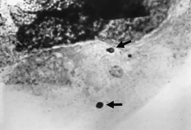
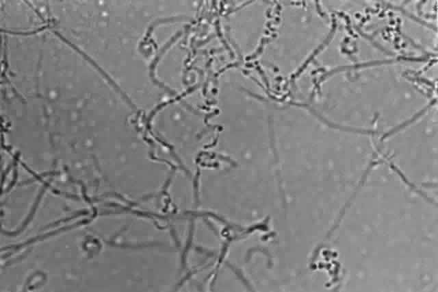
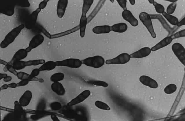
Growth matures within 2 to 6 days. Colonies are white, blue green, green, yellow, brown, or black. Reverse is white, gold, or brown. Colonies are flat, wrinkled or furrowed, with a velvety or cottony texture. Microscopic findings include large, septated hyphae. Simple, unbranched conidiophores arise from a foot cell and are enlarged at the tip (vesicle). Each conidiophore is covered partially or completely with one or two rows of flask-shaped phialides with chains of conidia (Fig. 1).

Fig. 1. Growth of Aspergillus fumigatus on malt agar showing chains of phialoconidia that form the head (X 25).

FUSARIUM SPECIES.

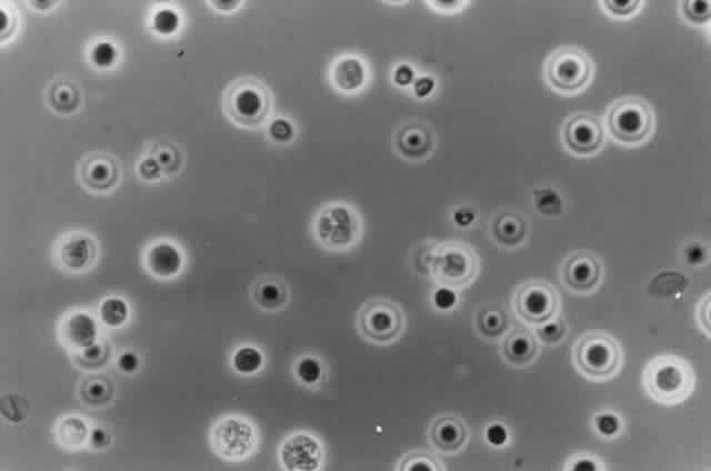
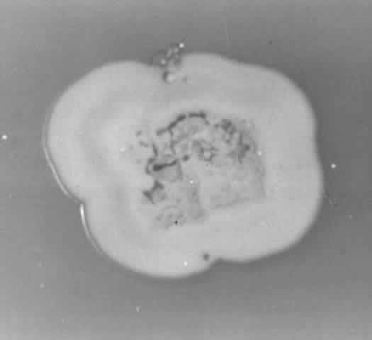
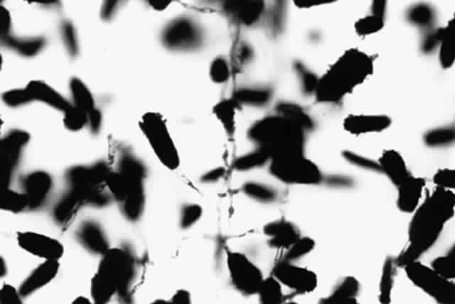
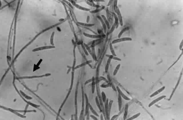
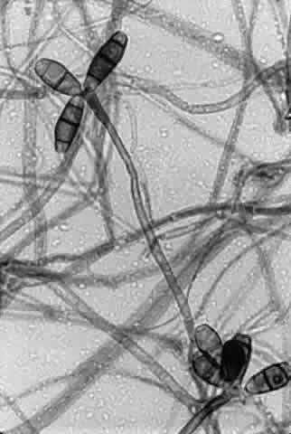
Growth matures within 2 to 6days. Colonies are white, blue, pink, purple, yellow, or green. Reverse is light in color. Texture is fluffy to woolly. Microscopic findings include small septated hyphae. Conidiophores are simple and either long or short, branched or unbranched. They produce single- or two-celled oval to cylindric microconidia (Fig. 2). Large banana-shaped multiseptated macroconidia are produced on potato dextrose agar in 4 to 6 weeks (Fig. 3). Chlamydospores may be numerous or rare.

Fig. 2. Lactophenol cotton blue mount of Fusarium solani from Sabouraud dextrose agar at 24 hours. Note presence of simple conidiophore (arrow) and two-celled microconidia (X 400).

Fig. 3. Lactophenol cotton blue mount of Fusarium solani from potato dextrose agar after 4 weeks' incubation. Note presence of large banana-shaped macroconidia (X 400).

MONILIA SITOPHILA.

Growth matures within 2 to 6 days. Colonies are white, then become salmoncolored. Microscopic findings include septated hyphae. Conidiophores produce branching chains of oval conidia, which are produced by continuous budding. Arthrospores are formed when the older hyphae break up.

PAECILOMYCES SPECIES.

Growth matures within 2 to 6 days. Colonies are white, pink, violet, yellowish brown, or greenish gold. Reverse is white to brown. Colonies spread and have a granular to velvety texture. Microscopic findings include slender delicate hyphae. Conidiophores are septated and have phialides with elongated necks containing long chains of small elliptic or oblong conidia (a penicillus).

PENICILLIUM SPECIES.

Growth matures within 2 to 6 days. Colonies are white, any shade of green to blue-green, pink, tan, yellow, or orange. Reverse is usually white but may be red or brown due to a diffusible pigment. Colonies are wrinkled with a velvety or granular texture. Microscopic findings include septated hyphae. Conidiophores are either unbranched or branched. The branches are termed metulae and they bear the flask-shaped phialides which contain the unbranched chains of either smooth or rough, round conidia. This entire structure--the metula, phialides and conidia--form the “penicillus.”

VERTICILLIUM SPECIES.

Growth matures in 2 to 6days. Colonies are first white, then become pinkish brown, red, green, or yellow. Reverse is either white or rust. Colony texture is either powdery or velvety. Microscopic findings include septated hyphae. Conidiophores are erect, simple or branched at several levels, and occur in whorls. Flask-shaped phialides arise from the apex of the branches. Conidia are single celled, oval to cylindrical, and appear singly or in clusters at the ends of the phialides.

DEMATIACEOUS FUNGI

ALTERNARIA SPECIES.

Growth matures within 2 to 6 days. Colonies are gray, grayish white, grayish green, grayish brown, or brown to greenish black with a light gray to white border. The reverse is black. Texture is woolly. Microscopic findings include golden brown to dark brown septated hyphae. Conidiophores are dark brown and septated. Conidia are dark brown, muriform with horizontal and longitudinal septa and are formed in long chains (Fig. 4).

Fig. 4. Lactophenol cotton blue mount showing dark brown, muriform, conidia of Alternaria species (X 400).

AUREOBASIDIUM PULLULANS (PULLULARIA PULLULANS).

Growth matures within 6 to 10 days. Colonies are at first white, they become black, shiny, and leathery with a grayish edge as the colony matures. The reverse is black. Microscopically, two types of hyphae are produced. The first type is hyaline, thin walled, and produces hyaline single-celled conidia directly from the hyphal wall. The second type is darkly pigmented, thick walled, closely septated, and produces hyaline single-celled conidia that arise from short projections (denticles) from the hyphal wall.

CLADOSPORIUM SPECIES.

Growth matures within 6to 10 days. Colonies are grayish green to dark olive, folded and heaped with a velvety texture. Reverse is black or dark brown. Most species are not capable of growing at 35 °C. Microscopic findings include dark brown septated hyphae. Conidiophores are branched with chains of budding conidia. Conidia are one celled with distinct scars on their ends (disjunctors). The large cells which bear the chains of conidia resemble shields (“shield cells”) when they are seen by themselves.

CURVULARIA SPECIES.

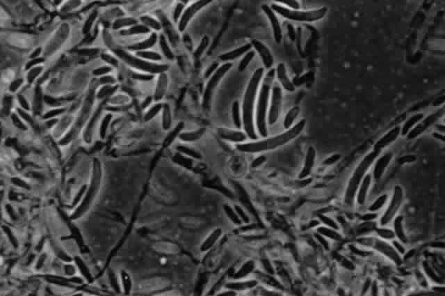
Growth matures within 2 to 6 days. Colonies are dark olive green or grayishbrown, becoming brown to black with a pinking gray surface. Reverse is dark brown to black. Texture is woolly. Microscopic findings include dark brown septated hyphae. Conidiophores are brown, septated, simple or branched, solitary or in groups. Conidia are pale to dark brown, usually curved, occasionally straight, and contain 2 to 6 septa with the first and last cell being lighter that the rest of the cells. One of the segments may be larger than the other segments giving the conidium a bent appearance (Fig. 5).

Fig. 5. Lactophenol cotton blue mount of Curvularia lunata on Sabouraud dextrose agar showing the four-celled conidia with the first and fourth cells being lighter (X 400).

DRESCHLERA SPECIES.

Growth matures within 2 to 6 days. Colonies are gray to black. Reverse is dark. Texture is velvety. Microscopically, brown septated hyphae are seen. Conidiophores are septated, brown, and twisted. The conidia are large, multiseptated, and cylindric and are produced in a zigzag fashion.

HELMINTHOSPORIUM SPECIES.

Growth matureswithin 2 to 6 days. Colonies are dark gray to black. Reverse is black. The texture is cottony to velvety. Microscopic findings include brown septated hyphae. Conidiophores are septated, brown to dark brown, erect, and parallel walled. Conidia are pale to dark brown, obclavate (widest at their base), usually containing more than six cells, and are produced laterally on the conidiophores.

ZYGOMYCETES

ABSIDIA SPECIES.

Growth matures within 1 to 5days. Colonies are white, later becoming gray. Reverse is white. Texture is coarse, woolly, cottony, resembling gray “cotton candy.” Microscopically are seen wide, aseptate, or rarely septated hyphae with rhizoids produced between the sporangiophores (internodal). The sporangiophores are branched and widen just below the sporangia. A septum is present below the sporangium. When the sporangium wall ruptures, the sporangiospores are released, and a short collarette remains on the sporangiophore. The sporangiospores are one celled, ovoid to slightly pear-shaped, smooth and hyaline to light black.

MUCOR SPECIES.

Growth matures within 1 to 4 days.Colonies are white, later becoming brown to black. Reverse is white. Texture is cottony, resembling “cotton candy.” Microscopic findings include wide aseptate, rarely septate, hyphae without rhizoids or stolons. Sporangiophores are long, often branched with a large spheric sporangium. A collarette may be present after the sporangium ruptures. Sporangiospores are smooth, round, or ellipsoid.

RHIZOPUS SPECIES.

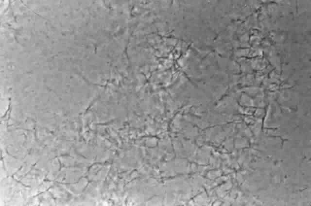
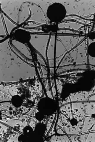
Growth matures within 1 to 4days. Colonies are white, later becoming gray or yellowish brown. Reverse is white. Texture is woolly, cottony, resembling “cotton candy.” Pathogenic species are capable of growing at 35°C. Microscopic findings include wide aseptate, rarely septate, hyphae with rhizoids produced directly below the sporangiophore. Sporangiophores are long and unbranched with dark globose sporangia. No collarette is present after the sporangium ruptures. Sporangiospores are one celled, ovoid, smooth to roughened and hyaline to brown (Fig. 6).

Fig. 6. Lactophenol cotton blue mount of Rhizopus showing dark sporangia borne on long sporangiophores (X 40).

SYNCEPHALASTRUM SPECIES.

Growth matures within 1 to 4 days. Colonies are white, later becoming dark gray to black. Reverse is white. Texture is woolly or cottony (“cotton candy”). Microscopically are seen wide aseptate, rarely septated, hyphae. Sporangiophores are short, branched with globose vesicles at their tips. Tubular sporangia containing sporangiospores are attached. Rhizoids may be present. Syncephalastrum species may be confused with Aspergillus species but can be separated based on the tubular sporangia and the absence of phialides.

Back to Top
YEASTS AND YEASTLIKE FUNGI
Colonies of yeast on blood and chocolate agar may be mistaken for staphylococcal colonies unless Gram stains are performed. Colonial and microscopic morphology are not important characteristics that can be used to identify yeasts. Important tests for identification purposes include the germ tube test, chlamydospore production, cornmeal agar morphology, urease production, pigment production, carbohydrate assimilation and fermentation, and phenol oxidase production (Table 4). A Dalmau plate (cornmeal agar with 1% Tween 80) is used to determine whether or not the yeast produces blastoconidia, arthroconidia, pseudohyphae, true hyphae, or chlamydospores.


TABLE FOUR. Characteristics of Yeast and Yeastlike Fungi


GeneraPigmentDalmau PlateUreaseInhibited by CycloheximideCarbohydrate AssimilationCarbohydrate Fermentation
CandidaNoBC, PHNo*VariableYesYes
CryptococcusYesBC, rare PHYesYesYesNo
RhodotorulaYesBC, rare PHYesYesYesNo
GeotrichumNoTH, ACNoYesYesNo
TrichosporonNoTH, PH, AC, BCYesNoYesNo
TorulopsisNoBCNoYesYesYes
MalasseziaNoRare TH, PHNoNoYesNo
ProtothecaNoSporangiaNoYesYesNo

*Candida krusei is urease positive.
BC = blastoconidia; PH = pseudohyphae; TH = true hyphae; AC = arthroconidia.


CANDIDA SPECIES.

Growth matures within 1 to 3days. Colonies are yeastlike, pasty, smooth, and cream-colored. Certain species of Candida may be inhibited by cycloheximide. They do not produce pigment. Only one species, C. krusei, is capable of producing urease. The genus can be speciated using carbohydrate assimilation testing or carbohydrate fermentation. Candida albicans produces pseudohyphae and large, thick-walled terminal chlamydospores on a Dalmau plate.

Species of Candida that have been reported as ocular pathogens include C. albicans, C. tropicalis, C. parapsilosis, C. pseudotropicalis, and C. guilliermondi.

CRYPTOCOCCUS SPECIES.

Growth matures within 1to 3 days. Colonies are flat or slightly raised, shiny, moist and may be mucoid. The colonies are cream but become tan with age. All species produce urease and are inhibited by cycloheximide. Species of Cryptococcus can be speciated using carbohydrate assimilation testing. None of the species are capable of carbohydrate fermentation. Microscopically visible are blastoconidia of varying size which may have a capsule present and rarely produce pseudohyphae.

The primary pathogen for humans is C. neoformans. A perfect state exists for C. neoformans--Filobasidiella neoformans. This organism is a basidiomycetous yeast, is heterophilic (appropriate mating types are necessary), and produces four chains of one-celled basidiospores on a swollen basidium located at the tip of the sporophore. True hyphae are present, which have clamp connections. Because of the requirement for mating pairs, F. neoformans is rarely isolated in the clinical microbiology laboratory. However, on direct examination, one may find the typical hyphae with the clamp connections.

RHODOTORULA SPECIES.

Growth matures within 1 to 4 days. Colonies are pink, coral, orange, red, or yellow. They are soft, yeastlike, and smooth. The majority of species are inhibited by cycloheximide. All species produce urease. Identification is made using carbohydrate assimilation testing only because they lack the ability to ferment carbohydrates. Microscopically, round or oval blastoconidia are produced. Rare pseudohyphae are produced, but no true hyphae are produced. A small capsule may be present.

GEOTRICHUM SPECIES.

Geotrichum species areyeast-like organisms. Growth matures within 1 to 4 days. Colonies are white, yeastlike, and moist with a ring of submerged hyphae at the periphery. Growth is better at 25 °C than at 35 °C. They do not produce urease, are inhibited by cycloheximide, and do not ferment carbohydrates. They can be speciated using carbohydrate assimilation testing. Microscopically, no blastoconidia are produced. They produce true hyphae which fragment to form cylindric arthroconidia. The arthroconidia separate at the junction of two septa.

TRICHOSPORON SPECIES.

Growth matures within 1 to 5 days. Colonies are cream colored, moist, andsoft. With age, the colony becomes yellowish gray, wrinkled, heaped, and more adherent to the agar surface. Members are not inhibited by cycloheximide and do not ferment carbohydrates. Some members produce urease. They can be speciated using carbohydrate assimilation testing. Microscopic findings include true hyphae, pseudohyphae, arthroconidia, and blastoconidia.

TORULOPSIS SPECIES.

Growth matures within 1 to 3 days. Colonies are small, white to cream in color,smooth, pasty, and yeastlike. Urease is not produced and growth is inhibited by cycloheximide. Species can be speciated based on carbohydrate assimilation and fermentation testing patterns. Microscopically, round to oval blastoconidia are produced. Pseudohyphae and true hyphae are not produced.

MALASSEZIA (PITYROSPORUM) SPECIES.

Rarely seenin the clinical microbiology laboratory because the majority of species require the presence of lipids in the medium or an olive oil overlay for their growth.

Malassezia furfur is the cause of pityriasis versicolor and can be recognized on direct preparations because of its bottlelike shape and the broad isthmus between the mother and daughter cell (Fig. 7).

Fig. 7. Giemsa stain of lid scraping showing budding yeast with a broad isthmus compatible with Malassezia furfur (arrows) (X 400).

Malassezia pachydermatis can be grown in the laboratory on standard fungal media. Growth matures in 1 to 3 days at 35 °C but is weak or scant when incubated at 25 °C. Growth can be inhibited by cycloheximide. Urease is not produced and carbohydrates are not fermented. Identification is accomplished by carbohydrate assimilation testing. Microscopically, pseudohyphae and true hyphae are absent. One conidium is produced at one end of a vegetative cell. A collarette is produced at the base of the conidium.

PROTOTHECA SPECIES.

Prototheca species are achlorophyllous algae that can be mistaken for yeast. They are capable of growing on standard fungal media, but they are inhibited by cycloheximide. Growth occurs within 1 to 3 days. Colonies are dull white to cream colored and yeastlike. Urease is not produced and carbohydrates are not fermented. Speciation is accomplished by carbohydrate assimilation testing. Microscopically seen are the presence of sporangia containing sporangiospores (Fig. 8). No pseudohyphae, true hyphae, or blastoconidia are present.

Fig. 8. Sporangia of Prototheca wickerhamii on Sabouraud dextrose agar (X 400).

Back to Top
AEROBIC ACTINOMYCETES
Aerobic actinomycetes belong to the Order Actinomycetales and are gram-positive bacteria that range in morphology from coryneform to well-developed filaments that resemble fungal hyphae. Some genera are acid fast or partially acid fast. The majority of genera grow in a temperature range of 25°C to 37°C but some are thermophilic, growing in temperatures up to 50°C.

They are commonly isolated on media used for fungal organisms but they are bacteria rather than fungi. They lack a nuclear membrane and have cell walls that contain muramic and diaminopimelic acids but do not contain chitin and glucans. They produce filaments that resemble fungal hyphae but the diameter of these filaments resembles the diameter of bacteria. Their growth is inhibited by antibacterial agents but not by antifungal agents because their cell walls do not contain fungal sterols.

Aerobic actinomycetes are ubiquitous in humans, animals, soil, and in compost. Human infections result from inhalation of the bacteria and from traumatic implantation of the organism. Aerobic actinomycetes that are frequently encountered as ocular pathogens in keratitis and endophthalmitis include Nocardia, Mycobacterium, Streptomyces, and Rhodococcus. Other members that are encountered in the clinical microbiology laboratory include Actinomadura, Nocardiopsis, Dermato-philus, and Oerskovia. Aerobic actinomycetes that are capable of growing on Sabouraud dextrose agar incubated at 30 °C include Nocardia, Streptomyces, Rhodococcus, Actinomadura, and Nocardiopsis.

Aerobic actinomycetes usually produce an “earthy” or “new mown hay” odor and are identified using colonial morphology and pigmentation; production of a diffusible pigment; morphology on tap water agar; acid fastness; decomposition of casein, tyrosine, and xanthine; urease production; and cell wall analysis. Tap water agar morphology is determined using tap water agar plates (2% granular agar in tap water). The organism is streaked in a straight line on the agar surface, incubated at 30 °C, and examined microscopically for 7 days using a 10X objective. The morphologic characteristics include the presence or absence of aerial and substrate hyphae, presence of typical bacterial coccoid or bacillary forms, and the presence of branched filaments or motile flagellated cells.

Antimicrobial susceptibility testing on the aerobic actinomycetes can be accomplished using either a modified Kirby-Bauer disk diffusion technique or an agar dilution system. Antibiotics to be tested include sulfonamides, trimethroprim-sulfamethoxazole, aminoglycosides, second-generation cephalosporins, imipenem, tetracycline, chloramphenicol, vancomycin, and erythromycin.

NOCARDIA SPECIES.

Growth occurs on Sabourauddextrose agar without antibiotics, Lowenstein-Jensen medium, and Middlebrook 7H11 agar and matures within 7 to 9 days. Colonies on Sabouraud dextrose agar are white to orange, raised, irregular or smooth (Fig. 9). Microscopically on tap water agar are seen thin, delicate, branching, beaded filaments that fragment into bacillary or coccoid forms (Fig. 10). They are gram positive and may be partially acid fast. There are three major pathogens to humans: N. asteroides, N. brasiliensis, and N. otitidiscaviarum.

Fig. 9. Colony of Nocardia asteroides on Sabouraud dextrose agar.

Fig. 10. Growth of Nocardia asteroides on tap water agar showing branching aerial mycelia (X 40).

STREPTOMYCES SPECIES.

Growth occurs on Sabouraud dextrose agar without antibiotics and matures within 4 to 10 days. Colonies are usually white, but occasionally gray, orange, rose, red, or green. Colonies are folded, hard, leathery, and may develop powdery or chalky appearing aerial hyphae. Filaments may be straight or spiraled and usually produce small rectangular conidia by segmentation (Fig. 11).

Fig. 11. Growth of Streptomyces species on tap water agar showing spiral aerial mycelia (X 40).

RHODOCOCCUS SPECIES.

Growth occurs on Sabouraud dextrose agar without antibiotics and matures within 2 to 10 days. Colonies are light pink, pink, orange, or coral red and are soft and bacterial-like. Microscopically are seen filaments that fragment into bacillary or coccoid forms. No aerial hyphae are present.

Back to Top
ANTIFUNGAL SUSCEPTIBILITY TESTING
Antifungal susceptibility testing, unlike antibacterial susceptibility testing, is not readily available in most hospital microbiology laboratories and must usually be sent to a reference laboratory for testing. Unlike bacterial susceptibility testing, there is no standard method for testing fungi. Problems encountered with fungal testing include solubilization of the antifungal agent, the type of medium that should be used, variances of temperature and duration of incubation, and most important, the size of the initial inoculum. Antifungal agents that can be tested include amphotericin B, 5-fluorocytosine, miconazole, ketoconazole, itraconazole, fluconazole, and pimaricin.

Unlike bacterial testing, there is no acceptable disk diffusion testing methods available in the United States. The most common method of testing is either a broth dilution method for yeast and yeast-like fungi and an agar dilution method for filamentous fungi. Data from antifungal susceptibility testing can be expressed as either the minimal inhibitory concentration (MIC) or as the minimal fungicidal concentration (MFC). Both are reported as μg/mL. The MIC is the lowest concentration of the antifungal agent that will inhibit the growth of the fungus tested. The MFC is the lowest concentration of the antifungal agent that will kill the fungus tested. Ideally, the MIC and the MFC should be within a twofold dilution of each other. There is little correlation between the results of antifungal susceptibility testing results and the patient's outcome.

Back to Top
REFERENCES

1. Rippon JW: Medical Mycology: The Pathogenic Fungi and The Pathogenic Actinomycetes, 3rd ed, pp 1–12. Philadelphia, WB Saunders, 1988

2. Brinser JH, Burd EM: Principles of diagnostic ocular microbiology. In Tabbara KF, Hyndiuk RA (eds): Infections of the Eye, pp 73–92. Boston, Little, Brown & Co, 1986

3. Brinser JH, Weiss A: Laboratory diagnosis in ocular disease. In Tasman W, Jaeger EA (eds): Duane's Clinical Ophthalmology, Vol 4, pp 1–14. Philadelphia, JB Lippincott, 1990

4. McGinnis MR: Laboratory Handbook of Medical Mycology. New York, Academic Press, 1980

5. Roberts GD: Laboratory methods in basic mycology. In Baron EJ, Finegold SM (eds): Bailey & Scott's Diagnostic Microbiology, pp 681–775. St. Louis, CV Mosby, 1990

6. Larone DH: Medically Important Fungi: A Guide to Identification. Washington, DC, American Society for Microbiology, 1993

Back to Top