Chapter 52
Gram-Negative Bacilli in Ocular Disease
NADA S. JABBUR
Main Menu   Table Of Contents

Search

PATHOGENESIS AND ANIMAL MODELS OF INFECTION
GRAM-NEGATIVE BACILLI AND MECHANISMS OF DRUG RESISTANCE
SUMMARY
REFERENCES

Bacteria can cause disease in host tissue by a variety of mechanisms. Gram-negative bacteria have a unique outer membrane containing lipopolysaccharides (LPS) that include molecular organization pathogenic to the host. Host interaction and colonization are facilitated by complex outwardly radiating organelles, flagella (arising from basal membrane structures and used for locomotion), and pili (used for adherence to the target cell). When present, capsules made of slimelike polysaccharides make phagocytosis difficult. The bacteria also produce a myriad of proteolytic enzymes aimed at destroying the host cell. The host cell surface carries glycoproteins and glycolipids that act as receptors for microbial attachment. Some bacteria have invasive properties that allow them to invade host defenses. Biofilm formation (when bacteria adhere to prosthetic devices), hypoxia, and the host's immune status are significant enhancers of pathogenesis. Finally, bacteria have developed different strategies for antibiotic resistance.

Gram-negative rods have been identified as major pathogens in major human organs, not the least of which is the eye. Species of Pseudomonas,1–6 Moraxella, 7–12 Acinetobacter,13–15 Haemophilus,16–19 Brucella,20 Francisella,21 Pasteurella,22 Capnocytophaga,23,24 Aeromonas,25,26 Alcaligenes,27 and the Enterobacteriaceae, including Escherichia,28 Shigella,29 Klebsiella,30 Proteus,31–33 Serratia,34–36 Yersinia,37,38 and Enterobacter,39,40 have all been clinically identified as human ocular pathogens.

Pseudomonas aeruginosa keratitis, the most common ocular infection caused by a gram-negative rod, can lead to blindness. The pathogenesis of Pseudomonas keratitis is multifactorial and has been studied in murine and rabbit models. Not many experimental models, however, have focused on other gram-negative rod ocular infections. To understand the pathogenesis of disease caused by gram-negative bacilli, researchers have indirectly gained knowledge from nonocular animal models such as those of Escherichia coli pyelonephritis41,42 and Klebsiella pneumoniae pneumonia.43

Back to Top
PATHOGENESIS AND ANIMAL MODELS OF INFECTION

MICROORGANISM VIRULENCE

Endotoxin

The unique outer membrane of gram-negative bacteria, containing phospholipids, LPS, and proteins, has been implicated as a major player in pathogenesis. The LPS portion of the envelope is both a major cell surface antigen and a major molecule triggering host response to bacterial infection. It is made up of three regions: the O-specific region (region I), responsible for the O or somatic antigenicity of the species and subspecies; the core polysaccharide (region II); and lipid A (region III), which is closest to the cell wall. The term “endotoxin” was used before discovering that it is the lipid A portion of the LPS. Different sugar sequences, linkage groups, and substituents cause different antigenic (serologic) specificities. The presence of O-antigen in Serratia marcescens enhances its adherence to inert and biologic surfaces.44 Region II is less variable than region I. Region III is highly conserved among eukaryotes, with little microheterogeneity among genera and species. Several members of the family Enterobacteriaceae express additional antigenic polysaccharides in the form of capsular or K antigens.

Lipid A is crucial in that it is responsible for the pathophysiologic effects associated with gram-negative bacterial infections and bacteremia, including pyrogenesis and hematologic, immune, and endocrinologic effects. Systemic exposures to endotoxin may result in hypotension, disseminated intravascular coagulation, and death. Most of these effects are mediated by cytokines and some by clotting and complement activation.

Capsule

Encapsulated strains of bacteria have capsules that consist of high-molecular-weight polysaccharides that form gels and allow bacteria to adhere to the host target cell surface. The capsule is shieldlike because it is hydrophilic and poorly immunogenic. First, the polysaccharide-rich composition of the capsule strongly inhibits phagocytosis by the hydrophobic surface of the host cell. Second, many capsules are poor immunogens45 and complement activators.46 If phagocytosis occurs, it requires specific opsonizing antibodies against the capsule. The thickness of the capsule in Francisella has been correlated with virulence, and in Brucella, encapsulated smooth colonies are ingested less readily than rough colonies.47 The virulence of Klebsiella43 and Yersinia48 species has also been correlated with capsular presence and protection. Immunologic diversity of the capsule within the same species explains the basis for serotyping. Although most ocular isolates of Haemophilus influenzae are nonencapsulated, the encapsulated form of type b is more virulent than the encapsulated type d.49

Flagella and Fimbriae (Pili)

When seen with transmission electron microscopy (TEM), the outer membrane of a gram-negative rod can take on a peritrichous appearance because of the flagella and more numerous pili that surround it. Flagella are larger than pili (they measure 16 to 18 nanometers in diameter) and allow for motility of the organism toward and within the host tissue. Ninety-five percent of clinical isolates of Pseudomonas are flagellated,50 and the burned-mouse model of P. aeruginosa has shown that flagella-deficient mutants are significantly less virulent.51 Flagellar proteins have been studied as vaccines. Rudner and associates52 showed that systemic or topical immunization of mice with strain-specific flagellar proteins or antiflagellar antibody homologous to the specific strain of bacteria protected them from pseudomonal keratitis.

TEM also reveals fimbriae, also known as pili, that measure 4 to 10 nanometers in diameter. These microfibrils can vary in number (2 to 12 in Pseudomonas) and can be classified according to their function as adhesins, lectins, evasins, aggressins, and sex pili. Fimbriated cells adhere to surfaces with specificity and thus allow colonization of a specific host tissue cell. Attachment to host cells is a first step in Pseudomonas keratitis. Stern and colleagues53 demonstrated that murine corneal trauma predisposes to ulceration, not by increasing the exposed area of de-epithelialized stroma for entry of the organisms, but by providing an injured epithelial edge to which P. aeruginosa can adhere. Purified Pseudomonas pili were found to compete with whole bacteria in saturating the binding receptors of host tissue.54 Knutton and coworkers,55 in their experiments with enterotoxigenic E. coli, showed that before invasion, these microorganisms adhere to the mucosal surface of intestinal cells with pili that allow them to hold on to specific mucosal receptors.

Proteolytic Enzymes

Exotoxin A,56 alkaline protease,57 exoenzyme S,58 phospholipase C,59 hemolysin,60 and elastase61 have all been implicated as players in the pathogenesis of Pseudomonas infections. Whereas endotoxins are part of the cellular wall, exotoxins (also known as cytolysins) are extracellular enzymes and are easily separated from the envelope. Most of the cytolysins that have been isolated and purified are made up of two components. One part binds to the target cell and allows the other part, the enzyme, to pass through the cell membrane. After preparing a specific combination of amino acids and a metal-chelating moiety as an inhibitor to Pseudomonas elastase (a zinc metalloendopeptidase), Kessler and associates61 demonstrated that intrastromal injection of the inhibitor first, followed by elastase, prevented corneal melting. Further, subconjunctival injections of the elastase and inhibitor only delayed corneal melting, suggesting that the reversal of corneal melt by the elastase is better carried intrastromally. Purified P. aeruginosa hemolysin injected into the corneas of rabbits was found to induce an extensive leukocytic invasion of the corneal stroma.60 In Moraxella angular conjunctivitis, the pathogenesis for lid maceration is caused by proteases from inflammatory cells rather than proteases elaborated by Moraxella.62 Both encapsulated and untypeable isolates of H. influenzae produce an IgA protease that degrades the protective secretory IgA elaborated by host mucosal surfaces.63,64

BACTERIAL RECEPTORS AND HOST OCULAR SURFACE

Host Surface Glycoproteins and Glycolipids

Adherence of bacteria to the corneal epithelium is a prerequisite for keratitis. Both P. aeruginosa and Staphylococcus aureus were found to bind to rabbit corneal epithelial cells in vitro. P. aeruginosa bound, in multiple layers, to the periphery of cells grown on glass slides, whereas S. aureus bound more randomly to the cell surface. E. coli did not bind significantly to those cells. The peripheral location of Pseudomonas binding is probably caused by its affinity to macromolecules of the cell surface involved in cell-cell interaction.65 Panjwani and coworkers66 also demonstrated that P. aeruginosa binds to rabbit corneal neutral glycosphingolipids. They later demonstrated that P. aeruginosa also binds to specific phospholipids (phosphatidylinositol and phosphatidylserine) extracted from rabbit corneal epithelium.67 These two molecules in ocular mucus or at the corneal surface may function as bacterial receptors and allow specific host-bacterium interaction and initial colonization.

Pseudomonas pili also bind to corneal epithelial receptors, which are glycoproteins.68 More specifically, such carbohydrate receptors have been studied and include sialic acid,69,70 N-acetylmannosamine,71 mannose,72 galactose,73 N-acetylglucosamine,74 and L-fucose.75 Although rabbit and human corneal epithelial cell models were not able to show detectable levels of the glycolipid asialo GM1,76 the incidence of murine P. aeruginosa keratitis was significantly reduced after mouse corneas infected with Pseudomonas were treated with a serum containing antibodies specific to asialo GM1, a glycolipid to which bacterial pili and LPS usually bind. This experiment, performed by Hazlett and associates,77 provides evidence that antibodies against host corneal receptors can significantly inhibit bacterial binding in vitro. In addition, when applied topically in vivo, the antibodies did confer some immunity and decreased the severity of the disease.

Endocytosis

In addition to the traditional extracellular pathogenesis of P. aeruginosa keratitis, the virulence of the infection has more recently been linked to an additional process that is intracellular. In those experiments, P. aeruginosa was seen inside keratocytes of athymic nude mice. TEM revealed that the invaded cells were present in membrane-bound vesicles, suggesting an endocytic process. At 24 hours, many of the bacteria were found free in the cytoplasm.78 In addition to the latter in vivo experiments, P. aeruginosa was found to invade several in vitro models. A temperature of 37°C allowed for a 10-fold increase in invasion of P. aeruginosa compared with experiments performed at 4°C. Further, host cell actin microfilaments may be required in the invasion, because cytochalasin D inhibited invasion.79

BIOFILM FORMATION

In suboptimal environmental conditions, as when nutrients are low, bacteria divide at a slower rate and behave differently. They secrete and live in an extracellular polymer matrix, also known as biofilm.80 Bacteria within a biofilm are relatively protected from the host's immune system and antibiotics and cause chronic and relentless infections. Biofilms are clinically important wherever biomaterials are used because they create a type of surface colonization.

Contact Lenses and Biofilm

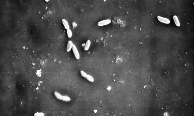
In ophthalmic practice, contact lenses and their cases are the most commonly used biopolymers. Since the advent of soft contact lenses, bacterial keratitis has occurred in association with infected cases and contact lens solutions,81,82 associated with poor patient compliance. Pseudomonal keratitis has also been linked to the use of saline soaking solutions prepared from distilled water and sodium chloride tablets.83 Extended overnight contact lens wear is a major risk factor for corneal ulcers,84–86 and among gram-negative rods, Pseudomonas and Serratia species have been the most common culprits (Fig. 1).87,88

Fig. 1. TEM showing P. aeruginosa adherent to the surface of a contact lens (× 5000). (Courtesy of Dr. Terrence P. O'Brien, The Wilmer Eye Institute, Lutherville, MD.)

P. aeruginosa is well adapted to grow in aquatic environments such as swimming pools, bathtubs, taps, and bottled and distilled water. Despite disinfectants that kill the single (planktonic) pseudomonal cells, some organisms adhere to the plastic, forming a biofilm. With time, the biofilm releases more planktonic cells, which can divide and replicate in a weaker disinfectant. Serratia species biofilm has been identified on the internal surface of plastic containers,89,90 and the microorganisms have been able to survive in the presence of benzalkonium chloride and 2% chlorhexidine. The ability of Serratia to survive chlorhexidine is attributed to changes in its outer bacterial cell membrane and increased adherence to polyethylene,91 a polymer from which plastic bottles and contact lens cases are manufactured.

Contact Lenses and Mucosal Immunity

The role of mucosal immunity in contact lensassociated bacterial keratitis may shed more light on one aspect of the underlying pathogenesis. Secretory IgA (sIgA) is the most predominant immunoglobulin in tears and plays an important role in preventing bacterial adherence to mucosal tissues.92 Hazlett and colleagues93 showed that sIgA inhibits binding of P. aeruginosa to cornea in a murine keratitis model. Further studies have revealed that the antipseudomonal response of sIgA was significantly lower in extended contact lens wearers than in controls. The mechanism for the lower response remains unclear. One possible explanation is that extended contact lenses cover the cornea and limbal conjunctiva, thus creating a barrier and reducing the chance of antigen presentation to the corneal antigen-presenting cells thought to be important in initiating the IgA response.94

Other Biomaterials

Gram-negative rod infections have been associated with other ophthalmic biopolymers such as ophthalmic sutures,95 intraocular lenses,5 sponges,96,97 and ganciclovir implants98 by means of an adhesive biofilm formation. Although gram-negative rods are isolated less commonly than gram-positive bacteria as causative organisms of suture abscesses, they can lead to endophthalmitis and loss of vision. A biofilm can develop around sutures, especially if loose. Because a suture abscess can lead to endophthalmitis after penetrating keratoplasty, all eroded (broken and loose) sutures should be removed as soon as they are identified.99,100 In vitro studies have demonstrated that P. aeruginosa adheres significantly better to intraocular lenses (IOLs) than does S. aureus (p < 0.05).101 Further, similar studies were performed with heparin-coated IOLs, and the results suggested that there is less P. aeruginosa attachment to the lens, and heparin may reduce the adherence by placing a highly hydrated layer between the bacteria and the IOL surface. The use of heparin-coated IOLs could diminish the incidence of endophthalmitis.102

Because new biomaterials continue to be introduced, research will continue to address ways to prevent biofilm formation and subsequently to reduce sight-threatening bacterial infections.

HYPOXIA

Contact Lenses and Oxygen Transmissibility

Corneal hypoxia has been linked to extended contact lens wear. The oxygen permeability, Dk, measures the amount of oxygen that can diffuse in a material. The oxygen transmissibility (Dk/L) determines how much oxygen a cornea receives after a contact lens of thickness L is placed on it. Overnight wear of contact lenses with poor oxygen transmissibility, especially those with Dk/L total values less than 50 × 10-9 (cm/sec)(ml O2/ml mm Hg), increases the risk of P. aeruginosa keratitis.103 Epithelial damage produced by contact lenses with low oxygen transmissibility worn overnight was shown to be the greatest risk factor for bacterial binding, not lens rigidity. Aswad and colleagues104 demonstrated that the extent of P. aeruginosa binding to extended-wear soft contact lenses was correlated with the number of focal deposits on the lenses, and that P. aeruginosa adhered more avidly to the focal deposits than did S. aureus. TEM further revealed that most of the binding of P. aeruginosa was to large focal deposits (more than 150 μm). These findings suggest that the corneal deposits may serve as points of attachment.

Lid Closure as a Risk Factor for Corneal Ulceration

Both clinical86 and experimental105 studies have suggested that the closed eye constitutes an important risk factor for infection when an extended-wear lens is worn. Aswad and associates105 found a significantly greater incidence of bacterial keratitis in rabbit eyes that had undergone lid closure after the placement of a contact lens as opposed to open eyes with a soft contact lens.

OTHER HOST FACTORS

Dry Eyes and Vitamin A Deficiency

Bacterium-host interaction is a key step in pathogenesis. In a P. aeruginosa keratitis model in New Zealand White rabbits and Wistar rats, Fleiszig and coworkers106 studied the effect of endogenous ocular surface mucus and found that its mucin fraction interfered with the adherence of the bacteria to intact corneal epithelial cells. The authors suggested that abnormal or reduced ocular mucus, as in dry eye conditions, may play a role in promoting P. aeruginosa keratitis.

Trauma and Immune Status

A variety of other host factors, including the immune status, can enhance gram-negative rod virulence. Trauma to the host surface allows easier bacterial adherence and penetration through the injured surface.53 Neutropenic and C3-depleted animals have been shown to have fewer polymorphonuclear leukocytes in the infected cornea and more fulminant infection.107–109

Vitamin A deficiency is associated with dry eyes and xerophthalmia. This represents a leading cause of blindness in developing countries. DeCarlo and associates110 studied pseudomonal keratitis in a vitamin A-deficient rabbit model and found that rabbits with severe xerophthalmia and epithelial keratinization had severe keratitis compared with those with initial mild punctate epithelial keratitis. Lack of mucus, as in certain forms of dry eye, has been experimentally shown to improve bacterial adherence to host tissue.106

Back to Top
GRAM-NEGATIVE BACILLI AND MECHANISMS OF DRUG RESISTANCE
Bacterial resistance to antibiotics is a problem of increasing concern. Different mechanisms of resistance have been identified, including enzyme modification, decreased antibiotic uptake, increased drug efflux, and target modification. The genetic basis of those mechanisms can be chromosome- or plasmid-related (Table 1). Plasmids incorporate foreign fragments of DNA or transposons that lead to the passage of the genetic code, conferring biochemical capabilities responsible for drug degradation.


TABLE 52-1. Resistance Mechanism of Gram-Negative Rods to Selected Antibacterial Agents


AntibioticsMechanismsGenetic BasisPresent in Pathogens
Beta-lactamsAltered penicillin-binding proteinChromosomalHaemophilus influenzae
   Escherichia coli
   Pseudomonas aeruginosa
 Reduced permeabilityChromosomalPseudomonas aeruginosa
   Enterobacter cloacae
   Serratig marcescens
   Klebsiella pneumoniae
   K. oxytoca
 Beta-lactamasePlasmid and chromosomalPseudomonas aeruginosa
   Enterobacteriaceae
   Moraxella
   Acinetobacter
FluoroquinolonesAltered DNA gyraseChromosomalEnterobacteriaceae
 Reduced permeabilityChromosomalPseudomonads
   Enterobacteriaceae
AminoglycosidesReduced uptakeChromosomalPseudomonas
   Enterobacteriaceae
 Modifying enzymesPlasmidEnterobacteroaceae
   Pseudomonads
ChloramphenicolAcetyltransferasePlasmid and chromosomalEnterobacteriaceae
TetracyclinesEffluxPlasmidEnterobacteriaceae
RifampinReduced DNA polymerase bindingChromosomalEnterobacteriaceae
   Pseudomonads
Folate inhibitorsAltered targetPlasmid and chromosomalEnterobacteriaceae
 Reduced permeabilityChromosomalPseudomonads
(Adapted from the MKSAP in Infectious Diseases. American College of Physicians, p. 218, 1997.)


ENZYME MODIFICATION

Certain gram-negative rods produce beta-lactamase, an enzyme capable of hydrolyzing the beta-lactam ring of penicillins, cephalosporins, monobactams, and carbapenems. The addition of clavulanate or sulbactam confers penicillinase resistance against beta-lactamase-producing strains of H. influenzae, E. coli, Proteus, and Klebsiella. Aztreonam and imipenem do not induce beta-lactamase activity and are poor substrates for drug-destructive enzymes.111

In addition to beta-lactamase, Pseudomonas, Proteus, and Escherichia can produce modifying enzymes that can inactivate antibiotics such as aminoglycosides by adenylation, phosphorylation, or acetylation. Aminoglycosides are polar drugs that are bactericidal and result in potentially low intraocular concentrations after systemic administration. Fortunately, when drug resistance to gentamicin and tobramycin occurs, most drug-resistant microorganisms are susceptible to amikacin because of its unique resistance to drug-inactivating enzymes.111 The higher the affinity of the modifying enzyme for the antibiotic, the more likely it is that the drug inactivation will occur at low concentrations.

Other enzyme modifications include point mutations in DNA gyrase in fluoroquinolone resistance,112 production of acetyltransferase in chloramphenicol resistance,113 plasmid encoding of esterase in erythromycin resistance,114 and other enzyme alterations in sulfonamide resistance (see Table 1).115

DECREASED ANTIBIOTIC UPTAKE

The passage of hydrophilic antibiotics through the bacterial outer membrane is enhanced by the presence of water-filled diffusion channels or porins that antibiotics can traverse. The lack of production of outer membrane proteins explains the resistance to aminoglycosides116 and imipenems.117 Gram-negative rod resistance to fluoroquinolones and folate inhibitors can also be explained by the reduced membrane permeability to those drugs.118 Alterations of outer membrane proteins in S. marcescens and Pseudomonas are responsible for resistance to quinolones.119

PROMOTION OF ANTIBIOTIC EFFLUX AND TARGET MODIFICATION

The primary mechanism for decreased accumulation of tetracycline in the host cell is active efflux of the antibiotic across the cell membrane.120 This mechanism has also been demonstrated in E. coli resistance to norfloxacin.121 Finally, resistance to folate inhibitors can occur by alteration of the target cell.

Back to Top
SUMMARY
Many species of gram-negative rods have been identified as human ocular pathogens and potential blinding agents. The key factors affecting visual outcome include the pathogenicity of the bacterium, the size of the inoculum, the immune status of the host, and the delay between clinical diagnosis and initiation of appropriate antibiotic therapy.
Back to Top
REFERENCES

1. Raber IM, Laibson PR, Kurz GH, Bernardino VB: Pseudomonas corneoscleral ulcers. Am J Ophthalmol 92:353, 1981

2. Brinser JH, Torczynski E: Unusual Pseudomonas corneal ulcers. Am J Ophthalmol 84:462, 1977

3. Nanda M, Pflugfelder SC, Holland S: Fulminant pseudomonal keratitis and scleritis in human immunodeficiency virus-infected patients. Arch Ophthalmol 109:503, 1991

4. Allen HF, Mangiaracine AB: Bacterial endophthalmitis after cataract extraction. Incidence in 36,000 consecutive operations with special reference to preoperative topical antibiotics. Arch Ophthalmol 91:3, 1974

5. Gerding DN, Poley BJ, Hall WH, LeWin DP, Clark MD: Treatment of Pseudomonas endophthalmitis associated with prosthetic intraocular lens implantation. Am J Ophthalmol 88:902, 1979

6. O'Brien T, Green WR: Periocular infections. In Mandell GL, Bennett JE, Dolin R (eds): Mandell, Douglas and Bennett's Principles and Practice of Infectious Diseases, p 1129. 4th ed. New York, Churchill-Livingstone, 1995

7. Marioneaux SJ, Cohen EJ, Arentsen JJ, Laibson PR: Moraxella keratitis. Cornea 10:21, 1991

8. Lipman RM, Deutsch TA: Late-onset Moraxella catarrhalis endophthalmitis after filtering surgery. Can J Ophthalmol 27:249, 1992

9. Sherman MD, York M, Irvine AR, Langer P, Cevallos V, Whitcher JP: Endophthalmitis caused by b-lactamase-positive Moraxella nonliquefaciens. Am J Ophthalmol 115:674, 1993

10. Schwartz B, Harrison LH, Motter JS, Motter RN, Hightower AW, Broome CV: Investigation of an outbreak of Moraxella conjunctivitis at a Navajo boarding school. Am J Ophthalmol 107:341, 1989

11. Garvey RJP, Reed TAG: Ophthalmia neonatorum due to Branhamella (Neisseria) catarrhalis. Case reports. Br J Vener Dis 57:346, 1981

12. Van Bijsterveld OP: The incidence of Moraxella on mucous membranes in the skin. Am J Ophthalmol 74:72, 1972

13. Marcovich A, Levartovsky S: Acinetobacter exposure keratitis. Br J Ophthalmol 78:489, 1994

14. Abel R, Shulman J, Boyle GL, Meltzer MA, Mirow DL, Leopold IH: Herellea vaginicola and ocular infections. Ann Ophthalmol 7:1485, 1975

15. O'Brien T, Green WR: Endophthalmitis. In Mandell GL, Bennett JE, Dolin R (eds): Mandell, Douglas and Bennett's Principles and Practice of Infectious Diseases, p 1120. 4th ed. New York, Churchill-Livingstone, 1995

16. Gregory JE, Henderson RW, Smith R: Conjunctivitis due to Haemophilus ducreyi infection [letter]. Br J Vener Dis 56:414, 1980

17. Hurwitz JJ, Rodgers KJA: Management of acquired dacryocystitis. Can J Ophthalmol 18:213, 1983

18. Brook I, Friedman EM, Rodriguez WJ, Controni G: Complications of sinusitis in children. Pediatrics 66:568, 1980

19. Taylor JRW, Cibis GW, Hantil LW: Endophthalmitis complicating Hemophilus influenzae type b meningitis. Arch Ophthalmol 98:324, 1980

20. Walker J, Sharma OP, Rao NA: Brucellosis and uveitis. Am J Ophthalmol 114:374, 1992

21. Guerrant RL, Humphries MK, Butler JE, Jackson RS: Tickborne oculoglandular tularemia. Case report and review of seasonal and vectorial associations in 106 cases. Arch Intern Med 136:811, 1976

22. Purcell JJ, Krachmer JH: Corneal ulcer caused by Pasteurella multocida. Am J Ophthalmol 83:540, 1977

23. Font RL, Jay V, Misra RP, Jones DB, Wilhelmus KR: Capnocytophaga keratitis: A clinicopathologic study of three patients, including electron microscopic observations. Ophthalmology 101:1929, 1994

24. Rubsamen PE, McLeish WM, Pflugfelder S, Miller D: Capnocytophaga endophthalmitis. Ophthalmology 100: 456, 1993

25. Carta F, Pinna A, Zanetti S, Carta A, Sotgiu M, Fadda G: Corneal ulcer caused by Aeromonas species. Am J Ophthalmol 118:530, 1994

26. Smith JA: Ocular Aeromonas hydrophila. Am J Ophthalmol 89:449, 1980

27. Tayeri T, Kelly LD: Alcaligenes faecalis corneal ulcer in a patient with cicatricial pemphigoid [letter]. Am J Ophthalmol 115:255, 1993

28. Park SB, Searl SS, Aquavella JV, Erdey RA: Endogenous endophthalmitis caused by Escherichia coli [review]. Ann Ophthalmol 25:95, 1993

29. Mark DB, McCulley JB: Reiter's keratitis. Arch Ophthalmol 100:781, 1982

30. Margo CE, Names RN, Guy JR: Endogenous Klebsiella endophthalmitis: Report of two cases and review of the literature. Ophthalmology 101:1298, 1994

31. Parunovic A: Proteus mirabilis causing necrotic inflammation of the eyelid. Am J Ophthalmol 76:543, 1972

32. Morris R, Camesasca FI, Byrne J, John G: Postoperative endophthalmitis resulting from prosthesis contamination in a monocular patient. Am J Ophthalmol 116:346, 1993

33. Heaven CJ, Mann PJ, Boase DL: Endophthalmitis following extracapsular cataract surgery: A review of 32 cases. Br J Ophthalmol 76:419, 1992

34. Lass JH, Haaf J, Foster CS, Belcher C: Visual outcome in eight cases of Serratia marcescens keratitis. Am J Ophthalmol 92:384, 1981

35. Duffey RJ: Bilateral Serratia marcescens keratitis after simultaneous bilateral radial keratotomy. Am J Ophthalmol 119:233, 1995

36. Al Hazzaa SA, Tabbara KF, Gammon JA: Pink hypopyon: A sign of Serratia marcescens endophthalmitis. Br J Ophthalmol 76:764, 1992

37. Saari KM, Laitineno O, Leirisalo M, Saari R: Ocular inflammation associated with Yersinia infection. Am J Ophthalmol 89:84, 1980

38. Crichton EP: Suppurative conjunctivitis caused by Yersinia enterocolitica [letter]. Can Med J 118:22, 1978

39. Milewski SA, Klevjer-Anderson P: Endophthalmitis caused by Enterobacter cloacae. Ann Ophthalmol 25:309, 1993

40. Chumbley LC: Canaliculitis caused by Enterobacter cloacae: Report of a case. Br J Ophthalmol 68:364, 1984

41. Iwahi T, Abe Y, Nakao M, Imada A, Tsuchiya K: Role of type I fimbriae in the pathogenesis of ascending urinary tract infection induced by Escherichia coli in mice. Infect Immun 39:1307, 1983

42. Winberg J, Mollby R, Bergstrom J et al: The PapG adhesin at the tip of P-fimbriae provides Escherichia coli with a competitive edge in experimental bladder infections of cynomolgus monkey. J Exp Med 182:1695, 1995

43. Kabha K, Nissimov L, Athamna A et al: Relationships among capsular structure, phagocytosis and mouse virulence in Klebsiella pneumoniae. Infect Immun 63:847, 1995

44. Palomar J, Leranoz AM, Vinas M: Serratia marcescens adherence: The effect of O-antigen presence. Microbios 81:107, 1995

45. Robbins JB, Schneerson R, Egan WB, Vann W, Liu DT: Virulence properties of bacterial polysaccharides-unanswered questions. In Smith H, Skehel JJ, Turner MJ (eds): The Molecular Basis of Microbial Pathogenicity, p 115. Weinheim, Verlag Chemie, 1980

46. Stevens P, Huang SN-Y, Welch WD, Young LS: Restricted complement activation by Escherichia coli with the K1 capsular serotype: A possible role in pathogenicity. J Immunol 121:2174, 1978

47. Joklik WK, Willett HP, Amos DB, Wilfert CM (eds): Brucella. In Zinsser Microbiology, p 609. 20th ed. Norwalk, Appleton & Lange, 1992

48. Friedlander AM, Welkos SL, Worsham PL et al: Relationship between virulence and immunity as revealed in recent studies of the F1 capsule of Yersinia pestis [review]. Clin Infect Dis (21 Suppl 2):S178, 1995

49. Roberts M, Stull TL, Smith AL: Comparative virulence of Hemophilus influenzae with a type b or type d capsule. Infect Immun 32:518, 1981

50. Ansorg RA, Knoche ME, Spies AF, Kraus CJ: Differentiation of the major flagellar antigens of Pseudomonas aeruginosa by the slide coagglutination technique. J Clin Microbiol 20:84, 1984

51. Montie TC, Drake D, Sellin H, Slater O, Edmonds S: Motility, virulence and protection with a flagella vaccine against Pseudomonas aeruginosa infection. Antibiot Chemother 39:233, 1987

52. Rudner XL, Hazlett LD, Berk RS: Systemic and topical protection studies using Pseudomonas aeruginosa flagella in an ocular model of infection. Curr Eye Res 11:727, 1992

53. Stern GA, Weitzenkorn D, Valenti J: Adherence of Pseudomonas aeruginosa to the mouse cornea. Arch Ophthalmol 100:1956, 1982

54. Rudner XL, Berk RS, Hazlett LD: Immunization with homologous Pseudomonas aeruginosa pili protects against ocular disease. Reg Immunol 5:245, 1993

55. Knutton S, Lloyd DR, Candy DC, McNeish AS: Ultrastructural study of adhesion of enterotoxigenic Escherichia coli to erythrocytes and human epithelial cells. Infect Immun 44:519, 1984

56. Pollack M: The role of exotoxin A in Pseudomonas disease and immunity [review]. Rev Infect Dis 5(Suppl 5):S979, 1983

57. Howe TR, Iglewski BH: Isolation and characterization of alkaline protease-deficient mutants of Pseudomonas aeruginosa in vitro and in a mouse eye model. Infect Immun 43:1058, 1984

58. Nicas TI, Bradley J, Lochner JE, Iglewski BH: The role of exoenzyme S in infections with Pseudomonas aeruginosa. J Infect Dis 152:716, 1985

59. Nicas TI, Iglewski BH: Toxins and virulence factors of Pseudomonas aeruginosa. In Sokatch JR (ed): The Bacteria. The Biology of Pseudomonas. New York, Academic Press, 1986

60. Johnson MK, Allen, JH: The role of hemolysin in corneal infections with Pseudomonas aeruginosa. Invest Ophthalmol Vis Sci 17:480, 1978

61. Kessler E, Spierer A, Blumberg S: Specific inhibitor of Pseudomonas aeruginosa elastase injected intracorneally in rabbit eyes. Invest Ophthalmol Vis Sci 24:1093, 1983

62. Van Bijsterveld OP: Bacterial proteases in Moraxella angular conjunctivitis. Am J Ophthalmol 72:181, 1971

63. Male CJ: Immunoglobulin A1 protease production by Hemophilus influenzae and Streptococcus pneumoniae. Infect Immun 26:254, 1979

64. Mulks MH, Kornfeld SJ, Frangione B, Plaut AG: Relationship between the specificity of IgA proteases and serotypes in Hemophilus influenzae. J Infect Dis 146:266, 1982

65. Panjwani N, Clark B, Cohen M, Barza M, Baum J: Differential binding of P. aeruginosa and S. aureus to corneal epithelium in culture. Invest Ophthalmol Vis Sci 31:696, 1990

66. Panjwani N, Zaidi TS, Gigstad JE, Jungalwad FB, Barza M, Baum J: Binding of Pseudomonas aeruginosa to neutral glycosphingolipids of rabbit corneal epithelium. Infect Immun 58:114, 1990

67. Panjwani N, Zhao Z, Raizman MB, Jungalwala F: Pathogenesis of corneal infection: Binding of Pseudomonas aeruginosa to specific phospholipids. Infect Immun 64:1819, 1996

68. Rudner XL, Zheng Z, Berk RS, Irvin RT, Hazlett LD: Corneal epithelial glycoproteins exhibit Pseudomonas aeruginosa pilus binding activity. Invest Ophthalmol Vis Sci 33:2185, 1992

69. Stern GA, Zam ZS: The effect of enzymatic contact lens cleaning on adherence of Pseudomonas aeruginosa to SCL. Ophthalmology 94:115, 1987

70. Hazlett LD, Moon MM, Berk R: In vivo identification of sialic acid as the ocular receptor for Pseudomonas aeruginosa. Infect Immun 51:687, 1986

71. Hazlett LD, Moon MM, Strejc M, Berk RS: Evidence for N-acetylmannosamine as an ocular receptor for P. aeruginosa adherence to scarified cornea. Invest Ophthalmol Vis Sci 28:1978, 1987

72. Spurr-Michaud SJ, Barza M, Gipson IK: An organ culture system for study of adherence of Pseudomonas aeruginosa to normal and wounded cornea. Invest Ophthalmol Vis Sci 29:379, 1988

73. Iida T, Kotoh M, Matsuo Y: Physical and chemical factors affecting the adherence of Pseudomonas aeruginosa to a rabbit corneal cell line (SIRC) cells. Hiroshima J Med Sci 34:201, 1985

74. Vishwanath S, Ramphal R: Tracheobronchial mucin receptor for Pseudomonas aeruginosa: Predominance of amino sugars in binding sites. Infect Immun 48:331, 1985

75. Doig P, Smith NR, Todd T, Irvin RT: Characterization of the binding of Pseudomonas aeruginosa alginate to human epithelial cells. Infect Immun 55:1517, 1987

76. Zhao Z, Panjwani N: Pseudomonas aeruginosa infection of the cornea and asialo GM1. Infect Immun 63:353, 1995

77. Hazlett LD, Masinick S, Barrett R, Rosol K: Evidence for asialo GM1 as a corneal glycolipid receptor for Pseudomonas aeruginosa adhesion. Infect Immun 61:5164, 1993

78. Fleiszig S, Zaidi TS, Fletcher EL, Preston MJ, Pier GB: Pseudomonas aeruginosa invades corneal epithelial cells during experimental infection. Infect Immun 62:3485, 1994

79. Fleiszig SM, Zaidi TS, Pier GB: Pseudomonas aeruginosa invasion of and multiplication within corneal epithelial cells in vitro. Infect Immun 63:4072, 1995

80. Elder MJ, Stapleton F, Evans E, Dart JK: Biofilm-related infections in ophthalmology. Eye 9(Pt 1):102, 1995

81. Krachmer JH, Purcell JJ Jr: Bacterial corneal ulcers in cosmetic soft contact lens wearers. Arch Ophthalmol 96:57, 1978

82. Pitts RE, Krachmer JH: Evaluation of soft contact lens disinfection in the home environment. Arch Ophthalmol 97:470, 1979

83. Wilson LA, Schlitzer RL, Ahearn DG: Pseudomonas corneal ulcers associated with soft contact lens wear. Am J Ophthalmol 92:546, 1981

84. Schein OD, Glynn RJ, Poggio EC, Seddon JM, Kenyon KR: The relative risk of ulcerative keratitis among users of daily-wear and extended-wear soft contact lenses: A case-control study. N Engl J Med 321:773, 1989

85. Wilhelmus KR: Review of clinical experience with microbial keratitis associated with contact lenses. CLAO J 13:211, 1987

86. Schein OD, Buehler PO, Stamler JF, Verdier DD, Katz J: The impact of overnight wear on the risk of contact-lens associated ulcerative keratitis. Arch Ophthalmol 112:186, 1994

87. Mondino BJ, Weissman BA, Farb MD: Corneal ulcers associated with daily-wear and extended-wear contact lenses. Am J Ophthalmol 102:58, 1986

88. Parment PA, Ronnerstam RA: Soft contact lens keratitis associated with Serratia marcescens. Acta Ophthalmol 59:560, 1981

89. Marrie TJ, Costerton JW: Prolonged survival of Serratia marcescens in chlorhexidine. Appl Environ Microbiol 42:1093, 1981

90. Nakashima AK, Highsmith AK, Martone WJ: Survival of Serratia marcescens in benzalkonium chloride and in multiple-dose medication vials: Relationship to epidemic septic arthritis. J Clin Microbiol 25:1019, 1987

91. Gandhi PA, Sawant AD, Wilson LA, Ahearn DG: Adaptation and growth of Serratia marcescens in contact lens disinfectant solutions containing chlorhexidine gluconate. Appl Environ Microbiol 59:183, 1993

92. Masinick SA, Montgomery CP, Montgomery PC, Hazlett LD: Secretory IgA inhibits Pseudomonas aeruginosa binding to cornea and protects against keratitis. Invest Ophthalmol Vis Sci 38:910, 1997

93. Kreger AS, Lyerly DM, Hazlett LD, Berk RS: Immunization against experimental Pseudomonas aeruginosa and Serratia marcescens keratitis. Invest Ophthalmol Vis Sci 27:932, 1986

94. Cheng KH, Spanjaard L, Rutten H, Dankert J, Polak BC, Kijlstra A: Immunoglobulin A antibodies against Pseudomonas aeruginosa in the tear fluid of contact lens wearers. Invest Ophthalmol Vis Sci 37:2081, 1996

95. Confino J, Brown SI: Bacterial endophthalmitis associated with exposed monofilament sutures following corneal transplantation. Am J Ophthalmol 99:111, 1985

96. Callanan D, Rubsamen PE: Moraxella infection of a scleral buckle [letter]. Am J Ophthalmol 114:637, 1992

97. Hahn YS, Lincoff A, Lincoff H: Infection after sponge implantation for scleral buckling. Am J Ophthalmol 87:180, 1979

98. Chen S, Stroh EM, Wald K, Jalkh A: Xanthomonas maltophilia endophthalmitis after implantation of sustained-release ganciclovir. Am J Ophthalmol 114:772, 1992

99. Leahey AB, Avery RL, Gottsch JD, Mallette RA, Stark WJ: Suture abscesses after penetrating keratoplasty. Cornea 12:489, 1993

100. Siganos CS, Solomon A, Frucht-Pery J: Microbial findings in suture erosion after penetrating keratoplasty. Ophthalmology 104:513, 1997

101. Lawin-Brussel CA, Refojo MF, Kenyon KR: In vitro adhesion of Pseudomonas aeruginosa and Staphylococcus aureus to surface-passivated polymethylmethacrylate intraocular lenses. J Cataract Refract Surg 18:598, 1992

102. Portoles M, Refojo MF, Leong FL: Reduced bacterial adhesion to heparin-surface-modified intraocular lenses. J Cataract Refract Surg 19:755, 1993

103. Imayasu M, Petroll WM, Jester JV, Patel SK, Ohashi J, Cavanagh HD: The relation between contact lens oxygen transmissibility and binding of Pseudomonas aeruginosa to the cornea after overnight wear. Ophthalmology 101:371, 1994

104. Aswad MI, John T, Barza M, Kenyon K, Baum J: Bacterial adherence to extended-wear soft contact lenses. Ophthalmology 97:296, 1990

105. Aswad MI, Barza M, Baum J: Effect of lid closure on contact-lens associated Pseudomonas keratitis. Arch Ophthalmol 107:1667, 1989

106. Fleiszig S, Zaidi TS, Ramphal R, Pier GB: Modulation of Pseudomonas aeruginosa adherence to the corneal surface by mucus. Infect Immun 62:1799, 1994

107. Chusid, MJ, Davis SD: Experimental bacterial keratitis in neutropenic guinea pigs: Polymorphonuclear leukocytes in corneal host defense. Infect Immun 24:948, 1979

108. Cleveland RP, Hazlett LD, Leon MA, Berk RS: Role of complement in murine corneal infection caused by Pseudomonas aeruginosa. Invest Ophthalmol Vis Sci 24:237, 1983

109. Hazlett LD, Berk RS: Effect of C3 depletion on experimental Pseudomonas aeruginosa ocular infection: Histopathological analysis. Infect Immun 43:783, 1984

110. DeCarlo JD, Van Horn DL, Hyndiuk RA, Davis SD: Increased susceptibility to infection to infection in experimental xerophthalmia. Arch Ophthalmol 99:1614, 1981

111. Pavan-Langston D, Dunkel EC: Antibiotics. In PavanLangston D, Dunkel EC (eds). Handbook of Ocular Drug Therapy and Ocular Side-Effects of Systemic Drugs, p 23. 1st ed. Boston, Little, Brown, 1991

112. Aoyama H, Fujimaki K, Sato K et al: Clinical isolate of Citrobacter freundii highly resistant to new quinolones. Antimicrob Agents Chemother 32:922, 1988

113. Gaffney DF, Foster TJ, Shaw WV: Chloramphenicol acetyl transferases determined by R-plasmids from gram-negative bacteria. J Gen Microbiol 109:351, 1978

114. Barthelemy P, Autissier D, Gerbaud G, Courvalin P: Enzymatic hydrolysis of erythromycin by a strain of Escherichia coli: A mechanism of resistance. J Antibiot 37:1692, 1984

115. Huovinen P: Trimethoprim resistance. Antimicrob Agents Chemother 31:1451, 1987

116. Goldstein FW, Gutmann L, Williamson R, Collatz E, Acar JF: In vivo and in vitro emergence of simultaneous resistance to both beta-lactam and aminoglycoside antibiotics in a strain of Serratia marcescens. Ann Microbiol 134A:329, 1983

117. Quinn JP, Dudek EJ, DiVincenzo CA, Lucks DA, Lerner SA: Emergence of resistance to imipenem during therapy for Pseudomonas aeruginosa infections. J Infect Dis 154: 289, 1986

118. Livermore DM: Interplay of impermeability and chromosomal beta-lactamase activity in imipenem-resistant Pseudomonas aeruginosa. Antimicrob Agents Chemother 36:2046, 1992

119. Sanders CC, Sanders WE Jr, Goering RV, Werner V: Selection of multiple antibiotic resistance by quinolones, beta-lactams, and aminoglycosides with special reference to cross-resistance between unrelated drug classes. Antimicrob Agents Chemother 26:797, 1984

120. McMurry L, Petrucci RE, Levy SB: Active efflux of tetracycline encoded by four genetically different tetracycline resistance determinants in Escherichia coli. Proc Natl Acad Sci USA 77:3974, 1980

121. Cohen SP, Hooper DC, Wolfson JS, Souza KS, McMurry LM, Levy SB: Endogenous active efflux of norfloxacin in susceptible Escherichia coli. Antimicrob Agents Chemother 32:1187, 1988.

Back to Top