Chapter 48
The Pathogenesis of Infectious Endophthalmitis
MICHELLE C. CALLEGAN , MICHAEL S. GILMORE, and DAVID W. PARKE II
Main Menu   Table Of Contents

Search

PREDISPOSING FACTORS AND SOURCE OF THE ORGANISM
THERAPEUTIC CHALLENGES OF ENDOPHTHALMITIS
PATHOGENIC MECHANISMS OF ENDOPHTHALMITIS
HYPOTHETICAL MODEL OF ENDOPHTHALMITIS PATHOGENESIS
ACKNOWLEDGMENTS
REFERENCES

Endophthalmitis is an intraocular inflammatory condition caused by the introduction of microorganisms into the posterior segment of the eye. Despite aggressive antibiotic, anti-inflammatory, and surgical intervention, endophthalmitis can result in loss of useful vision and, occasionally, loss of the eye. Recent experimental studies have begun to elucidate the interactions between the host and microorganism during endophthalmitis. The purpose of this chapter is to review the microbial sources, predisposing factors, and therapeutic challenges of endophthalmitis and to examine experimental findings from analyses of the pathogenic mechanisms of infectious endophthalmitis.
Back to Top
PREDISPOSING FACTORS AND SOURCE OF THE ORGANISM
Infectious endophthalmitis results from the seeding of microorganisms into the anterior or posterior segment as a consequence of intraocular surgery (postoperative) or penetrating injury of the globe (posttraumatic) or from metastatic spread into the eye from a distant infection site (endogenous). Etiologic agents of endophthalmitis range from relatively avirulent organisms found in the ocular environment (normal flora) to highly pathogenic environmental organisms.

Most reported endophthalmitis cases follow intraocular surgery. Therefore, the most common organisms involved are those found as normal flora of the eyelid margin and the tear film. Coagulase-negative staphylococci, commonly found as normal flora of the ocular environment, have been reported to cause up to 70% of acute postcataract surgery endophthalmitis.1–3 Other organisms involved include Staphylococcus aureus, viridans streptococci, other gram-positive bacteria, and gram-negative bacteria.1–3 Fungi are also a very common cause of postoperative endophthalmitis. Preoperative topical antibiotics are commonly used to decrease organism numbers in the tear film and surrounding tissues, but they do not sterilize the area.4 The low rate of clinical infection despite the predominance of microorganisms in or near the eye following surgery may be due to low inoculum levels or low pathogenicity of normal flora, in conjunction with a functional innate ocular defense system.

Delayed-onset postoperative endophthalmitis may occur weeks to several months following surgery. These chronic, indolent infections are likely due to either the sequestration of relatively avirulent organisms introduced at the time of surgery or from delayed inoculation of organisms that gain access to the eye through wound defects, sutures, vitreous wicks, or filtering blebs. In the case of filtering blebs, a higher prevalence of streptococci and enterococci are recovered.5,6 The organisms most commonly found in delayed-onset postoperative endophthalmitis are relatively avirulent organisms, such as Propionibacterium acnes and coagulase-negative staphylococci.7–9

Posttraumatic endophthalmitis occurs at a much higher rate than postoperative endophthalmitis (3% to 17% vs. 0.01%), and are caused by a greater variety of environmental organisms. In most series, staphylococci other than Staphylococcus epidermidis predominate, followed by Bacillus species.10–14 Gram-negative organisms also occur at a higher rate in posttraumatic cases (5 to 25%) than in cases following intraocular surgery.15 Of all the factors studied that were associated with trauma and subjected to multivariate analysis, only lens rupture was statistically associated with the development of endophthalmitis.16 In that same study, the presence of an intraocular foreign body (IOFB) approached but did not achieve statistical significance. Data compiled by the National Eye Trauma Study did indicate a statistical association between IOFBs and development of endophthalmitis, particularly when antimicrobial therapy was delayed.17,18

Endogenous endophthalmitis results from seeding of the eye by metastatic spread of organisms during bacteremia or fungemia. This type of endophthalmitis is relatively rare but can be devastating because of the likelihood of bilateral infection. Unlike postoperative and posttraumatic endophthalmitis in which the site of entry of microbes is anterior, in endogenous disease, the most frequent point of entry is the posterior segment vasculature. Therefore, endogenous endophthalmitis may present initially as a chorioretinitis without marked vitreitis. Populations at greatest risk included immunocompromised patients, those with prolonged indwelling devices, or intravenous drug users.19–21 Common causes of endogenous endophthalmitis include opportunistic and environmental pathogens such as S. aureus, Bacillus spp., gram-negative organisms, and fungi.19–22 Bacillus species and Candida are the most frequent causes of endogenous endophthalmitis.19–22

Back to Top
THERAPEUTIC CHALLENGES OF ENDOPHTHALMITIS
The goal of therapeutic management of infectious endophthalmitis is to sterilize the eye, reduce tissue damage from bacterial products and inflammation, and rescue useful vision. The recommended management of exogenous bacterial endophthalmitis in most cases includes intravitreal, periocular, and topical antimicrobials and, in severe cases, vitrectomy. Although there is no universal therapeutic regimen for infectious endophthalmitis, an adequate treatment strategy must account for the nature of the offending pathogen, as well as the unique anatomic and physiologic challenges posed by the interior of the eye.

The posterior segment is lined with highly sensitive photoreceptors and other retinal cells that can suffer irreversible damage not only from infection and its inflammatory sequelae but also from the high concentrations of antimicrobials needed to treat the infection.23–25 A second problem lies in the segregation of the interior of the eye from the systemic circulation by the blood-ocular fluid barrier. The vitreous and anterior chamber are not vascularized and, in concert with the blood-ocular fluid barrier, represent an area isolated from the rapid delivery of immune mediators, inflammatory cells, and systemically administered medications.26,27

On initial clinical presentation of endophthalmitis, the offending microbe is not typically known. Also, unfortunately, the clinical features of infection seldom give sufficient clues to specific microbial etiology.28 However, factors such as severity at presentation, pace of disease, appearance of wound, source of IOFB (if present), and corneal inflammatory ring infiltrates may in some cases provide useful predictive information.

The choice of empiric antibiotic therapy is, therefore, very important. Despite broad-spectrum antibiotic coverage, however, loss of vision remains a common result.24,29 The spectra and sensitivities of bacterial endophthalmitis isolates are reviewed in a number of recent clinical series.2,30–32 Currently, most ocular isolates are susceptible to ceftazidime and amikacin, which are commonly used in combination with vancomycin for therapy. However, the emergence of antibiotic-resistant organisms, particularly vancomycin-resistant gram-positive pathogens, may negatively affect future therapeutic strategies.33,34

The intraocular inflammation induced by microbial endophthalmitis is critical for removal of microorganisms, but collateral damage to delicate retinal tissues often results. The primary infiltrating cell type in infectious endophthalmitis is the polymorphonuclear neutrophil, which often liberates toxic oxygen metabolites and proteolytic enzymes close to sensitive intraocular tissues.35–38 The use of intravitreal anti-inflammatory agents, such as corticosteroids, to attenuate a vigorous host response is controversial. Experimental studies on the use of intravitreal anti-inflammatory agents in conjunction with antibiotic therapy are reported to be beneficial,39–43 detrimental,44 or ineffective.39,45–47 Despite these contradictory data and the paucity of clinical reports documenting the efficacy of intraocular corticosteroid use, dexamethasone is sometimes used as an adjunct to antibiotic therapy in endophthalmitis.

Vitrectomy is the surgical adjunct in endophthalmitis therapy. Surgical debridement of the infected vitreous cavity removes bacteria, inflammatory cells, and other toxic substances to facilitate vitreal diffusion, to remove vitreous membranes that may lead to retinal detachments, and to help speed recovery of vision.11 The Endophthalmitis Vitrectomy Study (EVS) demonstrated that in eyes with acute postcataract surgery endophthalmitis and better than light perception vision, immediate vitrectomy conferred no statistically significant visual benefit.24 However, in eyes with light perception vision, immediate vitrectomy was beneficial. Vitrectomy also plays an important role in the management of posttraumatic endophthalmitis, postoperative endophthalmitis following surgery other than cataract surgery, and postcataract surgery endophthalmitis eyes not responsive to initial nonvitrectomy management.24,48,49

Back to Top
PATHOGENIC MECHANISMS OF ENDOPHTHALMITIS
Data from the EVS were analyzed to identify clinical factors at presentation that were predictive of a poor visual outcome.24 Clinical indicators included the severity of the wound and infection at presentation; the responsible microbe; and ocular anatomic factors, such as intraocular pressure. Because of the range of inciting events to endophthalmitis, extent of ocular tissue injury, and the variety of organisms that contribute to endophthalmitis, the clinical spectrum varies broadly.

Endophthalmitis includes a relatively painless and easily treated intraocular inflammation caused by avirulent organisms such as coagulase-negative staphylococci, recurrent indolent infections caused by P. acnes, and an explosive and refractory panophthalmitis caused by Bacillus cereus.1,8,50 Certain organisms are typically associated with a predictable range of coincident inflammation. Pseudomonas aeruginosa, for example, is typically associated with more inflammation than S. epidermidis. There are always strain-specific exceptions, however. The offending microbe is not always predictive of the degree of inflammation engendered by the infection. A number of clinical reports detail severe endophthalmitis by relatively avirulent organisms, such as coagulase-negative staphylococci or Bacillus species other than B. cereus.51–55 Schematic comparisons of the infection courses of severe, avirulent, and chronic bacterial endophthalmitis are depicted in Figure 1.

Fig. 1. Representative courses of severe, avirulent, and chronic endophthalmitis. Data represent composite findings from experimental and clinical Staphylococcus epidermidis, Staphylococcus aureus, Enterococcus faecalis, Bacillus cereus, and Propionibacterium acnes endophthalmitis. The kinetics of pathologic parameters (bacterial growth, retinal response, and inflammatory cell influx) are shown relative to arbitrarily chosen theoretical maximums rather than absolute measures of each parameter. Top arrows represent the initiation of treatment, based on clinical recommendations.

Experimentally, equal numbers of different strains injected into the vitreous produce different clinical responses. However, the presence of microorganisms in the human eye does not always lead to tissue destruction. Therefore, different bacteria must possess different virulence factors. Current analysis of bacterial mutant strains deficient in individual and multiple toxins are defining specific roles for these proposed virulence factors in the pathogenesis of endophthalmitis. Additional putative virulence factors that are of interest are bacterial cell surfaces and their individual components, such as peptidoglycan, teichoic acid, polysaccharide capsules, lipopolysaccharide, and adhesive proteins. Because the bacterial cell surface is the foremost component of an organism to dialog with the host, its contribution to virulence must also be taken into account. The behavior of the organism in the eye during infection may also contribute to intraocular virulence. These aspects of endophthalmitis pathogenesis are discussed later.

STAPHYLOCCUS EPIDERMIDIS

Coagulase-negative staphylococci are a leading cause of posttraumatic and postoperative endophthalmitis, are found as normal flora of the ocular region, and are considered relatively avirulent.56 However, S. epidermidis and other coagulase-negative staphylococci have been reported to cause significant vision loss in some cases of endophthalmitis.30,51,52 In general, S. epidermidis does not secrete membrane-damaging toxins but does possess a number of adhesive proteins on its surface, including fibrinogen- and vitronectin-binding proteins, polysaccharide intercellular adhesin (PIA), and capsular polysaccharide adhesin (PS/A).57–60 The direct contribution of these adhesins in the pathogenicity of endophthalmitis must be determined.

S. epidermidis has emerged as one of the most important nosocomial pathogens often associated with nonophthalmic indwelling devices.61 In these types of infections, contamination of the implant likely occurs with a small inoculum from the patient's own normal flora during surgery. One likely source of S. epidermidis in postoperative endophthalmitis may, therefore, be organisms attached to implanted intraocular lenses (IOLs). In vitro adherence studies demonstrate that S. epidermidis adhere readily to most types of IOLs and that adherence depends on IOL materials, surface modifications, and extent of damage or irregularity of the IOL surface.62–66 Polypropylene haptics have been associated with an increased risk of postoperative endophthalmitis, presumably resulting from the increased bacterial adherence to this type of biomaterial and possibly from sequestration at the point of haptic attachment to the IOL.62 Polymethyl methacrylate (PMMA) lenses and haptics allowed less bacterial adherence than polypropylene haptics,62 whereas heparin-coated PMMA and Acrysoft lenses allowed even less adherence.63,64 Regarding the role of specific S. epidermidis adhesins in attachment to IOLs, a S. epidermidis ocular isolate containing the gene for PS/A and PIA (ica) adhered more readily to Acrysoft IOLs in vitro than a strain without the ica locus.65 It is not known what specific role these or other adhesins plays in S. epidermidis endophthalmitis pathogenesis.

Biofilm formation is considered to be an important virulence factor of coagulase-negative staphylococci and, in particular, S. epidermidis. Nosocomial infections are commonly associated with bacterial biofilm formation on implanted medical devices. Biofilm formation occurs as organisms replicate collectively on the surface of a biomaterial, where they become embedded in a multilayered mass of bacteria and extracellular polysaccharide.52,67 Organisms residing within the biofilm are usually sessile, and the biofilm as a whole is often resistant to antibiotic treatment and the host immune response. Because of the propensity for S. epidermidis to adhere to and form biofilms on several types of medical implant devices, it is reasonable to hypothesize that a similar situation could arise for contamination of IOLs and subsequent postoperative endophthalmitis.

As stated earlier, although relatively avirulent, S. epidermidis and other coagulase-negative staphylococci have been reported to cause fulminant endophthalmitis.30,51,52 Experimental models of S. epidermidis endophthalmitis have been initiated with a large number of bacteria (104 to 108),68–72 but virulent experimental infections have been achieved with as few as 50 organisms.72 In the absence of toxin production, precisely what causes such a robust inflammatory response remains in question. In experimental rat and rabbit models of S. epidermidis endophthalmitis, bacteria were readily cleared from the eye within 3 days of infection.37,69 This spontaneous sterilization coincided with intravitreal infiltration of neutrophils, plasma cells, B cells, and monocytes and measurable antibody to teichoic acid. No pathologic changes were noted in the retina.37 The complement system is also an integral participant in normal host defenses against experimental S. epidermidis endophthalmitis. Impaired host defenses against experimental staphylococcal endophthalmitis were observed in decomplemented guinea pigs.70 These data together suggest that antibodies and complement may assist the cellular immune response in bacterial clearing, leading to spontaneous sterilization in S. epidermidis endophthalmitis. The paradox of the significant number of microbe-positive anterior chamber taps recovered at the conclusion of human cataract surgery73–75 and the infrequency of endophthalmitis argues for the potency of the ocular immune response to control invasion by a limited number of avirulent microorganisms. Although these findings provide a hypothesis for relatively mild forms of S. epidermidis endophthalmitis, what triggers pathogenesis in more severe cases remains an open question.

STAPHYLOCOCCUS AUREUS

In contrast to S. epidermidis, S. aureus is a virulent intraocular pathogen. S. aureus endophthalmitis has been reported to result in final visual acuities of 20/400 or worse in more than half of the reported cases.76–79 Increased multiple antibiotic resistance in staphylococcal clinical isolates may also ultimately lead to an increased number of treatment failures.

S. aureus produces a number of putative virulence factors, namely adhesins, cytolytic toxins, and proteolytic enzymes, that are controlled by a system of global transcriptional regulators called staphylococcal accessory regulator (sar) and accessory gene regulator (agr).80 This quorum-sensing system recognizes the increase in bacterial numbers accompanying active infection and coordinately regulates the secretion of adhesins and other cell wall-associated proteins during the exponential phase of growth and the production of extracellular toxins and enzymes during the postexponential phase of growth. Mutants of S. aureus deficient in global regulation, therefore, produce cell wall-associated proteins and adhesins throughout the entire growth cycle but do not produce toxins. Such mutants have been analyzed in experimental models of S. aureus endophthalmitis in the rabbit and rat.81,82 Eyes infected with wild type S. aureus demonstrate focal retinal destruction with a rapid decline in electroretinogram (ERG) b-wave amplitude and a more intense intraocular inflammatory response than in eyes infected with a defective agr system.81,82 The intraocular virulence of S. aureus with mutations in both agr and sar global regulatory systems was almost completely attenuated.83 These studies suggested an important role for global regulation of extracellular toxins in S. aureus endophthalmitis pathogenesis.

The contribution of individual extracellular S. aureus toxins to endophthalmitis pathogenesis has also been analyzed using strains genetically engineered to be deficient in the specific toxins of interest. Analyses of S. aureus mutants defective in alpha-toxin (a membrane-damaging cytolysin), beta-toxin (a sphingomyelinase), and gamma-hemolysin (a bicomponent cytolysin) were analyzed in an experimental rabbit model of endophthalmitis.84 The absence of gamma-hemolysin did not alter the course or severity of endophthalmitis compared with that of infection caused by wild type S. aureus. However, declines in ERG b-wave amplitudes and overall retinal destruction on postinfection day 2 in untreated eyes infected with alpha-toxin-deficient or beta-toxin-deficient mutants were significantly less than that of eyes infected with wild-type S. aureus. There was a cumulative positive effect on the preservation of retinal function on postinfection day 2 in eyes infected with a triple mutant defective in alpha-toxin, beta-toxin, and gamma-hemolysin production. Differences in endophthalmitis severity were less clear after day 2, because significant progressive declines in ERG b-wave amplitudes occurred regardless of the S. aureus strain analyzed.84

Genomic analysis of S. aureus clinical ocular isolates demonstrated that all isolates tested possessed the gene encoding alpha-toxin.85 These data together suggest that globally regulated toxins, particularly alpha-toxin, are key virulence factors in S. aureus endophthalmitis. Arresting S. aureus toxin production by inactivation of global regulatory systems during the early stages of infection may, therefore, be a more viable therapeutic option than the targeting of individual toxins.

In addition to having detrimental effects on retinal function, experimental S. aureus endophthalmitis also incites significant intraocular inflammation. In experimental S. aureus endophthalmitis models in the rabbit and the rat, antibody levels to staphylococcal ribitol teichoic acid were detected, and neutrophils, lymphocytes, plasma cells, and monocytes infiltrated into the vitreous during the early stages of infection.35,36 Neutrophil adhesins E-selectin and intercellular adhesion molecule 1 (ICAM-1) were also expressed before leukocyte migration into the vitreous.86 As with experimental S. epidermidis endophthalmitis, this cellular response correlated with clearance of staphylococci from the vitreous and spontaneous sterilization.36 Although metabolically inactive S. aureus and purified cell wall sacculi have been shown to elicit significant intraocular inflammation in an experimental sterile endophthalmitis model,87 the specific cell wall components eliciting such are response have yet to be identified.

ENTEROCCUS FAECALIS

Enterococcus faecalis is a gram-positive facultative anaerobe commonly associated with antibiotic-resistant nosocomial infections. E. faecalis is most often isolated from infected filtering blebs following glaucoma surgery and is the causative agent in 4% to 8% of postoperative endophthalmitis.5,6,30,51,88 The visual outcomes of E. faecalis endophthalmitis are commonly poor, with as many as 80% of cases resulting in a final visual acuity of 20/200 or worse.30,51,88 The development of multidrug-resistant strains over the past two decades compounds the problem of treating E. faecalis infections. Fortunately, clinical reports of enterococci resistant to all antibiotics, including vancomycin, have yet to include ocular isolates.33,89

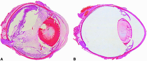
Approximately 50% of E. faecalis ocular isolates produce a cytolysin that disrupts mammalian and bacterial cell membranes.90 In an experimental rabbit endophthalmitis model, cytolysin-producing E. faecalis caused more fulminant and destructive changes in retinal architecture and a rapid decline in ERG b-wave amplitude than did noncytolytic E. faecalis.39,91 Transmission electron microscopy demonstrated retinal tissue damage occurring several hours before light microscopic detection or changes in retinal function in eyes infected with cytolytic E. faecalis. Experimental endophthalmitis with cytolytic E. faecalis was also refractory to intravitreal antibiotic and anti-inflammatory treatment, whereas noncytolytic E. faecalis endophthalmitis was resolved with a similar therapeutic regimen39 (Fig. 2). The results suggested that the presence of the cytolysin likely contributes to treatment failures and that treatment success may necessitate targeting of the cytolysin as an adjunct to therapy.

Fig. 2. Retinal function retained in eyes infected with cytolytic (red) and noncytolytic (blue) Enterococcus faecalis. Endophthalmitis caused by cytolytic E. faecalis was refractory to treatment with ampicillin (Amp), gentamicin (Gn), and dexamethasone (Dx), as indicated by the precipitous decline in retinal function by 72 hours. Noncytolytic E. faecalis was readily treated with Amp/Gn/Dx. Retinal function in these eyes was retained to preoperative levels at 72 hours. (Data compiled from Jett BD, Jensen JG, Atkuri RV et al. Evaluation of therapeutic measures for treating endophthalmitis caused by isogenic toxin-producing and toxin-nonproducing Enterococcus faecalis strains. Invest Ophthalmol Vis Sci 1995;36:9)

BACILLUS CEREUS

B. cereus is a ubiquitous gram-positive aerobic saprophyte found primarily in the soil and most commonly recognized as an agent of acute food poisoning. B. cereus, however, causes one of the most explosive and devastating forms of endophthalmitis, often resulting in loss of useful vision, and sometimes the eye, within a few days following inoculation. Bacilli are most commonly introduced into the anterior or posterior segment on contaminated projectiles following a penetrating injury or from the posterior segment vasculature as a consequence of bacteremia. Bacilli replicate and migrate rampantly throughout all parts of the eye, and an intense intraocular inflammatory response occurs in parallel with deteriorating retinal architecture.84,87 In the worst-case scenario, the infection spreads unhindered into periocular tissues, ocular architecture collapses, and the eye is lost. Although B. cereus is exquisitely sensitive to most antibiotics, this devastating consequence often occurs regardless of prompt and proper therapeutic and surgical intervention following clinical detection of infection.

Clinical reports have historically credited the unique virulence of B. cereus to toxins produced during infection.19,20,50,92,93 B. cereus produces a number of membrane-damaging cytolysins, enterotoxins, and proteases that are putative ocular toxin candidates.94 However, to date, three of these toxins (hemolysin BL, phosphatidylinositol-specific phospholipase C, and phosphatidylcholine-specific phospholipase C) have been shown individually to contribute little to the overall pathogenesis of experimental Bacillus endophthalmitis.95,96 It is likely, therefore, that if toxins are involved in the virulence of Bacillus endophthalmitis, they do so in a multifactorial fashion.

The Bacillus cell wall may also contribute to intraocular inflammation during endophthalmitis. Metabolically inactive B. cereus and purified B. cereus cell walls do not affect retinal function. However, these preparations are highly inflammogenic in experimental models of sterile endophthalmitis.87 Another significant virulence factor related to bacterial structure may be that of bacterial motility. The unusual motile capability of B. cereus in the eye appears to contribute significantly to experimental endophthalmitis pathogenesis. Bacillus strains genetically engineered to be deficient in motility could not migrate throughout the eye, did not grow as well in the vitreous, and were significantly less virulent than wild type motile bacilli97 (Fig. 3). These results highlight bacterial motility as a possible therapeutic target for Bacillus and perhaps other motile organisms in endophthalmitis.

Fig. 3. Whole-eye histology of motile (A) and nonmotile (B) experimental Bacillus endophthalmitis. Approximately 100 colony forming units (CFU) of a motile endophthalmitis isolate of Bacillus thuringiensis or a nonmotile isogenic mutant of B. thuringiensis was injected intravitreally into rabbit eyes. Eyes were harvested for histologic analysis at 18 hours. By 18 hours, in eyes infected with motile B. thuringiensis, retinal dissolution and detachment, severe inflammation, and corneal edema were apparent. The posterior and anterior segments were engorged with inflammatory cells and fibrin. Eyes infected with the nonmotile mutant of B. thuringiensis appeared normal throughout 18 hours postinfection. (Data compiled from Callegan MC, Cochran DC, Kane ST et al. The contribution of bacterial motility to Bacillus endophthalmitis pathogenesis. Association for Research in Vision and Ophthalmology Annual Meeting, 2001, Abstract 1382)

PROPIONIBACTERIUM ACNES

P. acnes is a ubiquitous gram-positive anaerobe found as normal flora of the eyelid and conjunctiva. It is also an opportunistic pathogen that has been associated with nosocomial prosthetic device infections and chronic inflammatory diseases such as acne vulgaris, arthritis, and osteomyelitis. P. acnes can cause an acute and mild form of endophthalmitis that is readily responsive to treatment but can also cause an unusual form of chronic and recurrent endophthalmitis following extracapsular cataract extraction and IOL implantation.7–9,98,99 P. acnes from the surrounding ocular flora can adhere to PMMA IOLs100 and, therefore, are a likely source of lens contamination. Despite adequate therapeutic and surgical intervention, late-onset treatment failures and persistent infections are common with P. acnes intraocular infection.

Chronic postoperative P. acnes endophthalmitis is characterized by a recurrent inflammation with granulomatous keratic precipitates and white plaques on the posterior capsule or IOL that may be observed weeks to months following surgery.99 These intracapsular plaques are thought to represent P. acnes sequestered inside the peripheral capsular bag.101–103 These sequestered plaques likely contribute to the host's inability to clear the infection and the failure of antibiotics to reach the sequestered bacterial plaques. Although there is little evidence that P. acnes exists as a biofilm in these infections, the resistance to killing by host inflammatory cells, the refractory nature of the infection to therapy, and plaque formation are consistent with the presence of a biofilm. The inherent slow replication rate of P. acnes is also thought to contribute to the indolent nature of the disease.104P. acnes does not produce membrane-damaging toxins but does produce proteases that may contribute to virulence during infection.105,106 Adherence factors of P. acnes have not been well studied, but P. acnes, like S. aureus and S. epidermidis, produce a fibronectin-binding protein that may aid in adhesion of P. acnes to IOLs, other types of ocular biomaterials, or intraocular structures.107 The role of P. acnes proteases and adhesion molecules in the pathogenesis of experimental endophthalmitis has yet to be analyzed.

GRAM-NEGATIVE PATHOGENS AND CANDIDA

Although clinical series vary in the frequency of isolation of gram-negative organisms, gram-positive isolates greatly outnumber gram-negative isolates in endophthalmitis. The paucity of experimental data on the pathogenic mechanisms of gram-negative bacterial endophthalmitis reflects the overall predominance of gram-positive organisms isolated from these cases. Gram-negative organisms accounted for only 6% of cases in the EVS study2 but up to 20% of posttraumatic cases of endophthalmitis.15 Common gram-negative endophthalmitis isolates include Klebsiella, Pseudomonas, and Hemophilus. The ocular pathogenesis of Pseudomonas is the best understood of all gram-negative pathogens because of the abundance of experimental data that exists for P. aeruginosa keratitis. It is unknown, however, whether the findings in experimental P. aeruginosa corneal infections extend to P. aeruginosa endophthalmitis. Many gram-negative organisms, including Pseudomonas, are avid biofilm producers, which may be a source of infection via adherence to IOLs in postoperative endophthalmitis. In endogenous gram-negative endophthalmitis, a number of cases reported were secondary to sepsis and had a moderately poor prognosis.108,109 Increasing reports of gram-negative endogenous endophthalmitis in diabetics highlight the danger of an immunocompromised state and metastatic infections with these opportunistic pathogens.110–112

Fungi are one of the most common causes of endogenous endophthalmitis. The prolonged use of intravenous devices, long-term antibiotic therapy, and the use of immunosuppressive agents have contributed greatly to the rise in frequency of fungemia and endogenous fungal endophthalmitis. Candida is the most common cause of endogenous endophthalmitis in immunocompromised patients, patients on long-term broad-spectrum antibiotics, and intravenous drug abusers.113–116 Incidence of Candida endophthalmitis in patients with disseminated candidiasis have been reported to range from 3% to as high as 40%, depending on the definition criteria of the study and the patient population included in the survey.117–120Candida has a particular predilection for the eye in cases of disseminated candidiasis. Endogenous Candida endophthalmitis generally results from metastatic spread to the choroidal and retinal vasculature with generation of fungal microabscesses, followed by extension of organisms into the vitreous.121,122 Although candidemia and subsequent Candida chorioretinitis and endophthalmitis are associated with immunocompromised and hospitalized patients, it is unclear why the eye is such a frequent site for deep tissue invasion. It is also unclear why patients with acquired immunodeficiency syndrome (AIDS) from human immunodeficiency virus (HIV) infection are often colonized with Candida but have a relatively low risk of Candida endophthalmitis. Candida chorioretinitis and endophthalmitis are also seen in nonimmunocompromised patients who experience multiple episodes of transient candidemia, as in the setting of intravenous drug abuse.

Back to Top
HYPOTHETICAL MODEL OF ENDOPHTHALMITIS PATHOGENESIS
In-depth analysis and understanding of the molecular and cellular events that occur during infectious endophthalmitis are in their infancy. However, recent experimental data provide relevant clues for the foundation of a working model of endophthalmitis pathogenesis. Following exogenous inoculation with a critical number of organisms, organisms can replicate relatively unchallenged in an immunologically privileged environment. In some cases wherein the size of the inoculum is below a certain threshold, and/or when the organism is avirulent, and/or when the natural host intraocular defense mechanisms are active and capable, spontaneous resolution of infection occurs with no adverse sequelae. Otherwise, clinical infection results.

Virulent organisms produce toxins and other factors during growth that may obstruct host immunity, whereas nontoxigenic organisms may be readily cleared with minimal ocular tissue damage by the host immune response and antimicrobial therapy. The extent of intraocular inflammation and retinal damage appears to depend on the relative virulence of the infecting organism. As indicated previously, this virulence may relate to the production of toxins and/or structural features of the microbe. The sheer presence of certain organisms in posterior segment may also stimulate intraocular inflammation. During growth and antimicrobial therapy, intact and disrupted bacterial cell walls or other cellular debris likely incite the intraocular immune response, resulting in the production of soluble immune mediators. Paradoxically, the same antibiotics that serve to disrupt and kill bacteria may liberate highly inflammogenic bacterial debris that may worsen inflammation.

As the infection progresses, the production of proinflammatory cytokines and other immune mediators initiates an inflammatory cascade, affecting the permeability of the blood-ocular fluid barrier. Influx of additional soluble mediators and cells exacerbates the inflammatory response. The unfortunate result is disruption and irreversible damage of delicate photoreceptor cells and significant collateral damage from the intraocular inflammatory response. Endophthalmitis resulting from avirulent organisms that are not immediately cleared may follow a similar course but at a reduced and more immunologically controlled rate that is more amenable to therapy.

A more detailed understanding of the interactions between pathogen and host is needed to more efficiently treat endophthalmitis and improve visual outcome. Novel therapies for endophthalmitis may target not simply bacterial destruction but toxin elaboration mechanisms, motility, and host response modulation.

Back to Top
ACKNOWLEDGMENTS
We would like to acknowledge Mary C. Booth, PhD (Athlone Institute of Technology, County Westmeath, Ireland), Daniel J.J. Carr, PhD, James Chodosh, MD, and Michael Engelbert, MD (University of Oklahoma Health Sciences Center, Oklahoma City OK), and Bradley D. Jett, PhD (Oklahoma Baptist University, Shawnee OK) for stimulating discussions. Portions of the work presented in this review were supported by U.S. Health Service Grants EY12985, EY08289, and EY11648, and an unrestricted grant from Research to Prevent Blindness, Inc.
Back to Top
REFERENCES

1. Aaberg TM, Flynn HW, Schiffman J et al. Nosocomial acute-onset postoperative endophthalmitis survey: A 10-year review of incidence and outcomes. Ophthalmology 1998;105:1004

2. Han DP, Wisniewski SR, Wilson LA et al. Spectrum and susceptibilities of microbiologic isolates in the Endophthalmitis Vitrectomy Study. Am J Ophthalmol 1996;122:1

3. Speaker MG, Milch FA, Shah MK et al. Role of external bacterial flora in the pathogenesis of acute postoperative endophthalmitis. Ophthalmology 1991;98:639

4. Ferro JF, DePablos M, Logrono MG et al. Postoperative contamination after using vancomycin and gentamicin during phacoemulsification. Arch Ophthalmol 1997;115:165

5. Soltau JB, Rothman RF, Budenz DL et al. Risk factors for glaucoma filtering bleb infections. Arch Ophthalmol 2000;118:338

6. Waheed S, Ritterband DC, Greenfield DS et al. New patterns of infecting organisms in late bleb-related endophthalmitis: A ten year review. Eye 1998;12:910

7. Aldave AJ, Stein JD, Deramo VA et al. Treatment strategies for postoperative Propionibacterium acnes endophthalmitis. Ophthalmology 1999;106:2395

8. Clark WL, Kaiser PK, Flynn HW et al. Treatment strategies and visual acuity outcomes in chronic postoperative Propionibacterium acnes endophthalmitis. Ophthalmology 1999;106:1665

9. Samson CM, Foster CS. Chronic postoperative endophthalmitis. Int Ophthalmol Clin 2000;40:57

10. Jonas JB, Knorr HL, Budde WM. Prognostic factors in ocular injuries caused by intraocular or retrobulbar foreign bodies. Ophthalmology 2000;107:823

11. Meredith TA. Posttraumatic endophthalmitis. Arch Ophthalmol 1999;117:520

12. O'Brien TB, Choi S. Trauma-related ocular infections. Int Ophthalmol Clin North Am 1995;8:667

13. Thompson ST, Parver LM, Enger CL et al. Infectious endophthalmitis after penetrating injuries with retained intraocular foreign bodies. Ophthalmology 1993;100:1468

14. Kunimoto DY, Dat T, Sharma S et al. Microbiologic spectrum and susceptibility of isolates: Part II. Posttraumatic endophthalmitis. Am J Ophthalmol 1999;128:242

15. Parrish CM, O'Day DM. Traumatic endophthalmitis. Int Ophthalmol Clin 1987;27:112

16. Thompson WS, Rubsamen PE, Flynn HW et al. Endophthalmitis after penetrating trauma: Risk factors and visual acuity outcomes. Ophthalmology 1995;102:1696

17. Dannenberg AL, Parver LM, Brechner RJ et al. Penetrating eye injuries in the workplace: The National Eye Trauma System Registry. Arch Ophthalmol 1992;110:843

18. Dannenburg AL, Parver LM, Fowler CJ. Penetrating eye injuries related to assault: The National Eye Trauma System Registry. Arch Ophthalmol 1992;110:849

19. Cowan CL, Madden WM, Hatem GF et al. Endogenous Bacillus cereus panophthalmitis. Ann Ophthalmol 1987;19:65

20. Shamsuddin D, Tuazon CU, Levy C et al. Bacillus cereus panophthalmitis: Source of the organism. Rev Infect Dis 1982;4:97

21. Greenwald MJ, Wohl LG, Sell CH. Metastatic bacterial endophthalmitis: A contemporary appraisal. Surv Ophthalmol 1986;31:81

22. Romero CF, Rai MK, Lowder CY et al. Endogenous endophthalmitis: Case report and brief review. Am Fam Physician 1999;60:510

23. Campochiaro PA, Lim JI. Aminoglycoside toxicity in the treatment of endophthalmitis. Arch Ophthalmol 1994;112:48

24. Endophthalmitis Vitrectomy Study. A randomized trial of immediate vitrectomy and of intravenous antibiotics for the treatment of postoperative bacterial endophthalmitis. Arch Ophthalmol 1995;113:1479

25. Wiechens B, Neumann D, Grammer JB et al. Retinal toxicity of liposome-incorporated and free ofloxacin after intravitreal injection in rabbit eyes. Int Ophthalmol 1998-1999;22:133

26. Streilein JW. Immunoregulatory mechanisms of the eye. Prog Retinal Eye Res 1999;18:357

27. Niederkorn JY. Anterior chamber-associated immune deviation. Chem Immunol 1999;73:59

28. Johnson MW, Doft BH, Kelsey SF et al. Relationship between clinical presentation and microbiologic spectrum. Ophthalmology 1997;104:261

29. Okhravi N, Towler HMA, Hykin P et al. Assessment of a standard treatment protocol on visual outcome following presumed bacterial endophthalmitis. Br J Ophthalmol 1997;81:719

30. Endophthalmitis Vitrectomy Study. Microbiologic factors and visual outcome in the Endophthalmitis Vitrectomy Study. Am J Ophthalmol 1996;122:830

31. Anand AR, Therese KL, Madhavan HN. Spectrum of aetiological agents of postoperative endophthalmitis and antibiotic susceptibility of bacterial isolates. Indian J Ophthalmol 2000;48:123

32. Kunimoto DY, Das T, Sharma S et al. Microbiologic spectrum and susceptibility of isolates: Part I. Postoperative endophthalmitis. Endophthalmitis Research Group. Am J Ophthalmol 1999;128:240

33. Gilmore MS, Hoch JA. Antibiotic resistance: A vancomycin surprise. Nature 1999;399:524

34. Jones DB. Emerging antibiotic resistance: Real and relative. Arch Ophthalmol 1996;114:91

35. Engstrom RE, Mondino BJ, Glasgow BJ et al. Immune response to Staphylococcus aureus endophthalmitis in a rabbit model. Invest Ophthalmol Vis Sci 1991;32:1523

36. Ravindranath RM, Mondino BJ, Adamu SA et al. Immunopathologic features of Staphylococcus aureus endophthalmitis in the rat. Invest Ophthalmol Vis Sci 1995;36:2482

37. Ravindranath RM, Hasan SA, Mondino BJ. Immunopathologic features of Staphylococcus epidermidis-induced endophthalmitis in the rat. Curr Eye Res 1997;16:1036

38. Giese MJ, Sumner HL, Berliner JA. Cytokine expression in a rat model of Staphylococcus aureus endophthalmitis. Invest Ophthalmol Vis Sci 1998;39:2785

39. Jett BD, Jensen JG, Atkuri RV et al. Evaluation of therapeutic measures for treating endophthalmitis caused by isogenic toxin-producing and toxin-nonproducing Enterococcus faecalis strains. Invest Ophthalmol Vis Sci 1995;36:9

40. Maxwell DP, Brent BD, Diamond JG et al. Effect of intravitreal dexamethasone on ocular histopathology in a rabbit model of endophthalmitis. Ophthalmology 1991;98:1370

41. Park SS, Vallar RV, Hong CH. Effect of intravitreal dexamethasone in treatment of pneumococcal endophthalmitis in rabbits. Arch Ophthalmol 1995;113:1324

42. Smith MA, Sorenson JA, D'Aversa G et al. Treatment of experimental methicillin-resistant Staphylococcus epidermidis endophthalmitis with intravitreal vancomycin and intravitreal dexamethasone. J Infect Dis 1997;175:462

43. Yoshizumi MO, Lee GC, Equi RA et al. Timing of dexamethasone treatment in experimental Staphylococcus aureus endophthalmitis. Retina 1998;18:130

44. Meredith TA, Aguilar HE, Drews C et al. Intraocular dexamethasone produces a harmful effect on treatment of experimental Staphylococcus aureus endophthalmitis. Trans Am Ophthalmol Soc 1996;94:241

45. Das T, Jalali S, Gothwal VK et al. Intravitreal dexamethasone in exogenous bacterial endophthalmitis: Results of a prospective randomized study. Br J Ophthalmol 1999;83:1050

46. Kim IT, Chung KH, Koo BS. Efficacy of ciprofloxacin and dexamethasone in experimental Pseudomonas endophthalmitis. Korean J Ophthalmol 1996;10:8

47. Shah GK, Stein JD, Sharma S. Visual outcomes following the use of intravitreal steroids in the treatment of postoperative endophthalmitis. Ophthalmology 2000;107:486

48. Abu el-Asrar AM, Al-Amro SA, Al-Mosallam AA et al. Post-traumatic endophthalmitis: Causative organisms and visual outcome. Eur J Ophthalmol 1999;9:21

49. Jonas JB, Budde WM. Early versus late removal of retained intraocular foreign bodies. Retina 1999;19:193

50. Schemmer GR, Driebe WT. Post-traumatic Bacillus cereus endophthalmitis. Arch Ophthalmol 1987;105:342

51. Driebe WT, Maandelbaum, Forster RK. Pseudophakic endophthalmitis: Diagnosis and management. Ophthalmology 1986;93:442

52. Huebner J, Goldmann DA. Coagulase-negative staphylococci: Role as pathogens. Annu Rev Med 1999;50:223

53. Lam SR, Devenyi RG, Berger AR et al. Visual outcome following penetrating globe injuries with retained intraocular foreign bodies. Can J Ophthalmol 1999;34:389

54. Maucour MF, Brugniart C, Ducasse A et al. Bacillary endophthalmitis: Four case reports. J French Ophthalmol 1999;22:371

55. Roy M, Chen JC, Miller M et al. Epidemic Bacillus endophthalmitis after cataract surgery I: Acute presentation and outcome. Ophthalmology 1997;104:1768

56. Bannerman TL, Rhoden DL, McAllister SK et al. The source of coagulase-negative staphylococci in the Endophthalmitis Vitrectomy Study. Arch Ophthalmol 1997;115:357

57. Nilsson M, Frykberg L, Flock JI et al. A fibrinogen-binding protein of Staphylococcus epidermidis. Infect Immun 1998;66:2666

58. Heilmann C, Hussain M, Peters G et al. Evidence for autolysin-mediated primary attachment of Staphylococcus epidermidis to a polystyrene surface. Mol Microbiol 1996;24:1013

59. Heilmann C, Schweitzer O, Gerke C et al. Molecular basis of intercellular adhesion in the biofilm-forming Staphylococcus epidermidis. Mol Microbiol 1996;20:1083

60. McKenney D, Hübner J, Muller E et al. The ica locus of Staphylococcus epidermidis encodes production of the capsular polysaccharide/adhesin. Infect Immun 1998;66:4711

61. Von Eiff C, Heilmann C, Peters G et al. New aspects in the molecular basis of polymer-associated infections due to staphylococci. Eur J Clin Microbiol Infect Dis 1999;18:843

62. Raskin EM, Speaker MG, McCormick SA et al. Influence of haptic materials on the adherence of staphylococci to intraocular lenses. Arch Ophthalmol 1993;111:250

63. Pinna A, Zanetti S, Sechi LA et al. In vitro adherence of Staphylococcus epidermidis to polymethyl methacrylate and Acrysoft intraocular lenses. Ophthalmology 2000;107:1042

64. Arciola CR, Caramazza R, Pizzoferrato A. In vitro adhesion of Staphylococcus epidermidis on heparin-surface-modified intraocular lenses. J Cataract Refract Surg 1994;20:158

65. Pinna A, Sechi LA, Zanetti S et al. Adherence of ocular isolates of Staphylococcus epidermidis to ACRYSOF intraocular lenses. Ophthalmology 2000;107;2162

66. Garcia-Saenz MC, Arias-Puente A, Fresnadillo-Martinez MJ et al. In vitro adhesion of Staphylococcus epidermidis to intraocular lenses. J Cataract Refract Surg 2000;26:1673

67. Heilmann C, Peters G. Biology and pathogenicity of Staphylococcus epidermidis. In Fischetti VA, Novick RP, Ferretti JJ et al. (eds). Gram-Positive Pathogens. Washington DC: ASM Press, 2000:442–449

68. Kim IT, Park SK, Lim JH. Inflammatory response in experimental Staphylococcus and Pseudomonas endophthalmitis. Ophthalmologica 1999;213:305

69. Meredith TA, Trabelsi A, Miller MJ et al. Spontaneous sterilization in experimental Staphylococcus epidermidis endophthalmitis. Invest Ophthalmol Vis Sci 1990;31:181

70. Giese MJ, Mondino BJ, Glasgow BJ et al. Complement system and host defense against staphylococcal endophthalmitis. Invest Ophthalmol Vis Sci 1994;35:1026

71. Smith MA, Sorenson JA, D'Aversa G et al. Treatment of experimental methicillin-resistant Staphylococcus epidermidis endophthalmitis with intravitreal vancomycin and intravitreal dexamethasone. J Infect Dis 1997;175:462

72. Alfaro DV, Runyan T, Kirkman E et al. Intravenous cefazolin in penetrating injuries: Treatment of experimental posttraumatic endophthalmitis. Retina 1993;13:331

73. Mistlberger A, Ruckhofer J, Raithel E et al. Anterior chamber contamination during cataract surgery with intraocular lens implantation. J Cataract Refract Surg 1997;23:1064

74. Tervo T, Ljungberg P, Kautiainen T et al. Prospective evaluation of external ocular microbial growth and aqueous humor contamination during cataract surgery. J Cataract Refract Surg 1999;25:65

75. Barza M, Pavan PR, Doft BH et al. Evaluation of microbiological diagnostic techniques in postoperative endophthalmitis in the Endophthalmitis Vitrectomy Study. Arch Ophthalmology 1997;115:1142

76. Kattan HM, Flynn HW, Pflugfelder SD et al. Nosocomial endophthalmitis survey: Current incidence of infection after intraocular surgery. Ophthalmology 1991;98:227

77. Phillips WB, Wong TP, Bergren RL et al. Late onset endophthalmitis associated with filtering blebs. Ophthalmic Surg 1994;25:88

78. Shrader SK, Band JD, Lauter CB et al. The clinical spectrum of endophthalmitis: Incidence, predisposing factors, and features influencing outcome. J Infect Dis 1990;162:115

79. Affeldt JC, Flynn HW, Forster RK et al. Microbial endophthalmitis resulting from ocular trauma. Ophthalmology 1987;94:407

80. Projan SJ, Novick RP. The molecular basis of pathogenicity. In Crossley KB, Archer GL (eds). The Staphylococci in Human Disease. New York: Churchill Livingstone, 1999:55–82

81. Booth MC, Atkuri RV, Nanda SK et al. Accessory gene regulator controls Staphylococcus aureus virulence in endophthalmitis. Invest Ophthalmol Vis Sci 1995;36:1828

82. Giese MJ, Berliner JA, Riesner A et al. A comparison of the early inflammatory effects of an agr-/sar- versus a wild type strain of Staphylococcus aureus in a rat model of endophthalmitis. Curr Eye Res 1999;18:177

83. Booth MC, Cheung AL, Hatter KL et al. Staphylococcal accessory regulator (sar) in conjunction with agr contributes to Staphylococcus aureus virulence in endophthalmitis. Infect Immun 1997;65:1550

84. Callegan MC, Engelbert M, Parke DW et al. Bacterial endophthalmitis: Epidemiology, therapeutics, and bacterium-host interactions. Clin Microbiol Rev 2002;15:111–124

85. Booth MC, Mahasreshti P, Pence L et al. Clonal associations among Staphylococcus aureus isolates from various sites of infection. Infect Immun 2001;69:345

86. Giese MJ, Shun DC, Rayner SA et al. Adhesion molecule expression in a rat model of Staphylococcus aureus endophthalmitis. Invest Ophthalmol Vis Sci 2000;41:145

87. Callegan MC, Booth MC, Jett BD et al. Pathogenesis of gram-positive bacterial endophthalmitis. Infect Immun 1999;67:3348

88. Mandelbaum S, Forster RK. Endophthalmitis associated with filtering blebs. Int Ophthalmol Clin 1987;27:107

89. Huycke MM, Sahm DF, Gilmore MS. Multiple-drug resistant enterococci: The nature of the problem and an agenda for the future. Emerging Infect Dis 1998;4:239

90. Booth MC, Hatter KL, Miller D et al. Molecular epidemiology of Staphylococcus aureus and Enterococcus faecalis in endophthalmitis. Infect Immun 1998;66:356

91. Jett BD, Jensen HG, Nordquist RE et al. Contribution of the pAD1-encoded cytolysin to the severity of experimental Enterococcus faecalis endophthalmitis. Infect Immun 1992;60:2445

92. Boldt HC, Pulido JS, Blodi CF et al. Rural endophthalmitis. Ophthalmology 1989;96:1722

93. David DB, Kirkby GR, Noble BA. Bacillus cereus endophthalmitis. Br J Ophthalmol 1994;78:577

94. Gilmore MS, Callegan MC, Jett BD. Enterococcus faecalis cytolysin and Bacillus cereus bi- and tri-component toxins. In JE Alouf, JH Freer (eds). The Comprehensive Sourcebook of Bacterial Protein Toxins. 2nd ed. London: Academic Press, 1999:419–434

95. Callegan MC, Jett BD, Hancock LE et al. Role of hemolysin BL in the pathogenesis of extraintestinal Bacillus cereus infection assessed in an endophthalmitis model. Infect Immun 1999;67:3357

96. Callegan MC, Cochran DC, Kane ST et al. Contribution of membrane-damaging toxins to Bacillus endophthalmitis pathogenesis. Infect Immun(submitted for publication)

97. Callegan MC, Cochran DC, Kane ST et al. The contribution of bacterial motility to Bacillus endophthalmitis pathogenesis. Association for Research in Vision and Ophthalmology Annual Meeting, 2001, abstract 1382

98. Meisler DM, Palestine AG, Vatine DW et al. Chronic Propionibacterium endophthalmitis after extracapsular cataract extraction and intraocular lens implantation. Am J Ophthalmol 1986;102:733

99. Meisler DM, Mandelbaum S. Propionibacterium associated endophthalmitis after extracapsular extraction: Review of the reported cases. Ophthalmology 1989;96:54

100. Doyle A, Beigi B, Early A. Adherence of bacteria to intraocular lenses: A prospective study. Br J Ophthalmol 1995;79:347

101. Meisler DM, Zakov ZN, Bruner WE et al. Endophthalmitis associated with sequestered intraocular Propionibacterium acnes. Am J Ophthalmol 1987;104:428

102. Sawusch MR, Michels RG, Stark WJ et al. Endophthalmitis due to Propionibacterium acnes sequestered between IOL optic and posterior capsule. Ophthalmic Surg 1989;20:90

103. Winward KE, Pflugfelder SC, Flynn HW et al. Postoperative Propionibacterium endophthalmitis: Treatment strategies and long-term results. Ophthalmology 1993;100:447

104. Hall GS, Pratt-Rippin K, Meisler DM et al. Growth curve for Propionibacterium acnes. Curr Eye Res 1994;13:465

105. Ingram E, Holland KT, Gowland G et al. Studies of the extracellular proteolytic activity produced by Propionibacterium acnes. J Appl Bacteriol 1983;54:263

106. Hoffler U, Gehse M, Gloor M et al. Enzyme production of Propionibacteria from patients with acne vulgaris and healthy persons. Acta Dermatol Venereol Stockh 1985;65:428

107. Yu JL, Mansson R, Flock JI et al. Fibronectin binding by Propionibacterium acnes. FEMS Immunol Med Microbiol 1997;19:247

108. Ang LP, Lee HM, Au Eong KG et al. Endogenous Klebsiella endophthalmitis. Eye 2000;14:855

109. Wong JS, Chan TK, Lee HM et al. Endogenous bacterial endophthalmitis: An east Asian experience and a reappraisal of a severe ocular affliction. Ophthalmology 2000;107:1483

110. Walmsley RS, David DB, Allan RN et al. Bilateral endogenous Escherichia coli endophthalmitis: A devastating complication in an insulin-dependent diabetic. Postgrad Med J 1996;72:361

111. Tierney MR, Baker AS. Infections of the head and neck in diabetes mellitus. Infect Dis Clin North Am 1995;9:195

112. Han SH. Review of hepatic abscess from Klebsiella pneumoniae: An association with diabetes mellitus and septic endophthalmitis. West J Med 1995;162;220

113. Essman TF, Flynn HW Jr, Smiddy WE et al. Treatment outcomes in a 10-year study of endogenous fungal endophthalmitis. Ophthalmic Surg Lasers 1997;28:185

114. Shankland GS, Richardson MD. Source of infection in Candida endophthalmitis in drug addicts. Br Med J Clin Res 1986;292:1106

115. Kronish JW, Johnson TE, Gilberg SM et al. Orbital infections in patients with human immunodeficiency virus infection. Ophthalmology 1996;103:1483

116. Dunn JP, Holland GN. Human immunodeficiency virus and opportunistic ocular infections. Infect Dis Clin North Am 1992;6:909

117. Donahue SP, Greven CM, Zuravleff JJ et al. Intraocular candidiasis in patients with candidemia: Clinical implications derived from a prospective multicenter study. Ophthalmology 1994;101:1302

118. Scherer WJ, Lee K. Implication of early systemic therapy on the incidence of endogenous fungal endophthalmitis. Ophthalmology 1997;104:1593

119. McDonnell PJ, McDonnell JM, Brown RH et al. Ocular involvement in patients with fungal infections. Ophthalmology 1985;92:706

120. Parke DW, Jones DB, Gentry LO. Endogenous endophthalmitis among patients with candidemia. Ophthalmology 1982;89:789

121. Edwards JE, Mongomerie JZ, Foos RY et al. Experimental hematogenous endophthalmitis caused by Candida albicans. J Infect Dis 1975;131:649

122. Edwards JE, Mongomerie JZ, Ishida K et al. Experimental hematogenous endophthalmitis due to Candida: Species variation in ocular pathogenicity. J Infect Dis 1977;135:294

Back to Top