|
| Chapter 37 The Immunologic Components of Tears PETER C. DONSHIK, WILLIAM H. EHLERS and MARK BALLOW Table Of Contents |
|
IMMUNOGLOBULINS COMPLEMENT TEAR MEDIATORS IN OCULAR ALLERGY TEAR IMMUNOGLOBULINS IN DISEASE STATES SUMMARY REFERENCES |
| Tears comprise the fluid found in the precorneal tear film and the conjunctival
fornices. The fluid volume of the tears is 5 to 10 μL.1 The lacrimal glands produce most of the tear fluid2; the goblet cells3 and the accessory lacrimal glands4 of the conjunctiva have a secondary role in tear production. This complex
fluid is composed of many different substances, such as proteins, enzymes, lipids, metabolites, and electrolytes. The protein level in the tears of normal, healthy persons has been reported to vary between 3 and 20 g/L, depending on the secretion rate and methods of collection.3 Vascular permeability also plays a role, and it is known that these protein levels can double during inflammatory conditions. The following proteins have been isolated in the tear fluid: albumin, lysozyme, lactoferrin, transferrin, ceruloplasmin, IgA, IgG, IgM, IgE, complement, glycoprotein, and antiproteinases.4 Tear immunoglobulins have been measured in normal and some diseased states. In the noninflamed eye, IgA, IgG, and IgE have been measured (Table 1).5 In the inflamed eye, these tear immunoglobulins are elevated and, on occasion, IgM is found to be present. Certain components of the complement system have often been found in normal and diseased states. Histamine, tryptase, and plasmin are other important mediators that have been found in the tears of allergic persons.
TABLE ONE. The Immunoglobulin Level in Normal Tears
IgE quantitated by the paper radioimmunosorbent test. IgG, IgM, IgA quantitated by an enzyme-linked immunosorbent assay. (Donshik PC, Ballow M: Tear immunoglobulins in giant papillary conjunctivitis induced by contact lenses. Am J Ophthalmol 96:460, 1983. Published with permission from The American Journal of Ophthalmology. (c) The Ophthalmic Publishing Company)
|
| IMMUNOGLOBULINS |
| The IgA class of serum immunoglobulins was first described in 19596 and was soon shown to be the major immunoglobulin present in external
secretions.7 Further studies by McClellan and co-workers8 and Donshik and Ballow9 have shown that IgA is the predominant immunoglobulin in the tear film, with
an average value of 17 mg/dL (range, 7 to 85 mg/dL). Tear IgA is an 11S immunoglobulin dimer with certain structural differences from serum IgA, which is a 7S immunoglobulin.10 The IgA consists of two heavy chains and two light chains either of the κ or λ type, but not both. Tear IgA has a molecular weight of 380,000 daltons and is composed of two 7S IgA molecules held in the dimer configuration by the J chain. Upon secretion by the submucosal plasma cells, this IgA dimer binds to secretory component (SC) cells of the lacrimal gland. Bound SC facilitates the transport of the IgA dimer and protects it from proteolytic digestion in the tear secretions.11 The tear secretory IgA is also probably produced by the accessory lacrimal gland, the only other gland in which the SC component has been identified. Sullivan and Allansmith12 provided further evidence that tear IgA is locally produced and does not originate in the serum. They were able to produce a chronic elevation in the serum IgA of rats by 20- fold, without obtaining an increase in the tear IgA. They also showed that incubation of the rats' exorbital gland in vitro led to the accumulation of IgA. It had been shown that this gland, like the human lacrimal gland, has a high density of IgA-containing plasma cells and produces significant amounts of SC. It is also possible that in addition to the local stimulation and production of IgA, there is a mechanism of remote stimulation resulting in the local production of tear immunoglobulins. For example, Burns and co-workers have shown that the oral administration of Streptococcus mutans (a nonocular bacterium) results in the formation of secretory IgA antibody in human tears and saliva.13 Furthermore, they demonstrated that the tear IgA did not leak from the serum, but was locally produced. The IgA in tears is believed to protect the mucosal surfaces of the eye by acting as an immunologic barrier to the adherence, colonization, and cellular entry of microbial organisms and antigenic macromolecules. IgG, the most abundant immunoglobulin in the serum, is present in only small amounts in normal tears.5,14 Donshik and Ballow9 found that an average of 1.0 mg/dL of IgG is present in the tears of normal persons. This immunoglobulin is composed of two heavy chains and two light chains. The four subclasses of IgG (i.e., IgG1, IgG2, IgG3, and IgG4) are classified on the basis of changes in the structure of the heavy chain. These subclasses have slightly different biologic, physical, and chemical properties. The exact function of IgG in tears is not known. In serum, however, it plays an important role in immune defense reactions against microbial organisms and toxins. Except for subclass IgG4, the IgG subclasses fix complement. IgG4 is thought to be important since it can act as a blocking antibody: when combined with allergens, it will prevent the allergens from interacting with the IgE bound to mast cells. IgM is found in smaller concentrations in the serum (60 to 200 mg/dL) and is undetected in normal tears.9,14 It has a sediment coefficient of 19S and a molecular weight of 900,000 d. It is composed of five 7S subunits. In serum, it is the most efficient immunoglobulin with regard to agglutinating particular antigens and fixing complement. It appears to be involved in the primary immune reaction, especially in combating bacterial infection. IgM forms the antibodies against polysaccharide antigens, as well as the antibody to natural forming blood groups and cold agglutinants. IgE is present in the serum in small amounts and can be detected in minute amounts in the tears of normal subjects. McClellan and co-workers have reported a value of 200 ng/mL,8 whereas Donshik and Ballow have reported a mean of 20 IU/mL in the serum of normal persons.9 Normal tear levels have been reported at a value of 1.1 IU/mL. Serum IgE has a molecular weight of 200,000 d; it does not fix complement. One of its important characteristics is that it can sensitize cells, especially mast cells, resulting in degranulation of these cells with a release of their vasoactive amines. IgE antibodies are known as reagins. They play an important role in the atopic allergic patient. |
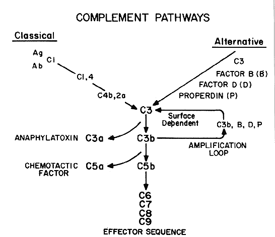
| COMPLEMENT | |
The complement (C) system is a complex network of proteins, enzymes, and
inhibitors. The biologic activities of this network are important in
both host defense and host reaction to inflammatory conditions (Fig. 1). The tears of normal persons have been shown to contain C3,8,14,15 hemolytic C4,16 hemolytic C,17 C5,18 and factor B.15,17 The complement system is present in the tears of normal persons, which
may have an important role in protecting the external eye against infection
or participating in the inflammatory action that could lead to
ocular damage.
|
| TEAR MEDIATORS IN OCULAR ALLERGY |
| In ocular allergy, one of the most widely studied mediators is histamine. Histamine is present in the granules of mast cells and basophils. When
mast cells are activated, histamine is released. It is present in tears
of normal individuals in very low concentrations.19 Elevated levels have been found, however, in patients with allergic conjunctivitis, and
especially in patients with vernal conjunctivitis (VC), reaching
levels of 100 ng/mL.20,21 Histamine can cause capillary dilatation, increased vascular permeability, and
smooth muscle contraction, which clinically results in conjunctival
redness, increased tearing, and ocular itching. Mast cells contain neutroprotease, and two types of mast cells with different neutroprotease composition have been reported. The MCt cells contain tryptase, and the MCtc cells contain both tryptase and chymase. The former has been shown to be a marker for T-lymphocyte-dependent mast cells, but the latter are not dependent on T-lymphocytes for normal growth.22 Irani and co-workers23 showed that normal conjunctival epithelium is devoid of mast cells, whereas the substantia propria contains an average of 11,054/mm3. Of these cells, 95% were classified as MCtc; thus, the predominant mast cells in the conjunctiva are those containing both tryptase and chymase. In patients with allergic conjunctivitis, however, mast cells were found in the substantia propria, but not in the epithelium. In patients with VC and giant papillary conjunctivitis (GPC), mast cells were found in both the epithelium and the substantia propria. In patients with VC, the MCt subtype was present in the substantia propria, but in patients with GPC and allergic conjunctivitis, the subtype was MCtc. Plasmin activity has been found in the tears of normal persons. Elevated levels have been found, however, in patients with allergic conjunctivitis following topical exposure to specific allergens.24 Elevated levels also have been reported in contact lens wearers.25 However, Vannas and co-workers26 found elevated plasmin levels only in wearers of soft and rigid extended-wear contact lenses, not in wearers of daily-wear contact lenses. They also found elevated plasmin activity levels after both short-term (1 hour) and long-term (8 hour) eye closure, with or without contact lens wear. Thus, they believed that the increased tear plasmin in their study was a factor of eye closure, rather than contact lens wear. Neutrophilic chemotactic factor, while present in tears, has been found to be significantly elevated in patients with GPC.27 Neutrophilic chemotactic factor is released from injured conjunctival tissue. Initial biochemical characterization indicates that this substance consists of proteins of both high and low molecular weight and has both anionic and cationic charges. It has also been shown that this substance is not related to interleukin-1, complement component C5a, or leukotriene B4.28 When neutrophilic chemotactic factor derived from rabbit conjunctival cells is then injected into the uninjured tarsal conjunctiva of rabbits, a morphologic change occurs that is clinically similar to that seen in GPC. Histologic analysis of the conjunctiva from these rabbits shows infiltrations of neutrophils, eosinophils, and plasma cells.29 |
| TEAR IMMUNOGLOBULINS IN DISEASE STATES | ||||||||||||||||||||
| The role of tear immunoglobulins in disease states is poorly understood. Investigators
have not entirely been in agreement as to whether the
changes in the tear immunoglobulins are due to local production or to
transudation from the blood plasma. There is no question that tears are important in preventing and fighting external ocular infections. The presence of antimicrobial substances, such as lysozymes, lactoferrin, and beta lysin, is well accepted.30 Secretory IgA, the predominant immunoglobulin in tears, prevents bacterial adherence to mucosal surfaces and can neutralize viruses.31 Patients with IgA deficiency have an increased incidence of recurrent and chronic conjunctivitis.32 Sen and Sarin33 found elevated IgA levels in patients with acute bacterial conjunctivitis, blepharoconjunctivitis, and keratoconjunctivitis. In patients with VC, phlyctenular conjunctivitis, and corneal ulcers, the values were similar to those in control subjects. However, the authors did not state whether the increased levels were due to local production of tear immunoglobulins or to leakage of immunoglobulins from serum secondary to the active inflammation. During the first months of wear, rigid gas-permeable contact lenses can cause a decrease in the tear levels of secretory IgA, accompanied by elevated levels of lysozyme, which then return to normal levels. Certain persons, however, can show marked decreases in tear secretory IgA, even after 1 year of using these lenses. Whether these persons are at a higher risk for ocular complication secondary to contact lenses has not been determined.34 Tear IgG is significantly elevated in patients with acute adenoviral conjunctivitis, whereas the IgM in these patients was only slightly elevated, and there was no significant change in the tear IgA.35 It was interesting to note that, in these patients, the serum IgA level was unchanged, but instead of an increase, there was a decrease in serum IgG and IgM as compared with control patients. The abnormalities of the immunoglobulins in both tears and serum returned to normal levels as the disease process abated. Tear immunoglobulin levels have been studied extensively in patients with VC. A bilateral, recurrent, often severe inflammatory condition of the conjunctiva, VC is characterized by itching, tearing, mucoid discharge, and photophobia. Papillary hypertrophy of the upper tarsal conjunctiva and the presence of eosinophils in the tear secretions are pathognomonic of this condition. IgE has been shown to be elevated in the tears of these patients.36 Samra and co-workers37 reported significantly elevated levels of IgE in tears of patients with VC and demonstrated that the IgE was locally produced by the outer eye. Ballow and Mendelson38 reported that specific IgE antibodies are present in the tears of patients with VC and noted elevated IgG and IgM levels in the tears of these patients.39 They used transferrin as a marker for the transudation of plasma proteins into tear secretions and found that IgG and IgM, as well as IgE, were mostly locally produced by the external eye. Furthermore, they reported the presence of pollen-specific IgG antibodies to rye grass and ragweed antigen E in the tears of patients with VC. These specific tear IgG antibodies were not found in the tears of normal persons or those with nonspecific conjunctivitis. As with the other tear immunoglobulins, these specific IgG antibodies were locally produced by the eye. Not all of the patients with VC had elevated tear IgG and IgE levels: There appeared to be different groups of patients with VC with regard to abnormalities in their tear immunoglobulins. Specifically, only 69% of patients who had elevated IgE had elevated specific IgG antibodies, whereas of those patients with VC who had undetectable levels of IgE, 82% had specific IgG antibodies. These results suggest that both IgE- and IgG-mediated immune mechanisms may be important in the pathogenesis of VC. Since IgG may interact with the complement system to generate biologically active products of the complement cascade, studies were undertaken by Ballow and co-workers15 to determine the levels of complement in the tears of VC patients. These investigators showed that the tears of these patients had increased levels of C3 and factor B. The C3 appeared to be locally produced by the tissues of the external eye. Furthermore, C3a des Arg (C3 anaphylatoxin), a product resulting from activation of the complement system, was also found to be elevated in VC patients. C3a causes a noncytolytic release of histamine from mast cells and basophils, as well as the contraction of smooth muscle and an increase in capillary permeability.40 Preliminary studies in our laboratory have shown that the IgG antibodies to rye grass and ragweed antigen E, at least in part, belong to the IgG4 subclass. The IgG4 could be acting as a reaginic immunoglobulin mimicking IgE-mediated diseases or as a blocking antibody for IgE-mediated mast cell activation.41 Thus, the pathophysiology of VC, as indicated by its tear immunologic components, is quite complex. IgG and IgE mechanisms, as well as the complement system, appear to be important. IgE and the generation of C3 anaphylatoxins are important in basophil-mast cell activation and the release of vasoactive mediators into the tears. Patients with allergic rhinoconjunctivitis have elevated tear IgE. The majority of these patients have specific IgE antibodies to inhalant pollens and antigens. There also appears to be a subgroup of patients with allergic-like conjunctivitis with different tear immunoglobulin abnormalities. These patients have the clinical features of allergic conjunctivitis, but they have a negative immediate skin reactivity, absent specific IgE antibodies to inhalant pollen antigens, and normal IgE levels in their tears. However, these patients have elevated pollen-specific IgG antibodies in their tear secretions, which are locally produced.41 GPC secondary to contact lenses is characterized by increased mucous production, blurry vision, itching, decreased contact lens tolerance, and the development of giant papillae on the upper tarsal conjunctiva.42,43 The histologic findings consist of plasma cells, lymphocytes, mast cells, eosinophils, and basophils and are consistent with an immunologic mechanism similar to that of VC.43,44 This hypothesis is supported by the abnormalities found in their tear immunoglobulin. Elevated IgE, IgG, and IgM (Table 2), as well as increased levels of C3, factor B, and C3a anaphylatoxin, have been shown to be locally produced, and their elevated levels are not a result of transudation from the serum secondary to ocular inflammation. Furthermore, the elevated IgG and IgE levels appear to correspond to the clinical severity of the disease process. This is in contrast to asymptomatic contact lens wearers, who have no significant tear immunoglobulin abnormalities.9,45
TABLE TWO. Tear Immunoglobulins in Giant Papillary Conjunctivitis
|
| SUMMARY | ||||||||||||||||||||||||||||||||||||||||||||||||||
| The tear film is a complex fluid containing a variety of substances. Tear
immunoglobulins are present in normal tears. In disease states, their
abnormalities reflect the eye's reaction to the particular pathologic
process. In VC and GPC, the external eye is capable of a localized
immune reaction, which is well illustrated by the increased levels
of locally produced immunoglobulins (e.g., IgG, IgE, IgM, and in VC, pollen-specific IgE and IgG antibodies). Furthermore, studies
have emphasized that the external eye can mount a local
immune response. The target organ mediates clinical symptomatology, even
in the absence, in some patients, of systemic immunity.28,29 Complement appears to play a role in the inflammatory process of VC and
GPC. The generation of complement-derived products (e.g., C3 anaphylatoxins), even by nonimmune mechanisms, may be important in
basophil-mast cell activation, resulting in many of the clinical features
of inflammation in these two ocular disorders. In other disease states, specific
tear immunoglobulins have been shown to be elevated (Table 3). Their exact function and origin, however, has not been well studied. Further
studies toward a better understanding of the role of elevated
tear immunoglobulins in health and disease are currently being conducted.
TABLE THREE. Tear Immunoglobulins in Ocular Inflammation
E = elevated; N = normal; ND = not detected; OE = occasionally elevated; SE = slightly elevated.
|