Chapter 32
Immunology of Infectious Systemic Diseases That Affect the Eye
MITCHELL H. FRIEDLAENDER
Main Menu   Table Of Contents

Search

BACTERIAL SYSTEMIC INFECTIONS
VIRAL SYSTEMIC INFECTIONS
FUNGAL SYSTEMIC INFECTIONS
PARASITIC SYSTEMIC INFECTIONS
REFERENCES

Infectious diseases are intimately associated with the functions of the immune system. Much of our knowledge about basic mechanisms in immunology has been acquired by the study of host interaction with infectious agents. Immune mechanisms probably evolved to protect the individual from the destructive effects of parasitic organisms and their products. If these protective mechanisms are successful, they eliminate the infectious agents. Under certain conditions, however, immune mechanisms can behave as pathogenic forms of hypersensitivity. The eye may be directly infected by a variety of agents that produce systemic disease, or it may be a focus of hypersensitivity reactions to these agents.
Back to Top
BACTERIAL SYSTEMIC INFECTIONS

TUBERCULOSIS

Mycobacterium tuberculosis is a facultative, intracellular, aerobic, acid-fast bacillus that is pathogenic only for humans. It produces mainly lung disease, but many extrapulmonary sites, including the eye, can also be affected. Cellular rather than humoral immune mechanisms are decisive in the host's recovery from tuberculosis, and in its diagnosis the tuberculin skin test (a manifestation of cellular immunity) is more useful than the measurement of serum antibody.

Immunopathology

M. tuberculosis can survive and proliferate within phagocytic cells. It may escape the bactericidal effect of the macrophages by preventing the fusion of enzyme-containing lysosomes with phagosomes that contain the organisms. Lipids and waxes of high molecular weight constitute up to 60% of the bacterium's cell wall and may be responsible for the responses of the host's tissues to the tubercle bacillus. They may also account for the impermeability of the organism to tissue stains. Tuberculoproteins are responsible for the induction of hypersensitivity and for the reaction of the skin to tuberculin. A substance known as “cord factor” causes the serpentine growth of the tubercle bacillus and may also inhibit leukocyte migration and stimulate granuloma formation.

The host deals with M. tuberculosis principally by the mechanism of cellular immunity. Cell-mediated immunity to tuberculin can be passively transferred to uninfected experimental animals along with living lymphoid cells but not with immune serum. Immunity can also be transferred with the leukocyte extract transfer factor. Although patients with agammaglobulinemia produce no demonstrable antibodies, they can develop delayed hypersensitivity responses and normal resistance to the tubercle bacillus. Patients with defects in delayed hypersensitivity are more susceptible to the disease than persons normal in this respect.

Tuberculosis may affect nearly all of the ocular tissues. A granulomatous uveitis is the lesion most frequently encountered and may be associated with mutton-fat keratic precipitates, Koeppe nodules, and vitreous opacities. Diffuse posterior uveitis may also occur, and although rare with modern treatment, the disease can affect the lids, sclera, cornea, and retina. With the recent increase in tuberculosis in AIDS patients, the ocular manifestations of tuberculosis are increasing.1–3

LEPROSY

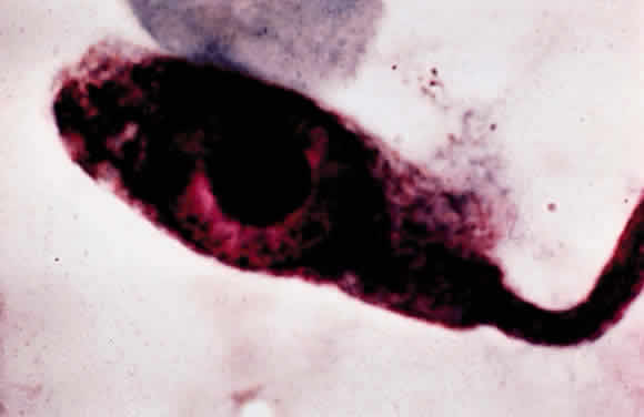
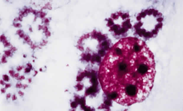
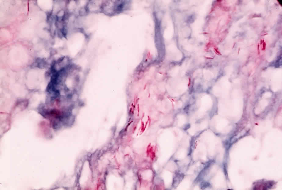
Leprosy is a chronic infectious disease caused by Mycobacterium leprae, an obligate, intracellular, acid-fast bacillus (Fig. 1). This organism can invade nerves and skin and give rise to widespread clinical manifestations. Ocular lesions occur in 90% of lepromatous leprosy cases, and nearly one third of these produce significant visual loss.

Fig. 1. Mycobacterium leprae in skin biopsy. (Courtesy Mr. M. Okumoto)

Immunopathology

The following three clinical types of leprosy are recognized: (1) lepromatous (nodular) leprosy is characterized by marked deficiency in cellular immunity and extensive infiltration of the tissues with M. leprae, (2) tuberculoid leprosy is distinguished by the preservation of immune responses and the absence of demonstrable bacilli within the tissues, and (3) intermediate or borderline leprosy has features of both the lepromatous and tuberculoid types. Patients with lepromatous leprosy are anergic to the lepromin skin test antigen, to which normal persons have a positive delayed hypersensitivity reaction. They also fail to develop contact allergy to dinitrochlorobenzene (DNCB) and other contact sensitizers, they have depressed delayed hypersensitivity reactions to common bacterial antigens, and they demonstrate prolonged skin allograft survival.4,5

Patients with lepromatous leprosy have significantly lower-than-normal levels of circulating T and B lymphocytes, and the T cells in the paracortical regions of their lymph nodes are severely depleted.6 Their peripheral lymphocytes show diminished blast transformation in the presence of the mitogen phytohemagglutinin. Animals thymectomized at birth, and therefore lacking cellular immunity, readily develop widespread infections with M. leprae.7 The infection is apparently related to the survival of the bacillus within macrophages. Recent electron microscopic studies have shown that the organism can escape from the phagolysosome and remain free in the macrophage cytoplasm.

In contrast to being deficient in cellular immunity, patients with lepromatous leprosy produce normal or greater-than-normal numbers of antibodies and may even produce autoantibodies. They have above-normal levels of both gamma globulin and the cryoproteins. Thyroglobulin antibodies and rheumatoid factor are found in 50% of patients and false-positive Venereal Disease Research Laboratory (VDRL) tests in about 10%. Lupus erythematosus (LE) cells occasionally appear, and antibodies to both basement membrane and intercellular substances have been found in the skin of infected patients. The similarity between leprosy and the collagen vascular diseases, particularly LE, is indicated by their sharing such clinical manifestations as butterfly facial rashes, arthritis, depressed cellular immunity, and autoantibody production.

In lepromatous leprosy, the lids may show granulomatous inflammation,8 and nodules may appear on the face and eyelids. A hyperemic conjunctivitis may develop and may be followed by symblepharon and lid deformities. Corneal nodules, interstitial keratitis, iridocyclitis, and chorioretinitis have also been reported.9

Diagnosis

The lepromin skin test was introduced by Mitsuda and is performed with extracts of skin nodules from leprosy patients. A positive intradermal test leads to a tuberculin type of skin reaction 24 to 48 hours later, and a nodular reaction (the Mitsuda reaction) appears in about 7 days and peaks in 3 or 4 weeks. The test is useful in differentiating the types of leprosy in patients already known to have the disease. A positive lepromin skin test indicates tuberculoid or nearly tuberculoid disease, and a negative skin test indicates progression to lepromatous leprosy. Positive skin tests are common in the normal population due to cross-reactivity with other mycobacterial antigens.

SYPHILIS

Syphilis is caused by the spirochete Treponema pallidum, a motile, highly infectious agent that functions predominantly as an extracellular pathogen. It occurs naturally only in humans and is most easily transmitted by sexual contact. Both humoral and cellular immunity are important in the defense of the host against syphilis. Three stages of the diseases are clinically identifiable, and the eye may be involved in all three. Although severe, destructive syphilitic lesions have been far less common since the discovery of penicillin, the disease must be considered in the differential diagnosis of many diverse clinical problems.

Immunopathology

Immunity to T. pallidum depends on a complex interaction between humoral and cellular immunity. Several lines of evidence indicate that antibody is important in this infection. Partial immunity develops in the rabbit after the passive transfer of serum from immune animals. Immobilizing antibodies against treponemes can be found in the serum of patients who have had syphilis, especially advanced syphilis. A number of nonspecific or reaginic antibodies are found in the sera of patients who have the disease, and these form the basis of many serologic diagnostic tests. Immune complex deposition may play a role in the nephropathy associated with secondary syphilis in adults, and IgG may be found along the glomerular basement membrane in patients with congenital syphilis.10

Delayed hypersensitivity directed against treponemal antigens is absent in primary and early secondary syphilis, develops late in the secondary disease, and is found regularly in latent and tertiary disease. Because syphilis progresses through the primary and secondary stages despite the presence of antibodies that immobilize treponemes, it follows that antibodies developing during the course of an infection are at best only partially protective.

When viewed microscopically, the typical lesion of syphilis is a granulomatous inflammation. Granulomas, which contain reticuloendothelial cells (including epithelioid and giant cells), are thought to represent an immune response to such poorly soluble substances as foreign bodies, insoluble antigens, and microorganisms that cannot be easily eliminated. Granulomatous reactions are clearly different from typical delayed hypersensitivity reactions: their onset is much slower, and they may require weeks or even months to develop. Many granulomatous lesions are accompanied by vasculitis, suggesting that there may be an associated immune complex disease.

Diagnosis

During primary and secondary syphilis, the spirochete can be identified microscopically by dark-field examination. Serologic tests do not become positive until 14 to 21 days after the initial infection. Because spirochetes are difficult to identify in the late stages of the disease, serologic tests are extremely important. Two categories of tests—for reaginic antibodies and for treponemal antibodies—are available. Reaginic antibodies bear no relation to IgE; rather, they are antibodies directed against cardiolipin, a tissue-derived substance thought to be a component of mitochondrial membranes. Beef heart is used as a source of cardiolipin in many reaginic antibody diagnostic tests, examples of which are the VDRL, Hinton, and Kahn tests. Unfortunately, false-positive results on these tests occur in such diseases as LE, leprosy, infectious mononucleosis, and hepatitis, probably because of the many tissues and microorganisms that have mitochondrial membranes.

Two tests for treponemal antibody are the T. pallidum immobilization test and the fluorescent T. pallidum antibody test (FTA). In the former, specific antibodies against the spirochete can be detected, whereas the latter uses spirochetes from infected rabbit testes as a substrate for the serum being tested. If positive, the bound antibody can be detected with fluoresceinated antihuman gamma globulin. Other nonpathogenic treponemes may be absorbed from the serum before testing, in which case the test is referred to as the FTA absorption test (FTA-ABS test).

The FTA-ABS test is positive in 80% to 100% of primary syphilis cases, the VDRL test in 50% to 70%.11 In secondary syphilis, both tests are nearly always positive. The VDRL test is less often positive after treatment, whereas the FTA-ABS test remains positive for many years; although false-positive VDRL results are common, one rarely encounters a false-positive FTA-ABS result, except in systemic lupus. In latent syphilis, the VDRL test is negative in up to one third of patients.

Treponemes are sometimes detected in the aqueous humor of patients with uveitis, but they may be nonpathogenic organisms such as Treponema microdentium.12 Aqueous humor aspiration, followed by the FTA technique, may be useful when a search for treponemes is being made.

A chancre may occur on the lids or conjunctiva in primary syphilis. A pinkish, macular rash may appear on the lids in secondary syphilis, and ulcerative blepharitis can cause scarring and loss of cilia. Iridocyclitis can occur in secondary syphilis, and chorioretinitis can be associated with late secondary and tertiary disease. Tertiary syphilis can produce distinctive lesions of the eye and central nervous system.

Interstitial keratitis is the hallmark of congenital syphilis and may be a hypersensitivity phenomenon.13 Other features of congenital syphilis are iridocyclitis, chorioretinitis, and malformation of the optic discs. HIV-positive patients may have an atypically dense vitritis14 or other forms of uveitis.15–17

TULAREMIA

Francisella tularensis, the organism that causes tularemia, is a pleomorphic gram-negative coccobacillus that requires special culture media for its isolation. As small a dose as 50 organisms of the more virulent type A form can produce infection in humans. Type B produces a milder disease. Infection is transmitted by intermediate rodent hosts, especially the rabbit. A substantial number of cases have also been reported in association with exposure to ticks. Other intermediate hosts are squirrels, cats, foxes, and raccoons. Tularemia is an occupational hazard for shepherds, mink ranchers, hunters, and butchers, all of whom may handle infected tissues. Purulent conjunctivitis and typical Parinaud's oculoglandular syndrome can follow inoculation of the eye with F. tularensis.

Immunopathology

Although the host's defenses against tularemia are not fully understood, cell-mediated immunity appears to be important. Immunity is associated with (1) a delayed hypersensitivity skin reaction to tularemia skin-test antigen, and (2) the greater-than-normal ability of rabbit macrophages to kill F. tularensis. In vitro tests for cell-mediated immunity are present in sensitized individuals. Inactivated vaccines have no protective effect, but a live, attenuated strain of the organism can protect both humans and animals. There is no evidence that antibodies are protective in tularemia.

Diagnosis

Diagnosis is based on isolation of the organisms and a rise in agglutinating antibody titers, which usually occurs after the first 2 weeks of illness but may take much longer. In most cases there is a fourfold or greater rise. A skin test, in which a lyophilized, ether-extracted bacterial antigen is used, may be helpful but is not always available.

BRUCELLOSIS

This is an infection acquired by humans through contact with infected tissue of cattle, pigs, sheep, and goats, or by ingestion of raw milk or milk byproducts. It is one of the most prevalent zoonoses in the Unites States and is caused by several species of the genus Brucella, a small gram-negative bacillus. The causal species are B. abortus, B. melitensis, B. suis, and rarely B. canis. Veterinarians and abattoir workers are often intermediate hosts. The organism is transmitted from infected tissues to humans through cuts in the skin, conjunctival contact, ingestion of uncooked meat, or inhalation of organisms. The ocular lesions associated with Brucella infection are nummular keratitis, scleritis, optic neuritis, and uveitis.

Immunopathology

Brucella has a predilection for the reticuloendothelial system. Once ingested, the organisms are rapidly taken up by neutrophils and transported to the histiocytes of the liver, spleen, and lymph nodes. There they enter a period of prolonged intracellular residence. They may remain intracellular for several weeks (rarely for months) while the phagocytes containing the organisms form noncaseating granulomas. Clinical symptoms occur when organisms are released from infected reticuloendothelial cells.

Immunity to Brucella seems to be cell-mediated. Delayed hypersensitivity develops during experimental infection. The skin test lacks specificity, however, and has been discontinued for routine use in the United States.

Although antibodies in Brucella infection are not protective, agglutinating and complement-fixing antibodies can be measured. An IgA antibody can act as a blocking factor and produce spuriously low agglutinin levels, but interference by this factor can be avoided by using a Coombs' test. Antibody measurement is important in the diagnosis of brucellosis, because the organism is fastidious and culture is difficult.

LYME DISEASE

Lyme disease is caused by the spirochete Borrelia burgdorferi and is transmitted by the deer tick.18,19 Systemic findings include a purpuric skin rash, “target” lesions, and sudden onset of arthritis involving the knees and ankle joints. Serologic and immunofluorescent tests are available for identifying the infection. Ocular findings include chronic intermediate uveitis,20 posterior uveitis,21 choriocapillaritis, neuroretinitis, retinal vasculitis, multifocal corneal infiltrates, and conjunctivitis.22,23 Treatment with antibodies, such as ceftriaxone, and steroids is generally indicated.24

Back to Top
VIRAL SYSTEMIC INFECTIONS

VARICELLA-ZOSTER

Varicella-zoster (V-Z) virus is the etiologic agent in varicella (chickenpox) and herpes zoster. Chickenpox is the more common clinical manifestation of the two. It is a common contagious disease of childhood characterized by fever, a papular, vesicular rash, and transmission by droplet infection. Herpes zoster, including herpes zoster ophthalmicus, is more common in middle and old age. It may occur in healthy individuals with no apparent precipitating cause and is then referred to as primary or spontaneous. Secondary, or symptomatic, herpes zoster develops in individuals whose natural immunity has been impaired by such factors as old age, malignancy, immunosuppressive therapy, chronic illness, or trauma. In HIV-infected patients, herpes zoster infections may be prolonged and chronic.25 The virus may also be a cause of acute retinal necrosis.26

Immunopathology

The V-Z virus is neurotropic and produces a skin rash with a dermatome distribution. Most herpes zoster infections are caused by a reactivation of latent V-Z virus residing in the dorsal root of a ganglion. This usually occurs at a time when the host's immunity is in some way compromised. The reactivated virus can travel along the nerve axons and produce the characteristic vesicular lesion of the skin in accordance with the distribution of the nerve. Infection can also follow re-exposure of the host to the virus by contact with a patient who has either chickenpox or zoster. In neither event can the affected immune-deficient individual respond adequately to ward off the viral infection.

It has been shown that varicella can be prevented by passive immunization with antibody to V-Z virus. The role of antibody in the prevention of zoster, however, and in the eventual recovery from either zoster and varicella is somewhat less clear. Hope-Simpson has suggested that there is a critical antibody level above which an individual is protected from latent virus in neuronal tissues, and that this level can be maintained by endogenous or exogenous stimulation by viral antigens.27

Complement-fixing antibody usually appears early in the course of V-Z infection, and some observers have speculated that patients lacking such antibody early in the course of zoster are more likely to have disseminated zoster infection. Although patients with disseminated zoster do not synthesize detectable levels of complement-fixing antibodies until 2 or 3 weeks after the disease begins, some of them have been shown to have V-Z antibody when tested by a new, more sensitive method. By this method it is possible (by immunofluorescence) to detect antibodies to membrane antigens of cells infected with the virus and to show a prompt rise in IgA, IgG, and IgM in both localized and generalized zoster.28 Dissemination may occur even in the presence of high levels of serum antibody. This means that a brisk serum antibody response to V-Z virus does not necessarily alter the course of the disease.

It has long been suspected that cellular immunity is important in the recovery from infection with V-Z virus.29 Patients with cell-mediated immunity deficiencies are more susceptible to zoster than patients with humoral immune deficiencies. Depression of cellular immunity from malignancy or treatment with immunosuppressive drugs also predisposes to the development of zoster. In vitro studies have shown that leukocytes from donors immune to varicella are more efficient in inactivating V-Z virus than leukocytes from donors susceptible to varicella.30 The leukocytes from the susceptible patients fail to reduce the V-Z virus titer. Thus, individuals who recover from V-Z virus infection have specific cell-mediated immunity against the virus. They seem also to have high local levels of interferon.

VACCINIA

Edward Jenner (1798) was the first to immunize a subject against smallpox by an inoculation with vaccinia. Vaccinia virus is only mildly pathogenic for humans and affords protection against infection with smallpox virus by cross-reacting with it. An ocular vaccinial lesion can occur after vaccination if the eye is accidentally inoculated, and generalized vaccinia can develop after the vaccination of immunodeficient subjects and patients with widespread dermatoses, especially atopic dermatitis.

Immunopathology

Approximately 8 days after an inoculation of the skin with vaccinia virus, a delayed hypersensitivity reaction develops at the inoculation site.31 The vaccinia virus contains antigens that cross-react with smallpox virus, protecting the vaccinated individual from smallpox. Vaccination of normal subjects produces no systemic disease unless the patient has been compromised.

The immune response to vaccinia virus is the result of an interaction between antibody, cellmediated responses, and interferon. A normal, immunocompetent subject responds to intradermal vaccination with a “primary take”: the host's immune defenses eradicate the virus at the site of inoculation. Subsequent revaccination in the same subject produces a milder skin reaction that peaks in 4 or 5 days. This reaction is probably due mainly to cell-mediated immune responses to the virus. Subjects with severe defects in cellular immunity almost always have a fatal response to smallpox vaccination. Subjects with defective antibody responses may develop severe necrotic skin reactions (vaccinia gangrenosum), but this can be treated successfully with vaccinia immune globulin or the drug thiosemicarbazone. These preparations, however, cannot prevent a fatal response to smallpox vaccination in patients with T-cell defects. It would seem, therefore, that both humoral and cellular immunity are important in the control of vaccinia virus infection. It may be that antibody participates in the reduction of the antigen load (i.e., the mass of virus particles), and that failure to do so may result in temporary immune paralysis of the T-cell system. If the T-cell system is inoperative, the virus will apparently not be eliminated despite the presence of antibody.

The immunization of rabbits with vaccinia virus vaccine by the intranasal route results in the appearance of IgA antibody activity in tears.32 If the vaccination is intradermal, however, the antiviral activity of the tears is associated with IgG. IgG is also the predominating serum immunoglobulin found in immunized animals. Antibody titers in both tears and serum can be raised by the interferon inducer poly I:C. Vaccination by either the intradermal or the intranasal route results in reduced shedding of the virus and a reduction in the rabbit's clinical disease.33 High levels of serum neutralizing antibody can be correlated with mild illness, but tear antibody is apparently not related to either illness or virus shedding. The lack of protection by neutralizing antibody in the tears suggests that the cellular immune mechanism plays a prominent role in vaccinia virus infection.

After the inoculation of the rabbit cornea with vaccinia virus, the cells within the deep layers of the epithelium are the first to show infection and to undergo degeneration.34 Viral replication is thought to occur within the stromal keratocytes.35

RUBELLA

The teratogenic potential of the rubella virus was first recognized by the Australian ophthalmologist Sir Norman Gregg.36 Infection during early pregnancy leads to congenital malformation of the eyes, heart, and ears of the fetus. The ocular defects in congenital rubella syndrome include cataract, microphthalmia, nystagmus, retinopathy, and transient corneal clouding. They develop in 30% to 60% of infants exposed fetally to rubella, with cataract and retinopathy occurring most often.

Immunopathology

The fetus is most susceptible to the effects of the rubella virus from day 20 to day 40 of gestation. The mechanism responsible for the malformations is not completely understood. It is believed that death of the infected cells, a change in rate of cell growth, or perhaps both of these mechanisms are important. The reason for the persistence of the virus in the fetus is not understood. Endogenous IgM antibodies and maternal IgG antibodies are present at birth, and the antibody responses of the fetus are apparently intact. Fetal cellular immunity is depressed, however, and this may be a factor in the inability of the fetus to clear virus from the tissues.

In postnatal rubella infection, antibodies to the virus increase rapidly, reaching maximum titers in 7 to 10 days. IgM antibody can be detected for approximately a month, but IgG antibody persists for many years.37 Antibodies to rubella virus can be assayed by such techniques as hemagglutination inhibition, neutralization of virus infectivity, complement fixation, indirect immunofluorescence, and immunoprecipitation. All of the assay methods (except complement fixation) yield similar results, with the titers peaking within 2 weeks and then gradually decreasing. Antibodies usually remain detectable for life, and individuals with antibodies in their sera are believed to be immune from reinfection.

A defect in cellular immunity is suggested when virus persists.38,39 Lymphocytes from some infants with congenital rubella do not show a blastogenic response to phytohemagglutinin, and rubella virus added experimentally to lymphocytes from normal subjects suppresses the blastogenic response to phytohemagglutinin.40–42 It remains uncertain whether viral persistence is due to a defect in the immune mechanism or to a prolonged survival of clones of infected cells.

CYTOMEGALOVIRUS INFECTION

Cytomegalovirus (CMV) infection is a significant form of congenital disease in newborns and an acquired infection in immunosuppressed adults. The causal virus (one of the family of herpesvirus) is not easily eliminated, persisting in host tissues for months, years, or even a lifetime. It produces a chronic infection with a variable incubation period, outcome, and course. It may produce chorioretinitis, optic atrophy, mental or motor retardation, and lesions of various other organ systems. Patients with AIDS are particularly susceptible to CMV infections, and many develop retinitis and sometimes disseminated infections.43 Two virostatic antiviral agents, ganciclovir and trisodium phosphonoformate dexahydrate (foscarnet) have been shown to be useful in inhibiting the progression of CMV retinitis. Ganciclovir has been evaluated more extensively than foscarnet and is currently the only clinically available effective mode of therapy for AIDS-associated CMV retinitis.44,45

Immunopathology

Cytomegalovirus (Fig. 2) infects the fetus in the uterus, whose immune system is incompletely developed. The virus also infects adults with malignancies, particularly of the hematopoietic and reticuloendothelial systems. In kidney transplant patients (who are treated routinely with immunosuppressive agents), CMV has been detected in up to 90% of cases, and active infection may predispose these patients to bacterial superinfection and transplant rejection.46–48 High antibody titers are believed to be protective.

Fig. 2. Cytomegalovirus inclusion. (Courtesy Mr. M. Okumoto)

CMV infection is associated with abnormal serum globulins and depressed cellular immunity; for example, experimentally it causes (1) suppression of primary and secondary immune responses to sheep red blood cells in mice, (2) prolonged skin graft survival, and (3) inhibition of lymphocyte responsiveness to phytohemagglutinin.48–50 (The degree of immune suppression is directly related to the size of the viral inoculum.)

Human CMV also causes alterations of the immune system. Abnormal immunoglobulins, including rheumatoid factor, cryoglobulins, cold agglutinins, antinuclear antibodies, and the Coombs' test antibody, have all been detected after CMV infection.49 Apparently the virus causes alteration in the immune response only during the acute phase of the infection. The mechanism by which it causes immunosuppression is not fully understood. It has been suggested that viral antigenic competition may inhibit antibody responses to other antigens. A more likely explanation is that infection of potential antibody-producing cells by virus may divert lymphoid cells from their normal immune function to the production of more virus.50 CMV infection (including retinitis) has been seen in patients with AIDS.

Diagnosis

Several different tests are available for the detection of antibodies to human CMV. Although a complement-fixation test is commonly used, a sensitive fluorescent-antibody test may prove more useful in the future.51 Because IgM of maternal origin does not pass the intact placenta, IgM antibody in the infant's serum indicates congenital infection. In the adult, neutralizing antibodies are also present. The appearance of complement-fixing antibody or a fourfold rise in titer or both, associated with the characteristic clinical features of the disease in a kidney transplant recipient, is usually diagnostic. Diagnosis is also possible by histopathologic examination of biopsy material or by the identification of characteristic cytomegalic cells in the sedimented urine. The virus can also be cultured in human fibroblasts, in which characteristic plaques are produced.

RUBEOLA

Rubeola (measles) is an acute, febrile disease of childhood. It is caused by a paramyxovirus and produces a maculopapular rash and catarrhal inflammation of the eye and respiratory tract. The ocular lesions are mucopurulent or pseudomembranous conjunctivitis, epithelial keratitis, and occasionally dacryocystitis and retinal edema.

Immunopathology

Vaccination with inactivated measles virus results in augmentation of delayed hypersensitivity to measles virus. Inactivated virus also causes a temporary elevation of serum IgG titers, which then wane. A rare postvaccination complication may occur: on natural re-exposure to measles, a hyperacute “atypical” measles syndrome, characterized by fever, pneumonia, pleural effusion, and severe hemorrhagic rash, may develop, perhaps as a result of an immune complex deposition that induces an Arthus response in the skin and respiratory tract.

Subacute sclerosing panencephalitis is a degenerative disease that occurs during the first and second decades of life. It is characterized by personality changes, dementia, seizures, and myoclonus. Measles virus antigen has been found in infected brain material by fluorescent-antibody staining, and the virus has been recovered from the brain by cocultivation techniques. Despite continued infection, large amounts of antibody to measles virus have been found in the cerebrospinal fluid of these patients, suggesting a defect in their cellular immunity. Several studies have indicated such a defect; others have not. Some patients with subacute sclerosing panencephalitis have a blocking factor, possibly antibody in the cerebrospinal fluid and blood.

HIV RETINITIS

The human immunodeficiency virus has been shown to infect retinal tissue, including capillary endothelium and neuroretinal cells.43 Although HIV has not been demonstrated to cause necrotizing retinitis similar to CMV, HIV capillary endothelial infection and associated immune complex deposition are believed to play a role in the formation of the “cotton wool spots” of AIDS retinopathy.52 This capillary damage in turn may facilitate penetration of CMV-infected cells into the retina.53 Once within the retina, CMV infection may be enhanced by concurrent HIV infection. It has not been clearly established whether AZT therapy decreases the incidence of cotton wool spots or CMV retinitis in AIDS patients. Attempts to prevent the development of CMV retinitis by photocoagulation of cotton wool spots has not been successful. Many patients with AIDS-associated microvasculopathy, cotton wool spots, and the like report photopsias and a general dimming of vision. Central visual acuity and peripheral visual fields are not affected, however, and specific therapy is neither available nor necessary.

Back to Top
FUNGAL SYSTEMIC INFECTIONS

HISTOPLASMOSIS

Histoplasmosis is an intracellular mycotic infection caused by the dimorphic fungus Histoplasma capsulatum. This organism can produce acute or chronic pulmonary inflammatory disease. There is also a large body of epidemiologic, clinical, and experimental evidence strongly supporting its etiologic role in the ocular condition known as presumed ocular histoplasmosis. Patients with this condition have hemorrhagic or nonhemorrhagic macular disciform lesions, peripheral and peripapillary choroidal atrophic scars, and positive histoplasmin skin tests.

Immunopathology

Infection with H. capsulatum induces both humoral and cellular immune responses. Although the measurement of antibody is useful in the diagnosis and prognosis of the infection, it is the host's cellular immunity that is critical in the defense against the organism, and it may be cell-mediated hypersensitivity that causes the inflammatory and necrotic lesions that accompany the pulmonary disease.

Cellular immunity develops rapidly after primary infection with H. capsulatum. The immunizing constituent is thought to be a glycoprotein complex, the components of which can confer delayed hypersensitivity as measured by skin tests with histoplasmin and by the production of migration inhibitory factor. Lymphocytes from patients with positive histoplasmin skin tests become activated in the presence of histoplasmin antigens. The granuloma formation and pulmonary tissue destruction in histoplasmosis are usually attributed to vigorous cell-mediated immune responses to the infection. Although cell-mediated immunity is operative in both acute and chronic histoplasmosis, it may fail to function in disseminated infections. This implies a failure of macrophage activation and of the host's normal cell-mediated responses in dealing with the organism. An analysis of lymphocyte markers showed a high incidence of CD38, a marker that may correlate with poor lymphocyte function.54

Humoral immune responses also develop rapidly in histoplasmosis, and a high antibody titer may indicate recent infection. Neutrophils cannot kill the fungi, and antibodies, instead of being protective, indicate progressive disease, high titers suggesting a worsening of the infection rather than improvement. Antibody titers fall when the disease regresses and increase when it disseminates.

In the pathogenesis of presumed ocular histoplasmosis, cellular immune responses play an important role. Patients with this condition have hyperreactive lymphocyte responses that include skin test reactions to a number of common antigens.55 The skin test to histoplasmin is strongly positive, especially in patients with inactive disciform scars. Skin tests with intermediate-strength tuberculin are also frequently positive.56 These patients also show more spontaneous lymphocyte transformation than control patients (in 5- and 7-day cell cultures), and compared with the lymphocytes of control subjects, their lymphocytes are hyperreactive with mitogens, with antigens derived from H. capsulatum, and with various other microbial antigens.57

A flare-up of macular disease may follow a skin test with histoplasmin, and the lymphocytes of subjects sensitized to H. capsulatum have responded in vitro to booster injections of histoplasmin antigen.58–61 Perhaps a heightened cellular immune response explains these macular flare-ups that can follow both skin testing and histoplasmin desensitization therapy.59,60 Acute macular lesions have been treated successfully by immunosuppression with corticosteroids and azathioprine.62,63

Although the cellular immunity that operates against H. capsulatum may be defective in patients with pulmonary histoplasmosis, it is probably intact in patients with presumed ocular histoplasmosis.64 Those with macular disciform lesions have a high incidence of healed pulmonary lesions. Males have a higher frequency of unilateral and bilateral involvement than females and a greater mean induration after histoplasmin skin testing.65

The pathogenesis of the disciform macular lesion is unknown. Local hypersensitivity responses of the choroid, secondary to histoplasmin antigen or to antigen-antibody complexes, have been suggested.57 Alternatively, cross-reacting fungal antigens or antigenic alteration of normal ocular tissue may be the decisive factor. When a patient with presumed ocular histoplasmosis has no systemic disease, local hypersensitivity or reinfection may be contributing to the disciform lesion. Viable lymphocytes have been found in clinically inactive scars in pathologic specimens from two patients.66 These lymphocytes may be T cells that become activated when antigen was reintroduced. Recently, transforming growth factor-beta 1 and basic fibroblast growth factor were identified in the subretinal renovascular membranes.67 These membranes are composed of fibrovascular tissue interposed between Bruch's membrane and the retinal pigment epithelium. They may represent a nonspecific healing response to a local stimulus or injury.68

Diagnosis

Immunologic tests are of little use in the diagnosis of systemic histoplasmosis. Skin tests are often positive in endemic areas, and serologic titers may be falsely elevated due to previous skin testing. The elevation in the complement fixation titer may last several months. Because histoplasmin complement-fixing antibodies are elevated in only about half of patients with disseminated histoplasmosis, many infected patients are bound to be missed when subjected to this test. Histoplasmin antigen also cross-reacts serologically with blastomycin and coccidioidin.

Despite these pitfalls, both skin tests and serology have some value in the diagnosis of presumed ocular histoplasmosis. In a nonendemic area, a positive histoplasmin skin test may be helpful, and the complement fixation test can be of value when the skin test produces a false-negative result (about 11% of cases). In all cases, serum should be drawn before a skin test is applied, and it should be kept in mind that only one third of patients with this condition have elevated antibody titers. In the systemic disease, culture of the fungus is the best way to make the diagnosis; in presumed ocular histoplasmosis, the characteristic clinical features are the major diagnostic clues.

CANDIDIASIS

Candida albicans is an opportunistic fungus that produces systemic disease (often with ocular complications) in immunologically compromised patients (Fig. 3). It can also produce ocular disease (corneal ulcer) in the compromised eye without systemic disease. Candidal infection can occur in debilitated patients, in patients receiving intravenous hyperalimentation therapy administered through indwelling venous catheters, and in patients with diabetes, neoplasms, or narcotic addiction and HIV infection.69 The ocular complications of the systemic disease—snowball opacities in the vitreous, white, cottony retinal lesions, and retinal hemorrhages—may require systemic treatment.

Fig. 3. Candida albicans from corneal scraping. (Courtesy Mr. M. Okumoto)

Immunopathology

C. albicans grows as a yeast on some media (Fig. 4) and forms pseudomycelia on other media. It has an affinity for mucous surfaces and under certain conditions can penetrate them. The growth of Candida is favored by certain local factors: (1) a low pH, which causes transferrin to release the iron that Candida requires, and (2) high concentrations of glucose, which may be a factor in the susceptibility of diabetic patients to candidiasis. Broad-spectrum antibiotics eliminate the normal bacterial flora that competes with Candida for glucose, and thus more glucose is available for the Candida.

Fig. 4. Candida albicans on Sabaroud's agar. (Courtesy Mr. M. Okumoto)

Humoral and cellular immunity collaborate to defend the host against candidal infection. Serum anticandidal clumping factors bring about candidal agglutination and increase the normal uptake of the organisms by phagocytes. Neutrophils, monocytes, and macrophages can all wipe out Candida.

Cellular immunity is extremely important in protecting the host against candidal infection. Patients with chronic mucocutaneous candidiasis have defects in their cellular immunity that apparently lead to chronic candidal infection. These patients develop superficial candidal infections of the nails, skin, and mucous membranes. This constitutes a syndrome also associated with various endocrine abnormalities (especially hypothyroidism and adrenal insufficiency). Candidal infections are also seen in Hodgkin's disease, in patients with severe combined immune deficiency, and in the DiGeorge and Nezelof syndromes. Patients with mucocutaneous candidiasis have cutaneous anergy to Candida skin test antigen and generalized defects in cellular immunity (e.g., delayed homograft rejection and inability to become sensitized to DNCB). Some patients also have defects in lymphocyte transformation and in the production of migration inhibitory factor, but in many others these in vitro indications of cellular immunity are normal.

Patients with mucocutaneous candidiasis have also been found to have a serum inhibitory factor that prevents lymphocytes from proliferating in response to antigenic challenge in vitro. They may also be deficient in IgA, unable to produce the usual number of anticandidal antibodies, and defective in neutrophil and mononuclear cell chemotaxis. However, the most significant finding in these patients is apparently related to their T-lymphocyte systems. Children are usually affected before the age of 2 and as a rule have widespread candidal infections. Although the prognosis must be guarded, some success is obtained by treating them with immunologic reconstitution, and the transfusion of normal homologous leukocytes, thymic transplantation, the administration of transfer factor, and antifungal agents have all been used successfully.

C. albicans tends to proliferate in patients who are taking corticosteroids or immunosuppressive drugs. Steroids raise blood glucose levels and impair both neutrophil function and chemotaxis. These factors may permit the fungus to overgrow superficially and gain access to the bloodstream. Once in the bloodstream, the organism can spread metastatically to a number of organs, including the eye. If there is leukocytopenia, as there is during cancer chemotherapy, the organism may disseminate dramatically. Granulocyte transfusions may then be of help.

Agglutinins and precipitins may be detectable in the sera of patients with candidal infection. Because immunosuppressed patients do not make antibodies well, their detection cannot be relied on for diagnosis, especially because what antibodies there are may have formed immune complexes with Candida antigen and thus be undetectable. The identification of candidal cell constituents by gas-liquid chromatography may be diagnostically useful once the technique has been perfected.

COCCIDIOIDOMYCOSIS

Coccidioidomycosis is a rare mycosis caused by the dimorphic fungus Coccidioides immitis (Fig. 5). It is endemic in the southwestern United States, Central America, and South America and is primarily a pulmonary disease. It may affect virtually any organ, however, and is a rare cause of granulomas of the skin and of granulomatous conjunctivitis associated with a grossly visible preauricular node (Parinaud's oculoglandular syndrome).

Fig. 5. Coccidioides immitis from optic nerve lesion. (Courtesy Mr. M. Okumoto)

The fungus forms mycelia and endospores. The mycelia may disintegrate into aerosol particles, and when the particles are inhaled, clinical outbreaks of the mycosis occur. The arthrospores convert to spherules within alveolar macrophages, the spherules enlarge, their outer walls thin, and they release endospores.

Immunopathology

Cellular immunity is important in handling the primary infection. In healthy individuals, spherules are ingested by macrophages, and granulomas form. The granulomas slowly diminish in size and become fibrotic. In the absence of intact cellular immunity, progressive pulmonary disease occurs, with necrotizing pneumonia and widespread dissemination. Alternatively, a chronic coccidioidal granuloma, containing viable spherules, may form.

Endospores are chemotactic for neutrophils, which attempt to ingest and destroy them. The neutrophils themselves are destroyed, however, and contribute to the massive pulmonary tissue necrosis. If the organism is successfully handled, the spherules are limited in the number of their growth cycles. Over several weeks, the host develops delayed hypersensitivity to coccidioidin (mycelial antigen) or to spherulin (spherule antigen). Positive skin reactions can be elicited in patients with either pulmonary disease or mild, active, disseminated disease. Extensive disease leads to anergy or to reaction only to very concentrated antigen. Positive tests become negative when the disease worsens, and negative tests become positive when it improves.

Coccidioidin is prepared from autolyzed mycelia, but its exact composition is unknown. The 1:100 dilution is more specific than the 1:10 dilution because the latter often cross-reacts with other fungi, such as H. capsulatum. Spherulin is a recently introduced alternative to coccidioidin skin test antigen. It is as specific as 1:100 coccidioidin but considerably more sensitive. In concentrated solutions, it too cross-reacts with histoplasmin. Lymphocyte activation and migration inhibitory factor production have also been used to test host immunologic responses.

Antibody does not play a protective role in this disease, but antibody assays can be helpful in the diagnosis of acute and chronic infections. Four serologic methods—the precipitin test, complement fixation, immunodiffusion, and latex agglutination—are in use. In the acute infection, the precipitin test is positive within 2 weeks of the onset of symptoms but reverts to negative within 6 months in more than 90% of infections. The latex agglutination test may become positive earlier than the precipitin test, but false-positive results occur in 10% of cases. When the precipitin antibody titer falls, the complement fixation titer rises and remains positive in chronic disease but is not quantitative and is used only for screening purposes.

CRYPTOCOCCOSIS

Cryptococcus neoformans is a yeastlike fungus that causes widespread infection in immunosuppressed individuals. The organism is most often transmitted to humans through avian droppings, particularly those of pigeons and starlings. Skin tests with cryptococcal antigen are often positive in pigeon breeders. Neurologic lesions, including papilledema and cranial nerve disease, are common.

Immunopathology

Both humoral and cellular immunity collaborate in defending the host against C. neoformans. The organism possesses a capsule with a polysaccharide component that impairs phagocytosis by neutrophils, but antibody and complement potentiate phagocytosis. (It has been suggested that the fungi escape to the central nervous system so as to avoid antibody- and complement-dependent opsonization, but this is strictly speculative). The role of cellular immunity in the host's defense is also important. Histologic examination shows a granulomatous inflammatory response and a virtual absence of neutrophils. Individuals with hypogammaglobulinemia are not unusually susceptible, but patients with Hodgkin's disease and HIV infection70 are more likely than others to become infected. The skin test response to cryptococcal antigen is the delayed type. Lymphocyte activation and migration inhibitory factor production can be demonstrated in cultures of lymphocytes from healthy donors, and activated macrophages are exceptionally able to destroy the organism.

Back to Top
PARASITIC SYSTEMIC INFECTIONS

TOXOPLASMOSIS

The protozoan parasite Toxoplasma gondii commonly produces infection in humans and is a leading cause of posterior uveitis. Serologic surveys have shown that up to 50% of the population of the United States has been infected with the organism. Most of the infections are asymptomatic and subclinical and are often not recognized. Most of the ocular disease associated with toxoplasmosis probably occurs as a result of reactivation of congenital infection. Factors that compromise the host's immunity appear to lead to reactivation of a dormant infection or to dissemination of the infectious process.

T. gondii is a common opportunistic infection of the central nervous system in AIDS patients, leading to seizures and focal neurologic signs.71 The diagnosis is usually suspected on CT scanning. Ocular infection may occur during the course of a newly acquired primary infection or from dissemination of toxoplasmal organisms to the retina from a latent extraocular site. Although toxoplasmic retinitis occurs infrequently in AIDS, when it is discovered, CT of the brain should be used to seek evidence of toxoplasmic encephalitis.

Symptoms of floaters and photophobia are accompanied by granulomatous anterior uveitis and moderate to severe vitritis. Focal areas of necrotizing retinitis can be observed anywhere in the retina and are not related to pre-existing chorioretinal scars. Lesions may be multifocal or bilateral, and usually there is minimal retinal or choroidal inflammation.

Toxoplasmic retinitis does not resolve spontaneously in AIDS patients as it does in healthy patients, and therefore antitoxoplasmal therapy is mandatory. Treatment consists of pyrimethamine, sulfa, or clindamycin. Recurrences are frequent, and many patients must continue maintenance antimicrobial therapy indefinitely.

Epidemiology

Toxoplasma is an obligate intracellular parasite with almost universal geographic distribution (Fig. 6). It can live in all cells except nonnucleated erythrocytes. Three forms of the organism are recognized: (1) the trophozoite, a rapidly multiplying form often responsible for acute infections, (2) the tissue cyst, which is usually present in chronic infections, and (3) the oocyst, which has been identified in the feces of cats. The cat is the definitive host of Toxoplasma, and it is in the cat intestine that sexual reproduction of the organism takes place.

Fig. 6. Toxoplasma gondii in HeLa cell culture. (Courtesy Mr. M. Okumoto)

Oocysts are shed in cat feces and are highly resistant to environmental conditions, often remaining infectious for 3 to 6 weeks. Other animals and humans can be infected systemically by eating dirt (and thus ingesting oocysts directly) or by eating raw or undercooked meat of animals that have ingested oocysts. Ingested oocysts release invasive organisms that can encyst in most animal tissues. Transmission can also be transplacental, the mother acquiring the infection (usually subclinical or misdiagnosed) during pregnancy. Transmission by this route is probably responsible for much of the ocular toxoplasmal disease. Infection is also transmitted by close contact between humans and their cats or as a result of changing cats' litter boxes.

Immunopathology

Infection with Toxoplasma results in the production of IgG and IgM antibodies (detectable by various serologic techniques), but the presence of antibody is not enough to protect the host from infection. Even when antibody levels are high, the parasite can persist. Recent work in several laboratories has shown conclusively that resistance to toxoplasmal infection is mediated by cellular immune mechanisms.72–74 The ability of macrophages to destroy infective trophozoites is greatly enhanced if the toxoplasmas are first exposed to antibody and complement. The organisms multiply within macrophages until the host cells are destroyed. When this occurs, the extracellular parasite comes into contact with antibody and can be destroyed more efficiently.

The proliferation of Toxoplasma is controlled by activated macrophages, which are probably dependent on sensitized T lymphocytes. These lymphocytes produce migration inhibitory factor and other lymphokines early in the course of a toxoplasmal infection. Interferon is also produced during infection with live organisms. The activated macrophages act both specifically and nonspecifically to inhibit the parasite's multiplication. Because of the importance of the lymphocyte-macrophage immune cell axis, drugs that suppress immunity, such as corticosteroids and cytotoxic agents, can induce reactivation of toxoplasmosis and lead to dissemination of the parasite. Graft recipients may develop life-threatening toxoplasmal infection, and patients with malignancies, particularly Hodgkin's disease, are unusually susceptible.75

Certain methods of immunologic enhancement may prove valuable in the treatment of toxoplasmal infection and may supplement drug therapy.76 Certain adjuvants, such as Freund's adjuvant, and preparations of various toxoplasmal antigens may activate macrophages and confer resistance to Toxoplasma infection. Monocytes and macrophages from normal or chronically infected individuals cannot destroy toxoplasmas in vitro, but when these leukocytes are incubated with certain lymphokines, they can then kill or inhibit multiplication of the organism.77,78 The relation between these properties and actual infection in humans remains to be defined.

The typical ocular lesion of toxoplasmosis is a necrotizing retinochoroiditis, and it has been suggested that the initial ocular lesion is the result of invasion of the retinal cells by rapidly multiplying organisms. After a phase of multiplication, the parasite enters a cystic phase and the lesions become quiescent. The recurrent chorioretinal lesions may be produced by the rupture of cysts, but the cause of the rupture is uncertain. Alternatively, recurrent uveitis may be a uveal and retinal hypersensitivity reaction.79 Some observers believe that both acute and recurrent types of necrotizing retinochoroiditis are due to the multiplication of Toxoplasma in the retina. The fact that the injection of soluble nonliving antigen into the suprachoroidal space of the rabbit eye does not produce the typical histopathologic lesions of ocular toxoplasmosis supports this supposition.80

As in the systemic infection, antibody alone does not appear to prevent recurrence of ocular toxoplasmosis. It is the cellular immune system that seems to be important in protecting the host.81 Recently it has been shown that antibody, together with complement, may influence the encystment of toxoplasmal organisms.82 It is believed that plasma cells and the specific antibody they produce at the site of infection may perpetuate the cystic stage. The encysted form of the parasite, which is the form usually found in subsiding inflammatory lesions of the retina, may be particularly difficult to eradicate with currently available chemotherapeutic agents.

Diagnosis

Serologic methods are used most commonly to diagnose toxoplasmosis. Because Toxoplasma antibody is detectable in a high percentage of the normal population, the usefulness of these tests is somewhat limited, but any titer of antibody is significant if the patient has an ocular lesion characteristic of toxoplasmosis. Patients with widespread ocular disease may test positively for antibody only in undiluted serum. There seems to be no correlation between the level of the serologic test and the activity of the ocular disease. In any event, fluctuations in titer are usually associated with systemic disease rather than with ocular disease.

The following serologic tests are useful in the detection of Toxoplasma antibodies. The Sabin-Feldman dye test was the first reliable test developed and is the standard by which newer tests are judged. It measures the capacity of immune serum to modify the cell wall of Toxoplasma and changes its staining characteristics. However, because live organisms are required, other tests have become increasingly popular.

Both the indirect fluorescent antibody (IFA) test and the indirect hemagglutination (IHA) test use a killed antigen, are technically simple, and pose no threat of infection to laboratory workers. The IFA test is the most widely used and is as sensitive and specific as the dye test. In the IHA test, antibody appears about 1 week later than it does in either the dye test or the IFA test, and titers often go as high and persist as long.

The complement fixation test makes possible the detection of antibodies that appear late in the disease, rise to a lower level than other antibodies, and usually disappear within 5 years. This test is useful in detecting a comparatively recent infection.

An immune adherence hemagglutination test, which has been developed for the detection of surface antigens to Toxoplasma, uses human erythrocytes and inactivated toxoplasmal organisms, and its results correlate well with the results of the dye test.83

A plate hemolysin test has also been developed for the rapid screening of Toxoplasma antibodies; it is useful in detecting hemolytic antibodies directed against Toxoplasma antigens bound to red blood cells.84

A recently developed test is the IgM-IFA test, with which IgM antibodies to Toxoplasma can be detected. Because IgM antibodies appear early and soon diminish or disappear, this test can be used to detect acute infections. In congenital toxoplasmosis, it is sometimes not clear whether the IgG antibodies are maternal or fetal in origin, but the demonstration of IgM antibody in cord or neonatal serum is an unequivocal indication of intrauterine infection.85

The indirect fluorescent antibody test, the hemagglutination test, and the enzyme-linked immunosorbent assay for toxoplasmosis are all roughly comparable to the dye test in sensitivity and specificity, and all three are being performed with increasing frequency by clinical laboratories throughout the country.86 These tests should be performed in undiluted serum for an accurate diagnosis. Polymerase chain reaction is particularly useful in detecting Toxoplasma antigen in AIDS patients.

TOXOCARA INFECTION

Toxocara canis is the common intestinal ascarid found in dogs, and Toxocara cati is the intestinal ascarid of cats. Both are true roundworms of the phylum Nematoda and measure 4 to 12 cm in length. T. canis is the most important etiologic agent in visceral larva migrans. The disease is caused by ingestion of infective ova, usually by eating dirt. The organism also produces ocular inflammatory disease, which takes one of the following three forms: a posterior pole granuloma, a peripheral granuloma, or diffuse endophthalmitis.

Immunopathology

The dog is the definitive host for T. canis and the cat of T. cati.87 Humans are an intermediate or accidental host in which the parasite cannot mature or reproduce. Infective ova are ingested by the dog (or cat) and enter the gastrointestinal tract. Second-stage larvae gain access to the mesenteric vessels, enter the bloodstream, and seed the liver, heart, brain, eyes, kidneys, and muscles. After a pulmonary phase, organisms are swallowed and descend to the gastrointestinal tract, where they mature into adult larvae and reproduce. Infective ova are then deposited in the feces. Small children may be infected by ingesting soil contaminated with infective ova or by close contact with infected dogs or cats. After the ova have been ingested, the larvae may enter the victim's bloodstream through the mesenteric vessels and migrate to various organs, including the liver, lung, brain, and eye, but the parasites do not become adult worms or complete their life cycles in humans.

Certain abnormalities detectable by laboratory study are found in patients with systemic visceral larva migrans, but in patients with the ocular infection, systemic lesions and serologic abnormalities are minimal or nonexistent. Because the ocular disease is difficult to diagnose, immunodiagnostic tests for the parasite have been eagerly sought.

Patients with visceral larva migrans usually have a leukocytosis in the range of 15,000 to 100,000 and often an eosinophilia of 30% to 90%. Hypergammaglobulinemia, mostly of the gamma fraction, and elevated levels of IgM and IgE are characteristic. IgE may be 10 to 15 times the normal level. The heterophile antibody may be detected, and anti-A and anti-B blood group antigens may be elevated.85 A skin test has been used in the past, but unfortunately it cross-reacts with the antigens of other nematodes.

Serologic tests, including the indirect hemagglutination and bentonite flocculation tests, are neither very sensitive nor very specific; positive results correlate with a very acute phase of the disease only.88 A capillary tube precipitin test is fairly sensitive and specific.89 Cross-reaction with Ascaris can be eliminated by absorption of the patient's serum with Ascaris antigen. Recently an enzyme-linked immunosorbent assay has been developed that shows promise for serodiagnosis in the future.

Back to Top
REFERENCES

1. Welton TH, Townsend JC, Bright DC et al: Presumed ocular tuberculosis in an AIDS patient. J Am Optom Assoc 67:350, 1996

2. Berinstein DM, Gentile RC, McCormick SA, Walsh JB: Primary choroidal tuberculoma. Arch Ophthalmol 115:430, 1997

3. Helm CJ, Holland GN: Ocular tuberculosis. Surv Ophthalmol 38:229, 1993

4. Waldorf DS, Sheagren JN, Trautman JR et al: Impaired delayed hypersensitivity in patients with lepromatous leprosy. Lancet 2:773, 1966

5. Bullock WE: Studies of immune mechanisms in leprosy. I. Depression of delayed allergic response to skin test antigens. N Engl J Med 278:298, 1968

6. Mendes NF, Kopersztych S, Mota NGS: T and B lymphocytes in patients with lepromatous leprosy. Clin Exp Immunol 16:23, 1974

7. Rees RJW: Enhanced susceptibility of thymectomized and irradiated mice to infection with Mycobacterium leprae. Nature 211:657, 1966

8. Guimaraes FC, Cruz AA: Eyelid changes in long-standing leprosy. Ophthal Plast Reconstr Surg 14:239, 1998

9. Lewallen S: Prevention of blindness in leprosy: An overview of the relevant clinical and programme-planning issues. Ann Trop Med Parasitol 91:341, 1997

10. Kaplan BS, Wigelsworth FW, Marks MI, Drummond KN: The glomerulopathy of congenital syphilis—an immune deposit disease [abstr]. J Pedriatr 81:1154, 1972

11. Tamesis RR: Ocular syphilis. Ophthalmology 97:1281, 1990

12. Smith JL, Israel CW: The presence of spirochetes in late seronegative syphilis. JAMA 199:126, 1967

13. Schwartz GS, Harrison AR, Holland EJ: Etiology of immune stromal (interstitial) keratitis. Cornea 17:278, 1998

14. Kuo IC, Kapusta MA, Rao NA: Vitritis as the primary manifestation of ocular syphilis in patients with HIV infection. Am J Ophthalmol 125:306, 1998

15. Shalaby IA, Dunn JP, Semba RD, Jabs DA: Syphilitic uveitis in human immunodeficiency virus-infected patients. Arch Ophthalmol 115:469, 1997

16. Margo CE, Hamed LM: Ocular syphilis. Surv Ophthalmol 37:203, 1992

17. McLeish WM, Pulido JS, Holland S et al: The ocular manifestations of syphilis in the human immunodeficiency virus type 1-infected host. Ophthalmology 97:196, 1990

18. Zaidman GW: The ocular manifestations of Lyme disease. Int Ophthalmol Clin 37:13, 1997

19. Bergloff J, Gasser R, Feigl B: Ophthalmic manifestations in Lyme borreliosis. A review. J Neuroophthalmol 14:15, 1994

20. Aaberg TM: The expanding ophthalmologic spectrum of Lyme disease. Am J Ophthalmol 107:77, 1989

21. Mikkila H, Seppala I, Leirisalo-Repo M et al: The etiology of uveitis: The role of infections with special reference to Lyme borreliosis. Acta Ophthalmol Scand 75:716, 1997

22. Kornmehl EW, Lesser RL, Jaros P et al: Bilateral keratitis in Lyme disease. Ophthalmology 96:1194, 1989

23. Lesser RL: Ocular manifestations of Lyme disease. Am J Med 98:60S-S, 1995

24. Suttorp-Schulten MS, Kuiper H, Kijlstra A et al A: Long-term effects of ceftriaxone treatment on intraocular Lyme. Am J Ophthalmol 116:571, 1993

25. Chern KC, Conrad D, Holland GN et al: Chronic varicella- zoster epithelial keratitis in patients with acquired immunodeficiency syndrome. Arch Ophthalmol 116:1011, 1998

26. Miller RF, Brink NS, Cartledge J et al: Necrotising herpetic retinopathy in patients with advanced HIV disease. Genitourin Med 73:462, 1997

27. Hope-Simpson RE: The nature of herpes zoster: A long-term study and a new hypothesis [abstr]. Proc R Soc Med 58:9, 1965

28. Brunell PA, Gershon AA, Uduman SA, Steinberg S: Varicella-zoster immunoglobulins during varicella, latency and zoster. J Infect Dis 132:49, 1975

29. Morrison WL: Viral warts, herpes simplex and herpes zoster in patients with secondary immune deficiencies and neoplasms. Br J Dermatol 92:625, 1975

30. Gershon AA, Steinberg S, Smith M: Cell-mediated immunity to varicella-zoster virus demonstrated by viral inactivation with human leukocytes [abstr]. Infect Immun 13:1549, 1936

31. Von Pirquet CF: Klinishce Studien Uber Vakzination and Vakzinale Allergie. Leipzig, Deuticke, 1907

32. Knopf HLS, Blacklow NR, Glassman MI et al: Antibody in tears following intranasal vaccination with inactivated virus. II. Enhancement of tear antibody production by the use of polyinosinic: polycytidilic acid (poly I:C) [abstr]. Invest Ophthalmol 10:750, 1971

33. Knopf HLS, Blacklow NR, Glassman MI: Antibody in tears following intranasal vaccination with inactivated virus. III. Role of tear and serum antibody in experimental vaccinia conjunctivitis [abstr]. Invest Ophthalmol 10:760, 1971

34. Matas BR, Spencer WH, Hayes TC et al: Morphology of experimental vaccinial superficial punctate keratitis. Invest Ophthalmol 10:348, 1971

35. Mizukawa T, Fujita N, Hara J: Autoradiographic and fluorescein antibody studies of susceptibility to virus of corneal stromal cells of the rabbit. Folia Ophthalmol Jpn 16:317, 1965

36. Gregg NM: Congenital cataract following German measles in the mother [abstr]. Trans Ophthalmol Soc Aust 3:35, 1941

37. Rawls WE: Virology and epidemiology of rubella virus. Int Ophthalmol Clin 12:21, 1972

38. Dent PB, Olsen GB, Good RA et al: Rubella virus-leukocyte interaction and its role in the pathogenesis of congenital rubella syndrome. Lancet 1:291, 1968

39. Rawls WE: Congenital rubella: The significance of virus persistence. Prog Med Virol 10:238, 1968

40. Marshall WC, Cope WA, Soothill JF, Dudgeon JA: In vitro lymphocyte response in some immunity deficiency diseases and in intrauterine virus infections. Proc R Soc Med 63:35, 1970

41. White LR, Leikin S, Villavicencio O et al: Immune competence in congenital rubella: Lymphocyte transformation, delayed hypersensitivity, and response to vaccination. J Pedriatr 73:229, 1968

42. Olsen GB, Dent PB, Rawls WE et al: Abnormalities of in vitro lymphocyte responses during rubella virus infection. J Exp Med 128:47, 1968

43. Pomerantz RJ, Kuritzkes DR, de la Monte SM et al: Infection of the retina by human immunodeficiency virus type 1. N Engl J Med 317:1643, 1987

44. Smith IL, Taskintuna I, Rahhal FM et al: Clinical failure of CMV retinitis with intravitreal cidofovir is associated with antiviral resistance. Arch Ophthalmol 116:178, 1998

45. Kuppermann BD: Therapeutic options for resistant cytomegalovirus retinitis [review]. J Acquir Immune Defic Syndr Hum Retrovirol 14(Suppl 1):S13, 1997

46. Anderson HK, Spencer ES: Cytomegalovirus infection among renal allograft recipients. Acta Med Scand 186:7, 1969

47. Hedley-Whyte ET, Craighead JE: Generalized cytomegalic inclusion disease after renal homotransplantation [abstr]. N Engl J Med 272:473, 1965

48. Howard RJ, Najarian JS: Cytomegalovirus-induced immune suppression. I. Humoral immunity [abstr]. Clin Exp Immunol 18:109, 1974

49. Kantor GL, Goldberg LS, Johnson BM et al: Immunologic abnormalities induced by post-perfusion cytomegalovirus infection [abstr]. Ann Intern Med 73:553, 1970

50. Howard RJ, Miller J, Najarian JS: Cytomegalovirus-induced immune suppression. II. Cell-mediated immunity [abstr]. Clin Exp Immunol 18:119, 1974

51. Betts RF, George SD, Rundell BB et al: Comparative activity of immunofluorescent antibody and complement-fixing antibody in cytomegalovirus infection. J Clin Microbiol 4:151, 1976

52. Cantrill HC, Henry K, Jackson B et al: Recovery of immunodeficiency virus from ocular tissues in patients with acquired immunodeficiency syndrome. Ophthalmology 95:1458, 1988

53. Skolnik PR, Kosloff BR, Hirsch MS: Bidirectional interactions between immunodeficiency virus type 1 and cytomegalovirus. J Infect Dis 157:508, 1988

54. Hodgkins PR, Lane AC, Chisholm IH et al: Immunophenotying in presumed ocular histoplasmosis-like retinopathy. Eye 9:56, 1995

55. Weber JC, Schlaegel TF: Delayed skin test reactivity of uveitis patients: Influence of age and diagnosis. Am J Ophthalmol 67:732, 1969

56. Ganley JP: Epidemiologic characteristics of presumed ocular histoplasmosis. Acta Ophthalmol [Suppl] 119:1, 1973

57. Ganley JP: The role of the cellular immune system in patients with macular disciform histoplasmosis. Int Ophthalmol Clin 15:83, 1975

58. Krause AC, Hopkins WG: Ocular manifestations of histoplasmosis [abstr]. Am J Ophthalmol 34:564, 1951

59. Makley TA, Long JW, Suie T, Stephan JD: Presumed histoplasmic chorioretinitis with special emphasis on present modes of therapy. Trans Am Acad Ophthalmol Otolaryngol 69:443, 1965

60. Schlaegel TF, Weber JB: Follow-up study of presumed histoplasmic choroiditis. Am J Ophthalmol 71:1192, 1971

61. Ganley JP, Smith RE, Thomas DB et al: Booster effect of histoplasmin skin testing in an elderly population [abstr]. Am J Epidemiol 95:104, 1972

62. Newell FW, Krill AE: Treatment of uveitis with azathioprine (Imuran). Trans Ophthalmol Soc UK 87:499, 1967

63. Schlaegel TF Jr: Histoplasmic choroiditis. Ann Ophthalmol 6:237, 1974

64. Spaeth GL: Absence of so-call Histoplasma uveitis in 134 cases of proven histoplasmosis. Arch Ophthalmol 77:41, 1967

65. Schlaegel TF, Weber JC, Helveston E, Kenney D: Presumed histoplasmic choroiditis. Am J Ophthalmol 63:919, 1967

66. Davidorf FH: The role of T lymphocytes in the reactivation of presumed ocular histoplasmosis scars. Int Ophthalmol Clin 15:111, 1975

67. Reddy VM, Zamora RL, Kaplan HJ: Distribution of growth factors in subfoveal neovascular membranes in age-related macular degeneration and presumed ocular histoplasmosis syndrome. Am J Ophthalmol 120:291, 1995

68. Saxe SJ, Grossniklaus HE, Lopez PF et al: Ultrastructural features of surgically excised subretinal neovascular membranes in the ocular histoplasmosis syndrome. Arch Ophthalmol 111:88, 1993

69. Hemady RK: Microbial keratitis in patients infected with the human immunodeficiency virus. Ophthalmology 102:1026, 1995

70. Claus JJ, Portegies P: Reversible blindness in AIDS-related cryptococcal meningitis. Clin Neurol Neurosurg 100:51, 1998

71. Holliman RE: Toxoplasmosis and acquired immune deficiency syndrome. J Infect Dis 16:121, 1988

72. Borges JS, Johnson WD Jr: Inhibition of multiplication of Toxoplasma gondii by human monocytes exposed to T-lymphocyte products. J Exp Med 141:483, 1975

73. Frenkel JK: Adoptive immunity to intracellular infection. J Immunol 98:1309, 1967

74. Jones TC, Len L, Hirsch JG: Assessment in vitro of immunity against Toxoplasma gondii [abstr]. J Exp Med 141:466, 1975

75. Miller DG: Immunologic Diseases. Boston, Little, Brown, 1965

76. Ruskin J, Remington JS: Toxoplasmosis in the compromised host. Ann Intern Med 84:193, 1976

77. Arjo JF, Remington JS: Effect of clindamycin on acute and chronic toxoplasmosis in mice. Antimicrob Agents Chemother 5:647, 1974

78. Anderson SE Jr, Remington JS: Effect of normal and activated human macrophages on Toxoplasma gondii. J Exp Med 139:1154, 1974

79. Frenkel JK: Pathogenesis of toxoplasmosis and of infections with organisms resembling Toxoplasma. Ann NY Acad Sci 64:215, 1956

80. Nozik RA, O'Connor GR: Studies on ocular toxoplasmosis in the rabbit. I. The effect of antigenic stimulation. Arch Ophthalmol 33:724, 1970

81. O'Connor GR: Ocular toxoplasmosis. Jpn J Ophthalmol 19:1, 1975

82. Shimada K, O'Connor GR, Yoneda C: Cyst formation by Toxoplasma gondii (RH strain) in vitro. Arch Ophthalmol 92:496, 1974

83. Shimada K, O'Connor GR: An immune adherence hemagglutination test for toxoplasmosis. Arch Ophthalmol 90:372, 1973

84. Jackson WB, O'Connor GR, Hall JM: Plate hemolysin test for the rapid screening of Toxoplasma antibodies [abstr]. Appl Microbiol 27:896, 1974

85. Remington JA, Miller MJ, Brownlee I: IgM antibodies in acute toxoplasmosis. Diagnostic significance in congenital cases and a method for their rapid detection. Pediatrics 41:1082, 1968

86. Rothova A, van Knapen F, Baarsma GS et al: Serology of ocular toxoplasmosis. Br J Ophthalmol 70:615, 1986

87. Overgaauw PA: Aspects of Toxocara epidemiology: Human toxocarosis. Crit Rev Microbiol 23:215, 1997

88. Brown DH: Ocular toxocara. I. Experimental immunology. Ann Ophthalmol 3:907, 1971

89. Defalla AA: The serodiagnosis of human toxocariasis by the capillary-tube precipitation test. Trans R Soc Trop Med Hyg 6978:146, 1975

Back to Top