Chapter 29
Immunology of Skin Diseases That Affect the Eye
MITCHELL H. FRIEDLAENDER
Main Menu   Table Of Contents

Search

ATOPIC DERMATITIS
CONTACT DERMATITIS
URTICARIA
HEREDITARY ANGIOEDEMA
PSORIASIS
TOXIC EPIDERMAL NECROLYSIS (RITTER'S DISEASE, LYELL'S DISEASE)
PEMPHIGUS
CICATRICIAL PEMPHIGOID
DRUG-INDUCED CICATRIZATION
BULLOUS PEMPHIGOID
DERMATITIS HERPETIFORMIS
ERYTHEMA MULTIFORME
ERYTHEMA NODOSUM
REFERENCES

The skin of the eyelids is susceptible to the same types of hypersensitivity that affect the skin of other parts of the body. The lids are, in fact, peculiarly subject to inflammation. Because the eyelid skin is thinner than any other, it is unusually sensitive to trauma, to penetration by allergens, and to toxic substances. The loose subcutaneous tissues permit an accumulation of fluid that becomes walled off from the surrounding structures by the orbital septum and that causes severe periorbital edema.

In this chapter, the 12 principal hypersensitivity diseases, or types of disease, that affect both skin and eye are discussed.

Back to Top
ATOPIC DERMATITIS
Atopic dermatitis is one of the eczematous skin eruptions (Fig. 1). It occurs most commonly but not exclusively in childhood. An incidence of 3% has been estimated in children younger than 5 years. Patients with atopic dermatitis often have a history of respiratory allergy or of allergic reactions to certain foods. In addition to other immunologic abnormalities, these patients seem to have skins that are abnormally reactive to various stimuli. This abnormal skin reactivity (probably a metabolic or biochemical defect) may be genetically determined. Although patients with atopic dermatitis undergo extensive allergic testing, it is often impossible to find any relationship between their dermatitis and a known allergen.

Fig. 1. Skin lesions in atopic dermatitis. (Korting G: The Skin and Eye. Philadelphia, WB Saunders, 1973)

Ocular findings include a chronic keratoconjunctivitis with papillary hypertrophy of the palpebral conjunctiva (Fig. 2). Atopic cataracts (Fig. 3) are sometimes observed and are typically shieldlike opacities located in the anterior portion of the lens. In contrast to vernal conjunctivitis, stellate or linear scarring of the conjunctiva may often be seen. An increased incidence of keratoconus is also associated with atopic keratoconjunctivitis. Tear film abnormalities and ocular surface disease have been reported.1

Fig. 2. Atopic keratoconjunctivitis with papillary response of the upper palpebral conjunctiva. (Courtesy of Dr. HB Ostler)

Fig. 3. Anterior shieldlike cataract of atopic dermatitis. (Courtesy of Dr. HB Ostler)

IMMUNOPATHOLOGY

The following abnormal and paradoxic skin responses are characteristic of patients with atopic dermatitis.1a

White Dermographism

When normal skin is stroked with a blunt instrument, the normal response is the erythema and wheal that characterize the “triple response of Lewis.” In patients with atopic dermatitis (and a number of other skin conditions), the erythema is often replaced by a white line surrounded by an area of blanching.

The Delayed-Blanch Phenomenon

When acetylcholine or methacholine is injected into the skin of normal individuals, vasodilation and erythema follow, but in patients with atopic dermatitis a white, spreading reaction occurs from 5 to 35 minutes after the injection and persists for as long as 60 minutes. This blanching in atopy was originally thought to be due to a paradoxic vasoconstriction.2 It is now believed, however, that vasodilation does occur and that the erythema is obscured by an outpouring of fluid into the skin. It is this exudation of serum from capillaries that is unique in atopic patients.3

Norepinephrine Levels

Patients with atopic dermatitis have lower than normal levels of norepinephrine in the blood and higher than normal levels in affected skin.4 There is some indication that atopic patients have a tendency to bind large concentrations of norepinephrine to the skin. They also have cold hands and pallor of the ears, nose, and perioral area.

Immunoglobulin Levels

Serum IgE concentrations are usually elevated in patients with active atopic dermatitis and decline strikingly when the clinical manifestations of the disease are in remission.5,6 High IgG and low serum complement levels have been noted in some cases, but they may have been the result of the chronic skin infections that often develop in patients with atopic dermatitis. In most cases the serum levels of IgA, IgM, and IgD are normal. Despite the elevation of serum IgE, most atopic patients have normal numbers of peripheral blood lymphocytes carrying IgE and other immunoglobulins. They may have an abnormally high number of peripheral eosinophils and B lymphocytes, however, and of lymphocytes with complement receptors.

Levels of Cellular Immunity

Evidence that patients with atopic dermatitis are deficient in cellular immunity has been uncovered. Delayed hypersensitivity skin responses to ubiquitous antigens, including Candida and streptokinase-streptodornase, are often poor.7 This form of delayed cutaneous anergy is most marked in children with severe dermatitis. Such patients may also fail to become sensitized to the topical application of dinitrochlorobenzene. The mean percentages of T cells in the peripheral blood of patients with eczema may be lower than in normal controls, and the T-lymphocyte response to low concentrations of the mitogen phytohemagglutinin may be significantly depressed.8 Perhaps it is because of a defect in their cellular immunity that atopic patients have a higher than normal susceptibility to viral and fungal infections.9,10 Atopic patients also seem to have a high incidence of staphylococcal lid disease and herpetic keratitis.

A further association between atopy and deficiencies in cellular immunity is suggested by certain well-studied immunodeficiency syndromes. Patients with Wiskott-Aldrich syndrome, ataxia-telangiectasia, and sex-linked hypogammaglobulinemia all have a form of eczema that is indistinguishable from atopic dermatitis.

Because T lymphocytes are important regulators of the synthesis of IgE and other antibodies, it has been postulated that a disorder of T-regulator cells is responsible for the failure of the cells to terminate IgE antibody response to certain antigens. IgE binds to mast cells in the skin, initiating the release of histamine and other chemical mediators during antigenic stimulation. The overly reactive skin of atopic patients may respond excessively to the effects of histamine and other chemical mediators.

It is now clear that cytokines play a key role in regulating antibody synthesis. Interleukin-4 seems to be particularly important in human IgE synthesis and may have an important role in controlling mast cells and IgE production in atopic dermatitis.11

Eosinophil activation and cell surface antigen expression are greater in atopic keratoconjunctivitis than in less inflammatory ocular allergic disorders.11a

HISTOLOGY

Atopic dermatitis lesions show interepithelial vesicles, vascular dilation in the dermis, and perivascular infiltration by inflammatory cells. Chronic lesions also show hyperkeratosis and acanthosis. Lysosomes have been found in the stratum granulosum of atopic skin by electron microscopy, suggesting the possibility that the disease has an autodigestive component. Hyperplasia of mast cells has been noted in the conjunctival epithelium and stroma.11b Strong staining for HLA-DR antigens has been found in the conjunctival epithelium.11c

Back to Top
CONTACT DERMATITIS
Contact dermatitis is probably the most common immunologic disease encountered by dermatologists. It results from the exposure of the skin to a wide variety of substances commonly found in the environment. These include drugs, dyes, plant resins, preservatives, cosmetics, and metals. There are two types of contact dermatitis: (1) irritant (the more common type) and (2) allergic. Irritant contact dermatitis is caused by damage to the skin from excessive moisture or from acids, alkalis, resins, or chemicals capable of injuring the skin if in contact with it long enough. Neither allergy nor hypersensitivity plays a role in irritant contact dermatitis. In contrast, allergic contact dermatitis occurs only in sensitized individuals, and the mechanism of cell-mediated immunity is brought into play. When an individual who has been sensitized to a given chemical or other sensitizing substance is re-exposed to the same chemical, an erythematous, delayed skin reaction is induced. Contact sensitivity affecting the eyelids is often due to topical ophthalmic preparations such as neomycin (Fig. 4) and atropine. Contact allergy to glaucoma drops, antibiotics, preservatives, cyanoacrylate, and latex have also been reported.11d Eyeglass frames, soaps, and cosmetics may also be responsible for contact sensitivity around the eyes.

Fig. 4. Contact allergy caused by neomycin. (Courtesy of Dr. RS Weinberg)

IMMUNOPATHOLOGY

Irritant dermatitis is provoked (1) by frequent defatting of the skin by excessive moisture or (2) by substances with highly irritant properties.12 Continued defatting or repeated irritant exposure is followed by edema, erythema, vesiculation, and scaling of the skin. While the eczematous skin is inflamed, it is in danger of developing a secondary allergic contact dermatitis. If the irritant is removed or if the habit that produced the inflammation is curtailed, the irritant dermatitis resolves.

In allergic contact dermatitis, the sensitizing substances are usually low-molecular-weight haptens that bind to dermal proteins and form complete antigens. The haptens do not significantly alter the configuration of these carrier proteins. When a contact sensitizer is first applied, most of it is rapidly removed by the bloodstream. Whether sensitization occurs in the draining lymph node, at a peripheral skin site, or elsewhere is not known for certain. It is known, however, that initial exposure results in the production of specifically sensitized lymphocytes capable of responding to the antigen when re-exposure occurs. A second application of the sensitizing substance produces an inflammatory response and an accumulation of mononuclear and other inflammatory cells characteristics of cell-mediated responses.

In the guinea pig, contact sensitivity can be transferred to unsensitized guinea pigs with mononuclear or lymph node cells. This was first shown in the classic experiments of Landsteiner and Chase in 1942.13 The chemical substances that mediate these erythematous reactions may be the lymphokines that are produced by sensitized lymphocytes on exposure to a specific antigen. The role of the prostaglandins in allergic contact dermatitis has also been studied with interest. Prostaglandin E can induce delayed-onset inflammation and a dusky erythema of the skin that persists for up to 10 hours.

The lesions of allergic contact dermatitis do not depend on humoral antibody and do not spare individuals with deficiencies of the humoral immune system. Patients with cell-mediated immune deficiencies, however, have little or no contact sensitivity. These are patients with certain malignancies, sarcoidosis, and certain immunodeficiency diseases such as the Wiskott-Aldrich syndrome. Langerhans' cells of the epidermis appear to trap contact allergens. These cells then interact with T lymphocytes to establish contact sensitivity.

HISTOLOGY

The lesions of contact dermatitis are characterized by mononuclear cell infiltration of the dermis. During the acute stages, the epidermis shows edema or spongiosis followed by intraepidermal vesiculation. During the chronic stages there may be irregular epidermal thickening and hyperkeratosis. Dvorak and Dvorak14,15 showed that contact dermatitis lesions in guinea pigs and humans contain large infiltrations of basophilic leukocytes.

Back to Top
URTICARIA
Urticaria is a cutaneous eruption induced by a variety of immunologic and nonimmunologic pathogenic mechanisms.15a,15b Its prevalence in the general population is high (between 10% and 25%). In 70% to 90% of cases, no specific cause can be found; in the other 10% to 30%, psychogenic, allergic, and physical factors can be identified. An etiologic classification of urticaria is presented in Table 1. The skin lesions are sharply circumscribed elevated areas of edema and may occur around the eyes (Fig. 5). If the swelling is extensive and affects the subcutaneous tissues, the term angioedema is used to describe it. Urticaria can be classified as acute or chronic. Acute urticaria is often associated with immunologic mechanisms; chronic urticaria, which lasts 8 weeks and more, often has no identifiable cause, although emotional or allergic factors are sometimes implicated.


TABLE 1. Causes of Chronic Urticarial Lesions

  Unknown (90% of cases)
  Drugs
  Immunologic drug reactions
  Nonimmunologic release of chemical mediators
  Food additives and preservatives
  Food allergens
  Infections
  Parasitic (e.g., ascariasis, hookworm disease, strongyloidiasis, filariasis, echinococcosis, schistosomiasis)
  Viral (e.g., coxsackievirus infection, infectious mononucleosis, viral hepatitis)
  Bacterial
  Contactants (penetrants)
  Psychogenic factors
  Urticaria pigmentosa
  Genetic abnormalities (e.g., hereditary urticaria, deafness, amyloidosis)
  Systemic diseases (e.g., collagen vascular disorders, Hodgkin's disease, malignancy, amyloidosis)
  Physical allergy
  Light (solar) urticaria
  Secondary to abnormality of protoporphyrin IX metabolism
  Heat urticaria
  Generalized (cholinergic)
  Localized
  Cold urticaria
  Acquired essential
  Acquired (secondary to cryoglobulinemia, cryofibrinogenemia, or cold hemolysin syndrome)
  Familial
  Traumatic urticaria
  Dermographic
  Pressure induced

(Adapted from Freedman SO: Clinical immunology of the skin. In Freedman SO, Gold P [eds]: Clinical Immunology. 2nd ed. Hagerstown, Maryland, Harper & Row, 1976, p 212)


Fig. 5. Urticaria. (Korting G: The Skin and Eye. Philadelphia, WB Saunders, 1973)

IMMUNOPATHOLOGY

The clinical signs of urticaria can be simulated by injecting histamine into the skin. This results in localized vasodilation, increased vascular permeability, and itching. Except in cold urticaria, however, the plasma histamine level is not elevated. This may be due to the normally rapid uptake of histamine by various tissues.16

The immunologic mechanisms in urticaria are not well understood. Cell-bound IgE is not increased, and serum IgE levels, although sometimes elevated, are often within normal limits.17 Occasionally, depressed levels of IgG, IgA, and IgM are found in children with both chronic urticaria and chronic infection.18

Urticaria may be associated with type II (cytotoxic) hypersensitivity reactions. This is the mechanism that plays a role in hemolytic transfusion reactions when IgG or IgM antibodies react with erythrocyte antigens and induce cell lysis. Urticaria may occur during serum sickness or in response to a drug allergy such as allergy to penicillin. Most of the immunologic causes of urticaria are related to infection, foods, and drugs.

When IgE antibody is not concerned in the urticarial reaction, cytotoxic mechanisms and immune (antigen-antibody) complexes often play a role. Activation of the complement system and production of the anaphylatoxins C3a and C5a may lead to histamine release from mast cells. Clinical examples of urticaria caused by immune-complex deposition are seen in serum sickness and cryoglobulinemia (cold urticaria). In human serum sickness, IgE reaginic antibodies are believed to mediate the urticarial lesions, whereas antigen-antibody complexes mediate the lesions of the heart, kidneys, and joints. Cold urticaria may be passively transferred to normal recipients with cold precipitable IgG. Although this event is normally attributed to aggregated IgG or mixed IgG-IgM cryoglobulins and activation of the complement system, it is possible that homocytotropic IgG antibodies also play a part.

Histamine is believed to be the principal mediator in urticaria, but its duration of action is thought to be too short to account for all the observed clinical manifestations. The precise role of other possible mediators such as slow-reacting substance of anaphylaxis, bradykinin, and the prostaglandins has yet to be established.

HISTOLOGY

Urticarial lesions are infiltrated by neutrophils and eosinophils, and dermal edema and vasodilation are prominent features. In chronic urticaria, electron microscopy has also shown a reduction in the density of mast cell granules.

Back to Top
HEREDITARY ANGIOEDEMA
Hereditary angioedema is characterized by repeated attacks of subepithelial edema affecting the skin, the respiratory tract, and the gastrointestinal tract. Lid edema is sometimes a prominent feature (Fig. 6). The skin lesions may be well demarcated, but there is no urticaria. Although hereditary angioedema was recognized in 1888, it is only recently that a biochemical abnormality in the complement system was found to be associated with it.19–22

Fig. 6. Angioneurotic edema. (Korting G: The Skin and Eye. Philadelphia, WB Saunders, 1973)

IMMUNOPATHOLOGY

Patients with hereditary angioedema have an inherited deficiency of C1 inhibitor, an α2-globulin that inhibits activation of the first component of complement. This leads to uncontrolled activation of the complement pathway; to the generation of a kinin-like substance; and to repeated episodes of angioedema affecting the skin, the respiratory tract, and the gastrointestinal tract. Pharyngeal or laryngeal edema may cause asphyxiation and death. In 85% of the patient's relatives, C1 inhibitor is deficient or absent. In the remaining 15%, the inhibitor is present in normal amounts but is functionally inactive.

C1 inhibitor acts in two places during the activation of C1. It inhibits mainly C1s but may also inhibit C1r.23 C1 inhibitor has many other physiologic functions and is known to inhibit the kinin system, plasma thromboplastin antecedent (factor XI), plasmin, and activated Hageman factor. These other functions may contribute to the role of C1 inhibitor in the manifestations of hereditary angioedema. The administration of C1-esterase inhibitor either in fresh-frozen plasma or in partially purified form has been useful in terminating acute attacks of the disease. Inhibitors of plasmin such as ε-aminocaproic acid and tranexamic acid have been effective when given prophylactically. Purified C1-esterase inhibitor is now available, and its effects last about 3 days.24

Back to Top
PSORIASIS
Psoriasis is a chronic skin disease characterized by epidermal hyperplasia and an accelerated rate of epidermal turnover. Although it is inherited as a simple autosomal dominant trait with incomplete penetrance, both genetic and environmental factors are important. Aside from the scaling, erythematous skin lesions, the disease is often associated with inflammatory arthritis. Ocular changes—lid lesions, conjunctival lesions, and corneal vascularization—are reported to occur in 10% of affected patients (Fig. 7). Peripheral corneal infiltrates are sometimes seen, and acute anterior uveitis is occasionally observed in psoriatic patients who possess the histocompatibility antigen HLA-B27. These changes are twice as frequent in males than in females and may be the only manifestations of the disease.25

Fig. 7. Nonspecific conjunctivitis associated with psoriasis. The skin is scaly and erythematous. (Courtesy of Dr. HB Ostler)

IMMUNOPATHOLOGY

Psoriatic patients have a threefold to fourfold greater than normal number of the histocompatibility antigens HLA-B13 and HLA-B17.26,27 In one third of the patients, especially those with concurrent arthritis, the IgM level is lower than normal.28 Studies have shown higher than normal levels of serum IgG, IgA, and IgE and higher than normal levels of salivary IgA in some patients.29 Anti-IgG antibodies have been found in 45% of patients with psoriasis, suggesting the possibility that the disease is an autoimmune disorder. Cell-mediated immunity may also be affected. It is difficult to induce hypersensitivity to chemical agents (e.g., dinitrochlorobenzene) in psoriatic patients, even in those not receiving immunosuppressive drugs, and the lymphocytes show a diminished response to mitogen phytohemagglutinin.30,31

HISTOLOGY

The skin lesions of psoriasis are characterized by epidermal thickening, acanthosis, and elongated dermal papillae. There may be increased mitotic activity of the epidermis, hyperkeratosis, a stratum granulosum, and an inflammatory infiltrate consisting of mononuclear cells in the superficial corium or of neutrophils in the epidermis.

Back to Top
TOXIC EPIDERMAL NECROLYSIS (RITTER'S DISEASE, LYELL'S DISEASE)
Toxic epidermal necrolysis is an acute bullous eruption of the skin and mucous membranes. It is believed to have two different causes. In infants and children younger than the age of 10 years, the most common cause is staphylococcal infection (Ritter's disease). The staphylococci produce a toxin that has a specific effect on the skin's stratum granulosum. This causes separation and a generalized peeling of the epidermis. The mucous membranes of the mouth, tongue, trachea, and gastrointestinal tract may also show necrotic changes. Because of the characteristic burnlike changes in the skin, cases produced by coagulase-positive Staphylococcus aureus are called staphylococcal scalded skin syndrome.

In older patients with similar clinical findings, the disease is apparently drug related. The drugs most commonly implicated are penicillin, phenytoin, sulfonamides, phenolphthalein, and phenylbutazone. The mortality in these patients may be as high as 40%.

The eye may be affected in toxic epidermal necrolysis (Figs. 8 and 9). A mucopurulent conjunctivitis is the most common lesion, but symblepharon, eyelid changes, and corneal complications may develop in severe cases. Other causes include viruses, malignancies, graft-versus-host disease, and vaccines.32

Fig. 8. Extensive “burnlike” skin lesions in toxic epidermal necrolysis. (Courtesy of Dr. HB Ostler)

Fig. 9. Eyelid involvement in toxic epidermal necrolysis. (Courtesy of Dr. HB Ostler)

IMMUNOPATHOLOGY

The childhood form of toxic epidermal epidermolysis appears to be associated with a toxin produced by S. aureus and is known as “exfoliatin.” This is a protein with a molecular weight of 10,000 to 50,000 that has a specific effect on the stratum granulosum of the skin. How it acts is still unknown; however, exfoliatin production is controlled by plasmids. These are extrachromosomal DNA elements that replicate independently of bacterial chromosomes.33

The pathogenesis of drug-related cases of toxic epidermal necrolysis is unknown. It has been proposed that an autoimmune mechanism, in which the drug responsible for the condition acts as a hapten, leads to the formation of antibodies against epidermis.34 Within the epidermis of one of four patients with this condition, complement and immunoglobulins have been demonstrated by immunofluorescence.35 Several investigators have observed a rise in γ-globulins and α2-globulins, and Stein and Turk36 reported a reduction in IgA and IgM during the acute phase of the disease. A rise in α1-antitrypsin, ceruloplasmin, β1c-globulin, and IgG has also been noted.35 A deficiency of the prealbumin protein, detected by immunoelectrophoresis, was believed by Husz and colleagues35 to be characteristic of severe disease and possibly an indication of an impending fatal outcome.

Back to Top
PEMPHIGUS
Pemphigus is a chronic progressive bullous disorder whose several forms depend on clinical morphology (Fig. 10). The most common and well-studied form is pemphigus vulgaris. A variant, pemphigus vegetans, is characterized by vegetative lesions in the intertriginous areas. Pemphigus foliaceus has a high prevalence in Brazil and is characterized by erythematous bullous lesions and generalized exfoliation of the skin in a leaflike pattern. Pemphigus erythematosus has a prolonged erythematous stage and a seborrhea-like eruption.

Fig. 10. Pemphigus vulgaris, Nikolsky's sign. (Courtesy of Dr. JU Dy-Liacco)

Pemphigus affects Jewish people more frequently than other groups but has been documented in all races and ethnic groups. Before the advent of corticosteroids, the disease was almost universally fatal because of fluid and electrolyte imbalance, cachexia, and sepsis.

IMMUNOPATHOLOGY

A useful technique for the study of pemphigus and other bullous dermatoses is direct and indirect immunofluorescence. Direct immunofluorescence uses a skin biopsy specimen from the patient. The tissue is incubated with fluorescein-conjugated antiserum specific for human IgG, IgM, IgA, IgE, or the complement components. Indirect immunofluorescence is used to detect antibodies in the patient's serum that are directed against normal skin components. Epithelial tissue, which may be derived from rat esophagus, is incubated with the patient's serum. After washing the tissues, fluorescein-conjugated antihuman IgG, IgA, or IgM is applied to the sections to detect the already fixed anti-tissue antibodies.

To understand the pathogenesis of the bullous diseases, one must recall some of the skin's anatomic features. The epidermis is composed of layers of epidermal cells that originate in the basal epidermal layer. As these cells migrate into the stratum corneum, they become keratinized and compacted. Epidermal cells are held together by cytoplasmic projections that end on desmosomes. An amorphous substance known as intercellular cement is found in the spaces between the epidermal cells. If this cement is interfered with, epidermal cohesiveness is lost and blisters may form. The basal layer of the epidermis rests on the epithelial basement membrane and is attached to the underlying dermis by structures known as half-desmosomes. If the basement membrane or the half-desmosomes are destroyed, the epidermis may separate from the dermis and subepidermal blisters may form. Bullae may, thus, form within the epidermis, as in pemphigus, or beneath the epidermis, as in cicatricial and bullous pemphigoid (discussed elsewhere in this chapter).

Direct immunofluorescence studies of pemphigus have shown that the immunoglobulins, particularly IgG, can be found in the intercellular spaces of the epidermis37 (Table 2). Complement components (C1, C4, and C3), properdin factor B (C3 proactivator), and, to a lesser extent, properdin have also been found in the intercellular spaces.29 Levels of complement in the blister fluid are markedly reduced, moreover, suggesting tissue deposition and utilization of complement. By means of intercellular staining, Bean and associates38 found IgG in the conjunctiva of a patient with pemphigus vulgaris.


TABLE 2. Patterns of Direct Immunofluorescent Staining in Dermatologic Diseases


Immune ReactantPemphigus VulgarisBullous PemphigoidDermatitis HerpetiformisSystemic Lupus Erythematosus
Direct intercellular staining    
IgG++++---
C3+---
Direct basement-membrane staining    
IgG-++++ (linear)+++++ (granular)
IgA--+++++
IgM--++
IgE-+--
C3-+++
C1gND*+++
C3PA†-+++
ProperdinIntercellularBasement membrane-- (circulating antibodies)
Indirect immunofluorescence    

*Not done;
†C3 proactivator (factor B).
(Freedman SO: Clinical immunology of the skin. In Freedman SO, Gold P [eds]: Clinical Immunology. 2nd ed. Hagerstown, Maryland, Harper & Row, 1976, p 222)


Approximately two thirds of all patients with pemphigus also possess a circulating IgG antibody with an affinity for the intercellular spaces of squamous epithelium. This antibody is believed to be directed against intercellular cement substance.39 The fact that it binds strongly to the intercellular substance suggests that it is a true autoantibody, but it also may be a secondary response to damage of the intercellular spaces caused by some other mechanism. The injection of circulating epithelial antibodies into monkeys produces acantholysis.40 The injection of primates with human pemphigus serum does not produce blisters characteristic of pemphigus, but it is followed by the localization of antibody in the epidermal intercellular spaces.41 It is possible that pemphigus autoantibodies initiate a pathologic process that is followed by complement deposition, influx of white blood cells, and destructive changes leading to blister formation.

HISTOLOGY

Pemphigus is characterized by loss of intercellular cement before any morphologic alteration in the desmosomal attachments. After the desmosomes are lost, the cells become acantholytic and round and drift into the blister cavity. Intraepidermal blister formation is the hallmark of pemphigus.

Back to Top
CICATRICIAL PEMPHIGOID
Cicatricial pemphigoid (benign mucous membrane pemphigoid, ocular pemphigoid) is a chronic bullous dermatosis affecting primarily the mucous membranes (Fig. 11). A related bullous dermatosis in which skin lesions rather than mucous membrane lesions predominate is known as bullous pemphigoid. The two diseases have similar histopathologic features and are considered by some observers to represent opposite ends of a single spectrum of disease. In tracing the history of these diseases, one finds that as early as 1794 Wichmann42 reported a case of chronic bullous disease in which the eye was affected and that in 1858 Cooper43 described a patient with blisters of the skin and conjunctiva. Von Graefe44 associated essential shrinkage of the conjunctiva with the end stage of pemphigus, and it was not until 1911 that Thost45 distinguished “cicatricial pemphigoid” from the various forms of pemphigus, a separation that was later emphasized by Civatte46 and Lever47 on the basis of histopathologic studies.

Fig. 11. Cicatricial pemphigoid. Bullous lesions of the oral mucosa. (Courtesy of Dr. HB Ostler)

Cicatricial pemphigoid is often a severe, debilitating, blinding disease. Characterized by symblepharon, obliteration of the fornices (Fig. 12), and corneal ulceration and vascularization, it is one of the most difficult ophthalmologic problems to manage successfully. Severe lid distortion and a dry-eye syndrome may ultimately develop.

Fig. 12. Cicatricial pemphigoid with symblepharon formation. (Courtesy of Dr. HB Ostler)

IMMUNOPATHOLOGY

In early studies, no antibodies to tissue antigens were found in cicatricial pemphigoid.48,49 In recent studies, however, with the use of more sensitive techniques, tissue-fixed basement membrane zone antibodies and occasionally circulating basement membrane zone antibodies in serum have been demonstrated.46,50–52 Direct immunofluorescence now shows basement membrane zone deposition of IgG (Fig. 13) or IgA in approximately 40% of patients with the disease and often also the deposition of IgM, components of the complement system (Clq, C4, C3), properdin, and fibrin.51,53 The deposition of alternative pathway components is believed to occur by way of the C3b amplification mechanism rather than by direct activation.

Fig. 13. Cicatricial pemphigoid. Basement membrane zone antibody demonstrated by immunofluorescence. (Courtesy of Dr. JU Dy-Liacco)

In the small percentage of patients with cicatricial pemphigoid in whom circulating basement membrane zone antibodies are found, the antibodies may be (1) IgG directed against the basement membrane zone of the patient's normal skin or (2) circulating IgA directed against the patient's normal buccal mucosa.51 Most of these patients are women with extensive disease. The antibodies are usually found in low titers and show more specificity for the patient's own tissues than for homologous tissues. In general, patients with cicatricial pemphigoid are less likely to have circulating antibodies than patients with bullous pemphigoid, in which 85% to 90% of patients test positively by indirect immunofluorescence.

Anti-nuclear antibodies have been found in two thirds of patients with the ocular manifestations of cicatricial pemphigoid.54 The anti-nuclear antibody levels do not appear to correlate with the clinical course of the disease. Circulating intercellular antibodies characteristic of pemphigus have been found occasionally in patients with typical cicatricial pemphigoid, and Bean55 has found circulating basement membrane zone antibodies in 3 of 10 of his patients. There must be some degree of specificity that allows antibody to react with one substrate tissue and not with another. Thus, one serum may react only with monkey esophagus and human skin but not with other tissues, and another serum may react only with the patient's own tissues. This antibody specificity exists in both pemphigus and pemphigoid, and it may partially account for the small percentage of cicatricial pemphigoid patients with circulating basement membrane zone antibodies.56 A more likely explanation, however, is that the amounts of antibody are small and that they bind to basement membrane instead of entering the circulation.56

The deposition of basement membrane zone antibody in both cicatricial and bullous pemphigoid and the fact that the two conditions occasionally present similar clinical pictures suggest a relationship between the two diseases. It may be that they represent a spectrum of disease with variable, overlapping manifestations. Although antibody directed against one's own tissues suggests an autoimmune pathogenesis, autoantibodies may be found in other types of disease associated with tissue destruction, such as viral hepatitis and myocardial infarction. Evidence for and against classifying the chronic bullous diseases as autoimmune has been presented, but the issue is not yet resolved.

In acute disease, the conjunctiva is heavily infiltrated with neutrophils, macrophages, Langerhans' cells, and CD8+ and CD4+ T lymphocytes.56a In chronic disease, the conjunctiva is infiltrated mainly by CD8+ T cells. Although many cytokines can be detected in the conjunctiva of pemphigoid patients, TGF-beta, a fibrogenic cytokine, may contribute to conjunctival scarring.56b

HISTOLOGY

The histopathologic features of cicatricial pemphigoid are similar to those of bullous pemphigoid. The earliest histologic change recognized by electron microscopy is intercellular and intracellular edema of the basal cells and subjacent endothelial cells. This is followed closely by eosinophil and histiocyte infiltration. The infiltrate in cicatricial pemphigoid is usually more massive, more diffuse, and less perivascular than the infiltrate in bullous pemphigoid, and in the cicatricial disease it may have a significant plasma cell component. Neutrophils may also be plentiful, but eosinophils, which are common in the skin lesions of bullous pemphigoid, are reported to be uncommon in the mucosal lesions that predominate in cicatricial pemphigoid.53 Recent studies suggest that eosinophil granule material may participate in tissue damage.56c

Back to Top
DRUG-INDUCED CICATRIZATION
A clinical picture similar to that of cicatricial pemphigoid can be induced with long-term topical drug therapy.57 When used for several years, echothiophate (Phospholine) can produce symblepharon, foreshortening and wrinkling of the conjunctiva, punctal occlusion, epidermalization, trichiasis, corneal opacification, and vascularization. Histologic examination shows basement membrane zone staining for IgG and an absence of goblet cells. Plasma cells and lymphocytes may appear in the conjunctiva, and there may be IgG deposition between epithelial cells. Topically applied epinephrine has also been implicated as a cause of this type of cicatrization, and possibly other drugs will be found equally culpable.58
Back to Top
BULLOUS PEMPHIGOID
Bullous pemphigoid, a chronic bullous disorder that occurs in middle-aged and older patients, is characterized by tense bullae on an erythematous base and affects preferentially the flexor areas of the body. It is a milder disease than pemphigus vulgaris and has subepidermal rather than intraepidermal blisters. Unlike cicatricial pemphigoid, the mucous membranes are not prominently affected, but the skin of the face and eyelids may be affected.

IMMUNOPATHOLOGY

Immunofluorescence of the lesions of bullous pemphigoid has shown immunoglobulins (IgG, IgA, IgM, IgD, and IgE) and components of the complement system (C1q, C4, C3, and C5) along the epidermal basement membrane.59–61 Along the same membrane, properdin, properdin factor B, and fibrin have also been found. Local activation of the complement system is suggested because complement levels in blister fluid are markedly reduced in bullous pemphigoid. Complement activation in this disease is thought to occur by both the classic and alternate pathways, because components of both systems have been detected by immunofluorescence.61 Some patients in whom the deposition of alternate pathway components can be demonstrated also show deposition of IgE on the basement membrane of the skin. Because IgE can activate the complement system, it may play a role in the disease.62 Serum IgE is elevated in 70% of patient with bullous pemphigoid.63

The immunofluorescent staining of the basement membrane in this disease is linear, in contrast to the granular staining demonstrable in a type III immune-complex disease such as lupus erythematosus. By means of indirect immunofluorescence, possibly 80% of these patients have a complement-fixing IgG antibody to skin basement membrane in their serum. Unlike the situation in pemphigus, the antibody titer does not seem to parallel the severity of the disease.64

It had been postulated that antibodies develop against basement membrane, that they fix complement, and that this releases chemotactic factors that bring polymorphonuclear leukocytes to the affected basement membrane.65 The polymorphonuclear cells release lysosomal enzymes that contain hydrolases and cause destruction of the basal lamina and the formation of fluid-filled blisters. (For a discussion of the histology of bullous pemphigoid, please refer to the section on histology of cicatricial pemphigoid.)

Back to Top
DERMATITIS HERPETIFORMIS
Dermatitis herpetiformis, another chronic bullous disease, is characterized by intense itching and by small groups of vesicles on the skin (Fig. 14). Its immunologic nature is supported by (1) the finding of IgA along the epidermal basement membrane in affected patients and (2) a predisposition to affect individuals with the HLA-B8 histocompatibility antigen.66,67 Lesions may occur about the face and eyelids.

Fig. 14. Dermatitis herpetiformis. Typical grouped arrangement of blisters. (Korting G: The Skin and Eye. Philadelphia, WB Saunders, 1973)

IMMUNOPATHOLOGY

Direct immunofluorescence studies have shown the granular deposition of IgA, and less frequently of IgG and IgM, at the dermoepidermal junction. C3 may also be found in areas in which IgA deposition occurs, but C1q and C4 are found only occasionally. This suggests activation of the complement system mainly by the alternate pathway. In a few patients, C1q has been detected in association with IgG or IgM.68 The relationship between IgA and the complement system in this disease is not well understood. IgA is incapable of activating the classic complement sequence, but aggregated IgA myeloma protein can activate the alternate complement pathway. It may be, therefore, that IgA and the alternate complement pathway are important in dermatitis herpetiformis.

Indirect immunofluorescence has not shown detectable anti-basement membrane antibodies in the sera of these patients, and serum complement levels are normal.

Possibly 90% of patients with dermatitis herpetiformis have the histocompatibility antigen HLA-B8, which is found in less than 30% of the general population. Of the patients with adult celiac disease, 90% also have this antigen. This suggests a genetic predisposition on the part of some individuals to develop these diseases.69

In addition to its skin manifestations, gastrointestinal abnormalities are found in patients with dermatitis herpetiformis.70 They include (1) intestinal malabsorption and abnormalities of the jejunal mucosa, (2) a greater than normal amount of IgA and IgM in the gastrointestinal fluid, and (3) a gluten-induced elevation of the synthesis of IgA in the gut tissues of affected patients in vitro. This last finding suggests a possible relationship to gluten sensitivity. (Serum IgA levels are often increased in dermatitis herpetiformis.)

The pathogenesis of the disease is unknown. Hypersensitivity to gluten may possibly develop in genetically susceptible individuals. Antigen entering the gut may stimulate the production of IgA antibody and lead to a patchy intestinal atrophy. This may allow IgA immune complexes to enter the systemic circulation and for some reason be deposited in the skin, where they activate the complement system and cause basement membrane damage. Another hypothesis is that there is cross reactivity between the inciting antigen and normal skin structures. These explanations are only speculative, however. The exact reason for the association between skin and gastrointestinal disease is unknown.

HISTOLOGY

The lesions of dermatitis herpetiformis show subepidermal bullae and eosinophilic microabscesses in the dermal papillae. Intestinal biopsy specimens have shown a patchy duodenal or jejunal atrophy that is indistinguishable from the atrophy in adult celiac disease.

Back to Top
ERYTHEMA MULTIFORME
Erythema multiforme is an acute bullous eruption that affects the skin and mucous membranes. The cause is unknown, but the disease has often been thought to be related to a drug or an infection. It has a minor and a major form. The minor form is milder and associated mainly with cutaneous lesions (Fig. 15). The more severe, major form often affects the conjunctiva (Fig. 16) and is of greater concern to the ophthalmologist. It is this latter form of the disease that is often referred to as the Stevens-Johnson syndrome.71 An acute conjunctivitis may be seen in the early stages of the disease, but symblepharon formation may develop and lead to trichiasis, corneal erosion, and a dry-eye syndrome. The end stage may be indistinguishable from advanced cicatricial pemphigoid. A small subset of these patients have recurrent conjunctival inflammation.72 These patients have a vasculitis or perivasculitis demonstrable in conjunctival biopsies, as well as infiltration with helper T cells, macrophages, and Langerhans' cells.

Fig. 15. Erythema multiforme. Erythematous rash involving the hands. (Courtesy of Dr. G Mintsioulis)

Fig. 16. Stevens-Johnson syndrome. Note conjunctival and skin involvement. (Courtesy of Dr. RF Dennis)

IMMUNOPATHOLOGY

Erythema multiforme has long been suspected of being a hypersensitivity disease. The precipitating causes may be infectious agents, reactions to drugs, x-radiation, malignancy, and physical factors such as sunlight and cold.

Of the associated infectious agents, one that is commonly mentioned is herpes simplex virus. Herpes simplex virus has been isolated from throat swabbings of patients with erythema multiforme, and there may be a rise in herpes simplex virus antibody titer in afflicted patients. In addition, the vesiculobullous lesions of erythema multiforme can be reproduced by the intradermal injection of antigen prepared from killed herpes simplex virus organisms.73 Other organisms have been implicated as well. Mycoplasma pneumoniae has been recovered from blister fluid, and complement-fixing antibodies to M. pneumoniae may be elevated. There is also an association between acute histoplasmosis and erythema multiforme; and other viruses (of mumps, variola, vaccinia, and poliomyelitis), various bacteria (Mycobacterium tuberculosis, Neisseria gonorrhoeae), fungi, and protozoa have at one time or another been considered as etiologic agents.74

The drugs most often associated with erythema multiforme are the sulfa agents. Long-acting sulfonamides for many years have been suspected of being etiologic agents of the Stevens-Johnson syndrome. Other commonly implicated drugs are the tetracyclines, penicillin, bromides, iodides, salicylates, barbiturates, phenylbutazone, cortisone, and vaccines against poliomyelitis, smallpox, influenza, diphtheria, and tetanus. Despite all the agents that have been impugned, however, none has been clearly established as a cause of erythema multiforme. The disease may represent pathophysiologic events that are precipitated by multiple causes.

HISTOLOGY

The definitive histopathologic lesion of erythema multiforme has not been clearly established. Some researchers believe that diffuse vasculitis and the release of necrotizing toxins within the epidermis may occur. The early bullae are formed subdermally and are similar to those of cicatricial pemphigoid. The basement membrane of the epidermis may be found in the roofs of the bullae. There may be severe dermal inflammation, and often the overlying epidermis shows necrosis. Circulating immune complexes and immunoreactant deposition in blood vessel walls have been noted in the dermis.75

Conjunctival scrapings in these patients show numerous inflammatory cells, including neutrophils and eosinophils, but as a rule no bacteria. Cultures usually grow only a normal flora.

Back to Top
ERYTHEMA NODOSUM
Erythema nodosum is a cutaneous complication of a wide variety of systemic disorders.3,76 Many of these disorders (e.g., sarcoid, Behçet's disease, leprosy, cat-scratch fever) have ocular manifestations. Erythema nodosum is characterized by acute, red, painful nodules on the extensor surfaces of the legs and forearms. Immunologic mechanisms are probably important in its pathogenesis.76a

During the 1930s, erythema nodosum was common among patients, particularly children, with active tuberculosis. Today it is often associated with sarcoid, especially in the Scandinavian countries and with β-hemolytic streptococcal and other upper respiratory tract infections in North America. A list of associated conditions is presented in Table 3.


TABLE 3. Conditions Associated with Erythema Nodosum

  Infectious diseases
  Streptococcal infection
  Tuberculosis
  Systemic fungal infections (e.g., coccidioidomycosis, North American blastomycosis, histoplasmosis)
  Psittacosis
  Lymphogranuloma venereum
  Yersinia infections
  Cat-scratch disease
  Systemic diseases
  Sarcoidosis
  Inflammatory bowel disease (e.g., ulcerative colitis, regional enteritis)
  Behçet's syndrome
  Drugs
  Oral contraceptive agents
  Sulfonamides
  Bromides
  Iodides

(From Blomgren SE: Erythema nodosum. Semin Arthritis Rheum 4:1, 1974)


IMMUNOPATHOLOGY

Erythema nodosum is characterized by a vasculitis. An Arthus reaction, with deposition of circulating immune complexes, has been suggested as a possible mechanism, but attempts to demonstrate IgG and IgM antibodies and complement components by immunofluorescence have not been successful. Patients with erythema nodosum have a factor in their sera that causes guinea pig macrophages to aggregate, suggesting the possibility that mediators of cellular immunity may be produced in excess.77

HISTOLOGY

Lesions of erythema nodosum are characterized histologically by an accumulation of leukocytes, by edema, and by the swelling of collagen bundles. The walls of veins are often edematous and infiltrated by inflammatory cells, and there may be endothelial cell proliferation and a narrowing of the vessel lumina.

Back to Top
REFERENCES

1. Dogru M, Nakagawa N, Tetsumoto K et al: Ocular surface disease in atopic dermatitis. Jpn J Ophthalmol 43:53, 1999

1a. Winkelman RK: Nonallergic factors in atopic dermatitis.J Allergy 37:29, 1966

2. Lobitz WC Jr, Campbell DJ: Physiologic studies in atopic dermatitis (disseminated neurodermatitis): I. The local cutaneous response to intradermally injected acetylcholine and epinephrine. Arch Dermatol 67:575, 1953

3. Blomgren SE: Erythema nodosum. Semin Arthritis Rheum 4:1, 1974

4. Solomon LM, Nadler NJ: Radioautography of nor-adrenaline-14 in atopic dermatitis. Can Med Assoc J 96:1147, 1967

5. Stone SP, Muller SA, Gleich GJ: IgE levels in atopic dermatitis. Arch Dermatol 108:806, 1973

6. Johannson SGO, Juhlin L: Immunoglobulin E in healed atopic dermatitis and after treatment with corticosteroids and azathioprine. Br J Dermatol 82:10, 1970

7. McGready SJ, Buckley RH: Depression of cell-mediated immunity in atopic eczema. J Allergy Clin Immunol 56:393, 1975

8. Rachelefsky GS, Opelz G, Mickey MR et al: Defective T cell function in atopic dermatitis. J Allergy Clin Immunol 57:569, 1976

9. Lobitz WC, Honeyman JF, Winkler NW: Suppressed cell mediated immunity in two adults with atopic dermatitis. Br J Dermatol 86:317, 1972

10. Wenner HA: Complications of infantile eczema caused by the virus of herpes simplex. Am J Dis Child 67:247, 1944

11. Brown MA, Hanifin JM: Atopic dermatitis. Curr Opin Immunol 2:531, 1990

11a. Hingorani M, Calder V, Jolly G et al:Eosinophil surface antigen expression and cytokine production vary in different ocular allergic diseases.J Allergy Clin Immunol 10:821, 1998

11b. Morgan SJ, Williams JH, Walls AF et al:Mast cell hyperplasia in atopic keratoconjunctivitis: An immunohistological study.Eye 5:729,1991

11c. Lee SJ, Rice BA, Foster CS: HLA class II expression in normal and inflamed conjunctiva.Reg Immunol 3:177,1990-91

11d. Bielory L: Contact dermatitis of the eye.Immunol Allergy Clin North Am 17:131, 1997

12. Dahl MV: Chronic, irritant contact dermatitis: Mechanisms, variables, and differentiation from other forms of contact dermatitis. Adv Dermatol 3:261, 1988

13. Landsteiner K, Chase MW: Experiments on transfer of cutaneous sensitivity to simple compounds. Proc Soc Exp Biol Med 71:516, 1942

14. Dvorak HF, Dvorak AM: Basophilic leukocytes: Structure, function and role in disease. Clin Haematol 4:651, 1975

15. Dvorak HF, Dvorak AM, Simpson BA et al: Cutaneous basophil hypersensitivity: II. A light and electron microscopic study. J Exp Med 132:558, 1970

15a. Cooper KD: Urticaria and angioedema: Diagnosis and evaluation. J Am Acad Dermatol 25:166, 1991

15b. Greaves M, Lawlor F, Angioedema: Manifestatons and management. Medicine 43:131, 1964

16. Beall GN: Urticaria: A review of laboratory and clinical observations. Medicine 43:131, 1964

17. Greaves MW, Plummer VM, McLaughlin P et al: Serum and cell bound IgE in chronic urticaria Clin Allergy 4:265, 1974

18. Buckley RH, Dees SC: Serum immunoglobulins: III. Abnormalities associated with chronic urticaria in children. J Allergy 40:294, 1967

19. Osler W: Hereditary angioneurotic edema. Am J Med Sci 95:362, 1888

20. Austen KF, Sheffer AL: Detection of hereditary angioneurotic edema by demonstration of a reduction in the second component of human complement. N Engl J Med 272:649, 1965

21. Donaldson V, Evans RR: A biochemical abnormality in hereditary angioneurotic edema. Am J Med 35:37, 1963

22. Ruddy S, Gigli I, Austen KF: The complement system of man. N Engl J Med 287:489, 545, 592, 632, 1972

23. Frank MM, Gelfand JA, Atkinson JP: Hereditary angioedema: The clinical syndrome and its management. Ann Intern Med 84:580, 1976

24. Sim TC, Grant JA: Hereditary angioedema: Its diagnostic and management perspectives. Am J Med 88:656, 1990

25. Kaldeck R: Ocular psoriasis. Arch Dermatol 68:44, 1953

26. Russell TF, Schultes LM, Kuban DJ: Histocompatibility (HL-A) antigens associated with psoriasis. N Engl J Med 287:738, 1972

27. White SH, Newcomer V, Mickey M et al: Disturbance of HL-A antigen frequency in psoriasis. N Engl J Med 287:740, 1972

28. Jordan RE, Beutner EH, Witebsky E: Basement zone antibodies in bullous pemphigoid. JAMA 200:751, 1971

29. Jordan RE, Schoeter AK, Rogers RS III et al:Classical and alternate pathway activation of complement in pemphigus vulgaris lesions. J Invest Dermatol 63:256, 1974

30. Epstein WL, Maibach HI: Immunologic competence of patients with psoriasis receiving cytotoxic therapy. Arch Dermatol 91:599, 1965

31. Levantine A, Brostoff J: Immunological responses of patients with psoriasis and the effect of treatment with methotrexate. Br J Dermatol 93:659, 1975

32. Dolan PA, Flowers FP, Arujo OE et al: Toxic epidermal necrolysis. J Emerg Med 7:65, 1989

33. Koblenzer PJ: Toxic epidermal necrolysis (TEN, Ritter's disease) and staphylococcal scalded skin syndrome (SSSS): A description and review. Clin Pediatr Phila 15:724, 1976

34. Manganotti G, Silvestri U: Contributo allo studio della sindrome di Lyell. Arch Ital Dermatol 32:192, 1964

35. Husz S, Berko G, Schneider I et al: Immunoelectrophoretic changes in Lyell syndrome. Int J Dermatol 13:205, 1974

36. Stein J, Turk E: Beitrag zum Epidermolyse-Syndrome (Lyell) im Kindesalter. Dtsch Ges Wes 22:2125, 1963

37. Beutner EH, Lever WF, Witebsky E et al: Autoantibodies in pemphigus vulgaris. JAMA 192:682, 1965

38. Bean SF, Holubar K, Gillett RB: Pemphigus involving the eyes. Arch Dermatol 111:1484, 1975

39. Beutner EH, Jordan RE: Demonstration of skin antibodies in serum of pemphigus vulgaris patients by indirect immunofluorescent staining. Proc Soc Exp Biol Med 117:505, 1964

40. Wood GW, Beutner EH, Chorzelski TP: Studies in immunodermatology: II. Production of pemphigus-like lesions by intradermal injection of monkeys with Brazilian pemphigus foliaceous sera. Int Arch Allergy 42:456, 1972

41. Sams WM, Jordan RE: Pemphigus antibodies: Their role in disease. J Invest Dermatol 56:474, 1971

42. Wichmann JE: Ibdeen zur Diagnostik: Beobachtenden Aertzen Mitgetheilet. Vol 1. Hanover, Germany, Helwing, 1794, p 89

43. Cooper W: Pemphigus of the conjunctiva. Ophthalmol Hosp Rep 1:155, 1858

44. Von Graefe A, cited in Taylor RF: Modern treatment of severe shrinkage of the conjunctiva. Br J Ophthalmol 51:31, 1967

45. Thost A: Der chronische Schleimhautpemphigus der oberen Luftwege. Arch Otorhinolaryngol 25:459, 1911

46. Civatte A: Le diagnostic des dermatoses buleuses au laboratoire. Arch Belg Derm Syph 5:273, 1949

47. Lever WF: Pemphigus: A histopathologic study. Arch Dermatol 64:727, 1951

48. Peck SM, Osserman KE, Weiner LB et al: Studies in bullous diseases: Immunofluorescent serologic tests. N Engl J Med 279:951, 1968

49. Jablonska S: Immunopathology of bullous diseases. Ann Clin Res 2:7, 1970

50. Bean SF, Waisman M, Michel B et al: Cicatricial pemphigoid. Arch Dermatol 106:195, 1972

51. Furey N, West C, Andrews T et al: Immunofluorescent studies of ocular cicatricial pemphigoid. Am J Ophthalmol 80:825, 1975

52. Herron BE: Immunologic aspects of cicatricial pemphigoid. Am J Ophthalmol 79:271, 1975

53. Person J, Rogers RS: Bullous and cicatricial pemphigoid: Clinical, histopathologic and immunopathologic correlations. Mayo Clin Proc 52:54, 1977

54. Waltman S, Yarian D: Circulating autoantibodies in ocular pemphigoid. Am J Ophthalmol 77:891, 1974

55. Bean SF: Cicatricial pemphigoid. Int J Dermatol 14:23, 1975

56. Cram DL, Griffith MR, Fukuyama K: Pemphigus-like antibodies in cicatricial pemphigoid. Arch Dermatol 109:235, 1974

56a. Elder MJ, Lightman S: The immunological features and pathphysiology of ocular cicatrical pemphigoid.Eye 8:196, 1994

56b. Bernauer W, Wright P, Dart JK et al:The conjunctiva in acute and chronic mucous membrane permphigoid: An immunohistochemical analysis. Ophthalmology 100:339, 1993

56c. Heilinghaus A, Schaller J, Mauss S et al:Eosinophil granule proteins expressed in ocular cicatricial pemphigoid. Br J Ophthalmol 82:312,1998

57. Patten JT, Cavanagh HD, Allansmith MR: Induced ocular pseudopemphigoid. Am J Ophthalmol 82:272, 1976

58. Norn MS: Pemphigoid related to epinephrine treatment. Am J Ophthalmol 83:138, 1977

59. Glowa JR, Barrett JE: Drug history modifies the behavioral effects of pentobarbital. Science 220:333, 1983

60. Beutner EH, Chorzelski TB, Jordan RE: Autosensitization in Pemphigus and Bullous Pemphigoid. Springfield, Illinois, Charles C Thomas, 1971, p 143

61. Provost TT, Tomasi TB: Immunopathology of bullous pemphigoid. Clin Exp Immunol 18:193, 1974

62. Ishizaka T, Sian CM, Ishizaka K: Complement fixation by aggregated IgE through alternate pathway. J Immunol 108:848, 1972

63. Arbesman CE, Wypych JI, Reisman RE: Serum IgE in human diseases. In Goodfriend L, Sehon AH, Orange R (eds): Mechanisms in Allergy (Reagin Mediated Hypersensitivity) Immunology (series). Vol 1. New York, Marcel Dekker, 1973, p 163

64. Sams WM, Jordan RE: Correlation of pemphigoid and pemphigus antibody titers with activity of the disease. Br J Dermatol 84:7, 1971

65. Sams WM: Pemphigus and pemphigoid: Dermatologic example of type II immune reactivity. Ann Allergy 37:255, 1976

66. Chlorzelski TP, Beutner EH, Jablonska S et al: Immuno-fluorescence studies in the diagnosis of dermatitis herpetiformis and its differentiation from bullous pemphigoid. J Invest Dermatol 56:373, 1971

67. Van Der Meer JB: Granular deposits of immunoglobulins in the skin of patients with dermatitis herpetiformis: An immunofluorescent study. Br J Dermatol 84:493, 1969

68. Provost TT, Tomasi TB: Evidence for the activation of complement via the alternate pathway in skin disease: II. Dermatitis herpetiformis. Clin Immunol Immunopathol 3:178, 1974

69. Gebhard RL, Falchuk ZM, Katz SI et al: Dermatitis herpetiformis: Immunologic concomitants of small intestinal disease and relationship to histocompatibility antigen HL-A8. J Clin Invest 54:98, 1974

70. Jablonska S, Chorzelski TP, Beutner EH et al: Dermatitis herpetiformis and bullous pemphigoid. Arch Dermatol 112:45, 1976

71. Stevens AM, Johnson FC: A new eruptive fever associated with stomatitis and ophthalmia: A report of two cases in children. Am J Dis Child 24:526, 1922

72. Foster CS, Fong LP, Azar D et al: Episodic conjunctional inflammation after Stevens-Johnson syndrome. Ophthalmology 95:4531, 1988

73. Shelley WB: Herpes simplex virus as a cause of erythema multiforme. JAMA 201:153, 1967

74. Whitmore PV: Skin and mucous membrane disorders. In Duane R (ed): Clinical Ophthalmology. Vol 5. Philadelphia, JB Lippincott, 1980, pp 1–27

75. Mondino B: Cicatricial pemphigoid and erythema multiforme. Ophthalmology 97:939, 1990

76. Atkin N, Nitzberg S, Dorsey J: Lysis of intracerebral thromboembolism with fibrinolysin: Report of a case. Angiology 15:346, 1964

76a. Mondino BJ: Cicatricial pemphigoid and erythema multiforme. Ophthalmology 97:939, 1990

77. Kruger GG, Weston WL, Thorne EG et al: A phenomenon of macrophage aggregation activity in sera of patients with 
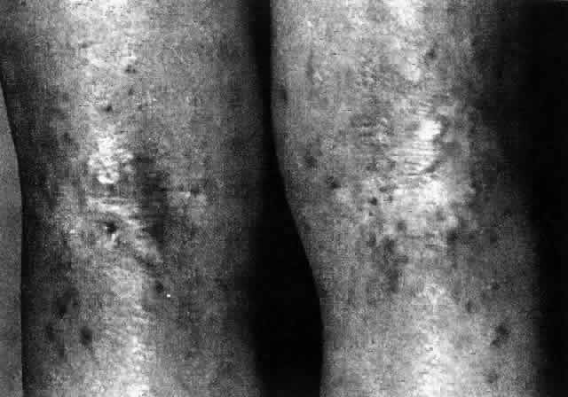
exfoliative erythroderma, erythema multiforme, and erythema nodosum. J Invest Dermatol 60:282, 1973

Back to Top