Chapter 27
Mediators of Ocular Inflammation
MATTHEW J. CHAPIN, PAULA J. WUN and MARK B. ABELSON
Main Menu   Table Of Contents

Search

PREFORMED MEDIATORS
OXIDATIVE PRODUCTS OF ARACHIDONIC ACID METABOLISM
COMPLEMENT
CYTOKINES AND LYMPHOKINES
ADHESION MOLECULES
SUMMARY
REFERENCES

Mediators are biologically active chemical compounds contained within inflammatory cells. On release from the cell, mediators act in a specific manner and at a specific site to induce a component of the inflammatory or immunologic process. Our understanding of the roles individual mediators play in the inflammatory and allergic disease has increased rapidly. This understanding allows us to identify rational and more effective means of therapy.

The mast cell is the principal effector cell for allergic reactions in the eye and other organ systems. The mast cell contains more than 30 preformed mediators, and mast cell degranulation stimulates the synthesis of many more. Mediators that are preformed and released by the mast cell, or are synthesized after mast cell activation, will be the primary focus of this review. However, other mediators with a role in ocular inflammation will also be discussed.

The mast cell surface has as many as 500,000 immunoglobulin-E (IgE) receptors, 10% of which are occupied in vivo.1 The Fc portion of the IgE molecule, the portion attached to the mast cell membrane, changes as a result of IgE cross-linking with the offending allergen, activating a serine esterase.2 This leads to an intracellular biochemical cascade causing mast cell degranulation and the subsequent release of preformed mediators, including histamine, eosinophil chemotactic factor of anaphylaxis, high-molecular-weight neutrophil chemotactic factor, and platelet-activating factor (Fig. 1). These mediators attract eosinophils and neutrophils, which restore homeostasis to the tissue. The signs and symptoms of an acute allergic reaction result from this intricate network of mediator interaction.

Fig. 1. Release of mast cell mediators in response to antigenic stimulation. (Abelson MB, Udell IJ: Ocular Allergy Update, p 8. Princeton, Excerpta Medica, 1981)

The inflammatory process is regulated internally by a negative feedback system. The interaction of histamine with mast cell surface histamine receptors elevates cyclic adenosine monophosphate (cAMP) concentrations, thereby “turning off” the mast cell.2 The second messengers, cAMP and cyclic guanosine monophosphate (cGMP), further control mediator release from mast cells and basophils.2 Increasing levels of cAMP block mediator release; increasing levels of cGMP stimulate mediator release. Beta-adrenergic receptor activation enhances cAMP levels; alpha-adrenergic receptor activation diminishes cAMP levels. Prostaglandins act by way of adenyl cyclase to increase cAMP levels. Phosphodiesterase degrades cAMP; thus, phosphodiesterase inhibitors can increase cAMP levels. Cholinergic stimulation results in increasing levels of cGMP and mediator release. Thus, allergic symptoms can be treated by increasing cAMP levels or decreasing cGMP levels. Pharmacologic modulation of these feedback mechanisms may provide a novel method for the treatment of allergic diseases.

Back to Top
PREFORMED MEDIATORS

HISTAMINE

The role of histamine as a mediator of acute inflammation has been clearly established—indeed, histamine represents the prototype of the inflammatory mediators. Histamine is an endogenous substance, widely distributed in mammalian tissue.3 It is stored in the secretory granules of tissue mast cells located primarily in connective tissue associated with blood vessels.4 Histamine is also found in platelets5 and in basophils.6,7 Histamine release can be triggered by both immunologic and nonimmunologic means. Re-exposure of sensitized individuals to an inciting antigen activates cell-bound IgE dimers, inducing mast cell and basophil degranulation and resulting in histamine release. Nonimmunologic mediator release can be induced by agents such as compound 48/80, dextran, and anaphylatoxins (e.g., C3a and C5a), or by trauma.8 Studies in a variety of immediate hypersensitivity models have shown that the release of immediate hypersensitivity mediators is both selective and noncytolytic.

Topical application of compound 48/80 produces the signs and symptoms of ocular allergy (itching, vasodilation, chemosis, and mucous discharge) in the eyes of guinea pigs, rabbits, and humans, and serves as a useful tool in screening anti-inflammatory agents.9 The degranulation of human conjunctival mast cells in response to topically applied compound 48/80, as demonstrated by light microscopy, has been confirmed by transmission electron microscopy.10 Mast cell granules enlarge in response to stimulation by compound 48/80; the number of enlarged granules corresponds to the extent of degranulation. Only cells with pink granules (by alkaline Giemsa stain) and granules enlarged to more than 1 μm showed definite evidence of exocytosis. Caulfield and colleagues, using an in vitro human anti-IgE model, have shown that granules enlarge, coalesce, become amorphous, and then discharge within 1 minute of stimulation.11 Only amorphous granules are capable of discharging their contents.11

Dale and Laidlaw12 used animal models to establish that injection of histamine results in collapsed arterial pressure, hypovolemia, and decreased body temperature. These symptoms are characteristic of traumatic or anaphylactic shock. Local application of histamine to skin results in redness, swelling, and flare from the local axon reflex,13 as well as itching from the stimulation of cutaneous nerve endings.14

To date, three different histamine receptor subtypes have been identified: H1, H2, and H3. Both H1 and H2 receptors have been identified in ocular tissue. When injected intradermally, histamine causes a localized triple response. First is the development of erythema immediately surrounding the injection site. Erythema results from vasodilation mediated by both H1 and H2 receptors.15,16 Second to develop is the cutaneous flare that occurs as an indirect response to stimulation of histamine receptors on afferent nonmyelinated nerve endings. Antidromic nerve conduction (the conduction of impulses in a direction opposite to the normal direction) initiates a reflex arc resulting in the release of various neuropeptides, including substance P and calcitonin gene-related peptide. These neuropeptides have a direct effect on arteriolar vasodilation.17 Finally, wheal results from the exudation of plasma through gaps between the vascular endothelium of postcapillary venules. This is mediated by H1 receptors.18 In addition to the triple response, intradermal injection of histamine causes the sensory response of itching.19

Topical application of histamine to the rabbit or human eye produces the signs and symptoms of an allergic reaction (itching, redness, and swelling) in a dose-dependent fashion.20 H1 receptor activation results in itching21 and redness,20 whereas H2 receptor activation leads to redness.22 The presence of a dual receptor system similar to that of skin has been demonstrated in the eye (Fig. 2). The highly selective H2 receptor agonist dimethylaminopropylisothiourea (dimaprit) produces diffuse vasodilation that can be blocked by cimetidine, an H2 antagonist, but not by antazoline, an H1 antagonist.22 Stimulation of the H1 receptor with the H1 agonist 2-(2-amino-ethyl)thiozole dihydrochloride produces itch and minimal vasodilation, which can be prevented by pheniramine maleate, an H1 antagonist, but not by cimetidine.21

Fig. 2. There are two histamine receptor sites important in allergic conjunctivitis: H1 (itch, burn, plus minimal vasodilation) and H2 (vasodilation). (Abelson MB, Udell IJ: Ocular Allergy Update, p 8. Princeton, Excerpta Medica, 1981)

Histamine levels have been described in normal human tears (5 to 10 ng/ml). In the past, these levels were not found to be consistently elevated in patients with allergic conjunctivitis, but only in the tears of patients with active vernal keratoconjunctivitis (VKC; 16 ng/ml).23,24 This elevation is probably the result of the extensive mast cell degranulation demonstrated in patients with VKC by light and electron microscopy.25 In addition, patients with VKC have four times as many mast cells in their conjunctiva as normal individuals,25 and the location of these mast cells is more superficial than in the normal conjunctiva.26 Such patients are, therefore, at greater risk for antigenic attack.

A 1990 study examined the presence of histaminase activity in human tears after in vivo conjunctival antigen challenge.27 Inactivation of histaminase resulted in a 15-fold elevation of histamine recovery. Enzyme-inactivated samples had histamine levels of 107.26 ng/ml, whereas untreated samples had histamine levels of 7.07 ng/ml. These results demonstrate the presence of histaminase activity in human tears and suggest that histaminase activity may have confounded the identification of histamine in ocular allergic disorders other than VKC. A 1995 study showed that the degradation of histamine was significantly lower in patients with VKC than controls in both tears and plasma.28

TRYPTASE

Tryptase is a preformed, tetrameric, serine endoprotease found in mast cells. It is stored in abundant quantities in its fully active form. Because tryptase is unique to mast cells, it is an excellent marker for them.29,30 Elevated levels of tryptase have been found in tears of patients after eye-rubbing. In addition, allergen challenge or provocation with compound 48/8031,32 also results in increased levels of tryptase in tears. Tryptase levels appear elevated during the early-phase reaction, but not during the late-phase reaction, after ocular allergen challenge.33 Tryptase is found elevated in patients with VKC, even during the remission phase.32,34

In addition to the eye, tryptase has been found in other biologic fluids such as serum,35 bronchial lavage fluid,36 nasal lavage secretions,37 skin,38 synovial fluid,39 and blisters.40 Tryptase has the ability to potentiate the effect of histamine, activate eosinophils and mast cells, and attract eosinophils and neutrophils. Because of these actions, tryptase inhibitors are being evaluated as potential therapies for asthma and have been shown to have effective anti-inflammatory properties. Mast cell stabilizers have been shown to reduce tryptase levels after allergen challenge.33 Tryptase also has the ability to degrade neuropeptides such as vasoactive intestinal peptide (VIP), peptide histidine-methionine, and calcitonin gene-related peptide. VIP is a prominent bronchodilator, and it is postulated that the destruction of VIP by tryptase results in the increased bronchomotor tone and bronchial hyperresponsiveness41 of asthma. The exact role of tryptase in ocular inflammation is not yet fully understood.

CHYMASE

Two mast cell subtypes have been described: T mast cells, which contain tryptase, and TC mast cells, which contain tryptase and chymase.29 Chymase is a serine endoprotease that is stored, preformed and fully active, in the TC mast cells. Unlike tryptase, chymase is inhibited by plasma proteinase inhibitors.42 The presence of chymase has not yet been demonstrated in the eye, although its presence is suggested by the large number of conjunctival mast cells of the MCtc phenotype.

HEPARIN

Heparin forms complexes with proteases and is released on degranulation of mast cells. Heparin has been shown to have anti-inflammatory properties43 and has been used for the treatment of allergic contact dermatitis,44 but there are only preliminary reports on the use of heparin in ocular allergy.45 Heparin may serve as an endogenous mediator that provides negative feedback on the release of proinflammatory mediators from the mast cell.

EOSINOPHIL CHEMOTACTIC FACTOR OF ANAPHYLAXIS

Eosinophil chemotactic factor of anaphylaxis (ECFA), a preformed mediator of immediate hypersensitivity, was first found in diffusates of guinea pig lung46 and human lung.47 It was subsequently extracted from rat mast cells,48 human leukemic basophils,7 human mast cell-rich lung,48 and nasal polyps.49 The activity of ECFA resides in two closely related tetrapeptides.50

ECFA is released during repeated mast cell challenge and degranulation in severe ocular allergic disease. ECFA is a potent chemotactic factor for eosinophils; therefore, the release of large amounts of ECFA is responsible for conjunctival eosinophilia.51 The presence of eosinophils in a conjunctival scraping is always indicative of ocular allergy. In fact, a conjunctival scraping containing two or more eosinophils per high-power field is considered diagnostic of ocular allergy.51 However, by definition, eosinophils do not have to be present in conjunctival scrapings to make the clinical diagnosis of ocular allergic disease, because they may be located only deep in conjunctival tissues.

ECFA is more selective than complementderived fragments,52 cocytotoxin,53 and peptides extracted from neoplasms.54 Interaction with ECFA reduces the chemotactic responsiveness of eosinophils,55 thereby retaining these cells at a specific site for regulatory purposes. Eosinophils release a number of substances that modulate the mast cell response and limit mediator activity: histaminase inactivates histamine,56–58 phospholipase inactivates platelet-activating factor,59 and aryl sulfatase inactivates leukotrienes.60–62

EOSINOPHIL GRANULE MAJOR BASIC PROTEIN

In addition to their beneficial role as modulators of inflammation, eosinophils are also responsible for tissue destruction, as noted in the cytotoxic effects of certain granular proteins on parasites and mammalian tissues.63 Eosinophil granule major basic protein (EMBP) accounts for more than 50% of the eosinophil granule protein and 25% of the total cellular protein.64,65

EMBP is a strongly cationic molecule with a molecular weight of 9,300 in humans and 11,000 in guinea pigs.66,67 A harmful role has been suggested for the eosinophil because of the markedly increased levels of EMBP is asthmatic sputum,68 and the ability of EMBP to mimic the pathologic changes seen in asthma.69 It has been demonstrated to elicit mast cell70 and basophil71 degranulation. Both VKC and contact lens-associated giant papillary conjunctivitis are associated with marked mast cell degranulation and eosinophil infiltration,72,73 providing further evidence that eosinophils play a role in tissue damage.

Increased tear levels of both EMBP and Charcot-Leyden crystal protein have been detected in patients with VKC. The increased EMBP levels appear to correspond to the severity of the disease.74 Also, immunofluorescence of conjunctival tissue from normal patients revealed little or no EMBP, whereas specimens from patients with VKC and contact lens-associated giant papillary conjunctivitis revealed significant EMBP deposition.75 However, in this study no correlation between the intensity of EMBP deposition and the severity of disease was documented. Thus, it appears that both VKC and contact lens-associated giant papillary conjunctivitis are characterized by eosinophil degranulation with the release of EMBP and other cytotoxic granule proteins that may further stimulate mast cell degranulation.75

The release of EMBP, a powerful epithelial toxic compound, may account for keratitis and shield ulcers in VKC. EMBP deposits were identified by immunofluorescence in the base and the mucus plug in two corneal shield ulcers removed by superficial keratotomy from two patients with VKC. EMBP may also contribute to sustained mast cell degranulation and thus the severe and protracted process associated with this condition.76

PLATELET-ACTIVATING FACTOR

Benveniste and colleagues named and characterized platelet-activating factor (PAF) as a phospholipase A2 (PLA2)-sensitive phospholipid,77–80 identified as 1-alkyl-2(R)-acetyl-glycero-3-phosphorylcholine.80,81 PAF is the most potent eosinophil chemotactic factor known and is approximately 100 times more effective than ECFA or leukotriene B4 (LTB4). It has been reported to mediate inflammation and vascular permeability.82 PAF has also been implicated in other pathophysiologic processes,83 such as aggregating platelets, chemotaxis and degradation of eosinophils and neutrophils, bronchoconstriction, increased bronchial responsiveness, and hypotension. Rabbit basophils have been shown to release PAF by an IgE-dependent process.84 Mast cells, eosinophils, monocytes, basophils, polymorphonuclear leukocytes, and macrophages have also been demonstrated to release PAF when stimulated with IgE, a calcium ionophore, or zymosan particles.84

Studies are available on the role of PAF as a mediator of ocular vascular permeability and ocular inflammation.84 Intravenous injection of PAF-acether in rabbits resulted in increased microvascular permeability of the retina characterized by intense ischemia and marked plasma leakage.85 The role of PAF as a chemical mediator in external ocular inflammation was examined in a dose-response study that involved topical application of PAF to rabbit and human eyes.86 Significant hyperemia and chemosis, very similar to the clinical picture of allergic conjunctivitis, were noted. Histologically, PAF was found to be chemotactic for conjunctival neutrophils and eosinophils, and it produced dramatic intravascular margination in the conjunctiva.

Back to Top
OXIDATIVE PRODUCTS OF ARACHIDONIC ACID METABOLISM
Calcium-requiring PLA2 and phospholipase C are rapidly activated during an allergic reaction, releasing cholesteryl esters. Cell membrane phospholipids are metabolized by the cholesteryl ester, leading to the production of arachidonic acid. Arachidonic acid is metabolized by two major pathways (the cyclooxygenase pathway and the lipoxygenase pathway), and the resulting metabolites are active inflammatory mediators. Topical application of arachidonic acid, at a concentration low enough to avoid cellular infiltration, iritis, or elevated intraocular pressure, produced marked chemosis, lid closure, and mild conjunctival vasodilation and mucous discharge in a rabbit model.87 Selective inhibition of the cyclooxygenase pathway with aspirin, indomethacin, and piroxicam significantly blocked both lid closure and chemosis, whereas pretreatment with the lipoxygenase blocker BW755C produced only minimal inhibition. Thus, cyclooxygenase seems to be the predominant pathway of arachidonic acid metabolism in the normal rabbit conjunctiva.

The addition of aspirin to the treatment regimen of patients with intractable VKC has produced dramatic improvement in conjunctival and episcleral redness, and resolution of keratitis and limbal infiltration, indicating that many of the signs and symptoms of VKC may be linked to products of the cyclooxygenase pathway.88

CYCLOOXYGENASE PRODUCTS

The cyclooxygenase pathway leads to the production of prostaglandins and thromboxanes (Fig. 3). Some of the effects attributed to these products are bronchospasm, coronary and pulmonary vasoconstriction, neutrophil chemoattraction, and augmentation of basophil histamine release (PGF2α, PGD2),89,90 pulmonary vasodilation and inhibition of platelet aggregation (PGI2),91 inhibition of macrophage spreading and surface adherence,92 and bronchodilation93 (PGE2). PGE1 and PGE2 produce vasodilation94 and erythema.95 Prostaglandins can also potentiate edema, enhance pain and fever caused by inflammatory stimuli, and sensitize nerve endings to other agents that induce pain.96

Fig. 3. Cyclooxygenase pathway of arachidonic acid metabolism leading to the formation of prostaglandins and thromboxanes. Inhibition sites of steroids and nonsteroidal anti-inflammatory agents (NSAI) are shown.

The prostaglandins PGE1, PGE2, PGF2α, and PGD2 have all been isolated from ocular tissue and aqueous humor,97 but PGD2 is the main prostaglandin produced by human mast cells in vitro98,99 and in vivo.100,101 After topical application to guinea pig or human eyes, PGD2 results in redness, conjunctival chemosis, tenacious mucous discharge, and eosinophilic infiltrate.102 Similar signs are seen in patients with ocular allergic disorders. PGE2 has been shown to increase blood flow independently and to synergize with histamine, bradykinin, and interleukin (IL)-1 to increase vascular permeability.103 Some evidence suggests that PGF may also be involved in allergic disease. Elevated tear levels of PGF have been detected in patients with VKC.104 Studies suggest that certain prostaglandins may also have anti-inflammatory actions. Thus, prostaglandins may also play a role in the negative feedback system that defines the allergic response as self-limiting.105 There are other products of the cyclooxygenase pathway: thromboxane (TXA2) and hydroxyheptadecatrienoic acid. TXA2 is a potent vasoconstrictor,106 platelet aggregator,107 and bronchoconstrictor.108 These actions suggest that TXA2 plays a role in bronchial asthma. The role of TXA2 in ocular allergy is unclear. Hydroxyheptadecatrienoic acid serves as a chemotactic factor for human granulocytes.109

LIPOXYGENASE PRODUCTS

Nonsteroidal anti-inflammatory agents such as aspirin and indomethacin block the cyclooxygenase pathway, but they do not inhibit the production of the lipoxygenase products of arachidonic acid. Steroids prevent the release of arachidonic acid from membrane phospholipids, perhaps by the formation of peptide inhibitors of phospholipase A2, thus blocking both the cyclooxygenase and lipoxygenase pathways. Therefore, steroids have the ability to block the production not only of prostaglandins and thromboxanes, but also of leukotrienes.110,111 Leukotrienes are products of arachidonic acid metabolism by the lipoxygenase pathway. The initial product 5-hydroperoxyeicosatetraenoic acid (5-HPETE, LTA4) can be converted to 5-hydroxy HETE, 5,12-d1-hydroxy HETE (LTB4), or the sulfidopeptide leukotrienes LTC4, LTD4, and LTE4. LTC4, LTD4, and LTE4 have been identified in tears after allergen challenge (Fig. 4).112 Like LTB4, the cysteine-containing leukotrienes LTC4, LTD4, and LTE4 exert proinflammatory effects. They are potent bronchoconstrictors and are capable of increasing the vascular permeability of postcapillary venules and stimulating mucus secretion in the lung, and thus are implicated in asthma.

Fig. 4. Lipoxygenase pathway of arachidonic acid metabolism leading to the formation of the slow-reacting substance of anaphylaxis leukotrienes LTC4, LTD4, and LTE4. Inhibition site of BW755C is shown.

LEUKOTRIENE LTB4

Probably the most studied leukotriene involved in ocular inflammation, LTB4 is produced predominantly by eosinophils and basophils but also by neutrophils, macrophages, and monocytes. LTB4 has been identified by gas chromatography—mass spectrometry113 as the lipoxygenase product responsible for the transient aggregation of human polymorphonuclear leukocytes induced by arachidonic acid in vitro.114,115 Thus, LTB4 acts as a potent neutrophil chemotactic agent and has platelet-aggregating activity in vitro. In addition, LTB4 exhibits both leukocyte chemokinesis and chemotaxis. In fact, LTB4 appears to be the most potent arachidonic acid metabolite derived by way of initial lipoxygenation, with respect to the in vitro effects on leukocyte aggregation and motility.116

Substantial in vivo data support the chemotactic activity of LTB4 in humans and other animals. These data include evidence of the accumulation of leukocytes at local sites of leukotriene administration (e.g., guinea pig peritoneum,117 rabbit aqueous humor,118 rabbit conjunctiva,119 and human skin120,121), direct observations on preparations (e.g., hamster cheek pouch122 and rabbit mesentery120), the production of neutropenia after intravenous injection in the rabbit,120 and increased plasma exudation in the presence of a vasodilator.123

LTB4 has been found in ocular tissue in rabbits and primates,124 as well as in tears after conjunctival allergen challenge. Topical application of LTB4 has been shown to cause both eosinophil and neutrophil chemotaxis in guinea pig and rat, although eosinophil chemotaxis predominated.125,126 When applied topically to guinea pig conjunctiva, LTB4 resulted in eosinophil chemotaxis.125 In humans, a subcutaneous injection of LTB4 causes a wheal and flare response, followed 2 to 3 hours later by a tender, indurated lesion consisting of a dermal infiltrate of mainly neutrophils. Conjunctival smears from patients with chronic VKC have shown that LTB4 and other leukotrienes are present,127 although their role in allergic conjunctivitis is probably limited. A study on the topical application of LTB4 on hamster conjunctiva did not demonstrate a change in the conjunctival vascular permeability.128 Application of LTB4 topically to human conjunctiva did not produce vasodilation, but biopsy revealed polymorphonuclear leukocyte infiltrates, and the subject reported migraine headaches lasting for more than 2 weeks (unpublished observation).

Both PGE2 and PGD2 act synergistically with LTB4 to enhance vascular permeability, resulting in edema formation, subsequent neutrophil infiltration, and also mast cell degranulation. The interactive effects of the products of arachidonic acid metabolism may have clinical significance in that the inhibition of the cyclooxygenase pathway by nonsteroidal anti-inflammatory agents could be beneficial or detrimental, depending on the altered profile of the arachidonic acid metabolites.

LTC4, LTD4, AND LTE4

The cysteine-containing leukotrienes, LTC4, LTD4, and LTE4, are potent bronchoconstrictors of peripheral airways in humans and other animals.129–133 Anaphylactic constriction of bronchi from atopic patients sensitive to birch pollen was inhibited by benoxaprofen and a prostacyclin derivative (U-60257), two lipoxygenase blockers. Mepyramine, an antihistamine, and indomethacin, a cyclooxygenase blocker, did not inhibit anaphylactic bronchoconstriction in these patients.134 LTD4 also causes smooth muscle contraction and increases vascular permeability. LTC4 and LTD4 are powerful depressants of myocardial contractile force, with marked negative inotropic effects evident when they are administered in the picogram and nanogram range.135,136 Both LTC4 and histamine increase tracheal insufflation pressure in anesthetized and artificially ventilated guinea pigs, but LTC4 is at least 100 times more potent than histamine and causes a longer-lasting increase.131,137 LTC4 and LTD4 induce mucus secretion in the submucosal glands of dog tracheas.138 These studies implicate these leukotrienes as major mediators of anaphylaxis in the lung. LTE4 has several systemic effects, including vasodilation and potentiating increased vascular permeability produced by histamine and bradykinin. LTD4 has been implicated in seasonal allergic rhinitis, and an LTD4 antagonist has been shown to reduce nasal symptoms in a seasonal allergic rhinitis study.139 However, another rhinitis study followed the rise and fall of inflammatory mediators during a nasal allergen challenge and found no link between clinical symptoms, drug efficacy, and the release of LTC4.140 These contradictory data demonstrate that the role of leukotrienes in ocular allergy, and the potential use of their antagonists as therapies, requires further study.

Lipoxygenase products have been identified in tears after allergen challenge,141 but topical application of LTC4 elicited no observable effect in rabbits or humans.142 This is in contrast to other mediators (e.g., LTB4 and PGD2), which are known to contribute to immediate hypersensitivity. These findings suggest that leukotrienes have little role in ocular allergies, although they may play a role in potentiating inflammatory mechanisms that have already started. Much research is needed to determine the role of LTC4, LTD4, and LTE4 in ocular immediate hypersensitivity.

HPETE AND HETE

In addition to leukotrienes, the lipoxygenase arm of arachidonic acid metabolism also produces HPETE and HETE.143 A study involving a hypoxia model of inflammation detected HETE in the cornea.144 Topical ocular application of HPETE and HETE to rabbit and human eyes had no visible effect on the conjunctiva (unpublished data). HPETE and HETE are known to be potent mucus-stimulating mediators in the lung,145 and it is possible that such a role may exist in the eye. The role of HPETE and HETE in ocular inflammatory conditions remains undefined; however, two HETE subtypes (HETE 1 and HETE 2) have been identified in tears of patients with VKC, pemphigoid, and rosacea.146

Back to Top
COMPLEMENT
Mast cells also participate in nonallergic inflammatory reactions, and type II hypersensitivity reactions provide an example of this. Type II hypersensitivity reactions require the participation of complement-fixing antibodies (IgG1, IgG3, or IgM) and complement. The antibodies are directed against antigens on the surface of specific cells. Thus, the damage caused by type II hypersensitivity reactions, and in turn by the complement system, is localized to a particular target cell or tissue. The mediators in type II hypersensitivity reactions include complement as well as macrophages and other leukocytes that liberate their enzymes.

The complement system consists of two major divisions: the classical pathway and the alternative pathway.147 Activation begins with the formation of antigen-antibody complexes and the subsequent generation of peptides that lead to a cascade of proteolytic events. IgG and IgM activate the classical pathway via C1.148 Thereafter, the sequential activation of C4, C2, C3, C5, C6, C7, C8, and C9 occurs. In the alternative pathway, IgA, IgE, IgG, endotoxin, zymosan, or other complex polysaccharides from microbial cell walls149 activate C3 and continue on as in the classical pathway. Cleavage of C3 causes the release of histamine from mast cells, neutrophil enzyme release, smooth muscle contraction, suppressor T-cell induction, macrophage IL-1, prostaglandin, and leukotriene secretion,150 and increased vascular permeability. C5 also produces mast cell activation and upregulation of LTB4, ECF, and other arachidonic acid metabolites. Of the other numerous products, C5, C6, and C7 are chemotactic factors, and C8 and C9 lead to cell lysis.151 Thus, the final step to both pathways is membrane damage leading to cell lysis. Complement-mediated cell lysis is also the result of type III hypersensitivity reactions, although ocular manifestations are known to be associated only with systemic type III hypersensitivity diseases. Although no ocular disease has been proven to be a type II hypersensitivity reaction, cicatricial pemphigoid is a candidate.152 Further, C1, C3, factor B, C4, C5, and C9 have been detected in tears153 and normal human corneas.154 Tear levels of C3 have been found to be higher in patients with allergic conjunctivitis. C3 has also been shown to be produced locally by conjunctival tissues in VKC and contact lens-associated giant papillary conjunctivitis.155 Thus, the complement system may play a role in ocular inflammation not related to systemic hypersensitivity reactions.

Back to Top
CYTOKINES AND LYMPHOKINES
Macrophages and T lymphocytes secrete lymphokines (e.g., IL-2) and cytokines (e.g., IL-1) and tumor necrosis factor. Lymphokines and cytokines are important in the inflammatory process by virtue of their powerful ability to induce specific actions on cells and to serve as chemoattractants. Interleukins represent another link in the redundancy of the inflammatory process. Interleukins are not technically mediators, because they are produced by lymphocytes. However, they are mentioned here to show how the mast cell is controlled by other cell lines. IL-3 is classified as a lymphokine and is produced by immunologically stimulated lymphocytes. Excessive production of this agent resulting from an imbalance between helper and suppressor T lymphocytes may be one of the basic defects leading to the allergic diathesis.

IL-3 was first defined as a lymphokine capable of inducing the T cell-associated enzyme 20 α-hydroxysteroid dehydrogenase (20αSDH) in cultures of splenic lymphocytes from nu/nu mice.156 Based on the association of 20αSDH primarily with T lymphocytes, it has been suggested that IL-3 may promote the differentiation of early T-cell precursors.157 Further, long-term cultures of splenic lymphocytes have shown that IL-3 supports the growth of a cell type resembling basophils or mast cells.156

Tremendous emphasis has been placed on identifying the role of interleukins in allergy and inflammation, but the research thus far has focused primarily on asthma. In brief, when the T-lymphocyte helper 2 cell (TH2) is activated by antigen, IL-3 and IL-5 are secreted, which in turn activate and recruit eosinophils; IL-4, also secreted by TH2 cells, activates B lymphocytes. Mast cells also release IL-5, which in turn activates eosinophils. IL-4 and IL-5 inhibitors are being studied as potential therapies for asthmatics. There are also interleukins that form a negative feedback loop on the proinflammatory interleukins. IL-12 and gamma interferon both are natural inhibitors of IL-4 and TH2 cell differentiation, and they modulate the inflammatory process. A recent study examined the role of IL-12 in a murine model of allergic conjunctivitis. The authors found that treatment with recombinant IL-12 increases cellular infiltration in the conjunctiva. However, treatment with high levels of IL-12 decreases cellular infiltration in the late phase reaction. In addition, after ocular challenge with allergen, both IL-12 knockout mice and mice pretreated with anti-IL-12 developed a moderate hypersensitivity reaction, but had minimal cellular infiltration. After allergen challenge in gamma interferon knockout mice, the immediate reaction and presence of eosinophils was enhanced.156a This series of experiments demonstrates the importance of IL-12 and interferon gamma in the regulation of ocular allergies and the possibility of their being targets for future therapy.

Back to Top
ADHESION MOLECULES
Adhesion molecules represent another class of proteins that play a role in the inflammatory process, although, like interleukins, they are not true mediators. Adhesion molecules are membrane-bound proteins that allow cells to interact with one another. Tumor necrosis factor, interleukins, antigen, histamine, leukotrienes, peroxide, and interferon may all serve as stimuli for adhesion molecules.158

There are several groups of adhesion molecules. The integrins include intracellular adhesion molecule-1 (ICAM-1). ICAM-1 is normally expressed on endothelium but can be induced on other tissues by cytokines. Although ICAM-1 is not found in normal eyes, expression was noted on conjunctival epithelium after antigen challenge.159 ICAM-1 has been used as a marker for immunotherapy efficacy in allergic rhinitis, and it may serve as a marker for inflammation.158 ICAM-1 may play a significant role in early- and late-phase cellular influx in allergic rhinitis. Many of the new antihistamines have been shown to reduce ICAM-1 in both eye and nasal allergy models.160,161

Selectins are another group of adhesion molecules. Elevated levels of selectins have been identified as contributing to cellular infiltrates in uveitis.162 In another study, patients with VKC were found to have significantly more ICAM-3 and vascular cell adhesion molecule-1 than controls, and endothelial leukocyte adhesion molecule-1 was noted on the vascular endothelial cells. Increased expression of adhesion molecules may play an important role in the pathogenesis of VKC.163

Chemokines are small proteins that combine with G protein-coupled receptors expressed on certain leukocytes. Chemokines are secreted by endothelial cells and leukocytes and function to convert selectins into integrins, which leads to extravasation of cells into the tissues. Eotaxin, monocyte chemotactic protein-3, and “regulated on activation, normal T-cell expressed and secreted” (RANTES) are key types of chemokines that are released by airway epithelium and skin keratinocytes. Elevated monocyte chemotactic protein levels have been found in the cornea in cases of herpetic keratitis.164

One study found monocyte chemotactic protein and RANTES in corneal keratocytes but not corneal epithelium,165 suggesting that the corneal stroma may be important in cell-mediated immunity. Another study found that human conjunctival epithelial cells were capable of producing RANTES in response to inflammatory stimuli, suggesting that RANTES may play a role in recruiting inflammatory cells such as eosinophils and T lymphocytes toward the ocular surface.166

Back to Top
SUMMARY
The components of inflammation are very complex and there appears to be much redundancy in the system, make it difficult to target inflammation in general. Taking advantage of endogenous negative feedback systems may be an opportunity for potential therapy. Further, studies of inflammation in systemic diseases, such as rhinitis and asthma, may provide direction for research in ocular inflammation. As the roles of more inflammatory mediators and their interactions are elucidated, more selective therapeutic approaches directed at the underlying disease process will undoubtedly emerge.
Back to Top
REFERENCES

1. Metzger H, Bach MK: The receptors for IgE in mast cells and basophils: Studies on IgE binding and on the structure of the receptor. In Bach MK (ed): Immediate Hypersensitivity: Modern Concepts Developments, p 561. New York, Dekker, 1978

2. Allansmith MR: Immunology of the eye. In: The Eye and Immunology, p 99. St. Louis, CV Mosby, 1982

3. Feldberg W: Distribution of histamine in the body. In Wolstenholme GEW, O'Connor CM (eds): Histamine (CIBA Foundation Symposium). Boston, Little, Brown & Co, 1956

4. Riley JR: The effects of histamine-liberators on the mast cells of the rat. J Pathol Bacteriol 65:471, 1953

5. Humphery JH, Jacques R: The release of histamine and 5-hydroxytryptamine (serotonin) from platelets by antigen-antibody reactions (in vitro). J Physiol 128:9, 1955

6. Graham HT, Wheelwright F, Parich HH et al: Distribution of histamine among the blood elements. Fed Proc 11:350A, 1952

7. Lewis RA, Goetzl EJ, Wasserman SI et al: The release of four mediators of immediate hypersensitivity from human leukemic basophils. J Immunol 114:87, 1975

8. Beaven MA: Histamine. Its role in physiological and pathological processes. Monogr Allergy 13:1, 1978

9. Udell IJ, Abelson MB: Animal and human ocular surface response to a topical nonimmune mast-cell degranulating agent (compound 48/80). Am J Ophthalmol 91:226, 1981

10. Udell JJ, Kenyon KR, Hanninen L et al: Conjunctival mast cell degranulation in compound 48/80 model (abstr). Invest Ophthalmol Vis Sci 20(suppl):9, 1981

11. Caulfield JP, Lewis RA, Hein A et al: Secretion in dissociated human pulmonary mast cells. J Cell Biol 85:299, 1980

12. Dale HH, Laidlaw PP: Histamine shock. J Physiol 52: 355, 1919

13. Lewis T: The blood vessels of the human skin and their responses. London, Shaw & Sons, 1927

14. Keele CA, Armstrong D: Substances Producing Pain and Itch. Baltimore, Williams & Wilkins, 1964

15. Robertson I, Greaves MW: Responses of human skin blood vessels to synthetic histamine analogues. Br J Clin Pharmacol 5:319, 1978

16. Harvery RP, Schocket AL: The effect of H1 and H2 blockade on cutaneous histamine response in man. J Allergy Clin Immunol 65:136, 1980

17. McCusker MT, Chung KF, Robert NM, Barnes PJ: Effects of topical capsaicin on the cutaneous responses to inflammatory mediators and to antigen in man. J Allergy Clin Immunol 83:1118, 1989

18. Smith JA, Mansfield LE, deShazo R, Nelson HS: An evaluation of the pharmacologic inhibition of the immediate and late cutaneous effects to allergen. J Allergy Clin Immunol 65:118, 1980

19. Abelson MB, Schaefer K: Conjunctivitis of allergic origin: Immunologic mechanisms and current approaches to therapy. Surv Ophth 38:115, 1993

20. Abelson MB, Allansmith MR: Histamine and the eye. In Silverstein AM, O'Connor GR (eds): Immunology and Immunopathology of the Eye, pp 362–364. New York, Masson, 1979

21. Weston JH, Udell IJ, Abelson MB: H1 receptors in the human ocular surface (abstr). Invest Ophthalmol Vis Sci 20(suppl):32, 1981

22. Abelson MB, Udell IJ: H2 receptors in the human ocular surface. Arch Ophthalmol 99:302, 1981

23. Abelson MB, Baird RS, Allansmith MR: Tear histamine levels in vernal conjunctivitis and other ocular inflammations. Ophthalmology 87:812, 1980

24. Abelson MB, Soter N, Simon M et al: Histamine in human tears. Am J Ophthalmol 85:417, 1977

25. Henriquez AS, Kenyon KR, Allansmith MR: Mast cell ultrastructure: Comparison in contact lens-associated giant papillary conjunctivitis and vernal conjunctivitis. Arch Ophthalmol 99:1266, 1981

26. Allansmith MR, Baird RS, Greiner JV: Vernal conjunctivitis and contact-lens associated giant papillary conjunctivitis and vernal conjunctivitis. Arch Ophthalmol 99:884, 1981

27. Berdy GJ, Levene RB, Bateman ST et al: Identification of histaminase activity in human tears after conjunctival antigen challenge. Invest Ophthalmol Vis Sci 31 (ARVO suppl):65, 1990

28. Abelson MB, Leonardi AA, Smith LM et al: Histaminase activity in patients with vernal keratoconjunctivitis. Ophthalmology 102(12):1958, 1995

29. Schwartz LB: Mediators of human mast cells and human mast cell subsets. Ann Allerg 58:226, 1987

30. Reiss J, Abelson MB, George MA, Wedner JH: Allergic Conjunctivitis. In Pepose J, Holland G, Wilhelmus K (eds): Ocular Infection and Immunity, p 347. Boston, Mosby, 1996

31. Butrus SI, Ochsner KI, Abelson MB, Schwartz LB: The level of tryptase in human tears: An indicator of activation of conjunctival mast cells. Ophthalmology 97:1678, 1990

32. Margrini L, Bonini S, Centofanti M et al: Tear tryptase levels and allergic conjunctivitis. Allergy 51:577, 1996

33. Bonini S, Schiavone M, Bonini S et al: Efficacy of lodoxamide eye drops on mast cells and eosinophils after allergen challenge in allergic conjunctivitis. Ophthalmology 104: 849, 1997

34. Fukagawa K, Saito H, Azuma N et al: Histamine and tryptase levels in allergic conjunctivitis and vernal keratoconjunctivitis. Cornea 13:345, 1994

35. Schwartz LB, Metcalfe DD, Miller JS et al: Tryptase levels as an indicator of mast-cell activation in systemic anaphylaxis and mastocytosis. N Engl J Med 316:1622, 1987

36. Wenzel S, Irani AA, Sander JM et al: Immunoassay of tryptase from human cells. J Immunol Methods 80:139, 1986

37. Castells M, Schwartz LB: Tryptase levels in nasal-lavage fluid as an indicatory of the immediate allergic response. J Allergy Clin Immunol 82:348, 1988

38. Schwartz LB, Atkins PC, Bradford TR et al: Release of tryptase together with histamine during the immediate cutaneous response to allergen. J Allergy Clin Immunol 141:821, 1988

39. Gruber BL, Schwartz LB, Rammamurthy NS et al: Activation of latent rheumatoid synovial collagenase by human mast cell 
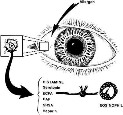
tryptase. J Immunol 140:3936, 1988

40. Deleuran B, Kristensen M, Larsen CG et al: Increased tryptase levels in suction-blister fluid from patients with urticaria. Br J Dermatol 125:14, 1991

41. Tam EK, Caughey GH: Degradation of airway neuropeptides by human lung tryptase. Am J Respir Cel Mol Biol 3:27, 1990

42. Schechter NM, Choi JK, Slavin DA et al: Identification of a chymotrypsin-like proteinase in human mast cells. J Immunol 137:962, 1986

43. Cahoalon L, Lider O, Schor H et al: Heparin disaccharides inhibit tumor necrosis factor-α production by macrophages and arrest immune inflammation in rodents. Int Immunol 9:1517, 1997

44. Ingber A, Trattner A, Cohen IR, Mekori YA: Low doses of low-molecular-weight heparin in vivo inhibits the elicitation of contact hypersensitivity. Acta Derm Venereol 7:454, 1994

45. Anderson W, Chan CC, Nussenblatt RB, Whitcup SM: Topical heparin inhibits compound 48/80 induced allergic conjunctivitis. Invest Ophthalmol Vis Sci 35:1291, 1994

46. Kay AB, Stechschult DJ, Austen KF: An eosinophil leukocyte chemotactic factor of anaphylaxis. J Exp Med 83: 602, 1971

47. Kay AB, Austen KF: The IgE-mediated release of an eosinophil leukocyte chemotactic factor from human lung. J Immunol 107:899, 1971

48. Wasserman SI, Goetzl EJ, Austen KF: Preformed eosinophil chemotactic factor of anaphylaxis (ECF-A). J Immunol 112:351, 1974

49. Kaliner M, Wasserman SI, Austen KF: Immunologic release of chemical mediators from human nasal polyps. N Engl J Med 289:277, 1973

50. Goetzl EJ, Austen KF: Purification and synthesis of eosinophilotactic tetrapeptides of human lung tissue: Identification as eosinophil chemotactic factor of anaphylaxis (ECF-A). Proc Natl Acad Sci USA 72:4123, 1975

51. Butrus SI, Abelson MB, Allansmith MR: Ocular allergic disorders. In Lockey RI, Bukantz SG (eds): Principles of Immunology and Allergy, p 166. Philadelphia, WB Saunders, 1987

52. Ward PA. Chemotaxis of human eosinophils. Am J Pathol 54:121, 1969

53. Wissler JH, Sorkin E, Stecher VJ: Regulation of serum derived chemotactic activity by the leukotactic binary peptide system. In Sorkin E (ed): Antibiotics and Chemotherapy, Vol. 19, p 442. Basel, Karger, 1974

54. Wasserman SI, Goetzl EJ, Ellman L et al: Tumor associated eosinophilotactic factor. N Engl J Med 290:420, 1974

55. Wasserman SI, Whitmer D, Goetzl EJ et al: Chemotactic deactivation of human eosinophils by the eosinophil chemotactic factor of anaphylaxis. Proc Soc Exp Biol Med 148: 301, 1975

56. Zeiger RS, Yurdin D, Colten HR: Histamine metabolism. II. Cellular and subcellular localization of the catabolic enzymes, histaminase and histamine methyl transferase in human leukocytes. J Allergy Clin Immunol 58:172, 1976

57. Lee D: Antihistamine activity of the eosinophil. J Pathol 99:96, 1969

58. Nilzen A: The antihistamine effect of human eosinophils. Allerg Asthmaforsch 16:24, 1970

59. Kater LA, Goetzl EJ, Austen KF: Isolation of human eosinophil phospholipase D. J Clin Invest 57:1173, 1976

60. Wasserman SI, Goetzl EJ, Austen KF: Inactivation of slow reacting substance of anaphylaxis by human eosinophil arylsulfatase. J Immunol 114:645, 1975

61. Wasserman SI, Goetzel EJ, Austen KF: Inactivation of human SRS-A by intact human eosinophils and by eosinophil arylsulfatase (abstr). J Allergy Clin Immunol 55:72, 1975

62. Orange RP, Murphy RC, Austen KF: Inactivation of slow reacting substance of anaphylaxis (SRS-A) by arylsulfatases. J Immunol 113:316, 1974

63. Butterworth AE, David JR: Current concepts: Eosinophil function. N Engl J Med 304:154, 1981

64. Gleich GJ, Loegering DA, Maldonado JE: Identification of a major basic protein in guinea pig eosinophil granules. J Exp Med 137:1459, 1973

65. Archer GT, Hirsch JG: Isolation of granules from eosinophil leukocytes and study of their enzyme content. J Exp Med 118:277, 1963

66. Gleich GJ, Loegering DA, Kueppers F et al: Physiochemical and biological properties of the major basic protein from guinea pig eosinophil granules. J Exp Med 140:313, 1974

67. Gleich GJ, Loegering DA, Mann KG et al: Comparative properties of the Charcto-Leyden crystal protein and the major basic protein from human eosinophils. J Clin Invest 57:633, 1976

68. Frigas E, Loegering DA, Solley SO et al: Elevated levels of the eosinophil granule major basic protein in the sputum of patients with bronchial asthma. Mayo Clin Proc 56: 345, 1981

69. Frigas E, Loegering DA, Gleich GJ: Cytotoxic effects of the guinea pig eosinophil major basic protein on tracheal epithelium. Lab Invest 42:35, 1980

70. Fahy G, Easty DL, Collum L et al: Double masked efficacy and safety evaluation of lodoxamide 0.1% ophthalmic solution versus opticrom 2%—a multicentre study. Ophthalmology Today p 341, 1988

71. Trocme SD, Kephart GM, Allansmith MR et al: Conjunctival deposition of eosinophil granule major basic protein in vernal conjunctivitis and contact lens-associated giant papillary conjunctivitis. Am J Ophthalmol 108:57, 1989

72. Allansmith MR, Baird RS, Greiner JV: Vernal conjunctivitis and contact lens-associated giant papillary conjunctivitis compared and contrasted. Am J Ophthalmol 87:544, 1979

73. Morgan G: The pathology of vernal conjunctivitis. Trans Ophthalmol Soc UK 91:467, 1971

74. Udell IJ, Gleich GJ, Allansmith MR et al: Eosinophil granule major basic protein and Charcot-Leyden crystal protein in human tears. Am J Ophthalmol 92:824, 1981

75. Trocme SD, Kephart GM, Allansmith MR et al: Conjunctival deposition of eosinophil granule major basic protein in vernal conjunctivitis and contact lens-associated giant papillary conjunctivitis. Am J Ophthalmol 108:57, 1989

76. Trocme SD, Kephart G, Bourne WM et al: Eosinophil major basic protein deposition in human corneal shield ulcers (abstr). Invest Ophthalmol Vis Sci 33 (suppl):94, 1992

77. Benveniste J, Chignard M, le Couedic JP et al: Biosynthesis of platelet-activating factor (PAF-acether). II. Involvement of phospholipase A2 in the formation of PAF-acether and lyso-PAF-acether from rabbit platelets. Thromb Res 25:375, 1982

78. Braquet P, Toqui L, Shen TY et al: Perspectives in platelet-activating factor research. Pharmacol Rev 39(2):97, 1987

79. Benveniste J, Le Couedic JP, Polonsky J et al: Structural analysis of purified platelet-activating factor by lipases. Nature (London) 269:170, 1977

80. Benveniste J, Tence M, Varonne P et al: Semi-synthesis and proposed structure of platelet-activating factor (PAF): PAF-acether an alkyl ether analog of lipophosphatidylcholine. CRC Acad Sci 289D:1037, 1979

81. Demopoulos CA, Pinckard RN, Hanahan DJ: Platelet-activating factor. Evidence for 1-alkyl-acetyl-sn-glyceryl-phosphorylcholine as the active component (a new class of lipid chemical mediators). J Biol Chem 254:9355, 1979

82. Tamura N, Agrawal D, Suliman FA et al: Effects of platelet-activating factor on the chemotaxis of normodense eosinophils from normal subjects. Biochem Biophys Res Commun 142:638, 1986

83. Rubin RM, Samples JR, Rosenbaum JT: Prostaglandin-independent inhibition of ocular vascular permeability by a platelet-activating factor antagonist. Arch Ophthalmol 106:1116, 1988

84. Braquet P, Toqui L, Shen TY et al: Perspectives in platelet-activating factor research. Pharmacol Rev 39(2):97, 1987

85. Braquet P, Vidal RF, Braquet M et al: Involvement of leukotrienes and PAF-acether in the increased microvascular permeability of the rabbit retina. Agents Actions 15: 82, 1984

86. George MA, Smith LM, Berdy GJ et al: Platelet activating factor induced inflammation following topical ocular challenge. Invest Ophthalmol Vis Sci 31 (ARVO Suppl):63, 1990

87. Klinman G, Butrus SI, Weston JH et al: Modulation of arachidonic acid metabolism in the rabbit conjunctiva (abstr). Invest Ophthalmol Vis Sci 24(suppl):200, 1983

88. Abelson MB, Butrus SI, Weston JH: Aspirin therapy in vernal conjunctivitis. Am J Ophthalmol 95:502, 1983

89. Wasserman M: Bronchopulmonary responses to prostaglandin F2 alpha, histamine, and acetylcholine in the dog. Eur J Pharmacol 32:146, 1975

90. Wasserman MA, DuCharme DW, Griffin RL et al: Bronchopulmonary and cardiovascular effects of prostaglandin D2 in the dog. Prostaglandins 13:255, 1977

91. Szczeklik A, Gryglewski RJ, Nizankowska E et al: Pulmonary and anti-platelet effects of intravenous and inhaled prostacyclin 
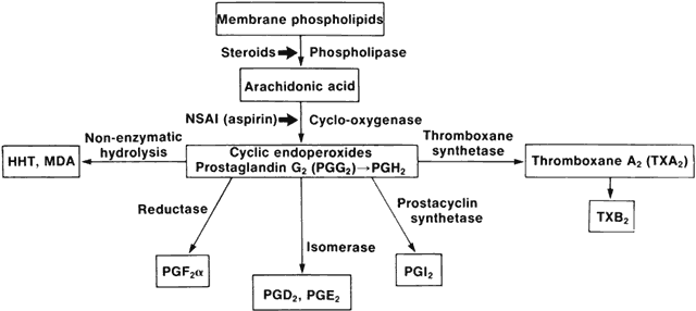
in man. Prostaglandins 16:651, 1978

92. Cantarow WD, Cheung HT, Sundharadas G: Effects of prostaglandins on the spreading, adhesion, and migration of mouse peritoneal macrophages. Prostaglandins 16:39, 1978

93. Mathe AA, Hedqvist P: Effect of prostaglandins F2 alpha and E2 on airway conductance in healthy subjects and asthmatic patients. Am Rev Respir Dis 111:313, 1975

94. Solomon LM, Juhlin L, Kirschenbaum MB: Prostaglandin on cutaneous vasculature. J Invest Dermatol 51:280, 1968

95. Crounkhorn P, Willis AL: Interaction between prostaglandins E and F given intradermally in the rat. Br J Pharmacol 41:507, 1971

96. Ferreira SH: Prostaglandins, aspirin-like drugs and analgesia. Nature New Biol 240:200, 1972

97. Leopold IH: Advances in ocular therapy: Noncorticosteroid anti-inflammatory agents. Am J Ophthalmol 78:759, 1974

98. Lewis RA, Holgate ST, Roberts LJ II et al: Preferential generation of prostaglandin D2 by rat and human mast cells. In Becker, Simon AS, Austen KF (eds): Biochemistry of the Acute Allergic Reactions (4th International Symposium), pp 239–254. New York, AR Liss, 1981

99. Lewis RA, Soter NA, Diamond PT et al: Prostaglandin D2 generation after activation of rat and human mast cells with anti-IgE. J Immunol 129:1627, 1982

100. Roberts LJ II, Sweetman BJ, Lewis RA et al: Increased production of prostaglandin D2 in patients with systemic mastocytosis. N Engl J Med 303:1400, 1980

101. Roberts LJ II, Sweetman BJ, Lewis RA et al: Markedly increased synthesis of prostaglandin D2 in systemic mastocytosis. Trans Am Assoc Physicians 93:141, 1980

102. Abelson MB, Madiwale NA, Weston JH: The role of prostaglandin D2 in allergic ocular disease. In O'Connor GR, Chandler JW (eds): Third International Symposium of the Immunology and Immunopathology of the Eye, pp 163–166. New York, Masson & Co, 1985

103. Davies P, MacIntyre DE: Prostaglandins and inflammation. In Gallin G, Goldstein I, Snyderman R (eds): Inflammation. Basic Principles and Clinical Correlates. New York, Raven Press, 1992

104. Dhir SP, Garg SK, Sharma YR et al: Prostaglandins in human tears. Am J Ophthalmol 87:403, 1979

105. Bhattacherjee P: The role of arachidonate metabolism in ocular inflammation. Prog Clin Biol Res 312:211, 1989

106. Ellis EA, Oilz O, Roberts LJ et al: Coronary arterial smooth muscle contraction by a substance released from platelets. Evidence that it is thromboxane A2. Science 193:1135, 1976

107. Hamberg M, Svensson J, Samuelsson B: Thromboxanes. A new group of biologically active compounds derived from prostaglandin endoperoxides. Proc Natl Acad Sci USA 72:2994, 1975

108. Reiss J, Abelson MB, George MA, Wedner JH: Allergic conjunctivitis. In Pepose J, Holland G, Wilhelmus K (eds): Ocular Infection and Immunity, p 347. Boston, Mosby, 1996

109. Goetzl EJ, Gorman RR: Chemotactic and chemokinetic stimulation of human eosinophil and neutrophil polymorphonuclear leukocytes by 12-L-hydroxy,8, 10-heptadecatrienoic acid (HHT). J Immunol 120:526, 1978

110. Blackwell GJ, Carnuccion R, DiRosa M et al: Macrocortin. A polypeptide causing the antiphospholipase effect of glucocorticoids. Nature 287:147, 1980

111. Hirata F, Schiffman E, Venkatasubramanian K et al: A phospholipase A2 inhibitory protein in rabbit neutrophils induced by glucocorticoids. Proc Natl Acad Sci USA 77:2533, 1980

112. Bisgard H, Ford-Hutchinson AW, Charleson S et al: Production of leukotrienes in human skin and conjunctival mucosa after 
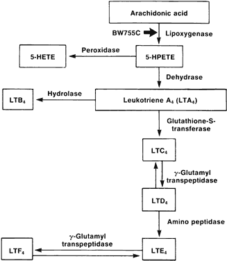
specific allergen challenge. Allergy 40:417, 1985

113. Ford-Hutchinson AW, Bray MA, Doig MV et al: Leukotriene B, a potent chemokinetic and aggregating substance released form polymorphonuclear leukocytes. Nature 286:264, 1980

114. O'Flaherty JT, Showell JH, Becker EL et al: Neutrophil aggregation and degranulation. Effect of arachidonic acid. Am J Pathol 95:433, 1979

115. O'Flaherty JT, Showell HJ, Becker EL et al: Role of arachidonic acid derivatives in neutrophil aggregation. A hypothesis. Prostaglandins 17:915, 1979

116. Smith MJH: Biological activities of leukotriene B4 (isomer III). Adv Prostaglandin Thromboxane Leukotriene Res 9:283, 1982

117. Smith MJH, Ford-Hutchinson AW, Bray MA: Leukotriene B: A potential mediator of inflammation. J Pharm Pharmacol 32:517, 1980

118. Bhattacherjee P, Eakins KE, Hammond B: Chemotactic activity of arachidonic acid lipoxygenase products in the rabbit eye. Br J Pharmacol 73:254P, 1981

119. Butrus SI, Corey EJ, Weston JH et al: The effect of leukotriene B4 in rabbit and guinea pig eyes (abstr). Invest Ophthalmol Vis Sci (Suppl):109, 1984

120. Bray MA, Ford-Hutchinson AW, Smith MJH: Leukotriene B4: An inflammatory mediator in vivo. Prostaglandins 22:213, 1981

121. Carr SC, Higgs GA, Salmon JA et al: The effects of arachidonate lipoxygenase products on leukocyte migration in rabbit skin. Br J Pharmacol 73:253P, 1981

122. Atherton A, Born GVR: Quantitative investigation of the adhesiveness of circulating polymorphonuclear leukocytes to blood vessel walls. J Physiol 222:447, 1972

123. Bray MA, Cunningham FM, Ford-Hutchinson AW et al: Leukotriene B4: A mediator of vascular permeability. Br J Pharmacol 72:483, 1981

124. Bisgard H, Ford-Hutchinson AW, Charleson S et al: Production of leukotrienes in human skin and conjunctival mucosa after specific allergen challenge. Allergy 40:417, 1985

125. Spada CS, Woodward DF, Hawley SB et al: Leukotrienes cause eosinophil emigration into conjunctival tissue. Prostaglandins 31:795, 1986

126. Trocme SD, Gilbert CM, Allansmith MR et al: Characteristics of the cellular response of the rat conjunctiva to topically applied leukotriene B4. Ophthalmic Res 21:297, 1989

127. Abelson MB: Lipoxygenase products in ocular inflammation (abstr). Invest Ophthalmol Vis Sci 25(suppl):42, 1984

128. Woodward DF, Ledgard SE: Comparison of leukotrienes as conjunctival microvascular permeability factors. Ophthalmic Res 17:318, 1985

129. Weiss JW, Drazen Jm, Coles N et al: Bronchoconstrictor effects of leukotriene C in humans. Science 216:196, 1982

130. Dahlen SE, Hedqvist P, Hammerstrom S et al: Leukotrienes are potent constrictors of human bronchi. Nature 288: 484, 1980

131. Hedqvist P, Dahlen SE, Gustafsson L et al: Biological profile of leukotrienes C4 and D4. Acta Physiol Scand 110: 331, 1980

132. Lewis RA, Austen KF, Drazen JM et al: Structure, function, and metabolism of leukotriene constituents of SRS-A. Adv Prostaglandin Thromboxane Leukotriene Res 9:137, 1982

133. Smedegard G, Hedqvist P, Dahlen SE et al: Leukotriene C4 affects pulmonary and cardiovascular dynamics in monkey. Nature 295:327, 1982

134. Hansson G, Bjorck T, Dahlen SE et al: Specific allergen induces contraction of bronchi and formation of leukotrienes C4, D4, and E4 in human asthmatic lung. Adv Prostaglandin Thromboxane Leukotriene Res 12:153, 1983

135. Burke JA, Levi R, Corey EJ: Cardiovascular effects of pure synthetic leukotrienes C and D. Fed Proc 40:1015, 1981

136. Levi R, Burke JA, Corey JA: SRS-A, leukotrienes, and immediate hypersensitivity reactions of the heart. Adv Prostaglandin Thromboxane Leukotriene Res 9:215, 1982

137. Drazen JM, Austen KF, Lewis RA et al: Comparative airway and vascular activities or leukotrienes C1 and D in vivo and in vitro. Proc Natl Acad Sci USA 77:4354, 1980

138. Johnson HG, Chinn RA, Chow AW et al: Leukotriene C4 enhances mucus production from submucosal glands in canine trachea in vivo. Int J Immunopharmacol 5:391, 1983

139. Donnelly AL, Glass M, Minkwitz MC, Casale TB: The leukotriene D4 receptor antagonist, ICI 204,219, relieves symptoms of acute seasonal allergic rhinitis. Am J Respir Crit Care Med 151(6):1734, 1995

140. Horak F, Toth J, Hirschwehr R et al: Effect of continuous allergen challenge on clinical symptoms and mediator release in dustmite-allergic patients. Allergy 53(1):68, 1998

141. Bisgard H, Ford-Hutchinson AW, Charleson S et al: Production of leukotrienes in human skin and conjunctival mucosa after specific allergen challenge. Allergy 40:417, 1985

142. Weston JH, Abelson MB: Leukotriene C4 in rabbit and human eyes (abstr). Invest Ophthalmol Vis Sci 26(suppl): 191, 1981

143. Udell IJ, Abelson MB: Chemical mediators of inflammation. Int Ophthalmol Clin 23(1):15, 1983

144. Vafeas C, Mieyal PA, Urbano F et al: Hypoxia stimulates the synthesis of cytochrome P450-derived inflammatory eicosanoids in rabbit corneal epithelium. J Pharmacol Exp Ther 287(3):903, 1998

145. Lundgren JD, Shelhamer JH, Kalimer MA: The role of eicosanoids in respiratory mucus hypersecreation. Ann Allergy 55:55, 1985

146. Abelson MB: Lipoxygenase products in ocular inflammation (abstr). Invest Ophthalmol Vis Sci 25(suppl):42, 1984

147. Reiss J, Abelson MB, George MA, Wedner JH: Allergic conjunctivitis. In Pepose J, Holland G, Wilhelmus K (eds): Ocular Infection and Immunity, p 347. Boston, Mosby, 1996

148. Cooper NR: Activation and regulation of the first complement component. Fed Proc 42:134, 1983

149. Pangburn MK, Morrison DC, Schreiber RD, Muller-Eberhard HJ: Activation of the alternative pathway. Recognition of surface structures on activators by bound C3b. J Immunol 124:977, 1980

150. Hugli TE: The structural basis for anaphyloxin and chemotactic function of C3a, C4a, C5a. CRA Crit Rev Immunol 2:231, 1981

151. Raisman MB: Cellular products. In Gallin JI, Goldstein IM, Snyderman R (eds): Inflammation: Basic Principles and Clinical Correlates. New York, Raven Press, 1992

152. Foster S: Hypersensitivity reactions. In Albert, Jakobiec: Principles and Practice of Ophthalmology. New York, WB Saunders, 1994

153. Willcox MD, Morris CA, Thakur A et al: Complement and complement regulatory proteins in human tears. Invest Ophthalmol Vis Sci 38(1):1, 1997

154. Mondino BJ, Ratajczak HV, Goldberg DB et al: Alternate and classical pathway components of complement in normal cornea. Arch Ophthalmol 98:346, 1980

155. Ballow M, Donshik PC, Mendelson L: Complement proteins C3 anaphylatoxin in the tears of patients with conjunctivitis. J Allergy Clin Immunol 76:473, 1985

156. Ihle JN, Pepersack L, Rebar L: Regulation of T cell differentiation. In vitro induction of 20 alpha-hydroxysteroid dehydrogenase in splenic lymphocytes is mediated by a unique lymphokine. J Immunol 126:2184, 1981

156a. Magone MT, Whitcup SM, Chan CC et al: IL-12 is essential for the induction of late phase cellular infiltration in a murine model of allergic conjunctivitis. FASEB 132 (Abstract): A339, 1999

157. Ihle JN, Rebar L, Keller J et al: Interleukin 3: Possible roles in the regulation of lymphocyte differentiation and growth. Immunol Rev 98:101, 1981

158. Passalacqua G, Senna G, Dama A et al: The relationship between clinical efficacy of specific immunotherapy and serum intercellular adhesion molecule-1 levels. J Invest Allerg Clin Immunol 8(2):123, 1998

159. Ciprandi G, Buscaglia S, Pesce GP et al: Allergic subjects express intercellular adhesion molecule-1 (ICAM-1 or CD54) on epithelial cells of conjunctiva after antigen challenge. J Allergy Clin Immunol 91:783, 1993

160. Canonica GW, Ciprandi G, Passalacqua G et al: Molecular events in allergic inflammation: Experimental models and possible modulation. Allergy 52(34 Suppl):25, 1997

161. Ciprandi G, Buscaglia S, Pesce G et al: Cetirizine reduces inflammatory cell recruitment and ICAM-1 (or CD54) expression on conjunctival epithelium in both early- and late-phase reactions after allergen-specific challenge. J Allergy Clin Immunol 95(2):612, 1995

162. Suzuma I, Mandai M, Suzuma K et al: Contribution of E-selectin to cellular infiltration during endotoxin-induced uveitis. Invest Ophthalmol Vis Sci 39(9):1620, 1998

163. Abu el-Asrar AM, Geboes K, al-Kharashi S et al: Adhesion molecules in vernal keratoconjunctivitis. Br J Ophthalmol 81(12):1099, 1997

164. Thomas J, Kanangat S, Rouse BT: Herpes simplex virus replication-induced expression of chemokines and proinflammatory cytokines in the eye: Implications in herpetic stromal keratitis. J Interferon Cytokine Res 18(9):681, 1998

165. Tran MT, Tellaetxe-Isusi M, Elner V et al: Proinflammatory cytokines induce RANTES and MCP-1 synthesis in human corneal keratocytes but not in corneal epithelial cells. Beta-chemokine synthesis in corneal cells. Invest Ophthalmol Vis Sci 37(6):987, 1996

166. Fukagawa K, Saito H, Tsubota K et al: RANTES production in a conjunctival epithelial cell line. Cornea 16(5): 564, 1997

Back to Top