Chapter 26
Immunology of Ocular Tissues
G. SMOLIN and MITCHELL H. FRIEDLAENDER
Main Menu   Table Of Contents

Search

EXTERNAL TISSUES
ANATOMY AND PHYSIOLOGY
IMMUNE RESPONSES
INTERNAL TISSUES
ANATOMY AND PHYSIOLOGY
IMMUNE RESPONSE
REFERENCES

EXTERNAL TISSUES
The tissues whose immune systems are to be discussed in this section are the eyelids, conjunctiva, lacrimal apparatus, cornea, and sclera.
Back to Top
ANATOMY AND PHYSIOLOGY

VASCULATURE

The lacrimal artery, the superior and inferior medial palpebral arteries, and the branches of the ophthalmic artery that supply the extraocular muscles and ciliary body form the rich blood supply of the conjunctiva, eyelids, and lacrimal gland (Fig. 1).1 This blood supply can be thought of as a natural immunologic defense. When the eyes are inflamed, the blood vessels dilate, fluid leaks into the extravascular spaces, and leukocytes migrate into these spaces from the bloodstream. More specifically, macrophages, polymorphonuclear leukocytes, lymphocytes, C-reactive protein, immunoglobulins, and so forth are the elements of the defense system that are brought to these ocular tissues during inflammation.2

Fig. 1. Schematic representation of the blood supply to the anterior segment of the eye.

Since many blood vessels are grossly visible in such tissues as the conjunctiva, immunologic inflammatory disease, which produces only subtle changes in buried organs such as the kidney, may cause conjunctival inflammation that is readily apparent early in its course.

There are neurogenic and nonneurogenic causes of vasomotor activity. The capillaries, precapillaries, and small arterioles are activated by chemical materials; the large vessels near the terminal arterioles are activated by the nervous system. The nonneurogenic causes are complicated but are basically (1) the vasoactive amines, including histamine and 5-hydroxytryptamine, (2) the serum proteases and polypeptides known as kinins, and (3) the prostaglandins. Most inflammatory reactions, in which the vessels dilate and their permeability increases, are biphasic: there is an immediate response (medicated by histamine-like substances) that reaches its maximum 8 to 10 minutes after application of the noxious stimulus, and there is a late response (mediated by kinins and prostaglandins) that occurs after an intervening normal period.

Since the normal cornea is devoid of blood vessels, the immunologic mechanisms are less effective and the tissue does not respond readily to antigenic insults (i.e., microorganisms, corneal grafts, and so forth). Necrosis and the subsequent accumulation of polymorphonuclear leukocytes vascularize the cornea and alter its privileged immune status.3

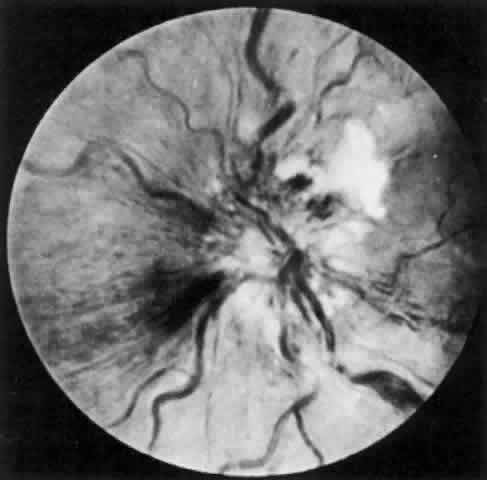
The behavior of corneal allografts is illustrative. When the results of one series were averaged, hosts with avascular corneal beds had rejected 3.5% of the grafts in 10 months, hosts with mildly vascularized corneal beds had rejected 13.3% in 4 months, and hosts with moderately vascularized corneal beds had rejected 65% in 2 months.4 It has also been shown that in experimental animals, 95% of lamellar corneal grafts and 75% of penetrating grafts placed in avascular beds survived for long periods of time. This was true even in animals sensitized by permitting them to reject large skin grafts from the same donors that provided the corneal buttons. These amply sensitized animals could not convey the sensitized lymphocytes to their avascular corneas.5 It is also significant that the graft rejection line usually starts at the site of maximum vascularization (Fig. 2).

Fig. 2. A rabbit eye with a mild corneal graft reaction characterized by neovascularization and opacification of the donor tissue. A significant part of the recipient cornea remains uninvolved in this reaction.

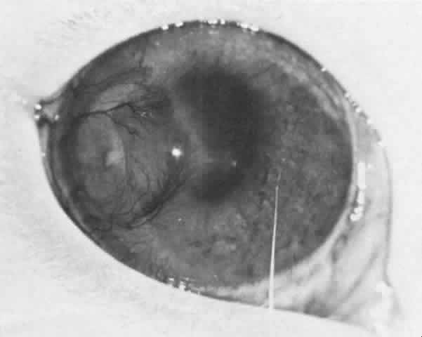
Trauma and non-graft-related inflammation (both of which dilate the vessels) and interrupted sutures left in place too long (causing neovascularization) can increase graft rejection by affecting the vasculature.

LYMPHATIC VESSELS AND REGIONAL LYMPH NODES

The lymphatics are arranged in pretarsal and posttarsal plexuses and are connected by cross channels. The posttarsal channels drain the conjunctiva and tarsal glands, and the pretarsal channels drain the skin and skin structures. Both groups drain from the lateral side into the preauricular and parotid lymph nodes, and from the medial side into the submandibular lymph nodes.

Although cell-lined lymphatic vessels do not occur in nonvascularized corneas, their presence in vascularized corneas seems to have been well established by the experimental work of Collin, Busacca, Mann, Aoki, and Smolin and their colleagues.6–10

Lymphatics and regional lymph nodes play a significant role in immunologic reactions (e.g., in transplantation). Certain tissues, such as hamster cheek-pouch, testes, brain, anterior chamber, lens, and avascular cornea, are devoid of conventional lymphatic drainage. Privileged in this respect, they support the continued growth of grafted tissue.11,12 In the conjunctiva and vascularized cornea, however, the lymphatics increase the host's ability to recognize nonself corneal antigen.

Langerhans' cells, which are more abundant peripherally in the cornea than centrally, may process the antigen, thus enhancing immune responsiveness. Depletion of these cells may prolong nonrecognition and graft survival. After the unilateral intracorneal injection of rabbit eyes with bovine gamma globulin, many antibody-forming cells are found in the homolateral draining lymph nodes, corneal limbus, and uveal tract. The first sites at which these antibody-forming cells can be detected are the draining nodes (Fig. 3).13

Fig. 3. A graph depicting the antibody-forming cells that appear in the draining node after an intracorneal injection of antigen.

TEAR FILM AND LACRIMAL APPARATUS

In addition to the blood vessels and lymphatic channels, there are other features of the external ocular tissue that affect the immunologic reaction. The intact epithelial surface, the low surface temperature, the low pH of the tear film, the presence of macrophages, and lid blinking are important as natural immunologic defense mechanisms. The tears themselves are also a natural immune defense. They not only wash away antigen and potentially pathogenic bacteria but contain bacteriostatic and bactericidal substances as well.

Sapse and co-workers identified the following seven proteins in human tears: specific tear prealbumin, serum albumin, ceruloplasmin, transferrin or lactoferrin, immunoglobulin A (IgA), immunoglobulin G (IgG), and lysozyme.14 The lysozyme comes from the type A cell in the lacrimal gland. (The type B cell supplies a neutral glycoprotein and sulfosialomucin.15) The tear prealbumin, IgA, lysozyme, and lactoferrin are the major tear proteins.16 The bacteriostatic substances in the tears are lactoferrin, lysozyme, and a nonlysozyme antibacterial agent (beta lysin?) that often appears to act in concert with other immunologic mechanisms.17,18 Lactoferrin has a concentration of about 2 mg/mL in tears and represents one of the major tear proteins.19 Lactoferrin, as well as other tear proteins (tear-specific prealbumin, lysozyme, secretory IgA), is synthesized and excreted by the lacrimal gland.20 Lactoferrin appears to act directly on certain strains of bacteria21 and it may interfere with the complement system and regulate granulocyte and macrophage colony-stimulating factors.22

The tears contain appreciable amounts of IgA, IgG, and complement, all of which are probably derived from the normal plasma cells lying beneath the conjunctival epithelium and in the stroma of the lacrimal gland.23,24 Although most of the plasma cells in and around the eye are located in the lacrimal gland, there are also significant numbers in the conjunctiva, fewer in the accessory gland, and insignificant numbers in the limbal tissue.25 Staining patterns indicate the presence of IgG, IgD, IgA, and IgE in the lacrimal gland. IgM is absent.

The conjunctiva may contain localized areas (similar to Peyer's patches) for processing antigen. The mucous membrane-associated tissue (MALT) may be part of an integrated system of MALT present in other tissue and allows for the “homing” of antibody to the ocular tissue, especially to the lacrimal gland. In the conjunctiva, these lymphoid collections are sometimes referred to as CALT.26

Although tear IgA undoubtedly comes from the plasma cells, the increase in tear IgG during acute inflammation, associated with increased vascular permeability, indicates that some of these immunoglobulins are derived from the circulating blood. Most of the tear IgA has an attached secretory piece (formed by the lacrimal gland epithelium) and is relatively resistant to proteolytic enzymes.27 In an environment rich in proteolytic enzymes, this allows it to act as antibody and to play a role in the regulation of the normal bacterial and viral flora of the mucous membranes.28

IMMUNOGLOBULIN DISTRIBUTION

The conjunctiva has a rich supply of immunoglobulins. Extracellular IgG, IgA, and IgM, all of which have been found in the conjunctiva's substantia propria, may come from the rich vascular supply, the abundant plasma cells, the tear film, the lacrimal gland, or more likely, from all four sources.29

McMaster and colleagues feel that most of the proliferation of lymphatic tissue in the conjunctiva is a response to infection by organisms in the environment.30

IgG and IgA are found in the cornea, at the same levels centrally and peripherally, and at one-half and one-fifth the serum levels, respectively; IgMis found less often and only rarely in the central cornea. Albumin is present in the cornea, with less of it centrally than peripherally.31 The immunoglobulins have been found almost exclusively in the stroma but sometimes also in the epithelium and endothelium.

Other serum proteins (alpha-globulin, beta-globulin) are found in the corneal stroma, and nonserum proteins are found in the corneal epithelium.32 In serologic studies some investigators have shown organ specificity for some corneal antigens and species specificity for others (Fig. 4).33

Fig. 4. A schematic representation of the immunoelectrophoresis pattern of bovine corneal antigen and bovine serum against antibovine serum. Using this technique an organ-specific corneal antigen was elucidated.

Back to Top
IMMUNE RESPONSES

ANAPHYLACTIC HYPERSENSITIVITY REACTIONS

Antigen reacts with the Fab fragment of a specific class of antibody (IgE or reaginic antibody) that is bound to the subepithelial mast cells through a specialized region of the Fc fragment. This leads to degranulation of the mast cells and the release of vasoactive amines (histamine, heparin, platelet activating factor, leukotrienes, kinins, and prostaglandins34 in the tissue (Fig. 5). This, in turn, causes dilatation of the small venules and increased permeability of the capillaries. The result is edema and hyperemia such as is seen in hay fever conjunctivitis, a condition that does not last long and can be treated successfully with vasoconstrictors, cyclic adenosine monophosphate enhancers, or by antihistamines, which compete for histamine (H) receptor sites.35 H1 and H2 receptor sites are probably present in the external ocular tissue.

Fig. 5. A schematic representation of the control of pharmacologic mediator release from the mast cell or basophil.

Vernal keratoconjunctivitis and atopic keratoconjunctivitis differ from hay fever conjunctivitis by virtue of their heavy round-cell infiltration. Although the reason for the accumulation of this cell type is unclear, it may be due to the participation of a cell-mediated immune response (cutaneous basophilic hypersensitivity reaction) in these conditions.

Bee stings of the cornea or adnexal tissue result in rapid edema and hyperemia of the affected tissue.36 In some cases the bee sting may be more complicated than a simple histamine reaction, probably because of its toxic components or the activation of complement and the attraction of leukocytes to the site.

CYTOTOXIC REACTIONS

The binding of antibodies to an antigen on the cell surfaces causes a phagocytosis of the cell by (1) opsonic or immune (C3b) adherence, (2) cytotoxicity by killer cells, or (3) lysis through the operation of complement (Fig. 6).

Fig. 6. The cytotoxic reaction can occur by the mechanisms noted in this figure. (Roitt I: Essential Immunology, p 131. Oxford, Blackwell Scientific Publications, 1975)

Certain drugs may elicit a cytotoxic reaction of the ocular adnexal tissue by acting as haptens attached to a cell membrane. The tissue necrosis that ensues causes marked inflammation and neovascularization. If the tissue necrosis is severe, there may be gross scarring of the conjunctival tissue. Cicatricial pemphigoid and Mooren's ulceration may be due to this type of reaction.

IMMUNE-COMPLEX-MEDIATED HYPERSENSITIVITY REACTIONS

The density, size, and angulation of the limbal vasculature may contribute to the deposition of antigen, antibody, or immune complexes into the corneal periphery. Catarrhal ulcerations and corneal peripheral lesions in Wegener's granulomatosis may be due to this deposition. If these immune complexes or the vasculitis associated with immunologic disease occlude the limbal vasculature, ulceration of the corneal periphery may supervene.

When a macroglobulin antibody directed against the patient's own tissue-fixed IgG unites with IgG, there is (1) a binding of complement at the site and (2) chemotaxis of leukocytes and platelets to the area. An occlusive vasculitis resulting from these events is thought to be the basis of rheumatoid nodules in the sclera and of the “melting” of corneal and scleral collagen.

Recurrent immunologic interstitial keratitis can be produced in the guinea pig by extraocular means, and circulating immune complexes can increase ocular vascular permeability in the rabbit.37,38 Scarring of the cornea and conjunctiva can occur in such immune-complex diseases as cicatricial pemphigoid and erythema multiforme.

CELL-MEDIATED IMMUNE REACTIONS

Although the first phlyctenule is always at the limbus, subsequent lesions may develop on the conjunctiva or cornea. Mononuclear cells predominate until necrosis occurs; neutrophilic leukocytes predominate thereafter. The ulcer heals within 12 to 14 days. There are no conjunctival sequelae, but if the phlyctenule is on the cornea, scarring can be anticipated. Other manifestations of this type of hypersensitivity reaction (e.g., allograft reaction, contact dermatitis) are discussed elsewhere.

Back to Top
INTERNAL TISSUES
The tissues whose immune systems are to be discussed in this section are the optic nerve, retina, uveal tract, lens, and vitreous and aqueous humors.
Back to Top
ANATOMY AND PHYSIOLOGY

VASCULATURE AND LYMPHATIC VESSELS

The uveal tract and retina have a rich blood supply. The lens is avascular, however, and is further isolated immunologically by being surrounded by aqueous and sealed within a capsule. The role of blood vessels in inflammatory and immunologic processes was discussed in the previous section. There are no true lymphatic vessels inside the eye. The lymph usually drains through the spaces that surround the veins, through the lamina cribrosa, into the lymphatic spaces of the optic nerve. In spite of the absence of lymphatic channels, however, antigen injected into the vitreous drains to the regional lymph nodes.39 Antigen also drains gradually directly into the bloodstream, especially in the highly vascularized uveal tract.

Antigen injected into the aqueous humor may be aberrantly processed by reaching the blood vessels before the lymphatics. This processing may lead to tolerance rather than sensitization. The anterior chamber has therefore been described as an immunologically privileged site.

ANTIGENS

As in the cornea, most of the lens antigens are organ-specific.40 Since they are neither species-specific nor individual-specific, it would be reasonable to conclude that lens protein would be autoantigenic if the antigens should escape from their immunologic isolation.

Some of the antibodies present in potent lens antisera cross-react with mitochondria, endoplasmic reticulum, contractile organelles, and cell nuclei. This may explain the reactivity of lens antisera with ocular (uveal tract) and extraocular structures.41

There are at least nine antigens in the human lens.42 Autologous lens proteins are only weakly antigenic and frequently fail to elicit an immune response even when liberated into the aqueous. Manski suggests that the majority of antigenic determinants on the crystallin molecules normally participate in protein-protein interactions and are therefore not available for the immune recognition system.43

Of the three types of lens crystallin, alpha crystallin is the most antigenic.44 It has been suggested that the beta and gamma crystallins interfere with the antigenicity of the alpha crystallin, blocking the antigenic sites by protein-protein interactions.

The lens capsule contains antigens comparable to those present in glomerular basement membranes, Descemet's membrane, and vessels of the retina and uveal tract.45 They are probably not concerned with lens-induced inflammation in humans. Lens epithelium is possibly strongly antigenic. In experimental rabbits, heterologous soluble lens protein evokes a strong antibody (IgG) response and a weak T-cell response.

The antigenicity of the retina can be attributed in part to its neural, glial, and vascular elements. The vascular antigens are probably not peculiar to the retina, since antibodies to retinal vessels cross-react with glomerular basement membranes, but a number of other antigens, located principally in the photoreceptor layer, are specific for the retina.46,47 Some of these antigens react immunologically and may be of importance in the pathogenesis of a number of retinal disorders.48 Specific antigens may also be present in the retinal pigment epithelium. T-cell sensitization to constituents of the retinal pigment epithelium may play a role in the pathogenesis of sympathetic ophthalmia.49

The uveal tract contains antigens that are specific for the uvea and are probably associated with pigment-containing cells. The uvea also contains plasma proteins and antigens shared with the crystallin proteins of the lens and retina.50 There are differences between the choroid and the anterior uvea, however, possibly as a result of their different embryologic origins. These differences may account for the frequent dissociation of choroiditis from the more common iridocyclitis.

The organ-specific uveal-tract antigens are also species-specific. Although heterologous uveal extracts stimulate antibody formation, the local uveal response is much greater when homologous extracts are used for immunization.51 The antigenicity of uveal proteins is low, however, and antibody responses in animals given homologous extracts rapidly disappear when the stimulus is withdrawn.

The optic nerve consists of myelinated nerve fibers and glial tissues intersected by fibrovascular septae. The antigenic determinants of myelin appear to be organ-specific, and autoimmune reactions to them are probably largely due to the cell-mediated immune response.52

IMMUNOGLOBULIN DISTRIBUTION

The iris is usually free of immunoglobulins, possibly because its peculiar double-walled vessels, with their tight junctions between endothelial cells, can prevent the intrusion of immunoglobulin molecules.

The stroma of the ciliary processes has immunoglobulins and albumin, but the pigmented and nonpigmented epithelial cells probably do not. The stroma of the choroid contains all five immunoglobulins and albumin. The lens has no immunoglobulin, and in general the retina has little or none, although in some cases small amounts have been detected in the rod and cone layer.

After a primary immunologic uveitis was induced in a rabbit eye with a single intravitreal injection of antigen, the antibodies in the uveal tract and retina were largely IgG (93% or more), with IgA and IgM contributing the rest. After a second injection, the antibody response was essentially the same.53

The antibody that forms in response to intravitreal immunization is specific; that is, the uveal tract cells and draining lymph nodes produce antibody only to the antigen(s) with which the eye has been injected.39

Back to Top
IMMUNE RESPONSE

ANAPHYLACTIC HYPERSENSITIVITY REACTIONS

Although uveitis due to drug sensitivity can be the result of medication applied externally to the eyes of sensitized patients, it is more frequently a result of inhalation or the injection of allergens.54

The iridocyclitis that has occurred rarely in patients with hay fever has been assumed to be causally related to the hay fever.55

CYTOTOXIC REACTIONS

The destruction by malignant melanoma of the choroid by autologous serum-containing tumor-specific antibodies is an example of a cytotoxic reaction. This type of reaction may also play a role in sympathetic ophthalmia.56

IMMUNE-COMPLEX-MEDIATED REACTIONS

The intravenous injection of large doses of horse serum into rabbits can result in circulating antigen-antibody complexes. These may be deposited in susceptible vascular beds where they activate complement and cause tissue damage. By means of this model, Wong and co-workers produced an immune-complex uveitis in association with lesions of the renal glomeruli.57 If there is damage to the uveal vessels (immunologic or nonimmunologic), the combination of newly introduced, intravenous antigen and preexisting antibody can be deposited in the uvea as immune complexes--a result of altered vascular permeability. The complexes bind complement and cause a uveitis (Auer reaction). This phenomenon could explain the recurrence of uveitis after a quiescent period.58

Lens-induced uveitis, Behçet's disease, and the occlusive vasculitis of the ganglion cell layer of the retina produced by lupus erythematosus may all be immune-complex-mediated reactions (Fig. 7). Both homologous and heterologous retinal extracts have been used to produce experimental endophthalmitis in animals, and immunization with these extracts (antigens) produces both antibody and T-cell responses.59 Eales disease may also be an immune-complex disease produced by antibody-dependent lymphocytes.60

Fig. 7. A patient with systemic lupus erythematosus showing an occlusive vasculitis in the retina.

CELL-MEDIATED IMMUNE REACTIONS

Both sympathetic ophthalmia and Vogt-Koyanagi-Harada disease (Fig. 8) are thought to be delayed hypersensitivities to melanin-containing structures.

Fig. 8. A young boy with Vogt-Koyanagi-Harada disease. The pigment alteration of the skin and the cataract OS secondary to the uveitis can be noted.END

Back to Top
REFERENCES

1. Wolff E: Anatomy of the Eye and Orbit. Philadelphia, WB Saunders, 1961

2. O'Connor GR: Basic mechanisms responsible for the initiation and perpetuation of anterior segment inflammation. Trans Am Acad Ophthalmol Otolaryngol 79:56, 1975

3. Collins HB: Limbal vascular response prior to corneal vascularization. Exp Eye Res 16:443, 1973

4. Ciba Foundation Symposium: Corneal Graft Failure. Amsterdam, Associated Scientific Publishers, 1973

5. Khodadoust AA, Silverstein AM: Studies on the nature of the privilege enjoyed by corneal allografts. Invest Ophthalmol 11:137, 1972

6. Collin HB: Lymphatic drainage of 131I albumin from the vascularized cornea. Invest Ophthalmol 9:146, 1970

7. Busacca A: Les vais seaux lymphatiques de la conjonctive bulbaire humaine étudiés par la méthode des injections vitales de bleutripan. Arch Ophthalmol (Paris) 8:10, 1948

8. Mann I, Pirie A, Pulinger BD: An experimental and clinical study of the reaction of the anterior segment of the eye to chemical injury. Br J Ophthalmol 13:1, 1948

9. Aoki K: An experimental study of the new growth of lymphatic vessels in the cornea. Kaibogaku Zasshi 24:142, 1951

10. Smolin G, Hyndiuk R: Lymphatic drainage from vascularized rabbit cornea. Am J Ophthalmol 72:147, 1971

11. Billingham RE, Silvers WB: Studies on the cheek pouch skin homografts in the Syrian hamster. In Ciba Foundation Symposium: Transplantation, p. 90. London, Churchill, 1962

12. Lance EM: A functional and morphologic study of intracranial thyroid allografts in the dog. Surg Gynecol Obstet 125:529, 1967

13. Smolin G, Hall JM: Afferent arc of the corneal immunologic reaction. II. Local and systemic response to bovine gamma-globulin. Arch Ophthalmol 90:231, 1973

14. Sapse AT, Stone W Jr, Sercarz EE: Proteins in human tears. Arch Ophthalmol 81:819, 1969

15. Allen M, Wright P, Reid L: The human lacrimal gland. Arch Ophthalmol 88:493, 1972

16. Broekhuyse RM: Tear lactoferrin: A bacteriostatic and complexing protein. Invest Ophthalmol 13:550, 1974

17. Friedland BR, Anderson DR, Forster RK: Non-lysozyme antibacterial factor in human tears. Am J Ophthalmol 74:52, 1972

18. Ford LC, Delange RJ, Petty RW: Identification of a non-lysozyme bactericidal factor in human tears and aqueous humor. Am J Ophthalmol 81:30, 1976

19. Kijlstra A, Jeurissen SHM, Koenig KM: Lactoferrin levels in normal human tears. Br J Ophthalmol 67:199, 1983

20. Janssen PT, van Bjisterveld PO: Origin and biosynthesis of human tear fluid proteins. Invest Ophthalmol Vis Sci 24:623, 1983

21. Kijlstra A, Jeurissen SHM: Modulation of classical C3 convertase of complement by tear lactoferrin. Immunology 47:263, 1982

22. Badgy GC: Interaction of lactoferrin monocytes and lymphocyte subsets in the regulation of steady-state granulopoiesis in vitro. J Clin Invest 68:56, 1981

23. McClellan B, Whitney CR, Newman LP, Allansmith MR: Immunoglobin in tears. Am J Ophthalmol 76:89, 1973

24. Chandler JW, Leder R, Kaufman HE, Caldwell JR: Quantitative determinations of complement components and immunoglobulins in tears and aqueous humor. Invest Ophthalmol 13:151, 1974

25. Allansmith MR, Kajiyama G, Abelson MB, Simon MA: Plasma cell content of main and accessory lacrimal glands and conjunctiva. Am J Ophthalmol 82:819, 1976

26. Franklin RM, Remus LE: Conjunctival-associated lymphoid tissue. Invest Ophthalmol Vis Sci 25:181, 1984

27. Franklin RM, Kenyon KR, Tomasi TB Jr: Immunohistologic studies of human lacrimal gland: Localization of immunoglobulins, secretory component and lactoferrin. J Immunol 11:984, 1973

28. Tomasi TB: The gamma A globulins. Hosp Pract 2:25, 1967

29. Allansmith MR, O'Connor GR: Immunoglobulins. Surv Ophthalmol 14:367, 1970

30. McMaster PRB, Aronson SB, Bedford M: Mechanism of the host responses in the eye. Arch Ophthalmol 77:392, 1967

31. Allansmith MR, McClellan B: Immunoglobulins in the human cornea. Am J Ophthalmol 80:123, 1975

32. Hall JM, Smolin G, Wilson FM II: Soluble antigens of the bovine cornea. Invest Ophthalmol 13:304, 1974

33. Nelken E, Nelken D: Serological studies in keratoplasty. Br J Ophthalmol 49:159, 1965

34. Proud D, Sweet J, Stein P et al: Inflammatory mediator release on conjunctival provocation of allergic subjects with allergen. J Allergy Clin Immunol 85:896, 1990

35. Majno G, Pallade GE, Schoefl GI: Studies on inflammation. J Biophys Biochem Cytol 11:607, 1965

36. Sobotka AK, Franklin RM, Adkinsen FN Jr et al: Allergy to insect stings. J Allergy Clin Immunol 57:29, 1976

37. Kopeloff LM: Recurrent immunogenic interstitial keratitis. Arch Pathol Lab Med 100:74, 1976

38. Howes EL, McKay DG: Circulating immune complexes. Arch Ophthalmol 93:365, 1975

39. Hall JM: Specificity of antibody formation after intravitreal immunization with bovine gamma globulin and ovalbumin. I. Primary response. Invest Ophthalmol 10:775, 1971

40. Halbert SP, Manski W: Organ specificity with special reference to the lens. Prog Allergy 7:107, 1963

41. Rahi AHS, Misra RN, Morgan G: Immunopathology of the lens. Br J Ophthalmol 61:164, 1977

42. Maisel H, Goodman M: Analyses of mammalian lens protein by electrophoresis. Arch Ophthalmol 71:671, 1964

43. Manski W: Immunological studies on normal and pathological lenses. In The Human Lens in Relation to Cataract Formation. CIBA Foundation Symposium. Amsterdam, Associated Scientific Publishers, 1973

44. Kida H: Experimental endophthalmitis phacoanaphylactica in rabbits sensitized with the purified bovine alpha crystallin. Fabia Ophthalmol Jpn 12:304, 1961

45. Nozaki M, Foster L, Sery TW: Uveal and other ocular tissue reactions to heterologous anti-lens-capsule antibodies. Invest Ophthalmol 2:641, 1963

46. Roberts D: Studies on the antigenic structure of the eye using the fluorescent antibody technique. Br J Ophthalmol 41:338, 1957

47. Barbanov BM, Mikhailov AT: Immunoelectrophoretic analysis of water-soluble antigens of the chick retina. Bull Exp Biol 8:73, 1970

48. Wacker WB, Lipton MM: The role of two retina antigens in production of experimental allergic uveitis and its suppression by mycobacteria. Int Arch Allergy 41:370, 1971

49. Marak G: Immunopathology of sympathetic ophthalmitis. In Proceedings of the First International Symposium on Immunology and Immunopathology of the Eye, Strasbourg, 1974. Basel, Karger, 1976

50. Maisel H, Harmison C: Immunoembryological study of chick iris. J Embryol Exp Morphol 11:483, 1963

51. Aronson SB: Experimental allergic uveitis. Arch Ophthalmol 80:235, 1968

52. Falk GA, Kies MW: Passive transfer of experimental allergic encephalomyelitis and delayed hypersensitivity to myelin basic protein. Fed Proc 27:544, 1968

53. Shimada K, Silverstein AM: Local antibody formation within the eye: A study of immunoglobulin class and antibody specificity. Invest Ophthalmol 14:573, 1975

54. Walker I: Atopic uveitis. S Afr Med J 30:125, 1956

55. Coles RS: In Theodore FH, Schlossman A (eds): Ocular Allergy. London, Balliere, Tindall & Cox, 1958

56. Rahi AHS: Autoimmune reactions in uveal melanoma. Br J Ophthalmol 55:793, 1971

57. Wong VG, Anderson R, O'Brien RJ: Sympathetic ophthalmia and lymphocyte transformation. Am J Ophthalmol 72:960, 1971

58. Gamble CN, Aronson SB, Brescia FB: Experimental uveitis. Arch Ophthalmol 84:321, 1970

59. Meyers RL: Experimental Allergic Uveitis. Basel, Karger, 1976

60. Chilman T: Specific and non-specific antibody activity in retinal vasculitis. Trans Ophthalmol Soc UK 93:193, 1973

Back to Top