Chapter 25
General Principles of Immunology
PETER BENINCASA, RICHARD LUKA and LEONARD BIELORY
Main Menu   Table Of Contents

Search

INNATE VERSUS ACQUIRED IMMUNITY
ANTIGEN
COMPONENTS OF THE IMMUNE SYSTEM
IMMUNOPATHOLOGY
REFERENCES

The immune response has historically been considered a protective response to infectious disease. A broader view would be to define immunity as a host system's response to molecules identified as foreign, without implying either positive or negative physiologic consequences. This concept would then encompass immune regulation, with “appropriate” responses resulting in beneficial outcomes for the host (i.e., control of infection, tumor surveillance) and “inappropriate” responses resulting in deleterious outcomes (i.e., autoimmune disease, immediate hypersensitivity).

There are four cardinal features of the immunity: (1) the specific recognition of molecules capable of evoking an immune response, (2) the capacity for memory, (3) the subsequent ability to amplify the response, and (4) the ability to discriminate self from nonself. Immune recognition will lead to either immunity or tolerance (i.e., specific unresponsiveness), which is crucial in preventing uncontrolled autoimmune disease.1–3

This chapter will introduce current concepts in basic immunology and provide a framework for understanding immune-mediated disease and the clinical applications of immunomodulation. For the sake of clarity, we will discuss many of the components of the immune system in isolation; however, we should remember that immune regulation is a balance of a myriad of cellular interactions, feedback loops, and molecular cascades.

Back to Top
INNATE VERSUS ACQUIRED IMMUNITY
The immune response has classically been divided into innate (nonspecific) and acquired (specific) immunity. Innate immunity is present before exposure to foreign molecules, such as microorganisms, and represents the first line of defense for the individual. It essentially has no capacity for memory and so its response is not amplified on re-exposure to the inciting agent. The chief components of innate immunity are the mononuclear phagocyte system, including cytokines, complement proteins, and physiochemical barriers.

Acquired immunity is defined by the specific recognition of foreign molecules by lymphocytes and their products. Before exposure to antigens, antigen-specific lymphocytes develop that are capable of recognition and response to foreign molecules. This represents the primary response. Activation of lymphocytes results in immunologic memory with the resultant capacity for dramatic amplification of specific and nonspecific effector function on reexposure to the offending molecules, more commonly known as the secondary response. The immune system is capable of recognizing more than 109 antigenic determinants. This remarkably large repertoire results from variability at the antigen-binding sites of immunoglobulins and T-cell receptors (TCRs).1

Acquired immunity can be further subdivided into humoral and cell-mediated immunity. Humoral immunity is primarily mediated by immunoglobulins, the products of B lymphocytes, either in secreted form or as membrane-bound cell surface receptors. T lymphocytes are primarily responsible for cell-mediated immunity. Recognize, however, that the interaction between B and T cells is crucial in both forms of immunity.

An acquired immune response typically follows exposure to a foreign antigen, conferring memory and the capacity for a secondary response. This has been termed active immunity. Passive immunity describes the transfer of pooled immunoglobulin from immunized individuals to immediately confer immunity without waiting the typical 1 to 4 weeks for the development of an active response, as seen with immunizations to vaccines such as hepatitis. Passive immunity rapidly wanes and does not produce memory.1

Back to Top
ANTIGEN
Antigens are molecules capable of specifically binding to lymphocyte receptors. More precisely, immunogens are those antigens whose binding evokes an immune response (although the term antigen still is commonly used in its place). Whereas immunoglobulins are capable of binding soluble antigens, TCRs bind only processed peptides presented in the context of specific cell surface proteins, collectively called the major histocompatibility complex (MHC) gene products. The specific site on an antigen that is bound by an antibody is termed a determinant or epitope. Haptens are simple molecules, commonly less than eight amino acids in size, that can bind to antibodies but cannot evoke an immune response unless bound to a macromolecule. In this instance, the hapten behaves like an exogenous determinant on the larger carrier protein.1,3

The typical immune response is downregulated by a number of mechanisms and so wanes with time. The success of the immune response in eliminating its antigenic stimulus, the natural end-differentiation of lymphocytes into memory cells or short-lived cells, and feedback inhibition in the form of cytokine regulatory cascades and perhaps the development of anti-idiotypic antibodies all play a role in this self-regulation. Tolerance, or learned immunologic unresponsiveness, is discussed later.

Back to Top
COMPONENTS OF THE IMMUNE SYSTEM
The cells and lymphoid organs that compose the immune system participate in a series of complex interactions to regulate the immune response (Fig. 1). In this section, we outline the essential components and their functions before a more explicit discussion of their role in immune-mediated disease later in this chapter.

Fig. 1. Grand scheme of adaptive immune system. (After Goodman JW. The immune response. In: Stites DP, Terr AI [eds]. Basic and Clinical Immunology, p 36. 7th ed. Norwalk, CT: Appleton and Lange, 1991.)

PHYSIOCHEMICAL BARRIERS

Physiochemical barriers are significant first-line elements of a host's defense. The skin and mucous membranes limit access to the host by invading microorganisms. Enzymes, such as lysozyme, nonspecifically inhibit microbial growth. In the eye, the conjunctiva and the tear fluid layer provide the primary barrier against environmental aeroallergens, chemicals, and infectious agents. The tear fluid layer contains both specific and nonspecific immunologically active proteins, including lysozyme, histamine, tryptase, lactoferrin, ceruloplasmin, vitronectin, immunoglobulin A (IgA), immunoglobulin G (IgG), immunoglobulin M (IgM), and immunoglobulin E (IgE).4

Lymphocytes

Lymphocytes are the cells responsible for the specificity of immune recognition and for coordination of the immune response. They are derived from a lymphoid progenitor cell in the bone marrow and are divided into three classes: T cells (lymphocytes), B cells (lymphocytes), and natural killer (NK) cells. T cells and B cells are morphologically indistinguishable, small 8- to 10-μm-in-diameter lymphocytes with large nuclei. They are functionally distinct and are easily differentiated by the cell surface proteins they express: T cells with CD3, CD4, and CD8 and B cells with CD-19 surface markers.1

NK cells are large lymphocytes with many cytoplasmic granules, distinct cell surface markers (CD16), and the ability to lyse cells directly, especially tumor cells or normal cells infected by virus. NK cells lack immunoglobulin or TCRs for antigen recognition yet do not kill their targets at random. Their means for target cell recognition is not well understood. NK cells do participate in antibody-dependent cellular cytotoxicity (ADCC) on the basis of their expression of a low-affinity receptor for the Fc portion of IgG (CD16). In ADCC, a target cell coated with IgG can be lysed directly by an NK cell after binding of the Fc portion of IgG with its low-affinity receptor CD16. This not only provides a means of recognition for the target by the NK cell but also serves to activate it to release its granule contents such as perforin, an enzyme that perforates the wall of the target cell, and secrete cytokines to augment the inflammatory response.1

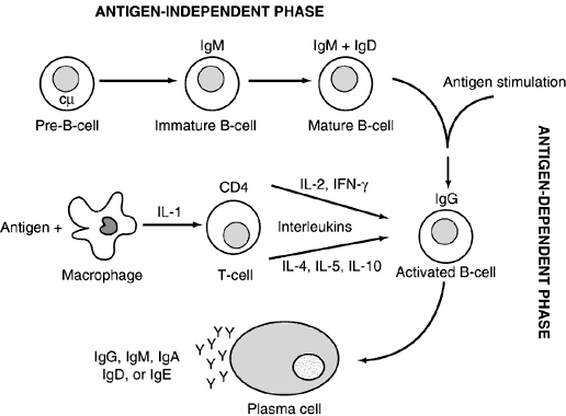
B cells develop from their precursors in the fetal liver and adult bone marrow. (They were first shown to mature in birds in the bursa of Fabricius, hence “B” cell.) They are primarily responsible for humoral immunity and are the exclusive producers of immunoglobulins, also known as antibodies, thus playing a vital role in the recognition and elimination of foreign antigen (further discussion on immunoglobulins below). Mature B cells can be divided into memory cells for the development of a rapid secondary response and plasma cells, which are totally committed to produce a single protein, an immunoglobulin. Plasma cells are terminally differentiated producers of large amounts of antibody (Fig. 2). B cells also interact closely with helper T cells through cell surface proteins such as CD40 and class II MHC and their complementary ligands.

Fig. 2. B-cell differentiation pathway. Pre-B cells characterized by intracytoplasmic immunoglobulin M (IgM) heavy chain (cμ) differentiate into immature B cells. These B cells are the first cells to reproduce light chains, which can then combine with the μ heavy chain to become expressed as IgM on the cell surface. The immature B cell differentiates into a mature B cell, which expresses both IgM and immunoglobulin D (IgD) on its cell surface. This part of the differentiation process does not require antigen and is referred to as antigen independent. On contact with the specific antigen via the surface immunoglobulin, the mature B cell becomes activated. Together with helper T cells (CD4+ ) and interleukins/cytokines derived from T cells and monocytes, the activated B cell matures into a plasma cell to produce immunoglobulins of a single isotype (Y). However, an antigen stimulates a polyclonal B-cell response, which results in many plasma cells and immunoglobulin production of several classes or isotypes. (After Medical Knowledge Self-Assessment Program [MKSAP]: Allergy and Immunology, Book 1, p 155. Philadelphia: American College of Physicians, 1993.)

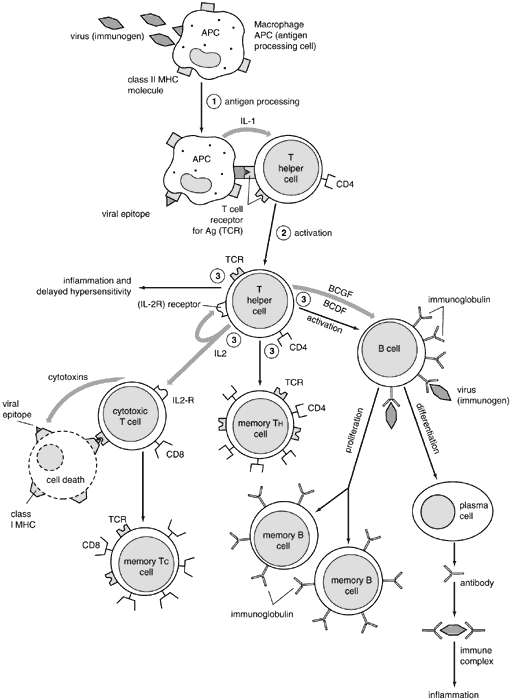
T cells develop from their bone marrow precursors in the thymus, where somatic gene rearrangement gives rise to functional TCR complexes as well as distinctive cell surface proteins. T cells can be functionally subdivided into either helper or cytolytic (CTL) cells. These functionally different populations express distinct cell surface proteins: CD4 on helper cells and CD8 on CTLs, which serve as ligands for the MHC gene products on antigen-presenting cells.5

CD4+ cells play a vital role in B-cell growth and differentiation, including the production of antibodies.6 They also are crucial in macrophage activation and in upregulating or downregulating the immune response. CD4+ cells can be further subdivided into TH1 and TH2 lymphocyte populations based on their cytokine-producing properties. Cytokines are small proteins produced by cells in response to a variety of inducing stimuli. Cytokines are produced by their producer cells and then influence the behavior on target cells. Hormones are classical polypeptides that also fulfill this definition but are not by convention classified as cytokines since they are produced by specific endocrine organs (e.g., the thyroid gland), whereas a cytokine may be produced by more than one cell type in a number of different tissues. Cytokines similar to hormones act on the cells that produce them (autocrine), on cells in a distant organ (endocrine), or on cells in the immediate vicinity of their production (paracrine) fashion to regulate lymphocytes, antigen-presenting cells, other inflammatory cells, and immunologically active molecules involved in cellular communication. Multiple studies have shown that cytokines also are characterized by their ability to act on different cells (pleiotropism), produce different effects on the same target cell, share properties with other cytokines (redundancy), and influence the production of other cytokines.1 It also has been discovered that among their many functions, cytokines can affect cell growth and differentiation similarly to those of the growth factors. Given the enormous diversity of cells producing cytokines and the numerous effects (many of which still are unknown) exerted by these molecules, it has been difficult to develop a satisfactory classification (Table 1). Cytokines also may be classified by cell of origin, but it is now clear that certain cytokines are produced by many cells, making these criteria impractical for classification.


TABLE 1. Cytokine Families

  Antiviral Agents

  Interferon <ga>, <gb>, <gg>


  Colony-stimulating factors

  CSF-1 or M-CSF
  GM-CSF
  G-CSF
  IL-3
  Stem cell factors
  Erythropoietin
  Thrombopoietin


  Lymphoid growth factors

  IL a,b
  IL-2-15
  Growth promoters
  Bone morphogeneic protein
  Ciliary neurotrophic factors
  Epidermal growth factors
  Fibroblast growth factors
  Hepatocyte growth factor
  Heregulin
  Glial growth factors
  Insulin-like growth factors I, II
  Nerve growth factors
  Platelet-derived growth factor
  Transforming growth factors <ga>, <gb>1---5
  Vascular endothelial cell growth factors


  Growth inhibitors

  Leukemia inhibiting factors
  Oncostatin M
  Mullerian inhibiting substance
  Transforming growth factor <ga>, <gb>1---5
  Tumor necrosis factor


  Chemotactic factors

  Complement (C3)
  Monocyte chemotactic protein-1
  Macrophage inflammatory protein 1 <ga>, <gb>
  MIF
  RANTES


CSF, colony-stimulating factor; GM-CSF, granulocytemacrophage colony-stimulating factor; IL-3, interleukin-3, MIF = macrophage migration inhibitory factor; RANTES = regulated on activation, normal T cell expressed and secreted.


TH1 lymphocytes express inflammatory cytokines that are involved in the effector functions of cell-mediated immunity. The primary cytokines of TH1 lymphocytes are interferon-γ (IFN-γ) and interleukin (IL)-2. TH2 lymphocytes produce cytokines such as IL-4 and IL-10, which often antagonize the inflammatory effects of IFN-γ and stimulate B-cell differentiation. IL-4 is required for the production of antibodies, IgA, and IgE, primarily associated with mucosal surfaces as this cytokine stimulates isotype switching to the α and ε heavy chains. TH1 lymphocytes appear to be more important in the defense against intracellular pathogens, whereas TH2 lymphocytes have an important role in the protection against parasitic disease. These two cell populations also are responsible for different pathologic states with TH1 responses involved in organspecific autoimmune disorders, including experimental autoimmune uveoretinitis, and TH2 cells involved in atopic disease.7

CD8+ lymphocytes are primarily responsible for cytolysis of virus-infected cells, malignant cells, and tissue allografts. This type of cell killing is highly specific as antigenic peptides are presented to the CD8+ cell in the context of class I MHC molecules and require direct contact of the CTL with the target cell. CD8+ cells also participate in cell-mediated immunity by activating macrophages. The role of CD8+ cells as “suppressor” cells is less clear, although a distinct subpopulation of non-CTL CD8+ cells may be distinguished.

Mononuclear Phagocytes

Cells of the mononuclear phagocyte system are derived from a common myeloid progenitor cell in the bone marrow. Their primary functions are phagocytosis and antigen presentation. In the peripheral blood, they are found as incompletely differentiated cells known as monocytes. Once in tissue, they mature and are known as macrophages.

Macrophages function as important effector cells of innate immunity. They are capable of phagocytizing foreign particles and microorganisms and killing them with lysosomal enzymes. They also produce and secrete cytokines that recruit other inflammatory cells and promote the inflammatory response. Among the cytokines typically expressed by macrophages are tumor necrosis factor (TNF)-α, IFN-γ, IL-1, and IL-6.

Macrophages also act as efficient antigen-presenting and costimulatory cells for T cells. T cells, in turn, may secrete cytokines, especially IFN-γ, which activate macrophages, upregulating their killing ability and antigen-presenting capacity.

Dendritic Cells

Dendritic cells are extremely efficient antigen-presenting cells with distinctive spinelike projections. Interdigitating dendritic cells are bone marrow derived and are found in the interstitium of most organs. In the skin and conjunctiva, they are known as Langerhans cells and bear the CD1 cell surface marker. Langerhans cells are extremely mobile, migrating from the skin to the peripheral lymph nodes, and are particularly effective at presenting antigen to CD4+ cells. Follicular dendritic cells are found in the germinal centers of lymph nodes, the spleen, and the mucosal lymphoid tissue. They are unrelated to interdigitating dendritic cells but also are extremely important antigen-presenting cells within lymphoid tissue.1

Neutrophils

Leukocytes containing abundant cytoplasmic granules play vital roles in the elimination of microorganisms and in the acute inflammatory response. Neutrophils comprise more than 90% of granulocytes and between 60% and 70% of the circulating leukocyte pool. They are extremely mobile, responding rapidly to chemotactic stimuli, adhering to specific ligands on the endothelial lining of blood vessels, and, via diapedesis, extravasating into tissue sites of injury. Chemoattractants for neutrophils include the byproducts of bacterial metabolism; C5a, which is a byproduct protein component of the complement cascade system; as well as leukotriene B4 and platelet- activating factor (PAF), both derived from cell membrane lipids.8

Various cellular processes including adhesion, migration, proliferation, differentiation, and activation are modulated by cell adhesion molecules (CAMs). Adhesion molecules are expressed on leukocytes, vascular endothelium, and epithelial cells and have been identified within various structures of the eye including the cornea, conjunctiva, choroid, uvea, and optic nerve.9 They are grouped in four major families: the integrins, the selectins, the cadherins, and the immunoglobulin supergene family.10 Endothelial CAMs important in regulating the infiltration of neutrophils into sites of inflammation include E-selectin and intercellular adhesion molecule-1 (ICAM-1). ICAM-1 binds to a complement receptor known as membrane attack complex type 1 (MAC-1) (i.e., complement receptor 3 or CR3) on the neutrophil, establishing firm adhesion and leading to extravasation.11 ICAM-1 also plays a role in ocular hypersensitivity reactions as reflected by its increased expression in atopic patients during allergen-specific conjunctival challenge.11,12

During phagocytosis, neutrophils engulf invading microorganisms, isolating them in phagosomes. Cytoplasmic granules containing a variety of cytotoxic enzymes fuse with the phagosome and destroy the pathogen. Most invading microorganisms are coated, or opsonized, with IgG or another complement protein byproduct known as iC3b. These can then bind to specific receptors on neutrophils (FcRII and MAC-1), facilitating phagocytosis.

The neutrophil has both oxygen-dependent and oxygen-independent antimicrobial systems. The oxygen-dependent system produces reactive oxygen metabolites (“respiratory burst”) that are toxic to microorganisms. The oxygen-independent system relies on the cationic peptides and enzymes released from cytoplasmic granules for killing.

Eosinophils

Eosinophils are important effector cells in atopic disease and parasitic infection. They contain a bilobed nucleus and distinctive acidophilic granules and are far more prevalent in tissue than in peripheral blood. Their cytoplasmic granules contain a number of basic proteins, including major basic protein (MBP), eosinophil cationic protein (ECP), eosinophil-derived neurotoxin (EDN), and eosinophil peroxidase (EPO), that are responsible for the eosinophils uptake of acid stains. MBP makes up more than 50% of total granule protein, is known to be extremely toxic to parasites, and is a vital component of eosinophil ADCC. MBP also is responsible for damage to epithelial cells, as seen in the respiratory epithelium in patients with asthma, and which is similar to the damage in the corneal epithelium in patients with vernal and atopic keratoconjunctivitis.

IL-5, produced by the TH2 subset of CD4+ cells and activated mast cells, is a potent stimulant for eosinophil production and activation. It also prolongs eosinophil survival and causes secretion of granule proteins. IL-4, another TH2 cytokine important in immediate hypersensitivity disease, causes endothelial cells to upregulate the expression of the adhesion molecule vascular cellular adhesion molecule (VCAM-1), which binds to its ligand VLA-4 on eosinophils. This results in eosinophil recruitment to sites of immediate and chronic immunologic hypersensitivity inflammatory reactions.

Mast Cells and Basophils

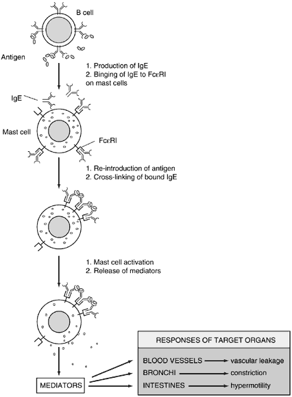
Mast cells and basophils play important roles in mediating immediate hypersensitivity. They each express large numbers of high-affinity IgE receptors (Fcε RI) on their cell surfaces. Mast cells are localized to mucosa, epithelial surfaces, and connective tissue. Basophils may be considered the peripheral blood counterpart to the mast cell.

Mast cells can be divided into two types based on their expression of secretory granules: tryptase mast cells (MCT) and tryptase-chymase mast cells (MCTC). MCT are found primarily in the lung and intestinal mucosa, whereas MCTC are localized to the skin and intestinal submucosa. The predominant form of mast cell found in the normal conjunctiva are MCTC; however, there is a noticeable increase in the MCT type in chronic conjunctival inflammatory conditions. It is estimated that more than 50 million mast cells are present in the conjunctiva.

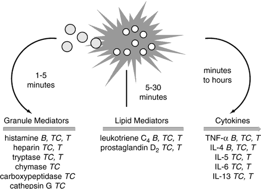
Mast cells and basophils are activated by the cross-linking of FcεRI molecules on their surface after the binding of multivalent antigen to sufficient IgE. Activated mast cells release their preformed mediators in a regulated fashion and then synthesize lipid-derived mediators of inflammation.

The prototype preformed vasoactive mediator released by mast cells is histamine. Histamine binding to its cell receptors results in vasodilation, endothelial cell contraction, and associated plasma leakage, as well as bronchial and intestinal smooth muscle constriction. Other preformed mediators include tryptase, chymase, carboxypeptidase-A, proteoglycans, eosinophil chemotactic factor, and neutrophil chemotactic factor. Lipid-derived mediators synthesized after activation include leukotrienes B4 (one of the most potent neutrophil chemoattractants known to the human body), C4, D4, and E4 (also known as SRS-A, a slow-releasing substance of anaphylaxis), prostaglandin D2, and PAF, which all have vasodilatory and smooth muscle constrictive effects (Fig. 3).4

Fig. 3. Time course of mediator release from human basophils (B), MCTC mast cells (TC), and MCT mast cells (T). Granule mediators are preformed; lipid mediators are newly generated; cytokines can be both preformed and newly generated. (After Medical Knowledge Self-Assessment Program [MKSAP]: Allergy and Immunology, p 23. 2nd ed. Philadelphia: American College of Physicians, 1997.)

Lymphoid Tissues

Organized lymphoid tissue allows for the concentration of lymphocytes and antigen-presenting cells at anatomically defined sites. The eye is immunologically unique in that it has no formed lymph nodes in the orbit or associated with the lacrimal gland, eyelids, or conjunctiva. Lymphocytes normally reside in the substantia propria of the acini of the lacrimal gland and the conjunctiva.

Lymph nodes situated along lymphatic pathways throughout the body provide a mechanism for the host to survey for foreign molecules. Each lymph node is divided into an outer cortex, which contains lymphoid follicles and is densely packed with cells, and the more sparsely populated medulla, which contains the vascular and lymphatic sinusoids. Follicles are B-cell zones and may develop germinal centers, where activated B cells proliferate and interact with follicular dendritic cells.

T cells are localized between the follicles and the deep cortex in the parafollicular areas. These cells are primarily CD4+ and interact with the interdigitating dendritic cells. The anatomic structure of the lymph node provides areas of interaction for B cells, T cells, and antigen-presenting cells. This structure expands or regresses in response to variable antigen exposure.

The spleen has an anatomic structure similar to that of the lymph node, is the primary site of immune response to blood-borne antigen, and appears to be the primary lymphoid site for intraocular antigenic stimulation. The white pulp of the spleen contains densely packed lymphoid tissue. T cells are localized to the periarteriolar lymphatic sheaths and B cells to the follicles and germinal centers.1

Other lymphoid tissue is found as aggregates of lymphocytes and accessory cells in the submucosa of the gastrointestinal and respiratory tracts. The skin contains lymphocytes and Langerhans cells in the dermis and epidermis.

Immunoglobulins

Immunoglobulins, or antibodies, are the glycoprotein products of antigen-stimulated B cells. The only function of specialized, terminally differentiated B cells, called plasma cells, is the production and secretion of large amounts of immunoglobulin, which is found in both membrane-bound (serving as B-cell surface receptors) and soluble forms. They are widely distributed in plasma and secretory fluids such as tears, milk, and mucous.

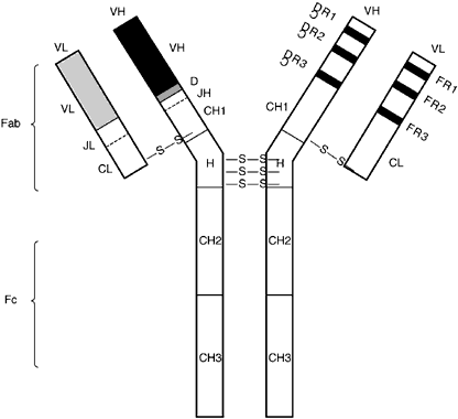
All immunoglobulins have the same basic structure with two identical heavy chains combining with two identical light chains. The two heavy chains are bound together by disulfide bonds, and each light chain is similarly attached to each heavy chain. Despite this similarity in basic structure, immunoglobulins may be divided into different classes based on certain physiochemical characteristics. The common physiochemical and antigenic properties of each class are based on shared regions of heavy-chain amino acid sequences. There are five basic classes, or isotypes: IgM, IgD, IgG, IgA, and IgE. There also are further subclasses: IgG1, IgG2, IgG3, IgG4, and IgA1 and IgA2. Different isotypes and subtypes mediate distinct effector functions. The two light-chain types, δ and μ, do not confer any effector function.

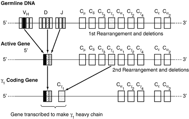
The diversity of the immunoglobulin repertoire results from the remarkable variability of the antigen-binding sites. These binding sites are composed of three hypervariable regions of both the light and heavy chains (Fig. 4). The variability in these regions is a result of somatic genomic recombination, multiple germline genes, junctional diversity, and somatic mutations (Fig. 5). The first immunoglobulin molecules expressed on mature B cells are IgM and IgD. IgD is almost exclusively membrane bound, contributing to less than 1% of total plasma immunoglobulin. Its function remains unclear.

Fig. 4. The immunoglobulin G molecule. The molecule is composed of two heavy-chain and two light-chain polypeptides joined by disulfide bonds (-S-S-) at the hinge (H) region. Both the heavy (H) and light (L) chains are composed of variable (V) regions at the N-terminus [the F(ab) fragment] and constant (C region domains at the C-terminus (one C region domain for the L chain, three C region domains for the H chain). The VH, CH, and CL region domains are depicted on one side of the F(ab) fragment, and the complementary determining regions (CDR) and framework (FR) areas on the other side of the F(ab) fragment. The FR regions are relatively conserved amino acid sequences of the variable regions that are interspersed among three highly variable regions called CDR. These hypervariable regions of the H and L chains interact to form a 3D structure, which constitutes the antigen-binding region. (After Medical Knowledge Self-Assessment Program [MKSAP]: Allergy and Immunology, Book 1, p 153. Philadelphia: American College of Physicians, 1993.)

Fig. 5. Immunoglobulin gene rearrangements in the production of γ1 heavy chain. In the germline DNA, there are approximately 200 variable region genes (VH), nine diversity genes (D), and six joining region genes (J). There also are nine constant region genes (C). The first rearrangement of the heavy-chain genes results in an active-gene complex that contains only one V, one D, and one J, the other VDJs having been deleted. When class-switching occurs to γ1, the constant region genes between the VDJ and C γ1 are deleted (i.e., Cμ, Cδ, and Cγ3). The γ1 heavy chain is now ready to be transcribed to synthesize mRNA for this particular γ1 heavy chain. (After Medical Knowledge Self-Assessment Program [MKSAP]: Allergy and Immunology, Book 1, p 157. Philadelphia: American College of Physicians, 1993.)

IgM plays a significant role in the primary immune response. Antigen contact with membrane-bound IgM initiates cell division, the production of secreted immunoglobulin, and the formation of memory B cells. In its secreted form, IgM usually is found as a pentamer, allowing for multiple contacts with antigen. It is predominantly confined to the intravascular pool and accounts for approximately 10% of immunoglobulins.

IgG is the most abundant immunoglobulin in normal human serum, accounting for approximately 75% of the total immunoglobulin pool. It is the primary mediator of immunoglobulin effector functions. IgG subclasses (except for IgG4) activate the classical pathway of the complement system. IgG helps mediate phagocytosis, ADCC, and cytokine secretion through different Fc receptors on a variety of cell types. IgG also is the only immunoglobulin to cross the placenta, thus conferring passive maternal immunity to the neonate. Thus, IgG ocular disorders are the only ones that the mother can transplacentally “passively” transfer to the infant at birth, such as myasthenia gravis.

Although thought to be a relatively unimportant component of systemic humoral immunity, IgA plays a major role in mucosal immunity. Dimeric IgA binds to specific Fc α receptors, “secretory components,” on epithelial cells of organs such as the intestine, as well as on the conjunctival surface. The secretory component shuttles the dimeric IgA through the cell until it is cleaved at the luminal side. Hence, dimeric IgA enters the mucosal lumen where it can neutralize pathogens. IgA is the predominant immunoglobulin in tear fluid, milk, saliva, and tracheobronchial secretions.1

IgE usually is found in small amounts in the serum of normal individuals but may be increased greatly in patients with atopic disease. It is responsible for immediate hypersensitivity reactions and for immunity to parasites. CD4+ TH2 cells produce IL-4, which promotes the production of IgE. IgE with specific epitopes to allergens has been isolated in the tear fluid of patients with atopic disorders (e.g., ragweed-specific IgE) in ragweed-sensitive patients.4

T-Cell Receptor Complex

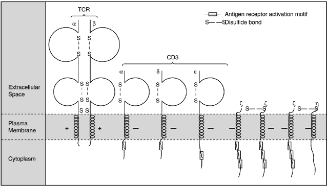
Antigen recognition by T cells is accomplished by specific TCR binding of antigenic peptide presented in the context of self-MHC molecules on antigen-presenting cells. TCR molecules do not recognize soluble antigens, native protein, or nonproteins as antigens. The TCR is a heterodimer of two polypeptide chains, α and β or γ and δ, linked by disulfide bonds. More than 90% of T cells in human peripheral blood, lymphoid tissue, and normal conjunctiva are TCR-α/β-positive.13 Both chains have variable and constant regions. The variable region shares many amino acid residues and a common tertiary structure with immunoglobulins. TCR molecules share enough sequence homology with immunoglobulin molecules to be considered members of the immunoglobulin supergene family.

As with immunoglobulins, the diversity of the TCR repertoire results from variability at the antigen-binding sites. This variability is a result of mechanisms that fundamentally resemble those for the immunoglobulin repertoire: multiple germline genes, somatic genomic recombinations, and junctional diversity. However, there is no evidence that somatic mutations result in a change of function or affinity of the TCR.

The TCR is noncovalently associated on the T-cell surface with CD3 and other proteins to form a functional TCR complex. The CD3 and these other proteins act as substrates for tyrosine kinases and actually function as the signal-transducing component for the TCR complex (Fig. 6).1

Fig. 6. Components of the T-cell receptor (TCR) complex. The γ, δ, and ε chains are present as monomers, noncovalently associated with the T-cell receptor αβ heterodimer and with one another. Þgz and Þgh are present as ÞgzÞgz homodimers or as ÞgzÞgh heterodimers, and these chains also may physically associate with the TCR or with other CD3 chains. Disulfide-bonded loops of immunoglobulin (Ig)-like domains are indicated in the extracellular regions of the TCR α and β chains and the CD3 γ, δ, and ε chains. The + and - symbols refer to charged residues in the transmembrane regions that probably mediate association of the chains. Antigen receptor activation motifs in cytoplasmic tails are conserved sequences that include sites of tyrosine phosphorylation. (After Abbas AK, Lichtman AH, Pober JS. Cellular and Molecular Immunology, p 145. 2nd ed. Philadelphia: WB Saunders, 1994.)

The Major Histocompatibility Complex

The MHC is a large, polymorphic genetic region located in humans on chromosome 6. It codes for a number of cell surface proteins, designated class I and II molecules, critical for distinguishing self from nonself. The MHC was discovered as the major antigenic system responsible for the rejection of tissue grafts from one individual to another (allografts). In humans, the MHC is known as the human leukocyte antigen (HLA) region based on the original demonstration of these proteins in humans on leukocytes. Class I MHC genes encode HLA-A, -B and -C surface antigens and are expressed on all nucleated cells. Class II genes encode HLA-D-related proteins (i.e., DR, DP, DQ) found on macrophages, other antigen-presenting cells, and B cells.

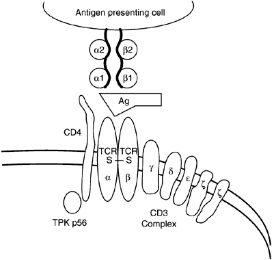
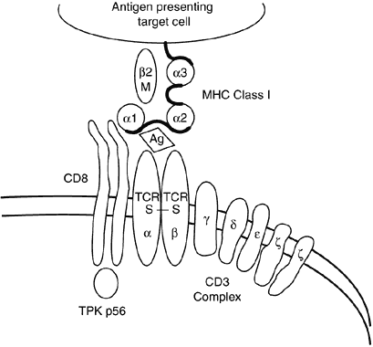
Class I and II molecules are responsible for mediating the interactions of T cells and antigen-presenting cells. CD4+ cells only recognize antigenic peptides in the setting of self MHC class II molecules (Fig. 7), whereas CD8+ cells only recognize peptides in the setting of self MHC class I molecules (Fig. 8). The peptide fragments that bind class II molecules generally are of extracellular origin, whereas endogenously derived peptides (viral particles) are bound by class I molecules.

Fig. 7. Antigen presentation: major histocompatibility complex (MHC) class II. Antigens (Ag) are modified and transported as antigenic determinants to the surface of antigen-presenting cells (APC). On the cell surface of the APC, processed antigen interacting with MHC class II determinants (α and β chains) binds to the T-cell receptor (TCR) of the CD4+ T cell. The CD4 determinant on the surface of the helper (CD4+ ) T cell enhances the binding of the TCR to the antigen-APC complex by binding to the MHC class II determinant. Binding of specific antigen to the TCR complex initiates T-cell activation and signal transduction by the CD3 complex. The CD4 molecule also may act as an accessory signal to enhance T-cell activation through tyrosine protein kinase (TPK p56) phosphorylation of the Þgz chain of the CD3 complex. (After Medical Knowledge Self-Assessment Program [MKSAP]: Allergy and Immunology, Book 1, p 160. Philadelphia: American College of Physicians [ACP], 1993.)

Fig. 8. Antigen presentation: major histocompatibility complex (MHC) class I. Antigen-presenting target cells, especially tumors or viral infected cells bearing MHC class I determinants (α chain and β2-microglobulin), present antigen to the CD8+ cytotoxic T cells through the T-cell receptor (TCR) complex. Binding of the Ag-MHC complex to the TCR leads to signal transduction via the CD3 complex. The CD8 molecule also may act as an accessory signal to enhance T-cell activation through tyrosine protein kinase (TPK p56) phosphorylation of the Þgz chain of the CD3 complex. (After Medical Knowledge Self-Assessment Program [MKSAP]: Allergy and Immunology, Book 1, p 161. Philadelphia: American College of Physicians [ACP], 1993, Fig. 28.)

Class I and II MHC products bind short stretches of peptides (class I, 9 to 11 amino acids in length; class II, 10 to 30 amino acids in length). The polymorphic amino acid residues in the peptide-binding region of the MHC molecules are responsible for both peptide binding and binding to the complementary ligand on the T cell.1,8

The Complement System

The complement system is a collection of functionally related proteins that interact with each other in a tightly regulated fashion to play a major role in host defense. A characteristic of the system is the sequential activation of proteolytic enzymes that allows for dramatic amplification of the activated molecules. Its principle functions are lysis of bacterial cell walls and enveloped viruses, opsonization, activation of the inflammatory response, and the solubilization and clearance of immune complexes.

Direct lysis of invading cells is mediated by the formation of the membrane attack complex (MAC) by terminal components of complement. The MAC inserts into the lipid bilayer of cell or viral membranes to form pores that allow for the rapid influx of water into the cell and consequent osmotic lysis.

Opsonization of microorganisms results from the binding of complement proteins C3 and iC3b to their surfaces. Neutrophils and macrophages express receptors for these proteins (CR1 to bind C3, MAC-1 and CR4 to bind C3bi), and thus phagocytosis of the opsonized pathogen is enhanced.

Activation of inflammation by complement is mediated by proteolytic fragments of early complement proteins (C3a, C4a, and C5a) termed the anaphylatoxins (Fig. 9). Anaphylatoxin binding to mast cell and basophils results in degranulation and the release of histamine and other vasoactive mediators as noted above. C5a also can act directly on vascular endothelium to cause contraction, vascular leak, and exocytosis.

Fig. 9. Overview of complement activation pathways. The classical pathway is initiated by C1 binding to antigen-antibody complexes, and the alternative pathway is initiated by C3b binding to various activating surfaces, such as microbial cell walls. The C3b involved in alternative pathway initiation may be generated in several ways, including spontaneously, by the classical pathway, or by the alternative pathway itself. Both pathways converge and lead to the formation of the membrane attack complex. In this and subsequent figures, bars over the letter designations of complement components indicate enzymatically active forms and dashed lines indicate proteolytic activities of various components. (After Abbas AK, Lichtman AH, Pober JS. Cellular and Molecular Immunology, p 296. 2nd ed. Philadelphia: WB Saunders, 1994.)

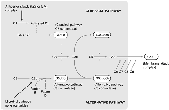
Large numbers of immune complexes may be formed in the setting of a vigorous immune response to high levels of circulating antigens. Their deposition in blood vessel walls can activate complement, resulting in inflammatory reactions that may damage local tissue. Complement binding to the Fc region of immunoglobulin molecules sterically inhibits the formation of immune complexes. The complement system also promotes the clearance of immune complexes from the circulation by the mononuclear phagocyte system.

The complement system contains two pathways, the classical and the alternative, that converge on the central complement protein C3. Complement activation along the classical pathway is initiated by antigen-antibody complexes, whereas activation along the alternative pathway occurs on microbial surfaces in the absence of antibody. Both pathways use C3 to produce enzymes that result in the formation of the MAC. Downregulation of the system is mediated by various soluble and membrane-bound proteins at steps throughout the cascade.1,4,14

Tolerance

Successful immune regulation requires mechanisms to generate specific immunologic self unresponsiveness, or tolerance. Tolerance to self is a learned process during which self-reactive lymphocyte clones are either prevented from developing or are inactivated after recognizing self-antigen. Loss of tolerance results in autoreactivity and, potentially, autoimmune disease.

The induction of self-tolerance in immature lymphocytes occurs in the generative lymphoid organs (i.e., the thymus for T cells and the bone marrow for B cells) and is termed central tolerance. Immature lymphocytes are more susceptible to the induction of tolerance than mature lymphocytes. If these cells encounter and recognize antigens before developing functional competence, they are deleted (clonal deletion) or rendered incapable of immunologic response (clonal anergy). The antigens encountered in the thymus and bone marrow are predominantly self-antigens, and thus the mature lymphocytes that emerge from these organs are depleted of self-reactive clones. Central tolerance in T cells results from clonal recognition of self-peptide-MHC complexes leading to either deletion or anergy in a process termed negative selection.1

Any self-reactive lymphocyte clones that escape this process and move into peripheral tissue subsequently may be inactivated as they encounter self-antigen under conditions that favor tolerance rather than immune activation. Conditions that influence this process, termed peripheral tolerance, include the type, amount, and portal of entry of antigen, the presence of competent antigen-presenting cells, and the type of responding lymphocyte.

The activation status of tissue antigen-presenting cells is a crucial determinant of peripheral tolerance. CD4+ cells that recognize antigen presented by antigen-presenting cells that cannot deliver a costimulatory signal (e.g., the absence of HLA B7 on a macrophage) become anergic. This period of anergy lasts for weeks, during which these cells will be unable to secrete IL-2 to signal autoproliferation. In addition, some antigen-presenting cells provide a direct inhibitory signal. CD4+ cells become unresponsive if antigen is presented to them by other class II MHC-bearing cells. The local cytokine milieu also influences peripheral tolerance. For example, T cells exposed to high concentrations of IL-2 at the time of antigen exposure die.

Peripheral B-cell tolerance occurs through similar mechanisms. B cells binding to antigen in the absence of T-cell help do not result in the production of antibody. In general, compared with the induction of anergy in T cells, B-cell anergy is shorter lived and requires higher concentrations of antigen to elicit.1

Transplantation immunologists have long recognized that certain sites, namely the anterior chamber of the eye, the brain, and the testes, are privileged in their ability to accept allografts. Immune privilege in the eye seems related to three factors: (1) the nature and function of ocular antigenpresenting cells; (2) the integrity of the blood-ocular (brain) barrier; and (3) the presence of an immunomodulatory intraocular environment. A unique form of systemic immune deviation exists after the inoculation of antigen into the anterior chamber, termed anterior chamber associated immune deviation (ACAID). In a simple experiment, the iris, ciliary body, and cornea were identified as secreting factors associated with the development of ACAID. The dendritic cell appears to be the messenger cell that is able to migrate via the aqueous humor through the trabecular meshwork into the blood and travel preferentially to the spleen. It appears that antigen is then presented in the context of class I MHC rather than the more usual class II MHC molecules. This induces a systemic suppressive response as reflected by depressed antigen-specific delayed-type hypersensitivity and an increase in the generation of cytotoxic T cells. It is believed that ACAID represents a physiologic adaptation to limit nonspecific inflammation and “bystander” cell injury related to delayed-type hypersensitivity, thus preserving the anatomically sensitive visual axis.15–17

Autoimmune disease results from the failure to maintain self-tolerance. This failure may result from the abnormal selection of self-reactive clones, the stimulation of lymphocytes that typically are anergic to self-antigen, or from the release of self-antigen from sites previously inaccessible to immune surveillance. Autoimmune disease also may develop when lymphocytes responding to foreign antigen recognize and cross-react to a self-protein in a process termed molecular mimicry.1

Back to Top
IMMUNOPATHOLOGY
The components of the immune system work together to ensure the destruction and the elimination of foreign organisms or potentially harmful substances globally seen by immunocompetent cells as antigens. In certain situations, the immune inflammatory response may be exaggerated and inappropriate, leading to damage to the organism's own tissue in the attempt to eliminate the offending agent. These abnormal hypersensitivity reactions can be divided into four major groups. The first three are primarily mediated by antibodies, whereas the fourth is primarily cell dependent.

TYPE I HYPERSENSITIVITY

Type I hypersensitivity, also known as mast cell- mediated hypersensitivity, is a term that describes a series of events that culminate in the activation of a unique set of effector cells known as mast cells, resulting in mediator release and consequent tissue inflammation. It is without a doubt one of the most rapidly occurring (i.e., seconds) and powerful effector mechanisms of the immune system with potentially devastating consequences. The initiating event is the interaction between an antigen and its specific IgE previously bound to the surface of tissue mast cells. This interaction, which is specific and requires prior sensitization, leads to mast cell activation and immediate release of preformed mediators responsible for the almost-instantaneous onset of symptoms. The initial response seems to be the first part of a more complex reaction that involves production of mediators de novo and their release at a later time to further extend the inflammatory reaction by recruiting other effector cells, primarily eosinophils. This constitutes the late-phase reaction and plays a central role in perpetuating the deleterious effects of immediate hypersensitivity.

IgE-mediated mast cell activation is the basic mechanism of allergic reactions and atopy and, as we shall see, may translate into a spectrum of clinical manifestations (Fig. 10).

Fig. 10. Sequence of events in immediate hypersensitivity. The initial contact with an antigen leads to specific immunoglobulin E (IgE) synthesis by B cells. Secreted IgE binds to mast cells or basophils through high-affinity Fcε receptors (FcεRI). On subsequent exposure to antigen, an immediate hypersensitivity reaction is triggered by cross-linking the IgE molecules. (After Abbas AK, Lichtman AH, Pober JS. Cellular and Molecular Immunology, p 296. 2nd ed. Philadelphia: WB Saunders, 1994.)

Why certain individuals have the propensity to produce specific IgE to environmental allergens is not completely understood. This predisposition, defined as atopy, is familial and occurs in approximately 30% of the general population. Atopic individuals have a higher incidence of allergic rhinitis, asthma, and atopic dermatitis, each of which may be present alone or in combination. In fact, atopic individuals are more prone to overreact to a variety of mediators released by mast cells or basophils with specific “shock organs” such as the eye, skin, and respiratory tract.

Environmental factors certainly contribute to the generation of clinically significant allergic reactions, but their role has yet to be defined.

A prerequisite to immediate hypersensitivity is the production of specific IgE by B lymphocytes after initial exposure to an antigen (sensitization or primary immune response). These immunoglobulins are subsequently bound to Fc receptors on the surface of mast cells and basophils. When the organism is re-exposed (secondary immune response) to the same antigen, this interacts with the surface-bound IgE, causing cell activation. For antigenantibody interaction to activate the cell effectively, cross-linking of at least two adjacent IgE molecules must occur (i.e., the antigen acts as a bridge between the antibodies, which, in turn, through their receptor, initiate a cascade of enzymatic reactions leading to degranulation and generation of newly formed mediators).

Preformed mediators (Table 2) are released from cytoplasmic granules by exocytosis. The type and number of mediators produced and released by mast cells seem to vary with their anatomic location. So-called mucosal mast cells, found in the gastrointestinal tract, contain chondroitin sulfate as their major granule proteoglycan and little histamine, whereas connective tissue mast cells, which are the predominant cell type of the periocular tissue, contain heparin and release large quantities of histamine. This vasoactive amine acts systemically and plays a dominant role in determining the clinical manifestations of immediate type hypersensitivity. It remains the primary target of most therapeutic interventions. Several enzymes present in the granules, which also are released in this phase of the reaction, may cause local tissue damage, and contribute to injury.1


TABLE 2. Mediators Involved in Type-I Hypersensitivity

  Preformed Mediators
  Biogenic amines

  Histamine
  Serotonin


  Granule macromolecules

  Tryptase
  Chymotryptase
  Serine proteases
  Aryl sulfatases
  Heparin
  Chondroitin sulfate


  Newly Synthesized Mediators
  Lipid mediators

  PGD2
  Leukotrienes (LTC4, LTD4, LTE4)
  PAF


  Cytokines

  TNF
  Interleukins (IL-1, IL-3, IL-4, IL-5, IL-6)
  Growth factors (GM-CSF)
  Interferon-<gg>


PGD2, prostaglandins; PAF, platelet-activating factor; TNF, tumor necrosis factor; GM-CSF, granulocyte-macrophage colony-stimulating factor.


Newly synthesized mediators (see Table 2) include lipid-derived molecules and cytokines. The lipid-derived mediators consist of (1) the prostaglandins synthesized from arachidonic acid via the cyclo-oxygenase pathway; (2) the leukotrienes, also derived from arachidonic acid by the action of 5-lipoxygenase; and (3) PAF. These mediators act locally as direct vasoconstrictors or smooth muscle constrictors and have been recognized as being of primary importance in tissue inflammation. The inhibition of these pathways by new therapeutic agents may prove to be an effective adjunct in controlling both immune and nonimmune-mediated inflammation.1

Over the past few years, it has become evident that cytokines represent the major medium by which cells communicate. The number of cytokines known to us continues to increase, and the complexity of cell-cell interaction is apparent. Mast cells are significant sources of cytokines including TNF, IL-1, IL-4, IL-5, IL-6, IL-3, and granulocyte macrophage-colony-stimulating factor (GM-CSF). These mediators clearly play a role in the late-phase reaction and account for the recruitment of other inflammatory cells to the site of reaction. IL-4, in particular, is responsible for the eosinophilic infiltrate typically associated with the late-phase reaction. It is thought to accomplish this through the induction of endothelial VCAM-1 and an eosinophil chemotactic substance. The eosinophilic infiltrate observed in the late-phase reaction represents the direct extension of the initial reaction in which activated eosinophils become effector cells and damage tissue by producing various substances such as MBP, EPO, ECP leukotriene C4, and PAF.1

The above-described sequence of events can occur wherever mast cells are located. These effects may be limited to small areas of tissue or may involve the whole organism, thus the clinical spectrum of anaphylaxis. This term refers to a specific immunologic reaction (i.e., IgE-mediated hypersensitivity) but has been variably used to describe a severe, often explosive reaction of unclear pathogenesis clinically manifested by cutaneous (urticaria/anaphylaxis), respiratory (laryngeal edema bronchospasm), gastrointestinal (nausea, vomiting, abdominal pain, diarrhea), and cardiovascular (hypotension, arrhythmias, cardiac ischemia) symptoms occurring singly or in combination. When these events are triggered by mechanisms other than antigen-IgE interaction, the term anaphylactoid reaction is preferred. This is discussed later.

An immediate cutaneous reaction is elicited when skin testing to an allergen is performed. This diagnostic tool is useful in determining the presence of specific IgE on mast cells in sensitized individuals. A small amount of antigen (i.e., protein from aeroallergens or foods) is injected either epicutaneously (prick or scratch test) or intradermally. If the individual has been sensitized previously and therefore carries specific IgE bound to mast cells, the antigen-antibody interaction leads to cross-linking of IgE, activation of mast cells, followed by the sequence of events described previously. The final result will be a localized area of inflammation clinically characterized by the “wheal and flare” reaction (erythema, edema, and itching) primarily mediated by histamine, as well as neuropeptides. Generalized urticaria is an example of a more diffuse cutaneous reaction that is often, but not always, IgE mediated.

Mast cells located in the upper and lower airways can be triggered to develop similar reactions in the nasal mucosa leading to allergic rhinitis or in the airways leading to bronchospasm and airway inflammation after inhalation of specific allergens. Activation of mast cells in the ocular mucosa leads to chemosis, angioedema, and conjunctivitis. Certain susceptible individuals with asthma may suffer exacerbations after exposure to allergens. Antigen may gain access to the immune system via several pathways such as parenterally or through direct contact with the mucosal membranes of the airways (aeroallergens), eye, or gastrointestinal tract (food and drug allergies). Intravenous exposure to antigens usually is associated with a brisk response and a more severe systemic reaction involving multiple target organs. The involvement of multiple organs is considered systemic anaphylaxis and is life threatening, requiring immediate intervention. The spectrum of limited reactions described may be present simultaneously: cutaneous symptoms characterized by urticarial eruptions and pruritus; respiratory symptoms characterized by severe bronchospasm and laryngeal edema; gastrointestinal symptoms characterized by cramping and abdominal pain; cardiovascular symptoms characterized by hypotension, bradycardia, and tissue hypoperfusion due to massive release of mediator; all of which can prove fatal.

The most common causes of anaphylaxis are medications such as penicillin. Certain foods (e.g., peanuts) as well as insect stings (e.g., bee, wasps, fire ants) also have been implicated as major causes of anaphylaxis (Table 3).4


TABLE 3. Immunoglobulin E—Mediated Agents

  Antibiotics

  Penicillin
  Cephalosporins
  Tetracyclines
  Vancomycin
  Neomycin
  Streptomycin
  Others


  Foreign proteins

  Heterologous serum
  Insulin
  Hymenoptera venom
  Chymopapain
  Trypsin
  Chymotrypsin
  Penicillinase
  Adrenocorticotropin hormone
  Antithymocyte globulins
  Rattlesnake venom
  Protamine
  Blood and blood products
  Latex


  Therapeutic agents

  Allergen extracts
  Benzylpenicilloylpolylysine (Pre-pen)
  Methylprednisolone
  Thiopental
  Vaccines
  Estradiol
  Streptokinase


  Foods

  Milk
  Egg white
  Shellfish
  Legumes
  Nuts
  Bananas
  Grains
  Fish
  Corn
  Others


ANAPHYLACTOID REACTIONS

Other mechanisms that lead to mast cell activation and degranulation exist. These alternative mechanisms do not require IgE or antigen-antibody interaction but lead to the same end result with both local and systemic manifestations. Examples of anaphylactoid reactions include direct mast cell activation by drugs such as aspirin and other nonsteroidal anti-inflammatory agents, opiates and derivatives, infusions of fluorescein,18–21 mannitol, radiocontrast media, and blood products (Table 4).4 Although the series of events leading to cellular activation is not clear in all instances, there are various proposed mechanisms.


TABLE 4. Non---Immunoglobulin E-Mediated Agents (Anaphylactoid)

  Immune complex or complement-mediated agents
  Blood and blood products

  Immunoglobulin
  Radiocontrast media (?)
  Menthotrexate
  Unsubstituted cellulose membrane dialyzers


  Modulator of arachidonic acid metabolism

  Acetylsalicylic acid
  Nonsteroidal anti-inflammatory agents
  Benzoates (presumed)
  Tartrazine (possibly)


  Direct histamine-releasing agents

  Opiates
  Curare, d-tubocurarine
  Radiocontrast media
  Intravenous fluorescein
  Dextran
  Iron-dextran
  Mannitol
  Pentamidine
  Many chemotherapeutic agents


The effects of aspirin on the arachidonic acid metabolic pathway have been known to inhibit the cyclo-oxygenase pathway, which causes shifting of the pathway toward leukotriene production. Leukotrienes are potent mediators of inflammation, in particular LTC4 and its breakdown products LTD4 and LTE4. This same mechanism may be the basis for aspirin-sensitive asthma. Activity of leukotrienes alone may account for up to 50% of pulmonary compromise and symptoms in asthma, and intervention at the level of production or receptor interaction by specific agents may play a key role in therapy.

Reactions to radiocontrast media or fluorescein are unpredictable and clinically mimic IgE-mediated anaphylaxis. These agents seem to activate mast cells directly by increasing the osmotic pressure in the surrounding environment. This is therefore not an immune-mediated reaction and the term anaphylactoid should be used. Patients who have had a reaction to contrast are at increased risk of adverse reaction on rechallenge. The risk is highest when contrast is given intravenously compared with intra-arterial administration. Use of nonionic dyes and premedication with antihistamines and steroids may reduce the risk of reactions to contrast. The infusion of blood, plasma, or immunoglobulins can potentially cause an anaphylactoid reaction. It is presumed that the formation of immune complexes causes complement activation and mast cell degranulation.

Drugs such as opiates, muscle relaxants, plasma expanders, and parenteral iron seem to have the ability to bypass all intermediate pathways and cause direct stimulation of mast cells. Release of histamine from both mast cells and circulating basophils can lead to any or all of the above-described clinical scenarios.

Anaphylactic and anaphylactoid reactions are clinically indistinguishable and should be treated similarly. Prevention remains the cornerstone of therapy for immediate hypersensitivity reactions. Avoidance of exposure to antigen in the sensitized individual, when possible, should be the primary goal. Unfortunately, many individuals are not aware of the sensitivity and anaphylactoid reactions do not require prior sensitization and therefore may occur on the first encounter with an offending agent. When possible, skin testing or a radioallergosorbent test can be used to determine specific sensitivities. The latter uses a solid-phase allergen immunosorbent in the first stage of the test to bind specific IgE in a serum specimen to be tested. After other components of the serum are washed away, the binding of radiolabeled anti-IgE antibodies to the allergen immunosorbent in the second stage permits detection of serum IgE bound in the first stage.

If a known anaphylactic is to be used and no alternatives are available, desensitization may be performed. This technique has intrinsic risks and has caused systemic reactions. The mechanism is through the induction of a state of tolerance by the incremental dosing of the offending protein. Both parenteral and oral protocols exist for a variety of medications, whereas parenteral (subcutaneous) is the only mode for insect sting hypersensitivity reactions.

As mentioned earlier, pretreatment with antihistamines and prednisone may be used in certain situations to reduce the risk or severity of reaction or both. Once anaphylaxis occurs, its immediate recognition is of prime importance. This represents a medical emergency requiring prompt administration of treatment, primarily directed at blocking the effects of the mediators being released. Subcutaneous administration of 0.3 to 0.5 ml of aqueous epinephrine 1:1000 is first-line therapy. This dose may be repeated if necessary. The effects of epinephrine are relaxation of bronchial smooth muscle and blood pressure support as well as prevention of mediator release. Inhaled bronchodilators also may be helpful in relieving bronchospasm, and airways should be assessed for evidence of laryngeal edema that may require endotracheal intubation.

Intravenous fluids should be administered for cardiovascular collapse, and additional support from pressor agents such as dopamine may be required. Other therapeutic methods include the administration of H1 and H2 (histamine receptors) blockers to counteract the effects of histamine. Parenteral steroids are effective in blunting the late-phase reaction, which typically occurs within 6 to 12 hours after the initial event.4

Severe refractory cases of anaphylactic shock can occur in patients taking β blocking agents, such as those used for patients with glaucoma. Atopic patients (asthma) with glaucoma should be given an alternate medication to control intraocular pressure.

TYPE II (CYTOTOXIC) HYPERSENSITIVITY

Type II hypersensitivity is another example of how immune mechanisms can, in certain situations, cause damage to the organism by initiating antiself reactions.

IgG, IgM, and, rarely, IgA mediate these reactions that are directed against antigens on the surface of one's own cells. The target cells may be circulating or components of fixed tissue and antibody-antigen interaction leads to cell lysis. The cytotoxic reactions may occur via three different mechanisms: complement-mediated lysis, opsonization and phagocytosis by macrophages or neutrophils, and cytolysis by NK cells bearing Fc γ receptors.

The clinical manifestations depend on the cells or the tissue targeted by the reaction. IgG-mediated immune hemolytic anemia can occur with high doses of penicillin. The drug or one of its metabolites binds to the surface of erythrocytes acting as a hapten. The drug-coated cells are targeted by specific IgG, and as a result complement activation causes cell death.

Erythrocytes also may be targeted by antibodies directed against surface alloantigens, giving rise to transfusion reactions. The ABO system of alloantigens present on the surface of human erythrocytes is responsible for incompatibility between individuals. All humans express the O antigen and are therefore tolerant to it. Individuals with an A or B genotype express A or B antigens on the cell surface and therefore tolerate foreign cells with A or B antigen, respectively. Anti-A IgM antibodies are present in the serum of individuals who do not express A antigen on self-erythrocytes, and anti-B IgM are present in the serum of individuals who do not express B antigen. As a result, if a patient is given a transfusion of erythrocytes from a donor who expresses an antigen not present on self-erythrocytes, the donor cells will be targeted by specific IgM and hemolysis will occur by complement activation.

It has been speculated that the naturally occurring alloantibodies directed against A and B antigens in the naive host might derive from exposure to cross-reactive microbial antigens.

Another clinical example of type II hypersensitivity involves the Rhesus (Rh) system. This is a complex system of antigens also present on the surface of erythrocytes. Individuals can be considered RhÞpl if they express the D antigen (85% of the population) and RhÞmi if they do not. If an RhÞmi patient is given a transfusion with erythrocytes containing the D antigen, these will be coated with antibody and lysed via complement activation.

Rh incompatibility between mother and fetus is at the base of the hemolytic disease of the newborn. An RhÞmi woman bearing an RhÞpl child may have anti-D antibodies develop late in pregnancy or at the time of delivery when RhÞpl fetal erythrocytes may enter the maternal circulation. If a second RhÞpl child is conceived, IgG antibodies, the only antibodies known to be able to cross the placenta, may be transferred from maternal to fetal circulation, causing hemolysis and fetal death in utero. This complication can be prevented by passive immunization of the mother soon after birth of her child with anti-D antibodies. These bind to the RhÞpl fetal erythrocytes in the maternal circulation, causing their destruction and preventing maternal sensitization to the D antigen.

Similar mechanisms have been invoked to explain the destruction of platelets in idiopathic thrombocytopenic purpura, but the initial event triggering the production of antibodies against self (autoantibodies) is not clear.

As mentioned earlier, fixed tissue components also can be the target of cytotoxic reactions. Virtually any tissue can be involved. Some notable examples are the thyroid leading to Hashimoto's thyroiditis, primary myxedema or Grave's disease, the pancreas resulting in insulin-dependent diabetes mellitus, and the kidney resulting in Goodpasture's disease. The production of autoantibodies in these conditions is tissue specific, and in many cases, the tissue antigen has been isolated and identified. Immunologic diseases of the kidney have been studied extensively. Antibodies, primarily complement-fixing IgG, are directed against the glomerular basement membrane. The predominant lesion is that of an infiltrative/proliferative glomerulonephritis.

Clinical manifestations may be limited to the kidney, but half of patients also have involvement of the lung basement membrane (Goodpasture's syndrome) or on rare occasions of the choroid plexus. This phenomenon is explained by the antigenic similarities of the basement membrane throughout the body. Deposition of antibodies and complement can be shown by immunofluorescence.

In pemphigoid of the eye (cicatricial pemphigoid), the desmosomal attachments between epithelial cells of the conjunctiva are lysed. IgG and IgA have been detected to bind to the basement membrane in 60% to 80% of the patients affected, and demonstration of antibody or complement at this level of the conjunctival epithelium is regarded as diagnostic.22,23 The result of these events is the formation of subepithelial blisters, an overgrowth of fibrous tissue, and severe corneal damage. The denuded conjunctival epithelium leads to adhesions (symblepherons), causing mechanical obliteration of eyelid movement and mucous producing goblet cells. Similar findings occur in linear IgA disease and drug-induced pemphigoid.24,25 Still another example of autoantibodies reacting to “self” proteins is found in pemphigus vulgaris mediated by complement fixing IgG to intercellular cement substance producing acantholysis and intraepithelial blistering all over the body, including the skin, mouth, and eyes. The target antigen in pemphigus vulgaris is desmoglein 3, a member of the desmoglein subfamily of cadherin adhesion molecules.26

TYPE III IMMUNE COMPLEX HYPERSENSITIVITY

Immune complexes are the result of the interaction between a soluble antigen and a specific antibody and represent the organism's attempt to neutralize and eliminate substances recognized as nonself. Under most circumstances, this mechanism is effective, and many cells actively participate in the process of immune complex clearing, particularly the mononuclear phagocyte system and complement proteins. Erythrocytes also have a role in clearing immune complexes by binding them through surface complement receptors and transporting them to the spleen and liver for removal by resident macrophages.

If large amounts of immune complexes are formed, this elimination mechanism may fail, causing accumulation of circulating complexes that may, in turn, be deposited in the tissue. It is the tissue deposition that leads to clinical manifestations and disease. Serum sickness was first described in 1911 when serum from horses immunized with diphtheria toxin was being injected in patients to passively immunize them against this disease. It was noted that these patients had joint inflammation, rash, and fever. The symptoms occurred even if the serum injected did not contain the antitoxin and therefore could not be attributed to the antidiphtheria antibody. The recipients had developed antibodies to horse serum proteins, and large amounts of immune complexes were formed and deposited in the skin, synovial tissue, and kidneys. Symptoms typically appear 1 week after the first injection but may appear sooner after subsequent injections.

Because of their anatomic structure, the complexes are deposited mainly in the arteries, glomeruli, and synovia, and therefore the common clinical and pathologic manifestations of immune complex diseases are vasculitis, nephritis, and arthritis.

Symptoms are acute when there has been transient antigen exposure, but they may become chronic with repeated exposure to that antigen or, as in the case of certain autoimmune disorders, when the antigen is a component of the organism itself.

One example of an autoimmune disease in which numerous autoantibodies are produced is systemic lupus erythematosus. Its clinical manifestations are proteiform and include arthritis and glomerulonephritis attributed to the deposition of immune complexes in these organs, giving rise to lesions similar to those found in chronic serum sickness. A decrease in serum levels of C3 and C4 reflects systemic activation of complement and is caused by consumption. Complexes also can deposit in the skin and blood-brain barrier, giving rise to cutaneous and neurologic manifestations. Autoantibodies directed against nuclear components are most characteristic of SLE. Anti-double-stranded DNA/ double-stranded DNA constitutes the major complex in this disease, although many other autoantibodies have been identified (e.g., ANA, anti-Sm).

Other examples of immune complex-mediated diseases are polyarteritis nodosa associated with prior hepatitis-B virus infection and poststreptococcal glomerulonephritis in which the immune complexes are composed, respectively, of hepatitis virus surface antigen and streptococcal antigen with their specific antibodies.27

An experimental model of this type of immune response is the Arthus reaction. This is a localized vasculitis induced by injecting antigen into the skin of a sensitized individual. Immune complexes are formed and deposited locally followed by complement activation, degranulation of mast cells, and recruitment of inflammatory cells with the consequent destructive events leading to tissue necrosis. These events are a common final pathogenic pathway that occurs wherever immune complexes are deposited and cause the clinical manifestations of the diseases described above.

A particular situation can occur in the lung of sensitized patients when specific antigens are inhaled. When high levels of IgG antibodies are present, immune complexes can form on the mucosal surface, initiating the chain of events described. This is the basis for diseases such as farmer's lung, pigeon fancier's disease, and furrier's lung, for example, collectively known as hypersensitivity pneumonitis.

TYPE IV DELAYED HYPERSENSITIVITY

Delayed-type hypersensitivity is a cell-mediated immune reaction, and unlike the other three types of hypersensitivity is not antibody dependent. Recognition of foreign substances resides in specific receptor molecules present on the membrane of T cells and not in humoral antibodies secreted by B cells.

This defines the two major branches of the immune response. However, as more information becomes available, it is evident that complicated interactions occur between humoral and cellular immunity.

The T-cell antigen receptor has been well characterized. It is a cell surface, disulfide-linked heterodimer composed of two glycoprotein chains. The α and β chains contain constant and variable regions similar to those found in immunoglobulins. Homology between T-cell receptors and immunoglobulins suggests that both these molecules derive from the same supergene family that also includes MHC class I and class II molecules.

Cell-mediated immunity is essential for eradicating intracellular microbes or viruses. It is in fact directed against cells that express foreign antigens on their surface and protects the organism against infections such as Listeria monocytogenes and mycobacteria. Furthermore, it represents the primary mechanism involved in allograft rejection.

After the cognitive phase, T cells are activated and secrete cytokines such as IL-2, TNF, and IFN-γ. These mediate inflammation by recruiting and activating effector cells, in particular the macrophage, whose function is enhanced by IFN-γ.

If the antigen is not a microbe, this reaction produces the damaging effects without the protective benefits. Contact sensitization with chemicals or environmental antigens can lead to delayed-type hypersensitivity as well as intradermal injection of microbial antigens in individuals with prior sensitization. Purified protein derivative prepared from Mycobacterium tuberculosis elicits a delayed response if injected in a patient previously infected with tuberculosis. The reaction evolves over 24 to 72 hours, which is the amount of time necessary for the cascade of events described to become clinically evident. Persistence of the antigen or microbe, as in granulomatous tuberculosis and leprosy, can lead to an ongoing T-cell response clinically characterized by granuloma formation, caseation, and fibrosis, whereas the lack of delayed-type hypersensitivity response to ubiquitous antigens such as Candida and Trichophyton is defined as anergy and may indicate T-cell dysfunction.

Cytokines

Cytokines are a diverse group of proteins that act as mediators of immune and inflammatory response. With advancing technology, it has been possible to identify, isolate, and characterize numerous molecules produced by cells of the immune system that serve as intercellular messages, not only for the regulation of immune cells, but also for interaction with other physiologic systems.

The number of cytokines continues to grow, and the complexity of the regulatory mechanisms and cellular interactions is now evident (see Table 1). The terms lymphokines and monokines frequently are used to describe cytokines produced by lymphocytes and mononuclear cells, respectively. Another term frequently encountered is interleukins, reflecting the initial hypothesis that cytokines were principally secreted by leukocytes to “talk” to other leukocytes. This hypothesis now appears grossly restrictive and inaccurate.

Transplant Immunology

The transfer of cells or tissue (graft) from one organism to another is called transplantation. The donor provides the graft and the individual who receives it is referred to as the recipient or host. If the donor and the recipient are the same, the graft is called an autologous graft such as the transfer of tissue from one location to another in the same organism. If the transplant occurs between two genetically identical individuals, it is a syngeneic graft, and if the two individuals are of the same species but genetically distinct, it is an allogeneic graft. Xenografts are transplanted between individuals of different species.

Although histology remains the major criterion for classifying rejection reactions, clinical features and the chronology of presentation are useful in determining the diagnosis.

The first rejection type, hyperacute rejection, is mediated by preexisting antibodies and occurs immediately after transplantation, usually within min-utes of anastomosis of the graft vessels to the host vasculature. The antibodies are directed against alloantigens present on the endothelium of the graft and initiate an immediate reaction involving the activation of complement. The endothelial cells are stimulated to secrete von Willebrand factor, causing platelet adhesion and aggregation. The end result is thrombotic occlusion of the graft vasculature and ischemic damage to the tissue. The antibodies that mediate this reaction are naturally occurring and may be IgM or IgG. One example of preformed antibodies is of those directed against antigens of the ABO blood group present on erythrocytes. These also are present on vascular endothelial cells. Others may be directed against MHC molecules or antigens yet to be identified. Donors and recipients are matched for the ABO blood group.

There is no treatment for hyperacute rejection that requires immediate graft removal.

The second rejection type, acute graft rejection, can be subdivided into two distinct entities. The first subtype is acute vascular rejection, which is characterized by an IgG-mediated vasculitic reaction against endothelial cell MHC molecules or other antigens with complement activation and direct lysis by T cells. The second subtype is acute cellular rejection, which is characterized by necrosis of parenchymal cells. The latter is complex and mediated by numerous cells and diverse mechanisms: CTL-mediated lysis, activated macrophage-mediated lysis, and NK cell-mediated lysis. (CD8+ CTLs probably are the most active cells in this type of reaction.) A lymphocytic and macrophage infiltrate often are present in the affected graft.

The third type is chronic graft rejection, which usually appears at least 60 days after transplantation. The pathogenesis of this type of rejection is not yet completely understood. It may represent the consequence of an earlier acute rejection leading to fibrosis and loss of normal organ architecture, although it is often seen in patients with no prior evidence of rejection. Other possibilities are chronic ischemic damage due to anatomic abnormalities and a form of chronic delayed hypersensitivity. These changes are not reversible with therapy.

The primary cause of late graft failure is from accelerated atherosclerosis and vascular occlusion. This usually develops months after transplantation and is caused by proliferation of intimal smooth muscle cells induced by growth factor secreted by macrophages activated by lymphocytes.1

Although some form of rejection almost always occurs in transplantation unless donor and recipient are genetically identical, this is greatly diminished by reducing the recipient's immune response with pharmacotherapy and by selecting donor and recipients based on the similarities of HLA alleles.

Corticosteroids in association with the purine nucleoside inhibitors (prototype: azathioprine) have been the primary immunosuppressive drugs for many years, but the introduction of cyclosporine A has improved the outcome of transplantation greatly. Corticosteroids also are used as first-line therapy in the treatment of allograft rejection. High-dose intravenous pulse therapy is the preferred regimen. Alternatively, antilymphocytic preparations such as OKT3 (IgG2a murine monoclonal antibody directed against lymphocytes) have been effective in the treatment of acute rejection refractory to steroids.

The two most important factors that determine the acceptance of a graft are blood group compatibility, in particular the ABO group, and HLA system compatibility.

Naturally occurring antibodies present in the host circulation bind antigens of the ABO group in the incompatible host, giving rise to a hyperacute reaction. This has largely been eliminated by matching blood groups in donor and recipient.

The histocompatibility antigens of the MHC (HLA) is the second group of substances that can trigger the rejection process. Matches at HLA-A, HLA-B, and HLA-DR alleles are important, and “tissue typing” is performed to determine the HLA molecules expressed. Graft survival is directly related to the number of loci matched.1

Corneal tissue can be transplanted with limited allograft rejection and is therefore considered an immunologically “privileged site.” This is primarily because of the avascularity of this structure, except for the corneoscleral limbus and the lack of immunocompetent cells. The abnormal proliferation of vessels in the cornea during a severe inflammatory response can compromise this immunologic privilege by giving access to cellular elements and their products initiating an inflammatory immune response.17

Back to Top
REFERENCES

1. Abbas AK, Lichtman AH, Pober JS (eds): Cellular and Molecular Immunology. 2nd ed. Philadelphia: WB Saunders, 1994

2. Paul WE (ed): Fundamental Immunology. 3rd ed. New York: Raven Press, 1993

3. Stites DP, Terr AI, Parslow TG: Basic and Clinical Immunology. 8th ed. Norwalk: Appleton & Lange, 1994

4. Middleton E, Reed CE, Ellis EF et al (eds): Allergy: Principles and Practice. 4th ed. St Louis: Mosby, 1993

5. Fink PJ, Bevan MJ: Positive selection of thymocytes. Adv Immunol 59:99–129, 1995

6. Micelli MC, Parnes JR: Role of CD4 and CD8 in T cell activation and differentiation. Adv Immunol 1993;53:59–122

7. Romagnani S: TH1 and TH2 in human diseases. Clin Immunol Immunopathol 80:225–235, 1996

8. Roitt I, Brostoff J, Male D (eds): Immunology. 4th ed. St Louis: Mosby, 1996

9. Ciprandi G, Buscaglia S, Pesce G et al: Allergic subjects express intracellular adhesion molecule-1 (ICAM-1 or CD54) on epithelial cells of the conjunctiva after allergen challenge.J Allergy Clin Immunol 91:783–792, 1993

10. Frenette PS, Wagner DD: Adhesion molecules—part 1. N Engl J Med 334:1526–1529, 1996

11. Elner SG, Elner VM: The integrin superfamily and the eye. Invest Ophthalmol Vis Sci 37:696–701, 1996

12. Schroth MK: Adhesion molecules in asthma and allergy. Immunol Allergy Clin 16:643–656, 1996

13. Soukiasian SH, Rice B, Foster CS et al: The T cell receptor in normal and inflamed human conjunctiva. Invest Ophthalmol Vis Sci 33:453–459, 1992

14. Frank MM, Austen KF, Claman HN, et al (eds): Samter's Immunologic Diseases. 5th ed. Boston: Little, Brown, 1995

15. Wilbanks GA, Streilein JW: Studies on the induction of anterior chamber associated immune deviation (ACAID). I. Evidence that an antigen-specific, ACAID-inducing cell associated signal exists in the peripheral blood. J Immunol 146:2610–2617, 1991

16. Streilein JW: Immune privilege as the result of local tissue barriers and immunosuppressive microenvironments. Curr Opin Immunol 5:428–432, 1993

17. Streilein JW, Bradley D, Sano Y: Immunosuppressive properties of the tissues of the ocular anterior segment. Ocular Immunol Inflammat 4:57–68, 1996

18. Lipson BA, Yanuzzi LA: Complications of intravenous fluorescein. Int Ophthalmol Clin 29:200–205, 1989

19. Kwiterovich KA, Maguire MG, Murphy RP et al: Frequency of adverse reactions after fluorescein angiography. Ophthalmology 98:1139–1142, 1991

20. Ellis PP, Schoenberger M, Rendi MA: Antihistamines as prophylaxis against side reactions to intravenous fluorescein. Trans Am Ophthal Soc 38:191–205, 1980

21. Stein MR, Parker CW: Reactions following intravenous fluorescein. Am J Ophthalmol 72:861–868, 1971

22. Foster CS: Cicatricial pemphigoid. Trans Am Ophthalmal Soc 1986;84:527–663

23. Merayo-Lioves JM, Dutt JE, Foster CS: Conjunctival cytokine expression in cicatricial pemphigoid and atopy, p 101. American Academy of Ophthalmology Annual Meeting, 1994

24. Leonard JN, Wright P, Williams DM et al: The relationship between linear IgA and benign mucous membrane pemphigoid. Br J Dermatol 110:307–314, 1984

25. Pouliquen Y, Paten A, Foster CS et al: Drug-induced cicatricial pemphigoid affecting the conjunctiva: Light and electron microscope features. Ophthalmology 93:775–783, 1986

26. Amagai M, Kapati S, Prussick R et al: Autoantibodies against the amino-terminal cadherin-like binding domain of pemphigus vulgaris antigen are pathogenic. J Clin Invest 90:919–926, 1984

27. Rich RR, Fleisher TA et al (eds): Clinical Immunology: Principles and Practice. St Louis: Mosby, 1996

Back to Top