Chapter 17
Evaluation of Visual Function
CHRIS A. JOHNSON
Main Menu   Table Of Contents

Search

VISUAL ACUITY
CONTRAST SENSITIVITY
VISUAL FIELD SENSITIVITY
CONCLUSION
ACKNOWLEDGMENT
REFERENCES

The stimulus for vision is light, which constitutes only a small portion of the electromagnetic spectrum for wavelengths between approximately 400 and 700 nm. Light is emitted by natural and artificial sources and is reflected by objects in our environment. The simplest eyes in nature function as basic light detectors, whereas the human visual system has evolved into an extremely complex mechanism for processing visual information. When light enters the eye, it is refracted by the optical media to form an inverted image on the retina. The photoreceptors convert this light energy into electrical signals that are processed by neural elements in the retina, optic nerve and higher visual centers of the brain. This processing of visual information is carried out by various subpopulations of neural mechanisms that are responsible for encoding stimulus features such as motion, form, color, depth, and other fundamental properties of the visual image.1–6 The relationship between the physical properties of light and the consequent perceptual and behavioral responses of the observer is known as visual psychophysics, which serves as the primary foundation for the clinical assessment of visual function.

Excellent reviews of color vision and binocular vision are available elsewhere in these volumes. The present chapter will therefore be limited to a discussion of three other aspects of visual function that are important parts of a clinical ophthalmic evaluation: visual acuity, contrast sensitivity, and measurement of visual field sensitivity (increment threshold sensitivity) by means of perimetry. These are all visual functions that have a long history of psychophysical and physiologic investigation devoted to them, but they also form an important part of the evaluation of the functional capabilities of patients with ocular disorders. Early detection of eye disease, differential diagnosis of various ocular and neurologic disorders, and monitoring the efficacy of therapeutic regimens are just several of the roles these visual function tests assume in a clinical setting. This chapter provides a brief overview of visual acuity, contrast sensitivity, and visual field testing and their application to the clinical evaluation of patients.

Back to Top
VISUAL ACUITY
Visual acuity is the most common measurement of visual function that is performed in a clinical setting. It serves as the primary means of assessing the integrity of the optics of the eye and of the neural mechanisms subserving the foveal region, because good visual acuity requires that both optical and neural components are functioning properly.7 Visual acuity is used to monitor the status of central visual function in patients and is an essential part of the procedure used to prescribe spectacle and contact lens corrections. In addition, visual acuity is important for many “real world” tasks, such as reading,8,9 face recognition,10 and other activities involving the discrimination of fine spatial detail.

Visual acuity is specified in terms of the visual angle subtended by fine spatial detail. An object's visual angle is determined by its physical size and its distance from the observer. For small angles, the visual angle of an object can be approximated by arc tan(object size/object distance). There are generally considered to be three types of visual acuity measurements: detection acuity, resolution acuity, and identification acuity. Detection acuity refers to the smallest stimulus object or pattern of elements that can be distinguished from a uniform field. The minimum angle of detection (MAD) is primarily limited by the contrast of the stimulus. For normal human foveal vision, Thibos and Bradley11 have recently demonstrated that optical components impose the primary limiting factor on detection acuity by means of contrast attenuation for small stimuli.

Resolution acuity refers to the smallest amount of spatial detail that can be discerned to allow one pattern to be distinguished from another. Typically, this type of acuity is measured with the use of a grating pattern of alternating light and dark stripes, and the observer's task is to determine the orientation (horizontal or vertical) of the grating. Resolution acuity is normally specified in terms of the minimum angle of resolution (MAR), and it is also limited primarily by contrast. For normal human foveal vision, resolution acuity is mainly constrained by the properties of the photoreceptor mosaic and underlying neural mechanisms.11

Identification acuity refers to the smallest spatial detail that can be resolved to allow one to recognize objects such as letters. It is this type of acuity measurement that is obtained in a clinical ophthalmic setting. Specification of identification or recognition acuity is usually in terms of the MAR, or values such as Snellen notation, which is based on MAR. For the letters typically used in eye charts, the MAR would correspond to the angle subtended by the thickness or stroke of a letter. The letters for most visual acuity charts are designed such that the overall size of the letter is five times larger than its thickness. An example of a typical eye chart used for clinical evaluation of visual acuity is shown in Figure 1. This design is basically the same as that originally introduced by Snellen in 1862,12 and the most common form of reporting visual acuity is still in terms of “Snellen notation.” This value consists of a fraction in which the numerator is the testing distance (usually 20 ft or 6 m) and the denominator is the distance at which a “normal” observer would be able to read the letter. Visual acuity charts use a reference of 1 minute of arc visual angle for the smallest resolvable detail (MAR) for a “normal” observer, which corresponds to a Snellen fraction of 20/20 (or 6/6). Similarly, a 20/40 (6/12) letter would be 10 minutes of arc high and have a thickness of 2 minutes of arc. It should be noted that the 20/20 reference for “normal” vision was developed more than 100 years ago. With today's high-contrast eye charts and better lighting sources, most normal individuals under the age of 50 years can be corrected to better than 20/20 visual acuity.

Fig. 1. An example of a typical Snellen-type eye chart for clinical measurement of visual acuity.

A new design of visual acuity charts has recently emerged.13,14 An example of one of these new eye charts (the Bailey-Lovie chart) is presented in Figure 2. This improved design has several advantages. First, the letters used are of approximately equal detectability, as compared with earlier charts that had some letters that were more legible than others. Second, each line has an equal number of letters, as compared with earlier charts with only one or two letters for the 20/100 and 20/200 visual acuity lines, and a large number of letters for the 20/20 and smaller visual acuity lines. Third, the spacing between letters is proportional to the letter size, as compared with an unequal spacing between letters for the older acuity charts. Finally, the change in visual acuity from one line to another is in equal logarithmic steps, as compared with very small changes for different lines at the small-letter end of the chart and rather large changes for the big-letter end of the chart for older eye charts. This new eye chart thus permits the ability to better specify visual acuity in terms of Snellen notation, MAR, or log MAR values. In addition, new methods of scoring responses to this type of visual acuity chart can provide greater sensitivity and reliability of the measure.15,16 The Early Treatment Diabetic Retinopathy Study (ETDRS) visual acuity chart, which is based on the design of the Bailey-Lovie eye chart, has now been used in several multicenter clinical trials sponsored by the National Eye Institute.17–19

Fig. 2. An example of the Bailey-Lovie chart, incorporating an equal number of letters per line, letters of approximately equal legibility, proportional spacing between letters, and a logarithmic change in letter size from one line to another.

A number of stimulus parameters are known to affect visual acuity measurements in normal individuals, including background adaptation luminance,20–22 contrast,22,23 refractive error,24–26 pupil size,27,28 retinal eccentricity,29–31 duration of presentation,32–34 the type of optotype used,35–37 and interaction effects from adjacent visual contours, or “crowding.”38–40 In clinical patient populations, factors such as “crowding,” luminance level, contrast, and type of optotype used can sometimes have an even greater influence on visual acuity measurements than they can for normal individuals, depending on the particular ocular disorder. If test parameters such as background luminance are varied in a consistent manner, they can be potentially useful for diagnostic purposes. However, the unintentional variation of background luminance or other test parameters (from one examination room to another or from one visit to another) can be problematic for monitoring the visual status of patients.

The measurement of visual acuity in special populations (e.g., young children and handicapped individuals) is not always possible with a letter chart. For these populations, “preferential looking” techniques, oculomotor responses such as optokinetic nystagmus, and electrophysiologic measures such as the visual evoked potential can be used to estimate visual acuity.41–44 In addition, a number of new eye charts and behavioral test procedures have been developed to assess visual acuity in nonverbal or handicapped individuals.45 These tests use patterned stimuli and caricatures of faces or common objects with “critical detail” to obtain an indication of the individual's level of visual acuity. Haegerstrom-Portnoy has provided an excellent review of many of these new behavioral procedures.45

Back to Top
CONTRAST SENSITIVITY
Although visual acuity is able to specify the smallest spatial detail that can be resolved for high-contrast stimuli, it does not provide any information about the visual system's ability to detect and discriminate objects of different sizes and contrasts. The most commonly used stimuli for assessing the contrast sensitivity characteristics of the human visual system are sinusoidal gratings, consisting of an alternating pattern of light and dark bars whose luminance varies sinusoidally in the direction perpendicular to the grating's orientation. A graphical representation of the luminance profile of a sinusoidal grating is presented in Figure 3.

Fig. 3. Graphical representation of the luminance profile of a sinusoidal grating stimulus.

The appearance of a sinusoidal grating is an alternating series of fuzzy light and dark lines. The size of the light and dark bars of the grating is specified according to spatial frequency, which is the number of cycles (pairs of light and dark bars) of the grating pattern per degree of visual angle. Typically, the range of spatial frequencies examined is between 0.5 and 30 cycles per degree. The contrast of the sinusoidal grating is determined by the luminance of the peaks (Lmax) and troughs (Lmin) of the luminance profile, according to the following equation:

Contrast can thus vary from a minimum of 0 for a uniform field (i.e., Lmax and Lmin are equal) to a maximum of 1 (where Lmin is equal to zero). For a particular spatial frequency, the contrast threshold is the minimum amount of contrast needed to detect the presence of the grating pattern. Contrast response properties are typically reported in terms of contrast sensitivity, which is the reciprocal of the contrast threshold (sensitivity = 1/threshold). The human visual system varies in the amount of contrast needed to detect a grating pattern for different spatial frequencies, or sizes of light and dark bars. This can be observed in Figure 4, which presents a sinusoidal distribution of light and dark bars in which spatial frequency varies logarithmically along the abscissa and contrast varies logarithmically along the ordinate. At a standard reading distance, it should be observed that intermediate spatial frequencies (medium-sized bars) are visible at lower contrast levels (i.e., the light and dark bars extend farther vertically) than are low spatial frequencies (wide bars) and high spatial frequencies (narrow bars), giving an inverted U-shaped appearance to the pattern.

Fig. 4. Illustration of contrast characteristics of human visual system for sinusoidal gratings. Contrast varies logarithmically along the ordinate, and spatial frequency varies logarithmically along the abscissa.

The use of sinusoidal gratings to evaluate spatial contrast sensitivity properties of the human visual system has several advantages. First, optical defocus does not change the shape or appearance of the sinusoidal grating, other than to reduce its contrast. Thus, only one attribute of the stimulus is altered by blur. Second, the contrast sensitivity function provides a means of characterizing the overall response properties of the visual system to a wide variety of complex stimuli and visual images. In the past, the contrast sensitivity function for vision has been compared to the audiogram for characterizing hearing ability.46 The audiogram specifies the minimum amount of sound energy necessary to detect pure tones (temporal sinusoidal waveforms) for various frequencies of pitch. This information can then be used to determine an individual's ability to hear complex sounds that consist of compound waveforms. In a similar fashion, the spatial contrast sensitivity function for vision presumably makes it possible to determine an individual's ability to process spatial information from complex visual scenes.

Fourier analysis demonstrates that any complex waveform can be decomposed into a series of sine waves of various frequencies, amplitudes, and phase (position) relationships.47 Thus, any complex visual scene can theoretically be broken down into a specific combination of sinusoidal distributions of light of different spatial frequencies, amplitudes, and phases in the vertical and horizontal dimensions. If the contrast response characteristics of the human visual system to various spatial frequencies are known, then it should be possible to determine how complex visual scenes will be processed by the visual system. Note, however, that the contrast sensitivity function describes the behavior of the visual system at threshold contrast levels, which is not necessarily representative of the visual system's characteristics for processing suprathreshold contrast information.48–50

An illustration of a normal contrast sensitivity function for the human visual system is presented in Figure 5. Contrast sensitivity is greatest for a spatial frequency of about four to five cycles per degree, with progressive reductions in contrast sensitivity for higher and lower spatial frequencies. This inverted U-shaped function reflects the broad bandpass characteristic of the human visual system's contrast response properties. This shape has been reported to represent an envelope of the response characteristics of multiple quasi-independent channels or neural mechanisms with narrower bandpass response characteristics,51 as illustrated in Figure 6. Indeed, neurophysiologic studies support the notion that the visual system uses a form of spatial frequency analysis in the early processing of visual information.52–54

Fig. 5. Typical spatial contrast sensitivity function for normal human foveal vision.

Fig. 6. Typical spatial contrast sensitivity function and underlying narrow-bandwidth channels. The envelope of the composite sensitivity of the narrow-bandwidth channels is felt to be the underlying basis of the contrast sensitivity function.

The use of sinusoidal gratings to study human visual function was first introduced by Schade in 195655 and was followed by a series of important studies by several investigators that established the theoretic and empirical basis for the contrast sensitivity function of the normal human visual system.56–59 These investigations were instrumental in defining the optical and neural components underlying the normal contrast sensitivity function. A number of factors affect the measurement of the normal contrast sensitivity function, including background adaptation luminance,59–61 stimulus field size,62–64 retinal eccentricity,65–68 pupil size,28,56 temporal characteristics,60,69,70 stimulus orientation,71,72 and various optical factors such as defocus, dioptric blur, diffusive blur, and astigmatism.11,37,56,73,74

Clinical applications of contrast sensitivity testing have been quite extensive. A number of investigators have used the contrast sensitivity function to evaluate optical properties of the human eye, including refractive error and defocus,11,37,56,73,74 corneal diseases,75–83 cataracts,84–90 refractive surgery,91,92 intraocular lenses,93–95 and normal aging properties of the optics of the eye.87,88,96–98 In many instances, contrast sensitivity deficits have been reported for patients with normal visual acuity and optical conditions that produce subtle changes in the quality of their vision. A number of retinal disorders have also been evaluated with the contrast sensitivity function, including diabetic retinopathy,99–101 age-related macular degeneration,102–104 and a variety of other retinal disorders.105–107 Several investigators have also reported foveal contrast sensitivity deficits in patients with glaucoma.108–114 Additionally, measurement of the contrast sensitivity function has also been reported to be effective in revealing subtle visual losses in optic neuritis and multiple sclerosis,17,115–118 other optic neuropathies and cerebral abnormalities,115,119–123 amblyopia,124 and other conditions such as Alzheimer's disease,98 Parkinson's disease,115 and cystic fibrosis.125

The contrast sensitivity function can also be measured in infants and young children, with use of either electrophysiologic or behavioral techniques.126–131 There is accumulating evidence that these procedures are able to provide valuable clinical information about the functional visual status of infants and children. As these techniques become more refined and easier to use, they are increasingly being incorporated into clinical settings.

In addition to providing useful information about the functional integrity of the visual pathways, there is also evidence that the contrast sensitivity function may be helpful in predicting the performance of various “real world” tasks, such as those involving the identification of objects such as aircraft132 or tanks,133 recognition of highway signs,134 reading ability,135–137 face recognition,138,139 and mobility skills of patients with subnormal vision.140 Thus, not only is the contrast sensitivity function useful for revealing subtle visual deficits associated with ocular disorders, but it also appears to be helpful in identifying problems that a patient is likely to encounter during daily activities.

Recently, there have been several new approaches to the evaluation of contrast sensitivity using wall charts in a manner similar to that in which clinicians measure visual acuity. One of these methods is represented by the Vistech chart, shown in Figure 7.141 This chart has a series of five rows (A through E), each with a different spatial frequency. Each row consists of a group of nine circular targets containing a sinusoidal grating that is either vertical, tilted to the left, or tilted to the right. From the left side of the chart to the right, there is a successive reduction in the contrast of the grating. Patients are positioned at the 10-ft viewing distance and are asked to “read” each row from left to right and indicate the orientation of the grating.

Fig. 7. The Vistech contrast sensitivity chart.

Another method of testing contrast sensitivity uses a typical letter chart for testing visual acuity, but with a lower contrast for the letters and the background. An example of a low-contrast version of the Bailey-Lovie chart is presented in Figure 8. Regan and Neima142,143 have also developed a series of visual acuity charts at several different contrast levels. Although normal observers will demonstrate a modest reduction in visual acuity for low-contrast targets, patients with early or subtle abnormalities will sometimes demonstrate quite profound reductions in visual acuity for low-contrast targets, in spite of good visual acuity for standard high-contrast letters. Thus, there is reasonable evidence that this approach to contrast sensitivity testing is able to reveal early disturbances of the visual pathways that are not revealed by standard visual acuity testing. The advantage of a low-contrast acuity chart in a clinical setting is that it is a test procedures that is highly familiar to most patients.

Fig. 8. A low-contrast version of the Bailey-Lovie visual acuity chart.

A third method of testing contrast sensitivity using a chart has been introduced by Pelli, Robson, and Wilkins and is known as the Pelli-Robson chart.144 As shown in Figure 9, the chart consists of letters of a fixed size that vary in contrast. Each line consists of six letters, with the three leftmost and three rightmost letters having the same amount of contrast. The patient reads the chart in a manner similar to that for a standard visual acuity chart, and the minimum contrast at which the letters can be detected is recorded. Several studies have reported that this method of testing contrast sensitivity is highly reproducible145–147 and is capable of detecting functional losses that are not evident with standard visual acuity testing. The Optic Neuritis Treatment Trial used the Pelli-Robson charts for evaluation of contrast sensitivity as an outcome measure for comparison of treatment groups.17

Fig. 9. The Pelli-Robson contrast sensitivity letter chart.

The use of sinusoidal gratings versus low-contrast letters for the measurement of contrast sensitivity is controversial. A number of arguments supporting each approach have been presented.148–150 Irrespective of the advantages and disadvantages of each method, it is important to note that different results are sometimes obtained by the two procedures. Different stimulus conditions in observers with normal visual function can result in different contrast sensitivity measures being obtained by gratings versus low-contrast letters.37 In addition, patients with certain visual disorders can produce different contrast sensitivity values for gratings versus letters.36

Back to Top
VISUAL FIELD SENSITIVITY
Perimetry and visual field testing are also tests of visual function that are commonly performed in a clinical setting. The basic technique is founded on the increment or differential light threshold, which refers to the minimum amount of light that must be added to a stimulus for it to be distinguished from a uniform background field. The relationship between the increment threshold (ΔL) and background luminance (L) is shown in Figure 10 on a log-log plot. At low levels, there is no influence of the background luminance on the increment threshold (i.e., a minimum amount of light is needed to detect the stimulus). In this region, the increment threshold is constant (ΔL = C [a constant]), as indicated by the horizontal portion of the curve in Figure 10. At higher levels, the increment threshold increases as a function of higher background luminances. In this region, a doubling of the background luminance requires a doubling of the stimulus increment for it to be detectable. This is denoted in Figure 10 by the portion of the curve with a slope of 1, which indicates that ΔL/L = C. This relationship (ΔL/L = C) is known as Weber's law or the Weber fraction in honor of the German scientist who was the first to describe it in 1832.151

Fig. 10. The log increment threshold (ΔL) as a function of log background luminance. At low backgrounds, ΔL is a constant, whereas at higher backgrounds ΔL/L is a constant. This region is known as Weber's law and has a slope of 1.

The background luminance that is typically used for perimetry and visual field testing is 31.5 apostilbs (10 candelas per square meter), which is approximately one third of the way up the portion of the curve in Figure 10 that has a slope of 1 (Weber's law). This is important, because changes in pupil size or other factors that affect retinal illumination will equally influence the background and stimulus, and thus will shift sensitivity up and down the Weber's law portion of the curve. Because ΔL/L = C in this region, increment threshold sensitivity remains unaffected. However, in some situations (e.g., elderly glaucoma patients with lenticular opacities and iatrogenic miosis), retinal illumination may be reduced to the nonlinear portion of the curve, confounding interpretation of responses.

The increment (differential) threshold varies as a function of visual field location. With the standard background luminance of 31.5 apostilbs (10 cd/m2), the fovea has the lowest increment threshold. There is a progressive increase in the increment threshold with increasing eccentricity from the fovea. By convention, representation of visual field data is presented in terms of sensitivity, which is the reciprocal of threshold. In this view, the highest sensitivity (for the standard background luminance) is at the fovea, with a progressive decrease in sensitivity with increasing peripheral eccentricity. A three-dimensional representation of visual field sensitivity for a normal visual field is shown in Figure 11. Note that visual field sensitivity and the total extent of the visual field are greater for the temporal visual field than for the nasal visual field, and sensitivity and visual field extent are greater for the inferior visual field than for the superior visual field. Because of its characteristic shape, this representation of visual field sensitivity has previously been referred to as the “hill of vision” or the “island of vision in the sea of blindness.” It is important to keep in mind that visual field locations are inversely related to retinal locations. Stimuli in the superior visual field project to the inferior retina and vice versa. Similarly, stimuli in the temporal visual field project to the nasal retina and vice versa.

Fig. 11. Three-dimensional representation of the differential light sensitivity of the visual field, often referred to as the “hill of vision” or the “island of vision in the sea of blindness.” A left eye is represented in the drawing. The black oval represents the physiologic blind spot, whose anatomic basis is the optic disc.

Both the background luminance level and the size of the target used to perform visual field testing can affect the shape of the “island of vision.”152,153 In general, the slope of the visual field sensitivity contour is steeper for higher background luminances and/or smaller targets, and it is flatter for lower background luminances and/or larger targets. At very low background luminances, the fovea becomes less sensitive than surrounding regions, producing a contour resembling a “volcano of vision in the sea of blindness.”

Three main techniques are used clinically to perform visual field testing: kinetic perimetry, static perimetry, and suprathreshold static perimetry. The most common form of kinetic perimetry uses a Goldmann perimeter, which consists of a white hemispheric perimeter bowl of uniform luminance onto which a small illuminated stimulus is projected. The patient's eye to be tested is aligned to the center of the hemispheric bowl. With the nontested eye occluded with an eye patch, the patient is instructed to maintain steady fixation on a small central target. The examiner is able to monitor the observer's eye position and fixation behavior by means of a telescope viewer. A stimulus of a selected size and luminance is projected onto the perimeter bowl and is moved from a nonseeing region in the far periphery toward the fixation point at a rate of approximately 4° per second.154 The patient presses a response button when the stimulus is first detected, and this location is marked on a chart. This procedure is repeated for a series of meridians around the visual field, each kinetic scan directed from a nonseeing region toward the fixation point. These locations of equal sensitivity are connected by a curved line referred to as an isopter. Because of the characteristic “hill of vision” shape of the visual field sensitivity profile, it is possible to evaluate different regions of the visual field by changing the size and luminance of the moving stimulus. To examine the far periphery, one would use a large, high-luminance moving stimulus that is readily detectable by locations with low sensitivity. Reducing the size or luminance of the stimulus makes it less detectable; therefore, the stimulus must be moved nearer to the central fixation point before it is detected by more sensitive regions of the visual field. Typically, a number of isopters are determined by the use of different stimulus sizes and luminances. Localized areas of nonseeing inside of isopters are called scotomas and are represented by areas that are solidly filled in on the visual field chart. The technique for performing quantitative kinetic perimetry is a highly acquired skill, requiring instruction and practice. A detailed discussion of kinetic perimetric technique is beyond the scope of this chapter. However, Anderson's book provides an excellent and thorough discussion of the intricacies of kinetic perimetric testing.155 A thorough kinetic perimetry evaluation of the visual field typically requires about 15 minutes for normal eyes, and up to 20 to 30 minutes for eyes with complex visual field loss.

Representation of the results of kinetic perimetry is typically in the form shown in Figure 12A, which depicts the findings for a normal left eye. Figure 12B depicts the three-dimensional representation of the hill of vision and shows its relationship to the isopter plot for kinetic perimetry. Basically, the visual field representation for kinetic perimetry is a type of topographic relief map of the hill of vision. Pathologic changes to the visual pathways are denoted by specific patterns of visual field sensitivity loss. On the kinetic perimetry plot, sensitivity losses are depicted by displacement or constriction of isopters from their normal shape or the presence of localized scotomas within isopters.

Fig. 12. Representation of an isopter graphical depiction of the visual field (A) and its relationship to the three-dimensional “hill of vision” (B).

Static perimetry, as its name implies, uses a stationary target whose visibility is altered by adjusting its luminance. In recent years, static perimetry has been popularized through its use by automated perimeters,156 and automated static perimetry is now considered to be the “gold standard” for visual field testing. With the fellow eye occluded, the observer is instructed to maintain steady fixation on a small central stimulus, and to respond by pressing a button each time a flash of light is detected in the peripheral visual field. If a stimulus is seen, its luminance is reduced for the next presentation; stimulus luminance is increased for the next presentation if it is not seen. A staircase or “bracketing” procedure is used to determine the increment or differential threshold, with a reduction in the size of the luminance increase or decrease each time there is a transition from seeing to nonseeing, or vice versa (i.e., a threshold “crossing”). For automated static perimetry, two threshold crossings are typically employed. Single stimulus presentations are pseudo-randomly distributed throughout the visual field so that the observer does not have any cues as to the location of the next stimulus, thus reducing the likelihood of eye movements. The status of staircase procedures for a number of locations (typically between 50 and 80) is thus tracked by the internal computer system. An equally-spaced grid of test locations is the most popular stimulus presentation pattern employed by automated perimeters.

There are several advantages to the use of automated static perimetry.157 First, the procedures and test conditions are standardized, making it possible to compare results from one visit to another or from one clinic to another. Second, on the most commonly used automated perimeters, the test results of an individual are automatically compared to an internally stored age-related normative database. This permits a point-by-point determination of whether visual field sensitivity is within normal limits. Third, summary statistics, known as visual field indices, are generated to give an overall evaluation of diffuse or widespread sensitivity loss, localized sensitivity loss, losses that adhere to specific patterns associated with glaucoma and other disorders, and the degree of response fluctuation exhibited by the observer. Finally, a number of “catch” trials are included in the test procedure to check for fixation losses, false-positive responses, and false-negative responses. These are known as reliability indices.

Representation of the results of static perimetry is usually in the form of a grey-scale graphical form, as depicted in Figure 13A. Areas of high sensitivity are denoted by lighter shadings, and areas of low sensitivity are represented by darker shadings. The relationship of the grey-scale representation to the three-dimensional “hill of vision” is shown in Figure 13B. As with the isopter visual field representation for kinetic perimetry, the grey-scale representation for static perimetry is designed to reflect the topographic characteristics of the hill of vision to reveal areas of normal and reduced sensitivity to light.

Fig. 13. Representation of a grey-scale graphical depiction of the visual field (A) and its relationship to the three-dimensional “hill of vision” (B).

Suprathreshold static perimetry is a rapid screening procedure for evaluation of the peripheral field of view.156 Rather than determining a threshold, suprathreshold static perimetry presents targets that are above threshold at a number of visual field locations. It therefore has very limited quantitative capabilities, although it is able to give a quick determination of whether there are any abnormalities in the visual field and, if so, their location. Three basic procedures have been used for suprathreshold static perimetry. The first merely presents a stimulus of a fixed luminance at each of the visual field locations. This is a simple procedure that can be performed quickly, but because visual field sensitivity varies with eccentricity, some visual field locations will be more suprathreshold than others. A second method adjusts the suprathreshold stimulus to a fixed amount above the average age-adjusted normal threshold. Thus, the stimulus luminance is different for various eccentricities and is presumably a fixed amount above the normal threshold. This is a better screening procedure, but it is a bit more complex. A third method performs a threshold evaluation for one to four visual field locations; it uses this information to estimate threshold values for the entire visual field and adjusts the suprathreshold stimulus to a fixed amount above this value for each visual field location. This is a more accurate screening method, but it requires more time and is more complicated. It has been demonstrated that automated suprathreshold static perimetry can be used to screen very large populations and provide useful information about the status of the visual field.158–161

There are several aspects of perimetry and visual field testing that are of clinical value. First, it is a means of detecting early functional deficits produced by a wide variety of eye diseases, many of which have their initial effects on peripheral vision while having little or no effect on foveal vision. Glaucoma is perhaps the most notable of these types of disorders. Second, the patterns of sensitivity loss in the visual field are of value for differential diagnostic purposes. Specific features of localized regions of reduced sensitivity can help define the locus of pathology in the visual pathways and the type of disease entity that is likely to be present. Third, patients are often unaware of peripheral vision loss, especially if it has progressed gradually. In a study of 10,000 driver's license applicants, it was found that approximately 3% had some loss of peripheral vision in one or both eyes, and more than half of them were unaware that they had any problem with their vision.159 Glaucoma, retinal disorders, and cataracts were the most common reasons for their peripheral vision loss. And finally, the peripheral visual field is important for driving159,162 and other mobility tasks.140

It is beyond the scope of this chapter to provide a detailed description of all the patterns of visual field loss that are associated with various disorders of the visual system. There are several excellent sources available that provide numerous examples of visual field deficits associated with lesions at specific loci in the visual pathways.155,163–167 Harrington's book is a particularly good source for examples of visual field loss and clinicopathologic correlations,164 and Frisen's book includes a comprehensive bibliography of relevant perimetry literature.163 In general, the patterns of visual field sensitivity loss correspond to the anatomic arrangement of nerve fibers at various sites in the visual pathways. Several examples, based on automated static perimetry with use of a grey-scale representation, are shown in the following figures. An arcuate nerve fiber bundle defect that is typical of glaucoma is presented in Figure 14. Note that the area of sensitivity loss “respects” the horizontal meridian in the nasal visual field (i.e., the defect stops abruptly at the horizontal midline). In addition, the defect “fans” out in an expanding arc from the blind spot. The blind spot corresponds to the location of the optic nerve head and is centered at 15° eccentricity on the horizontal meridian in the temporal visual field. The pattern of sensitivity loss shown in Figure 14 is consistent with damage to ganglion cell nerve fibers at the optic nerve head, as produced by glaucoma.

Fig. 14. Example of a glaucomatous nerve fiber bundle defect. See text for explanation.

Figure 15 presents an example of a bitemporal visual field deficit that is characteristic of lesions of the optic chiasm. At the chiasm, fibers from the nasal retina of each eye cross over to join fibers from the temporal retina of the other eye. Damage to these crossing nasal retinal fibers produces visual field loss in the temporal visual field of both eyes. Because the nasal and temporal retinal nerve fibers separate along the vertical midline, visual field loss resulting from chiasmal lesions demonstrates a “vertical step,” which is a distinct transition in sensitivity that occurs along the vertical meridian. The example shown in Figure 15 is the result of a pituitary tumor compressing the optic chiasm. The central 30° visual field representation for the left eye shows a nearly complete loss of sensitivity for the temporal visual field. In the right eye, there is nearly complete loss of sensitivity in the superior temporal quadrant, with partial sensitivity loss in the inferior temporal quadrant. Note that in both eyes, the areas of sensitivity loss “respect” the vertical meridian, demonstrating an abrupt transition in sensitivity at the vertical midline.

Fig. 15. Example of the left and right eye visual fields for a patient with a pituitary adenoma affecting the optic chiasm, which produces a bitemporal hemianopic defect. See text for explanation.

An example of a right homonymous hemianopsia, produced by a lesion to the optic radiations beyond the chiasm, is shown in Figure 16. The term homonymous refers to the fact that the defect is on the same side of vision, the right side, in both eyes. Thus, it is the temporal visual field (from nasal retinal fibers) in one eye and the nasal visual field (from temporal retinal fibers) in the other eye. The term hemianopsia refers to the fact that the defect involves half of the visual field, or a hemifield. A deficit involving only a quarter of the visual field (quadrant) is referred to as quadrantanopsia. Note that again the vertical meridian is respected in both eyes. This vertical step is a characteristic feature of lesions affecting the chiasmal and postchiasmal visual pathways.

Fig. 16. Example of the left and right eyes showing a postchiasmal lesion producing a right homonymous hemianopsia.

There are many additional patterns and features of visual field loss that are associated with disorders at specific sites in the visual pathways and particular disease entities. The examples presented above are just a few illustrative examples of these patterns. It is beyond the scope of this chapter to provide a detailed account of patterns of visual field sensitivity loss associated with all types of visual disorders. Several excellent sources are available for those readers interested in a more extensive description of specific types of visual field sensitivity loss patterns.155,163–167

In recent years, a number of new test procedures have been adapted for clinical visual field testing to examine properties other than achromatic increment threshold characteristics of the peripheral field of view. The rationale underlying the development of these test procedures is that they may reveal information about the pathophysiology of specific ocular disorders, as well as provide earlier detection of functional changes associated with particular eye diseases. A brief review of many of these new perimetric test procedures has recently been published.168

Short wavelength automated perimetry (SWAP) uses the two-color increment threshold technique of Stiles169 to isolate and measure the activity of short-wavelength (blue) sensitive mechanisms. This is achieved by using a high-luminance yellow background and a large (Goldmann size V) blue stimulus. Recent longitudinal investigations have shown that SWAP reveals earlier and more extensive losses in glaucoma and is able to predict the development and progression of glaucomatous optic nerve damage.170–181

High-pass resolution perimetry (HRP) uses a series of “ring” targets of different sizes that are displayed at various locations on a video monitor. The ring targets are thus similar to visual acuity optotypes such as the Landolt C and consist of a dark ring surrounding a light center region. The HRP ring targets are generated by performing a high-pass spatial frequency filtering of the stimulus. Frisen designed the stimuli in this manner so that the thresholds for detection and resolution of the stimulus are essentially identical (i.e., the ring can be resolved at the same target size at which the stimulus is first detected).163,182 Recent studies of patients with glaucoma, retinal disease, and various neuro-ophthalmologic disorders of the optic nerve and chiasm indicate that HRP provides useful information about early functional losses.163,182–187

Pattern discrimination perimetry (PDP) is a new visual field test procedure developed by Drum that also uses a video monitor.188 The test consists of the detection of an alternating checkerboard pattern of black and white dots on a dynamic background of random black and white dots. The visibility of the stimulus is varied by changing the percentage of dots adhering to the alternating checkerboard pattern (coherence). Thus, the coherence of the stimulus can be varied from 100% (complete checkerboard) to 0% (completely random). Studies of PDP to date indicate that it detects a greater number of deficits than does standard automated perimetry in glaucoma patients.188–193

Flicker and temporal modulation perimetry (TMP) are procedures that evaluate the temporal processing characteristics of the visual system at various locations throughout the visual field. Lachenmayr has introduced one form of flicker perimetry that measures critical flicker fusion (CFF) for a 100% modulation target.194–196 This method thus measures the highest temporal frequency for which flicker can be detected. Another procedure described by Casson measures the amount of flicker modulation or contrast needed to detect flicker for low (2 Hz), medium (8 Hz), and high (16 Hz) frequencies of flicker.197,198 Recent studies indicate that both techniques are useful for early detection of glaucomatous damage. TMP has additionally been shown to be predictive of future glaucomatous visual field loss, and has also been effective in demonstrating early functional losses in other optic neuropathies.199

Back to Top
CONCLUSION
New tests of visual function are constantly being developed and introduced into the clinical ophthalmologic environment. To be useful as a clinical evaluation method, a new test must evaluate fundamental sensory properties of vision and have “face validity,” demonstrate high sensitivity and specificity for distinguishing subtle abnormalities from normal responses, demonstrate high reliability and reproducibility, be robust to variations in test administration and test conditions in a clinical setting, be easy to administer and interpret, and offer advantages over existing test procedures. Very few evaluations of visual properties are able to satisfy all of the above criteria for a good clinical test of visual function. Visual acuity, contrast sensitivity, and evaluation of the peripheral field of view are three techniques that have established a role in the clinical evaluation of visual function.
Back to Top
ACKNOWLEDGMENT
The author and editors wish to acknowledge Dr. Lawrence F. Jindra, the author of the previous Chapter 17, “Evaluation of Vision.” Some of his material has been incorporated into the present chapter.
Back to Top
REFERENCES

1. Livingstone M, Hubel D: Segregation of form, color, movement and depth: Anatomy, physiology and perception. Science 240:740, 1988

2. Livingstone M, Hubel D: Psychophysical evidence for separate channels for the perception of form, color, movement and depth. J Neurosci 7:3416, 1987

3. Lennie P: Parallel visual pathways: A review. Vision Res 20:561, 1980

4. Shapley R: Visual sensitivity and parallel retinocortical channels. Annu Rev Psychol 41:635, 1990

5. Merigan WH, Maunsell JH: How parallel are the primate visual pathways? Annu Rev Neurosci 16:369, 1993

6. van Essen DC, Anderson CH, Fellman DJ: Information processing in the primate visual system: An integrated systems 
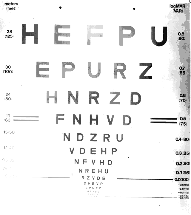
perspective. Science 255:419, 1992

7. Westheimer G: Visual acuity. In Moses RA, Hart WM (eds): Adler's Physiology of the Eye, 8th ed, p 415. St. Louis, CV Mosby, 1987

8. Goodrich GL, Apple LE, Frost A et al: A preliminary report on experienced closed-circuit television users. Am J Optom Physiol Opt 53:7, 1976

9. Bullimore MA, Bailey IL, Wacker RT: Contrast sensitivity and functional performance in low vision. Invest Ophthalmol Vis Sci (suppl) 31:598, 1990

10. Bullimore MA, Bailey IL, Wacker RT: Face recognition in age-related maculopathy. Invest Ophthalmol Vis Sci 32:2020, 1991

11. Thibos LN, Bradley A: New methods for discriminating neural and optical losses of vision. Optom Vis Sci 70:279, 1993

12. Snellen H: Probebuchstaben zur Bestimmung der Sehscharfe. Utrecht, PW van de Weijer, 1862

13. Bailey IL, Lovie JE: New design principles for visual acuity letter charts. Am J Optom Physiol Opt 53:740, 1976

14. Ferris FL, Kassoff A, Bresnick GH, Bailey IL: New visual acuity charts for clinical research. Am J Ophthalmol 94:91, 1982

15. Bailey IL: New procedures for detecting early vision losses in the elderly. Optom Vis Sci 70:299, 1993

16. Bailey IL, Bullimore MA, Raasch TW, Taylor HR: Clinical grading and the effect of scaling. Invest Ophthalmol Vis Sci 32:422, 1991

17. Optic Neuritis Study Group. The clinical profile of optic neuritis: Experience of the Optic Neuritis Treatment Trial. Arch Ophthalmol 109:1673, 1991

18. Early Treatment Diabetic Retinopathy Study Research Group: Early Treatment Diabetic Retinopathy Study Design and Baseline Patient Characteristics. ETDRS Report Number 7. Ophthalmology 98:741, 1991

19. Early Treatment Diabetic Retinopathy Study Research Group: Early photocoagulation for diabetic retinopathy. ETDRS Report Number 9. Ophthalmology 98:766, 1991

20. Shlaer S: The relation between visual acuity and illumination. J Gen Physiol 21:165, 1937

21. Simpson TL, Barbeito R, Bedell HE: The effect of optical blur on visual acuity for targets of different luminances. Ophthalmic Physiol Opt 6:279, 1981

22. Sloan LL: Measurement of visual acuity. A critical review. AMA Arch Ophthalmol 45:704, 1951

23. Regan D, Neima D: Low-contrast letter charts as a test of visual function. Ophthalmology 90:1192, 1983

24. Prince JH, Fry GA: The effect of errors of refraction on visual acuity. Am J Optom Arch Am Acad Optom 33:353, 1956

25. Peters HB: The relationship between refractive error and visual acuity at three age levels. Ann Am Acad Optom 38:194, 1961

26. Borish IM: Clinical Refraction, pp 368–371. Chicago, The Professional Press, 1975

27. Leibowitz HW: The effect of pupil size on visual acuity for photometrically equated test fields at various levels of luminance. J Opt Soc Am 42:416, 1952

28. Westheimer G: Pupil size and visual resolution. Vision Res 4:39, 1964

29. Mandelbaum J, Sloan LL: Peripheral visual acuity. Am J Ophthalmol 30:581, 1947

30. Kerr JL: Visual resolution in the periphery. Perception and Psychophysics 9:375, 1971

31. Johnson CA, Keltner JL, Balestrery FG: Effects of target size and eccentricity on visual detection and resolution. Vision Res 18:1217, 1978

32. Bartley SH, Ball RJ: Effects of intermittent illumination on visual acuity. Ann Am Acad Optom 45:458, 1968

33. Baron WS, Westheimer G: Visual acuity as a function of exposure duration. J Opt Soc Am 63:212, 1973

34. Graham CH, Cook C: Visual acuity as a function of intensity and exposure duration. Am J Psychol 49:654, 1937

35. Sloan LL, Rowland WM, Altman A: Comparison of three types of test target for the measurement of visual acuity. Q Rev Ophthalmol 8:4, 1952

36. White JM, Loshin DS: Grating acuity overestimates Snellen acuity in patients with age-related maculopathy. Optom Vis Sci 66:751, 1989

37. Herse PR, Bedell HE: Contrast sensitivity for letter and grating targets under various stimulus conditions. Optom Vis Sci 66:774, 1989

38. Flom MC, Weymouth FW, Kahneman D: Visual resolution and contour interaction. J Opt Soc Am 53:1026, 1963

39. Flom MC, Heath GG, Takahashi E: Contour interaction and visual resolution: Contralateral effects. Science 142:979, 1963

40. Stuart J, Burian HM: A study of separation difficulty: Its relationship to visual acuity in normal and amblyopic eyes. Am J Ophthalmol 55:47, 1962

41. Dobson V, Teller D: Visual acuity in human infants: A review and comparison of behavioral and electrophysiological studies. Vision Res 18:1469, 1978

42. Norcia AM, Tyler CW: Spatial frequency sweep VEP; visual acuity during the first year of life. Vision Res 25:1399, 1985

43. Fern KD, Manny RE: Visual acuity of the preschool child: A review. Am J Optom Physiol Opt 63:319, 1986

44. Preston KL, McDonald M, Sebris SL et al: Validation of the acuity card procedure for assessment of infants with ocular disorders. Ophthalmology 94:644, 1987

45. Haegerstrom-Portnoy G: New procedures for evaluating vision functions of special populations. Optom Vis Sci 70:306, 1993

46. Ginsberg AP: Introduction. In Proenza LM, Enoch JM, Jampolsky A (eds): Clinical Applications of Visual Psychophysics, pp 4–10. Cambridge, Cambridge 
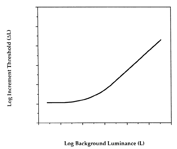
University Press, 1981

47. Campbell FW, Robson JG: Application of Fourier analysis to the visibility of gratings. J Physiol (Lond) 197:551, 1968

48. Bradley A, Ohzawa I: A comparison of contrast detection and discrimination. Vision Res 26:991, 1986

49. Hess RF, Bradley A: Contrast perception above threshold is only minimally impaired in human amblyopia. Nature 287:463, 1980

50. Legge G, Kersten D: Contrast discrimination in peripheral vision. J Opt Soc Am 4:1594, 1987

51. Ginsberg AP: Spatial filtering and vision: Implications for normal and abnormal vision. In Proenza LM, Enoch JM, Jampolsky A (eds): Clinical Applications of Visual Psychophysics, pp 70–106. Cambridge, Cambridge University Press, 1981

52. Enroth-Cugell C, Robson JG: The contrast sensitivity of retinal ganglion cells of the cat. J Physiol (Lond) 187:517, 1966

53. Bishop PO: Processing of visual information within the retinostriate system. In Darian-Smith I (ed): Handbook of Physiology. Baltimore, American Psychological Society, 1984

54. Kaplan E, Shapley RM: The primate retina contains two types of ganglion cells, with high and low contrast sensitivity. Proc Natl Acad Sci USA 63:2755, 1986

55. Schade OH: Optical and photoelectric analog of the eye. J Opt Soc Am 46:721, 1956

56. Campbell FW, Green DC: Optical and retinal factors affecting visual resolution. J Physiol (Lond) 181:576, 1965

57. Westheimer G: Modulation thresholds for sinusoidal light distributions on the retina. J Physiol (Lond) 152:67, 1960

58. DePalma JJ, Lowry EM: Sine-wave response of the visual system. II. Sine wave and square-wave contrast sensitivity. J Opt Soc Am 52:328, 1962

59. Van Nes FL, Bouman MA: Spatial modulation transfer in the human eye. J Opt Soc Am 57:401, 1967

60. Kelly DH: Adaptation effects on spatiotemporal sine-wave thresholds. Vision Res 12:89, 1972

61. Koenderink JJ, Bouman MA, Bueno de Mesquita AE, Slappendel S: Perimetry of contrast detection thresholds of moving spatial sine wave patterns. IV. The influence of mean retinal illuminance. J Opt Soc Am 68:860, 1978

62. Hoekstra J, Van der Groot DPA, Van den Brink G, Bilsen FA: The influence of the number of cycles upon the visual contrast threshold for spatial sine wave patterns. Vision Res 14:365, 1974

63. Savoy RL, McCann JJ: Visibility of low frequency sine wave targets: Dependence on number of cycles. J Opt Soc Am 65:343, 1975

64. Koenderink JJ, Bouman MA, Bueno de Mesquita AE, Slappendel S: Perimetry of contrast detection thresholds of moving spatial sine wave patterns: III. The target extent as a sensitivity controlling parameter. J Opt Soc Am 68:854, 1978

65. Daitch JM, Green DG: Contrast sensitivity of the human peripheral retina. Vision Res 9:947, 1969

66. Koenderink JJ, Bouman MA, Bueno de Mesquita AE, Slappendel S: Perimetry of contrast detection thresholds of moving spatial sine wave patterns: II. The far peripheral visual field (eccentricity 0–50 deg). J Opt Soc Am 68:850, 1978

67. Koenderink JJ, Bouman MA, Bueno de Mesquita AE, Slappendel S: Perimetry of contrast detection thresholds of moving spatial sine wave patterns: I. The near peripheral visual field (eccentricity 0–8 deg). J Opt Soc Am 68:845, 1978

68. Hilz R, Cavonius CR: Functional organization of the peripheral retina: Sensitivity to periodic stimuli. Vision Res 14:1333, 1974

69. Henning GB: Spatial-frequency tuning as a function of temporal frequency and stimulus motion. J Opt Soc Am [A] 5:1362, 1988

70. Olzak LA, Thomas JP: Seeing spatial patterns. In Boff K, Kaufmann L, Thomas J (eds): Handbook of Perception and Human Performance. New York, John Wiley & Sons, 1986

71. Campbell FW, Levinson J: The effect of orientation on the visual resolution of gratings. J Physiol (Lond) 187:437, 1966

72. Zemon V, Gutowski W, Horton T: Orientational anisotropy in the human visual system: An evoked potential and psychophysical study. Int J Neurosci 19:259, 1983

73. Legge GE, Mullen KT, Woo GC, Campbell FW: Tolerance to visual defocus. J Opt Soc Am 4:851, 1987

74. Bradley A, Thomas T, Kalaher M, Hoerres M: Effects of spherical and astigmatic defocus on acuity and contrast sensitivity: A comparison of three clinical charts. Optom Vis Sci 68:418, 1991

75. Zadnik K, Mannis MJ, Johnson CA, Rich D: Rapid contrast sensitivity assessment in keratoconus. Am J Optom Physiol Opt 64:693, 1987

76. Hess RF, Garner LF: The effect of corneal edema on visual function. Invest Ophthalmol Vis Sci 16:5, 1977

77. Carney LG, Jacobs RJ: Mechanisms of visual loss in corneal edema. Arch Ophthalmol 102:1068, 1984

78. Mannis M, Zadnik K, Johnson CA: The effect of penetrating keratoplasty on contrast sensitivity in patients with keratoconus. Arch Ophthalmol 102:1513, 1984

79. Carney LG: Visual loss in keratoconus. Arch Ophthalmol 100:1282, 1982

80. Hess RF, Carney LG: Vision through an abnormal cornea: A pilot study of the relationship between visual loss from corneal distortion, corneal edema, keratoconus and some allied corneal pathology. Invest Ophthalmol Vis Sci 18:476, 1979

81. Zadnik K, Mannis MJ, Johnson CA: An analysis of contrast sensitivity in identical twins with keratoconus. Cornea 3:99, 1984

82. Mannis MJ, Zadnik K, Johnson C, Adams C: Contrast sensitivity after epikeratophakia. Cornea 7:280, 1988

83. Carney LG: Contact lens correction of visual loss in keratoconus. Acta Ophthalmol (Copenh) 60:795, 1982

84. Hess RF, Woo G: Vision through cataracts. Invest Ophthalmol Vis Sci 17:428, 1978

85. Elliott DB, Hurst MA: Simple clinical techniques to evaluate visual function in patients with early cataract. Optom Vis Sci 67:822, 1990

86. Adamsons I, Rubin GS, Vitale S: The effect of early cataracts on glare and contrast sensitivity. A pilot study. Arch Ophthalmol 110:1081, 1992

87. Whitaker D, Elliott DB: Simulating age-related optical changes in the human eye. Doc Ophthalmol 82:307, 1992

88. Morrison JD, Jay JL: Changes in visual function with normal ageing, cataract and intraocular lenses. Eye 7:20, 1993

89. Elliott DB, Hurst MA, Weatherill J: Comparing clinical tests of visual function in cataract with the patient's perceived visual disability. Eye 4:712, 1990

90. Chylack LT, Jakubicz G, Rosner B et al: Contrast sensitivity and visual acuity in patients with early cataracts. J Cataract Refract Surg 19:399, 1993

91. Krasnov MM, Avetisov SE, Makashova NV, Mamikonian VR: The effect of radial keratotomy on contrast sensitivity. Am J Ophthalmol 105:651, 1988

92. Tomlinson A, Caroline P: Effect of radial keratotomy on the contrast sensitivity function. Am J Optom Physiol Opt 65:803, 1988

93. Hess RF, Woo GC, White PD: Contrast attenuation characteristics of iris clipped intraocular lens implants in situ. Br J Ophthalmol 69:129, 1985

94. Akutsu H, Legge GE, Showalter M et al: Contrast sensitivity and reading through multifocal intraocular lenses. Arch Ophthalmol 110:1076, 1992

95. Olsen T, Corydon L: Contrast sensitivity as a function of focus in patients with the diffractive multifocal intraocular lens. J Cataract Refract Surg 16:703, 1990

96. Owsley C, Sekuler R, Siemsen D: Contrast sensitivity throughout adulthood. J Opt Soc Am 23:689, 1983

97. Rohaly AM, Owsley C: Modeling the contrast-sensitivity functions of older adults. J Opt Soc Am 10:1591, 1993

98. Cronin-Golomb A, Rizzo JF, Corkin S, Growdon JH: Visual function in Alzheimer's disease and normal aging. Ann N Y Acad Sci 640:28, 1991

99. Sokol S, Moskowitz A, Skarf B et al: Contrast sensitivity in diabetics with and without background retinopathy. Arch Ophthalmol 103:51, 1985

100. Trick GL, Burde RM, Gordon MO et al: The relationship between hue discrimination and contrast sensitivity deficits in patients with diabetes mellitus. Ophthalmology 95:693, 1988

101. Sala SD, Bertoni G, Somazzi L et al: Impaired contrast sensitivity in diabetic patients with and without retinopathy: A new technique for rapid assessment. Br J Ophthalmol 69:136, 1985

102. Kleiner RC, Enger C, Alexander MF, Fine SL: Contrast sensitivity in age-related macular degeneration. Arch Ophthalmol 106:55, 1988

103. Lennerstrand G, Ahlstrom CO: Contrast sensitivity in macular degeneration and the relation to subjective visual impairment. Acta Ophthalmol 67:225, 1989

104. Sjostrand J, Frisen L: Contrast sensitivity in macular disease. A preliminary report. Acta Ophthalmol 55:507, 1977

105. Wolkstein M, Atkin A, Bodis-Wollner I: Contrast sensitivity in retinal disease. Ophthalmology 87:1140, 1980

106. Higgins KE, Meyers SM, Jaffe MJ et al: Temporary loss of foveal contrast sensitivity associated with panretinal photocoagulation. Arch Ophthalmol 104:997, 1986

107. Marmor M: Contrast sensitivity and retinal disease. Ann Ophthalmol 13:1069, 1981

108. Arden GB, Jacobson JJ: A simple grating test for contrast sensitivity: Preliminary results indicate value in screening for glaucoma. Invest Ophthalmol Vis Sci 17:23, 1978

109. Stamper RL: Psychophysical changes in glaucoma. Surv Ophthalmol 33:309, 1989

110. Sample PA, Juang PSC, Weinreb RN: Isolating the effects of primary open-angle glaucoma on the contrast sensitivity function. Am J Ophthalmol 112:308, 1991

111. Wood JM, Lovie-Kitchin JE: Evaluation of the efficacy of contrast sensitivity measures for the detection of early primary open-angle glaucoma. Optom Vis Sci 69:175, 1992

112. Wood JM: Contrast sensitivity deficits in subjects with glaucoma-like discs using an oscilloscope-based method. Acta Ophthalmol 70:610, 1992

113. Ross JE, Bron AJ, Clarke DD: Contrast sensitivity and visual disability in chronic simple glaucoma. Br J Ophthalmol 68:821, 1984

114. Atkin A, Bodis-Wollner I, Wolkstein M et al: Abnormalities of central contrast sensitivity in glaucoma. Am J Ophthalmol 88:205, 1979

115. Storch RL, Bodis-Wollner I: Overview of contrast sensitivity and neuro-ophthalmic disease. In Nadler MP, Miller D, Nadler DJ (eds): Glare and Contrast Sensitivity for Clinicians. New York, Springer-Verlag, 1990

116. Arden GB, Gucukoglu AG: Grating test of contrast sensitivity in patients with retrobulbar neuritis. Arch Ophthalmol 96:1626, 1978

117. Regan D, Whitlock JA, Murray TJ, Beverly KI: Orientation-specific losses of contrast sensitivity in multiple sclerosis. Invest Ophthalmol Vis Sci 19:324, 1980

118. Regan D, Silver R, Murray TJ: Visual acuity and contrast sensitivity in multiple sclerosis--Hidden visual loss. Brain 100:563, 1977

119. Bulens C, Meerwaldt JD, van der Wildt GJ, Keemink CJ: Spatial contrast sensitivity in unilateral cerebral ischaemic lesions involving the posterior visual pathway. Brain 112:507, 1989

120. Bodis-Wollner I: Visual acuity and contrast sensitivity in patients with cerebral lesions. Science 178:769, 1972

121. Bodis-Wollner I, Diamond SP: The measurement of spatial contrast sensitivity in cases of blurred vision associated with cerebral lesions. Brain 99:695, 1976

122. Wall M: Sensory visual testing in idiopathic intracranial hypertension: Measures sensitive to change. Neurology 40:1859, 1990

123. Katz B, Melles RB, Schneider JA: Contrast sensitivity function in nephropathic cystinosis. Arch Ophthalmol 105:1667, 1987

124. Hess RF: Application of contrast-sensitivity techniques to the study of functional amblyopia. In Proenza LM, Enoch JM, Jampolsky A (eds): Clinical Applications of Visual Psychophysics, pp 11–41. Cambridge, Cambridge University Press, 1981

125. Leguire LE, Pappa KS, Kachmer ML et al: Loss of contrast sensitivity in cystic fibrosis. Am J Ophthalmol 111:427, 1991

126. Salapatek P: Behavioral and electrophysiological evaluation of the infant contrast sensitivity function. In Proenza LM, Enoch JM, Jampolsky A (eds): Clinical Applications of Visual Psychophysics, pp 130–147. Cambridge, Cambridge University Press, 1981

127. Banks M, Salapatek P: Contrast sensitivity function of the infant visual system. Vision Res 16:867, 1976

128. Norcia AM, Tyler CW, Hamer RD: Development of contrast sensitivity in the human infant. Vision Res 30:1475, 1990

129. Norcia AM, Tyler CW, Hamer RD, Wesemann W: Measurement of spatial contrast sensitivity with the swept contrast VEP. Vision Res 29:627, 1989

130. Wilson HR: Development of spatiotemporal mechanisms in infant vision. Vision Res 28:611, 1988

131. Swanson WH, Birch EE: Infant spatiotemporal vision: Dependence of spatial contrast sensitivity on temporal frequency. Vision Res 30:1033, 1990

132. Ginsberg AP, Evans DW, Sekuler R, Harp SA: Contrast sensitivity predicts performance in aircraft simulators. Am J Optom Physiol Opt 59:105, 1982

133. Norman J, Erlich S: Spatial frequency filtering and target identification. Vision Res 27:87, 1987

134. Evans DW, Ginsberg AP: Contrast sensitivity predicts age-related differences in highway sign discriminability. Hum Factors 27:637, 1985

135. Rubin GS, Legge GS: Predicting low-vision reading rates from measures of contrast sensitivity. In Optical Society of America: Technical Digest, Topical Meeting on Non-Invasive Assessment of the Visual System, WB5-1, Washington, DC, 1985

136. Brown B: Reading performance in low vision patients: Relation to contrast and contrast 
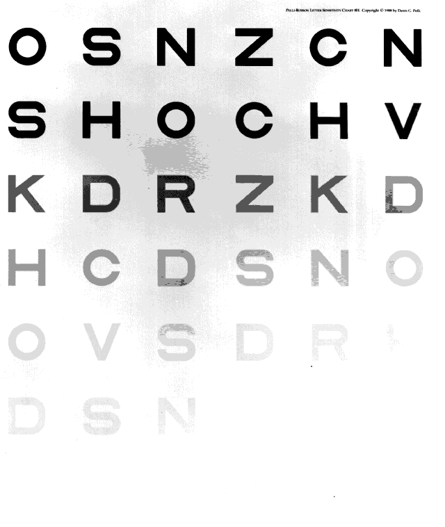
sensitivity. Am J Optom Physiol Opt 58:218, 1981

137. Leat SJ, Woodhouse JM: Reading performance with low vision aids: Relationship with contrast sensitivity. Ophthalmic Physiol Opt 13:9, 1993

138. Owsley C, Sekuler R, Boldt C: Aging and low contrast vision: Face perception. Invest Ophthalmol Vis Sci 21:362, 1981

139. Owsley C, Sloane ME: Contrast sensitivity, acuity and the perception of “real-world” targets. Br J Ophthalmol 71:791, 1987

140. Marron JA, Bailey IL: Visual factors and orientation-mobility performance. Am J Optom Physiol Opt 59:413, 1982

141. Ginsberg AP: A new contrast sensitivity vision test chart. Am J Optom Physiol Opt 61:403, 1984

142. Regan D, Neima D: Low contrast letter charts as a test of visual function. Ophthalmology 90:1192, 1983

143. Regan D, Neima D: Low-contrast letter charts in early diabetic retinopathy, ocular hypertension, glaucoma and Parkinson's disease. Br J Ophthalmol 68:885, 1981

144. Pelli DG, Robson JG, Wilkins AJ: The design of a new letter chart for measuring contrast sensitivity. Clinical Vision Science 2:187, 1988

145. Rubin GS: Reliability and sensitivity of clinical contrast sensitivity tests. Clinical Vision Science 2:169, 1988

146. Elliott DB, Bullimore MA, Bailey IL: Improving the reliability of the Pelli-Robson contrast sensitivity test. Clinical Vision Science 6:471, 1991

147. Elliott DB, Whitaker D: Clinical contrast sensitivity chart evaluation. Ophthalmic Physiol Opt 12:275, 1992

148. Leguire LE: Do letter charts measure contrast sensitivity? Clinical Vision Science 6:391, 1991

149. Regan D: Do letter charts measure contrast sensitivity? Clinical Vision Science 6:401, 1991

150. Pelli DG, Robson JG: Are letters better than gratings? Clinical Vision Science 6:409, 1991

151. Weber EH: In Boring EG: A History of Experimental Psychology. New York, Appleton-Century-Crofts, 1950

152. Aulhorn E, Harms H: Visual perimetry. In Jameson D, Hurvich LM (eds): Handbook of Sensory Physiology, Vol VII/4. Berlin, Springer-Verlag, 1972

153. Johnson CA, Keltner JL, Balestrery FG: Static and acuity profile perimetry at various adaptation levels. Doc Ophthalmol 50:371, 1981

154. Johnson CA, Keltner JL: Optimal rates of movement for kinetic perimetry. Arch Ophthalmol 105:73, 1987

155. Anderson DR: Perimetry With and Without Automation. St. Louis, CV Mosby, 1987

156. Johnson CA: The test logic of automated perimetry. ACTA:XXIV International Congress of Ophthalmology, pp 151–155. Philadelphia, JB Lippincott, 1983

157. Johnson CA: Role of automation in new instrumentation. Optom Vis Sci 70:288, 1993

158. Katz J, Tielsch JM, Quigley HA et al: Automated suprathreshold screening for glaucoma: The Baltimore Eye Survey. Invest Ophthalmol Vis Sci 34:3271, 1993

159. Johnson CA, Keltner JL: The incidence of visual field loss in 20,000 eyes and its relationship to driving performance. Arch Ophthalmol 101:371, 1983

160. Keltner JL, Johnson CA: Screening for visual field abnormalities with automated perimetry. Surv Ophthalmol 28:175, 1983

161. Johnson CA, Keltner JL: Automated suprathreshold static perimetry. Am J Ophthalmol 89:731, 1980

162. Hedin A, Lovsund P: Effects of visual field defects on driving performance. Doc Ophthalmol Proc Ser 49:541, 1987

163. Frisen L: Clinical Tests of Vision. New York, Raven Press, 1990

164. Harrington DO: The Visual Fields: A Textbook and Atlas of Clinical Perimetry. St. Louis, CV Mosby, 1981

165. Reed H, Drance SM: The Essentials of Perimetry, Static and Kinetic. London, Oxford University Press, 1972

166. Tate GW, Lynn JR: Principles of Quantitative Perimetry: Testing and Interpreting the Visual Field. New York, Grune & Stratton, 1977

167. Ellenberger C: Perimetry: Principles, Techniques, and Interpretation. New York, Raven Press, 1980

168. Johnson CA: Modern developments in clinical perimetry. Current Opinion in Ophthalmology 4:7, 1993

169. Enoch JM: The two-color threshold technique of Stiles and derived component color mechanisms. In Jameson D, Hurvich LM (eds): Handbook of Sensory Physiology, Vol VII/4. Berlin, Springer-Verlag, 1972

170. Sample PA, Weinreb RN, Boynton RM: Isolating color vision loss of primary open angle glaucoma. Am J Ophthalmol 106:686, 1988

171. Heron G, Adams AJ, Husted R: Central visual fields for short wavelength sensitive pathways in glaucoma and ocular hypertension. Invest Ophthalmol Vis Sci 29:64, 1988

172. Johnson CA, Adams AJ, Lewis RA: Automated perimetry of short-wavelength sensitive mechanisms in glaucoma and ocular hypertension. Preliminary findings. In Heijl A (ed): Perimetry Update 1988/89, pp 31–37. New York, Kugler and Ghedini, 1989

173. Sample PA, Weinreb RN: Color perimetry for assessment of primary open angle glaucoma. Invest Ophthalmol Vis Sci 31:1869, 1990

174. Weinreb RN, Sample PA: Short-wavelength visual field testing in eyes with primary open angle glaucoma. In Krigelstein GK (ed): Glaucoma Update IV, pp 146–155. Berlin, Springer-Verlag, 1991

175. Adams AJ, Johnson CA, Lewis RA: S cone pathway sensitivity loss in ocular hypertension and early glaucoma has nerve fiber bundle pattern. Proceedings of the 10th Symposium of the International Research Group on Colour Vision Deficiencies, pp 535–542. The Netherlands, Kluwer Academic Publishers, 1991

176. Sample PA, Weinreb RN: Progressive color visual field loss in glaucoma. Invest Ophthalmol Vis Sci 33:240, 1992

177. Sample PA, Taylor JDN, Martinez G et al: Short wavelength color visual fields in glaucoma suspects at risk. Am J Ophthalmol 115:225, 1993

178. Sample PA, Martinez GA, Weinreb RN: Color visual fields: A 5 year prospective study in eyes with primary open angle glaucoma. In Mills RP (ed): Perimetry Update 1992/93, pp 467–473. New York, Kugler Publications, 1993

179. Johnson CA, Adams AJ, Casson EJ, Brandt JD: Blue-on-yellow perimetry can predict the development of glaucomatous visual field loss. Arch Ophthalmol 111:645, 1993

180. Johnson CA, Adams AJ, Casson EJ, Brandt JD: Progression of early glaucomatous visual field loss for blue-on-yellow and standard white-on-white automated perimetry. Arch Ophthalmol 111:651, 1993

181. Johnson CA, Adams AJ, Casson EJ: Blue-on-yellow perimetry: A five year overview. In Mills RP (ed): Perimetry Update 1992/93, pp 459–466. New York, Kugler Publications, 1993

182. Frisen L: A computer graphics visual field screener using high-pass spatial frequency resolution targets and multiple feedback devices. Doc Ophthalmol Proc Series 49:441, 1987

183. Wall M, Conway MD, House PH, Allely R: Evaluation of sensitivity and specificity of spatial resolution and Humphrey automated perimetry in pseudotumor cerebri patients and normal subjects. Invest Ophthalmol Vis Sci 32:3306, 1991

184. Wall M: High-pass resolution perimetry in optic neuritis. Invest Ophthalmol Vis Sci 32:2525, 1991

185. Bynke H: Evaluation of high-pass resolution perimetry in neuro-ophthalmology. In Mills RP, Heijl A (eds): Perimetry Update 1990/91, pp 143–149. Amsterdam, Kugler Publications, 1991

186. Lindblom B, Hoyt WF: High-pass resolution perimetry in neuro-ophthalmology. Ophthalmology 99:700, 1992

187. Sample PA, Ahn DS, Lee PC, Weinreb RN: High-pass resolution perimetry in eyes with ocular hypertension and primary open-angle glaucoma. Am J Ophthalmol 113:309, 1992

188. Drum B, Breton M, Massof R et al: Pattern discrimination perimetry: A new concept in visual field testing. Doc Ophthalmol Proc Series 49:433, 1987

189. Stewart WC, Kelly DM, Hunt HH: Long-term and short-term fluctuation in pattern discrimination perimetry. Am J Ophthalmol 114:302, 1992

190. Drum B, Bissett R: Optimizing dot size and contrast in pattern discrimination perimetry. In Mills RP, Heijl A (eds): Perimetry Update 1990/91, pp 373–380. Amsterdam, Kugler Publications, 1991

191. Nutaitis MJ, Stewart WC, Kelly DM et al: Pattern discrimination perimetry in patients with glaucoma and ocular hypertension. Am J Ophthalmol 114:297, 1992

192. Drum B, Severns M, O'Leary D et al: Pattern discrimination and light detection test different types of glaucomatous damage. In Heijl A (ed): Perimetry Update 1988/89, pp 341–347. Amsterdam, Kugler Publications, 1989

193. Drum B, Severns M, O'Leary D et al: Selective loss of pattern discrimination in early glaucoma. Appl Opt 28:1135, 1989

194. Lachenmayr BJ, Drance SM, Douglas GR, Mikelberg FS: Light-sense, flicker and resolution perimetry in glaucoma: A comparative study. In Mills RP, Heijl A (eds): Perimetry Update 1990/91, pp 351–356. Amsterdam, Kugler Publications, 1991

195. Lachenmayr BJ, Drance SM, Chauhan BC et al: Diffuse and localized glaucomatous field loss in light-sense, flicker and resolution perimetry. Graefes Arch Clin Exp Ophthalmol 229:267, 1991

196. Lachenmayr B, Rothbacher H, Gleissner M: Automated flicker perimetry versus quantitative static perimetry in early glaucoma. In Heijl A (ed): Perimetry Update 1988/89, pp 359–368. New York, Kugler and Ghedini, 1989

197. Casson EJ, Johnson CA, Shapiro LR: Longitudinal comparison of temporal-modulation perimetry with white-on-white and blue-on-yellow perimetry in ocular hypertension and early glaucoma. J Opt Soc Am 10:1792, 1993

198. Casson EJ, Johnson CA: Temporal modulation perimetry in glaucoma and ocular hypertension. In Mills RP (ed): Perimetry Update 1992/1993, pp 443–450. Amsterdam, Kugler Publications, 1993

199. Casson EJ, Osako M, Johnson CA, Hwang P: Temporal and spatial response properties of optic neuritis patients manifesting statokinetic dissociation. Appl Opt 30:2136, 1991

Back to Top