Chapter 15
Light and Photometry
RAYMOND E. RECORDS
Main Menu   Table Of Contents

Search

NATURE OF LIGHT
PHOTOMETRY
LUMINANCE
CHARACTERISTICS OF COMMON LIGHT SOURCES
LIGHT AND THE EYE
TOXICITY OF LIGHT
TOXIC EFFECTS OF LIGHT ON THE EYE
REFERENCES

NATURE OF LIGHT
The eye is a massive, highly specialized exteroceptor that provides some 40% to 50% of the total sensory input reaching the cerebral hemispheres. The sole stimulus for vision is reflected or transmitted light, although certain x-rays or even external pressure on the globe that distorts the retina can produce circular but otherwise unformed visual sensations. In the absence of light, the entire visual system, including the refractive and neural portions of the eyeball itself, the neuromuscular mechanism, the visual pathways, and the visual receptive and association areas in the occipital cortex, ceases to function in any meaningful way. Light, the basic stimulus for vision, is a small portion of the electromagnetic spectrum of energy. Such electromagnetic radiation is a form of energy that can be transmitted through a vacuum and that produces a continuous spectrum of enormous dimensions extending from short cosmic rays, a few femtometers (10- 15 m) in wavelength, to power transmission, in which a single wavelength may be as long as 1.6 × 10⁸ m (Fig. 1). Light is defined as that portion of the electromagnetic spectrum that can stimulate the photoreceptors of the human retina to produce a visual response. The shortest wavelengths detectable by the human retina are generally about 380 nm, whereas the longest wavelengths end at approximately 760 nm. Electromagnetic radiation (light) falling within these limits passes through the cornea, lens, and other media of the eye to reach the retina. Upon reaching the outer or photoreceptor layer of the retina, light is converted into an electrical signal by polarization of N-retinylidene the visual chromophore present as a component of the four light-sensitive pigments contained in the photoreceptor outer segments.1 Light will make visible to the human eye anything from which it is either emitted or reflected, provided that the quantity reaching the retinal photoreceptors exceeds the stimulation threshold for those receptors at that exact moment.

Fig. 1. The electromagnetic spectrum of energy extending from very short cosmic rays through the extremely long wavelengths involved in electric power transmission. Visible light makes up only a small portion of the total energy spectrum. (Modified from Kaufman JE, Christensen JF [eds]: I.E.S Lighting Handbook, 5th ed. New York, Illuminating Engineering Society, 1972)

A basic characteristic of all electromagnetic energy, including light, is that it is produced by electrons that have been excited by an external force and have just reverted to a more stable position, thereby releasing energy. Such electromagnetic energy travels in a straight line (rectilinear propagation) from one point to another as a series of waves alternating perpendicular to one another and transverse to the direction of propagation (Fig. 2). Light and all other electromagnetic radiation travel at the same speed, which is approximately 3 × 1010 cm/sec in a vacuum. The speed of propagation is only slightly slower in clear air, and the decrease in velocity is inconsequential for physiologic purposes. In denser transparent media, such as the cornea, water, or optical glass, the velocity is much less, and it is this slowing of light rays by the denser media that is described by Snell's law or the refraction law. The ability of glass, plastic, and other transparent material to slow the progress of a beam of light makes possible the construction of lens systems to bring rays of light to a focus or diverge them to correct optical abnormalities of the human eye. The same principles apply to the anterior segment of the eye in its ability to take parallel or diverging rays of light and focus them on the retina to produce a sharp image rather than only an unformed sensation of lightness or darkness. The refractive indices of some common transparent materials are shown in Table 1.

Fig. 2. The wavelength of light can be measured from peak to peak or from any other position as desired (upper illustration). Light is constantly vibrating in a plane normal (90°) to the direction of propagation (middle figure). If a cross section of a ray of light is made, unpolarized light is found vibrating in all planes normal to the direction of propagation (A). Partially polarized light has a vibration in one plane that is greater than that in another (B). Plane polarized light is vibrating in only two directions normal to the direction of propagation (C).


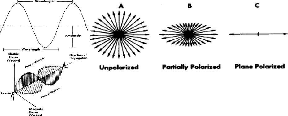
TABLE 1. Refractive Indices of the Eye and Various Common Substances


SubstanceRefractive Index
Cornea1.376
Aqueous humor1.336
Lens1.410
Vitreous humor1.336
Ice at 0°C1.312
Distilled H2 O at 20°C1.33
Polymethyl methacrylate1.491
Light crown glass1.515
Flint glass1.570–1.751
Diamond2.417


Visible light has three important characteristics, one of which is wavelength. This is the distance between any two similar given points on a single wave of light. It is usually recorded as the distance from one crest to the next adjoining crest (see Fig. 2). Wavelength is abbreviated lambda (λ), and of all the physical dimensions it is the most important in determining the color of light. Another important characteristic is the speed of light, which was discussed earlier. The third is frequency, which is usually abbreviated V and is the number of complete cycles moving past a specific point over a given period of time. Velocity, wavelength, and frequency are related in the following manner (equation 1):


Velocity (V) = wavelength (λ) × frequency (ν)

Frequency is a constant, and consequently wavelength and velocity are reciprocally related. If velocity decreases as a ray of light passes through crown glass, then the wavelength must shorten so that the frequency will remain constant (Fig. 3).

Fig. 3. Wavelength, velocity, and frequency characteristics of light in air and glass. (Riggs LA: Wavelength, velocity, and frequency characteristics of light in air and glass. In Graham CH [ed]: Vision and Visual Perception, Chapter 1. New York, John Wiley & Sons, 1965)

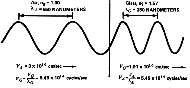
The speed of light can be measured in a variety of ways. One simple and direct method of such a measurement is the use of mirrors rotating at a (known speed and separated by a known distance. Wavelength can be measured by an interferometer. This somewhat cumbersome and delicate laboratory instrument uses an interference grating and will measure exactly the wavelength of any given test light. If the wavelength and velocity are known, the frequency is easily calculated from equation 1.

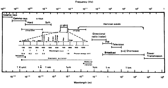
The exact nature of light is still unknown, although a number of theories that have been deduced from the physical behavior of light are useful for descriptive purposes. The foremost theories are those of Newton and Huygens. Newton in 1672 held that light is a particle or corpuscle that travels in a straight line from its source to the eye or from its source to an object, where it is then reflected into the eye. Newton's concept of light is still quite useful today when an attempt is made to describe the physical effects of light that result in transformation of the energy contained in the light ray. An example of this energy transformation is light falling on photographic film or the photochemical effect of light on the retinal pigments or on the basal cells of the skin. The toxic effects of light and its carcinogenic properties are also best explained in terms of particles impacting on a molecule and producing a chemical change therein. Such energy transformation can be considered in terms of light as a nonmaterial particle of minimal energy, a single unit of which is termed a photon or quantum. These terms were coined by Planck, who believed that light energy was released and absorbed in discrete quanta or photons. The retinal photoreceptors do seem to behave as true quantum detectors rather than as simple energy detectors, in that a single quantum of light absorbed by one molecule of retinal pigment located in a photoreceptor outer segment containing some 2 × 10⁸ other unaffected molecules is a sufficient stimulus to activate the receptor.2 The energy for a given quantum of light is least for the longest wavelengths and greatest for the shortest (see Fig. 1). Therefore, the energy contained in a given number of quanta is directly proportional to their frequency and inversely proportional to their wavelength. The energy (E) so contained can be calculated by the following formula (equation 2):


E = h·ν

where h is Planck's constant 6.6256 × 10⁻²⁷ joule-seconds, and ν is the frequency in hertz. The amount of available energy from the short violet end of the spectrum is about twice that from the longer red end (see Fig. 1). This is an important consideration when determining the heat-generating properties of light. When considering stimulation of the retinal photoreceptors, the total energy present in any given quantum of light is unimportant. The critical factor is the wavelength of the incident light and the ability of the photopigment to absorb it. If a specific wavelength of light can be absorbed by photopigment, the resultant reactions will be of the same magnitude regardless of whether the wavelength is long or short. Generally, Newton's concept (with Planck's modification) of light as a particle or corpuscle is most useful in describing the events that occur in photochemical reactions, photoelectric cell function, and other light-induced chemical or physical reactions.

The wave theory suggested by Huygens in 1678 states that light is generated by molecular vibrations in a luminous material and that these vibrations are transmitted through a transparent medium as waves whose movements are perpendicular to the direction of propagation. Maxwell in 1865 proposed that such wave motion is electromagnetic in nature and that light is similar in nature to all other electromagnetic energy forms that can be transmitted through a vacuum.3 The wave theory is most useful in describing the behavior of light passing through gratings or other small openings and thereby generating interference phenomena. It is also useful in describing the refraction of light by media of varying optical densities, including optical lenses, the cornea, and the lens of the eye. As noted previously, Snell was the first to describe the behavior of a ray of light passing from a medium of one optical density into or through a medium of dissimilar optical density. Snell's description of the behavior of light under these conditions is known as Snell's law or the refraction law. It states that the ratio of the sine of the angle of incidence to the sine of the angle of refraction of a ray of light is equal to the ratio of the velocity in the first medium to the velocity in the second medium. The refractive index of any given optical medium is the ratio of the speed of light in a vacuum to the speed of light in the material in question. Using the refractive index in Snell's equation rather than the velocity greatly simplifies the resulting calculation (equation 3):


n1 sine1 = n2 sine2

Here, n1 is the index of refraction of the first medium, sine1 is the sine of the angle of incidence, n2 is the index of refraction of the second optical medium, and sine2 is the sine of the angle of the refracted ray. Snell's law is one of the fundamental principles of geometric and physiologic optics.

Back to Top
PHOTOMETRY
Wavelength, frequency, and speed are all important physical characteristics of light. However, none of these physical measurements is very useful in determining the value of any given light source for performing visual tasks, photographic applications, or useful interactions in nonoptical biologic systems such as skin tanning or growing plants. Photometry is a branch of physical optics that is primarily concerned with the standardization and measurement of visible light. To a lesser degree, it is also involved with near and far ultraviolet and infrared energy measurement. The photometric literature also suggests standards for the illumination (light) needed to best carry out visual tasks such as suturing a cataract extraction wound, drafting a blueprint or other engineering drawing, reading a book, or illuminating a runway to land a heavy transport aircraft on a dark, foggy night. Table 2 lists standard units, symbols, and defining equations for fundamental photometric and radiometric quantities. Photometric units are applied to light, whereas radiometric units measure quantities of radiant energy.


TABLE 2. Standard Units, Symbols, and Defining Equations for Fundamental Photometric and Radiometric Quantities*


Quantity†Symbol†Defining EquationUnitSymbolic Abbreviation
Radiant energyQ ergerg
   jouleJ
   caloriecal
   kilowatt-hourkWh
Radiant densityww =dQ/dVjoule per cubi meter‡J/m3
   erg per cubic centimetererg/cm3
Radiant fluxΦΦ = dQ/dterg per seconderg/s
   watt‡W
Radiant flux density at a surface    
 Radiant exitance (Radiant emitance)§MM = d Φ/dAwatt per square centimeterW/cm2
   watt per square meter,‡.etc 
IrradianceEE = d Φ/dAwatt per square centimeterW/m2
   watt per square meter,‡.etc 
Radiant intensityII = d Φ/d ωwatt per steradian‡W/sr
  (ω = solid angle through which flux from point source is radiated)  
RadianceLL = d2Φ/dω(dA cos )watt per steradian and square centimeterW/sr · cm2
   = dI/(dA cos θ)  
  (θ = angle between line of sight and normal to surface considered)watt per steradian and square meter‡W/sr · m2
Emissivityεε = M /M blackbodyone (numeric) 
  (M and Mblackbodyare respectively the radiant exitance of the measured specimen and that of a blackbody at the same temperature as the specimen)  
Absorptanceαα = Φaione (numeric) 
Reflectanceρρ = Φrione (numeric) 
Transmittanceττ = Φtione (numeric) 
Luminous energy (quantity of light)QQv=K(λ)Qdλlumen-hourlm · h
   lumen-second‡ (talbot)lm · s
Luminous densityωω = dQ/dVlumen-hour per cubic centimeter‡ lm · h/cm3
Luminous fluxΦΦ = dQ/dtlumen‡lm
Luminous flux density at a surface    
 Luminance exitance (Luminous emitance)§MM = d Φ/dAlumen per square footlm/ft2
Illumination (Illuminance)EE = d Φ/dAfoot-candle (lumen per square foot)fc
   lux (lm/m2)‡lx
   phot (lm/cm2)ph
Luminous intensity (candlepower)II = d Φ/d ωcandela‡ (lumen per steradian)cd
   (ω = solid angle through which flux from point source is radiated)  
Luminance (photo metric brightness)LL = d 2 Φ/d ω(dA cos )candela per unit areacd/in2, etc.
   = dI/(dA cos θ)stilb (cd/cm2)sb
   (θ = angle between line of sight and normal to surface considered)nit (cd/m2‡)nt
   foot lambert (cd/πft2)fL
   lambert (cd/πcm2)L
   apostilb (cd/πm2)asb
Luminous efficacyKK = Φv/Φelumen per watt‡lm/W
Luminous efficiencyVV = K/Kmaximumone (numeric) 
  (Kmaximum = maximumvalue of K (λ) function)  

* The symbols for photometric quantities are the same as those for the corresponding radiometric quantities. When it is necessary to differentiate them, the subscripts v and e, respectively, should be used (e.g., Qv and Qe).
† Quantities may be restricted to a narrow wavelength band by adding the word spectral and indicating the wavelength. The corresponding symbols are changed by adding a subscript λ (e.g., Qλ) for a spectral concentration or a λ in parentheses (e.g., K[λ]) for a function of wavelength.
‡ International System (SI) unit.
§ To be deleted.
Φi, incident flux, Φa, absorbed flux, Φr, reflected flux, Φt, transmitted flux.


Light, by definition, is that portion of the electromagnetic spectrum that can be absorbed by the photopigments of human retina to result in a visual sensation. It is apparent by this definition that the best metric of light should be the human eye, and this is generally true for light that is used to perform visual tasks. When measuring light that is not used for vision, such as turning on and off a street light or operating a photodetector system, other standards of measurement may be used. To quantify the brightness of any light, standards must be applied so that one can describe the amount of light being emitted from any system or falling on a surface over a given period of time. When considering light-measuring systems, several important principles must be kept in mind. The first is the inverse square law. This principle states that the amount of radiation (E) reaching the surface varies inversely with the square of the distance (d) from the source of the radiation (I) (equation 4).

Another important principle is Lambert's cosine law. This states that if the source of illumination (I) is not normal (90°) to the surface upon which it falls, the radiant energy reaching that surface varies directly with the cosine of the angle of the incidence () (equation 5) (Fig. 4B):

Fig. 4. A. The inverse square law, which states that the amount of light continually diverges as one moves away from the source, thereby covering a greater area with lower intensity the greater the distance from the source. B. Lambert's cosine law: parallel rays of light falling on a plane surface will illuminate this surface directly in proportion to the cosine of the angle as measured normal to the irradiated surface. (Modified from Kaufman JE, Christensen JF [eds]: I.E.S. Lighting Handbook, 5th ed. New York, Illuminating Engineering Society, 1972)


E = I cosine θ

Both of these important concepts can be combined in a single formula (equation 6):

Photometers are instruments designed to measure visible light. They generally fall into two major categories: subjective instruments in which the human eye acts as the receptive and quantitative element, and photoelectric (objective) instruments in which light activates a photoelectric system that then generates an electric current that may be measured on a meter calibrated in various scale units. Among the former instruments are the Macbeth illuminometer and S.E.I. (Salford Electric Instruments) photometer (Fig. 5). The Macbeth illuminometer uses the inverse square law, in that both the standard light and the light to be tested are observed through the instrument with a sharp border between the two. The standard light is then moved closer or farther away from the screen until the brightness of the two lights balance. This comparison-matching scheme is known as minimum border contrast matching. A reading is then taken from a graduated scale that is part of the instrument housing. The S.E.I. photometer uses two neutral density wedges that are moved over or away from the standard light source by a vernier system to balance the standard and test lights (see Fig. 5).

Fig. 5. Top. Basic design of the Macbeth illuminometer. (E, eye position; D, comparison scale; K, rack and pinion; R, calibrated scale; T, test plate, S, standard lamp). Bottom. Schematic diagram of a Salford Electric Instrument Company photometer. (Hardy AC, Perrin FH: The Principles of Optics. New York, McGraw-Hill, 1932)

Other types of visual photometers include the Bunsen “grease spot” photometer, in which the screen is a spot of translucent paraffin. The Luckiesch-Taylor photometer uses a standard that is balanced against the test field by rotating a disk of increasing neutral optical density. The Lummer-Brodhun photometer uses an internal projection system in which the test and standard are projected onto a white opaque screen and the illumination of the standard source is adjusted until the border between the two disappears, again using subjective minimum border contrast matching as the end point.

Photoelectric photometers have largely replaced these older visual photometers. These newer instruments are simple to operate and convenient to use. They eliminate individual observer bias and variations among observers. Two major types are available. Photovoltaic cell meters operate from a photocell that directly converts incident light into an electric current that then operates a microammeter or galvanometer. The instrument can be calibrated directly in foot-candles or other units. Photoemissive or photoelectric meters also operate by use of a light-emittive photocell; however, power, instead of being generated by the photocell, is supplied by a battery or other external power source. Calibration can be in direct units such as foot-candles or other units as desired.

If such photoelectric instruments are used to measure light needed for various visual tasks and applications, then the spectral sensitivity of the instrument must closely match that of the photopic human retina. Some of these devices can also be used in low-light ranges that would be appropriate for scotopic vision. Here, a filter must be placed over the window through which light enters the instrument to shift the spectrum appropriately toward the shorter wavelengths, to more nearly match the scotopic light sensitivity with that of the human retina.

Most common light sources are supplied with a rating of the power consumed and not the amount of light produced: for example, a 25-watt incandescent light bulb or a 40-watt fluorescent tube. Large industrial lamps such as those used in search lights, flood lights, and airway beacons are often rated as to both the power consumed in watts and the light produced, usually expressed as candle power. The amount of light produced is difficult to relate directly to the power consumed because various sources have different degrees of efficiency: for example, an incandescent bulb is much less efficient in producing visible light than a fluorescent tube per watt of power expended.

A number of units are available to describe and quantify visible light illuminance. Some of these units, such as the foot-candle, are self-explanatory, whereas others, such as the nit, offer no clue as to their derivation or possible application (Table 3).


TABLE 3. Conversion Factors for Luminance Units


Multiply number of             
To obtain number of ↓ByStilbLambertCandle/in.2Candle/ft2Foot lambertMillilambertNitApostilbMilli-foot lambertMicrolambertMicro-foot lambertMicromillilambertMicromicrolambert
StilbCandle/cm213.183 × 10-11.550 × 10-11.076 × 10-33.426 × 10-43.183 × 10-410-43.183 × 10-53.426 × 10-73.183 × 10-73.426 × 10-103.183 × 10-103.183 × 10⁻¹³
LambertLumen/cm23.141614.869 × 10-13.382 × 10-31.076 × 10-310-33.1416 × 10-410-41.076 × 10-610-61.076 × 10-910-910-12
 Candle/in.26.4522.05416.944 × 10-32.210 × 10-32.054 × 10-36.452 × 10-42.054 × 10-42.210 × 10-62.054 × 10-62.210 × 10-92.054 × 10-92.054 × 10⁻¹²
 Candle/ft29.290 × 1022.957 × 1021.440 × 10213.183 × 10-12.957 × 10-19.290 × 10-22.957 × 10-23.183 × 10-42.957 × 10-43.183 × 10-72.957 × 10-72.957 × 10-10
Foot lambertLumen/ft22.919 × 1039.290 × 1024.524 × 1023.141619.290 × 10-12.919 × 10-19.290 × 10-210-39.290 × 10-410-69.290 × 10-79.290 × 10-10
Millilambert 3.1416 × 1031034.869 × 1023.3821.07613.1416 × 10-110-11.076 × 10-310-31.076 × 10-610-610-9
NitCandle/m21043.183 × 1031.550 × 101.076 × 103.4263.18313.183 × 10-13.426 × 10-33.183 × 10-33.426 × 10-63.183 × 10-63.183 × 10⁻⁹
Apostilb (asb)Lumen/m23.1416 × 1041044.869 × 1033.382 × 101.076 × 10103.141611.076 × 10-210-21.076 × 10-510-510-6
Milli-foot lambert 2.919 × 1069.290 × 1054.524 × 1053.1416 × 1031039.290 × 1022.919 × 1029.290 × 1019.290 × 10-110-39.290 × 10-19.290 × 10-7
Microlambert 3.1416 × 1031064.869 × 1053.382 × 1031.076 × 1031033.1416 × 1021021.07611.076 × 10310-310-6
Micro-foot lambert 2.919 × 1099.290 × 1084.524 × 1083.1416 × 1061069.290 × 1052.919 × 1059.290 × 1041039.290 × 10219.290 × 1019.290 × 10-2
Micromilli lambert 3.1416 × 1091094.869 × 1083.382 × 1061.076 × 1061063.1416 × 1051051.076 × 1031031.076110-3
Micromicro lambert 3.1416 × 101210124.869 × 10113.382 × 1091.076 × 1091093.1416 × 1081081.076 × 1061061.076 ×1031031

Example: X Foot lamberts = X × 3.426 × 10⁻⁴ Stilbs


There are four commonly used properties of light that are closely interrelated. They include (1) luminous intensity, (2) luminance (brightness), (3) luminous flux (light flow), and (4) illumination. Each of these measurable properties may have a particular value in describing the brightness of a light source such as a photocoagulator bulb, the amount of light given off per unit source of area (the sun), or the quantity of light passing through a fiberoptic cable connecting a laser tube with a slit lamp delivery system.

LUMINOUS INTENSITY

One of the measurable properties of a light source is its intensity. A source of light can be quite bright without being very intense, such as a penlight flashlight pointed toward an observer in a dark room. The light so produced is extremely bright, but it is very feeble in illuminating the room for any useful purpose. Conversely, a mercury vapor street light can be quite intense and illuminate a large area brightly without being very bright itself. Luminous intensity is a comparison measurement, and the worldwide standard is the candela (cd), for which the accepted abbreviation symbol is I. Candelae are calculated on the basis of a perfect or “ideal black-body” irradiation equal to solidifying platinum at a temperature of 2042°K. The luminous intensity of such a system is 60 candelae/cm2 when measured at the surface. Luminous intensity can be measured in a variety of ways, including a visual subjective direct match with a known standard light or with a visual or photoelectric photometer. For lights of differing color, one can use flicker photometry, which is based on temporal discrimination, or minimum border contrast matching, which is based on spatial discrimination. If the two lights are of quite different spectral value, one can successively match one wavelength band to another in stepwise fashion, so-called cascade matching.

Back to Top
LUMINANCE
Luminance is a characteristic of surface illumination. When the incident light increases in brightness, the perceived hues change. Long wavelength colors become more yellow, whereas shorter wavelength colors become more blue or green. This is known as the Bezold-Brücke hue shift. The apparent purity of any given hue, its saturation, or its chromatic strength increases with increasing brightness to reach a peak that is wavelength dependent.4

Luminance as a measure of surface illumination can be measured by a direct-reading photoelectric photometer. If the surface of the object to be measured is extremely bright, a glowing incandescent filament whose temperature is known can be used for comparison. An instrument that uses this principle is an optical pyrometer, which is used for measuring the temperature of large incandescent masses such as molten metal inside a blast furnace or other extremely hot systems. Such instruments are usually graduated in degrees so that the worker can apply the reading directly for purposes of regulation, casting, and other activities. Other than these highly specialized needs, a much more useful concept of light measurement is luminous flux.

LUMINOUS FLUX

The term luminous flux is an expression of the flow or transfer of light from a point or a given surface that may be uniform or nonuniform, normal to the direction of propagation. It is usually measured in lumens (lm). One lumen is the luminous flux radiating from a point source of light having an intensity of 1 candela within a unit solid angle (1 steradian). A steradian is a square projected on the surface of a sphere, each side of which is equal to the lengths of the radius of the sphere (Fig. 6). Luminous flux measured in lumens can be related to both power consumed and power irradiated, as well as to the luminous efficiency of the human eye. In relation to power consumed, most standard commercially available incandescent light bulbs irradiate approximately 15 to 20 lumens/watt of power consumed. Much of the power so used is expended as heat (Fig. 7). The concept of luminous flux is useful when considering how bright a light source must be to provide the illumination needed to carry out a specific visual task.

Fig. 6. Illustration of a hollow sphere having a total surface area of 12.57 (4π) square feet. The solid angle subtended by lines A, B, C, and D is 1 steradian. The source of the illumination is 1 foot from the inner wall of the sphere; thus, there is a luminous flux of 1 lumen falling on each square foot. The luminous intensity, therefore, is 1 candela. The illumination at any point on the sphere is 1 foot-candle (one lumen per square foot). It is assumed that the light source is a uniform point source of 1 candela located at the exact center of the sphere and that the surface of this sphere has zero reflectance. (Modified from Kaufman JE, Christensen JF: I.E.S. Lighting Handbook, 5th ed. New York, Illuminating Engineering Society, 1972)

Fig. 7. The emission spectrum of a common incandescent (tungsten filament) light bulb is compared with a “cool white fluorescent tube.” The peak irradiation of the incandescent bulb lies much farther in the nonvisible infrared area of the spectrum than does the wavelength of the maximal irradiation of the fluorescent tube. This illustrates the relative efficiency of fluorescent lighting in producing visible light, whereas incandescent bulbs produce a much greater portion of nonusable heat.

ILLUMINATION

Illumination of a surface (E) is the luminous flux (φ) falling on the surface per unit of area (a) (equation 7):

The commonly used units and conversion factors of illumination are shown in Table 4. If the illumination of the surface is not uniform, then one must consider each dissimilar element individually.


TABLE 4. Illumination Conversion Factors


1 lumen = 1/673 lightwatt1 watt-second = 103 ergs
1 lumen-hour = 60 lumen- minutes1 phot =1 lumen/cm2
1 foot-candle = 1 lumen/ft21 lux = 1 lumen/m2 =1 meter-candle
Multiply Numberof →   
To Obtain Number of ↓By    
 Foot- candlesLuxPhotMilliphot
Foot-candles10.09299290.929
Lux10.76110,00010
Phot0.001080.000110.001
Milliphot1.0760.11,0001


For practical everyday purposes in any given illuminated environment, the difference between the highest and the lowest illumination should not be more than 0.3. Lights should be placed at least 20° above the line of vision and often are best placed behind and also to each side of the worker. Because freedom from shadows is often an important consideration, several moderately bright sources of light are much better than a single brilliant source. This is easily tested by holding a pencil 10 cm above the work space. If a distinct shadow forms, the light source is not diffuse enough. Light placement should be such that no direct specular reflections strike the eye. This can be easily tested by placing a mirror on the work area.

Of interest to clinical practitioners is the spectral quality, the color, and the intensity of the light needed to adequately and comfortably carry out specific tasks.

LUMINOUS EFFICIENCY

The maximal sensitivity of the light-adapted (photopic) human eye is at approximately 550 nm (Fig. 8). Light at this wavelength is yellow green and is most efficient in stimulating the retinal cones. The efficiency of light as a photopic retinal stimulus decreases rapidly as one moves toward the shorter or longer wavelengths. In the dark-adapted (scotopic) eye, the peak of the sensitivity curve is shifted to shorter wavelengths, reaching its maximum at 507 nm (see Fig. 8). Cones are not stimulated by low light levels that stimulate the rods during scotopic vision. Rods are relatively insensitive to longer wavelengths of light toward the red end of the spectrum. Therefore, red goggles can be used to carry out photopic activities while allowing the retina simultaneously to undergo scotopic adaptation. Such adaptation goggles are often used by radiologists, fluoroscopists, and others who must do their work under scotopic conditions.

Fig. 8. Relative photopic and scotopic efficiency curves for the average normal human eye.

The four qualities of light can be measured in the environment and are useful for considering questions of general illumination or specialized applications of light energy. None of these directly addresses in any way the amount of illumination that reaches the retina or the image formation thereon. For purposes of standardization, comparison, and investigation, the illumination of the retina must be specified in some quantitative manner. The greatest variable when considering retinal illumination is pupillary area, which can vary by a factor of approximately 80 to 1 between maximal mydriasis and maximal miosis. The standard unit of retinal illumination is the troland. One troland is the illumination passing through the pupil of the human eye when viewing a 1-m2 flat surface with a luminance of 1 candela through a pupil 1 mm2 in area. Measuring retinal illumination in trolands makes no allowance for light loss in the cornea, the lens, or the vitreous. An elderly person with marked nuclear sclerosis or other cataractous change will have much lower retinal illumination because of light scattering and absorption than will a child with a crystal-clear lens.

Another important consideration is the Stiles-Crawford effect. Light entering the eye through the physiologic center of the pupil parallel to the visual axis will be maximally effective in stimulating the central retinal cones, resulting in the best contour discrimination, the best contrast, and therefore the best visual acuity. As light deviates from the physiologic center of the pupil, these functions are degraded by the alteration of the light ray-receptor orientation so that the foveal cones no longer act as efficient wave guides. By moving the light point of entry only 3 mm from the physiologic center of the pupil, photopic retinal image resolution is reduced by more than one-half that obtained when the light rays enter through the physiologic center of the pupil. This effect appears to be present only for the retinal cones, in that light entering obliquely has only a minor effect in decreasing scotopic stimulus values.5 The Stiles-Crawford effect greatly decreases the significance of pupil size for photopic visual functions.

Amblyopic eyes show maximum sensitivity near the pupillary margin. After retinal detachment surgery, the foveal cones seem to realign themselves with the physiologic pupillary center.6

Back to Top
CHARACTERISTICS OF COMMON LIGHT SOURCES

NATURAL LIGHT SOURCES

Sunlight is the most abundant source of natural illumination. Approximately 75% of the energy present in incident sunlight reaches the earth's surface. On a clear day, sunlight can illuminate the earth's surface in excess of 10,000 foot-candles. On overcast days, this may drop to 1000 foot-candles. The spectrum of sunlight at the earth's surface and that for photopic vision are shown in Figure 9. Atmospheric water vapor, ozone, dust, smoke, and other air impurities tend to absorb and reflect many of the shorter wavelengths. Trees, buildings, hills, cliffs, and other tall objects cast shadows, and these shadows are commonly termed shade. The illumination present in a shady area on a bright day is roughly comparable to that found on a cloudy day when there are no visible shadows, a value of approximately 1000 foot-candles.7

Fig. 9. The average spectral value for sunlight compared with that for photopic vision. The sharp dips in the sunlight curve toward the longer wavelengths are associated with absorption by smoke and water vapor.

Skylight is the sun's irradiation reaching the earth's surface that has been scattered by the atmosphere. Much of this scattering is due to the molecules that make up air, and the size of these molecules is quite small relative to the wavelengths of the scattered light. Such scattering varies inversely as the fourth power of the wavelength, and this results in the blue color of the clear sky and the red color of the sun when it is low on the horizon. Light scattering by larger molecules such as water vapor is about equal for all wavelengths of light; therefore, clouds, steam, and mist appear white.

Moonlight is reflected sunlight reaching the earth from the moon's surface. The moon is an inefficient reflector for the sun, and the brightest illumination at the earth's surface from incident moonlight is only approximately 0.04 foot-candles.

A small personal dosimeter with the detector head mounted on the bow of eyeglass frames has been developed. It measures visible light as well as ultraviolet B.8

ARTIFICIAL LIGHT SOURCES

Manmade light sources include hundreds of light-emitting systems ranging from a candle flame to gas-discharge tubes, lasers, and a host of other sources. A complete discussion of these artificial sources is beyond the scope of this chapter, but several common lighting systems are of interest to the clinician. Incandescent filament lights, light bulbs, are a universal source of artificial illumination. They function by passing an electric current through a filament. The current heats the filament to approximately 600°C, thereby producing visible light. The filament in most incandescent bulbs is tungsten, and the radiation characteristics of this element are shown in Figure 7.

Fluorescent lights are usually shaped as a tube and can be straight or bent into circular or other configurations. These are electron-discharge sources, in which electrons are passed through a vapor of mercury or some other element, resulting in nonvisible ultraviolet irradiation. This irradiation strikes a phosphor coated on the inside of the tube, which is then activated to produce visible light. The color of the light depends on the blend of chemicals making up the phosphor. A wide variety of emitted light colors are available, ranging from blue and standard white to warm white, pink, and red. The spectral emission for a cool white fluorescent tube is shown in Figure 7. Fluorescent tubes are much more efficient than tungsten filament bulbs in the production of visible light per watt of power applied.

Light-emitting diodes are used to form numbers or letters or to signal that an action is about to take place or has taken place. These are formed of pure semiconductor material containing at least two controlled impurities, one of which has an excess of electrons and the other of which has a shortage of electrons. When a direct current is passed through the system, electrons meet at the junction of the two impurities, where they combine, thereby emitting photons of visible light. Light-emitting diodes usually contain gallium phosphide, which produces red light, whereas those made of silicon carbide emit yellow light. The output of visible light is usually quite low, around 0.015 candela; however, this is ample to provide a lighted letter or number that is bright enough to be easily seen even in a brightly lighted room or in a sunlit environment such as the cockpit of an airplane flying at high altitude on a bright day. The light value is low enough that it can still be easily read in a dark environment, such as a darkened examination room, without dazzling the observer.

Satisfactory indoor illumination for most visual needs ranges from 50 to 100 foot-candles. This is only approximately 10% of the illumination present in the shade of a tree on a bright, clear day.7

Back to Top
LIGHT AND THE EYE
The eye is continually bombarded by all forms of electromagnetic radiation; however, only those wavelengths capable of absorption by the retinal photopigments with subsequent generation of a nerve impulse are termed visible light. The cornea is generally opaque to electromagnetic radiation shorter than 295 nm. Penetration through the cornea begins at about a wavelength of 310 nm. Most of this energy is absorbed by the lens, which does not begin to transmit light waves until their wavelength reaches approximately 380 nm (Fig. 10).9 The transmission curve then rises rapidly until wavelengths of approximately 500 nm and above are transmitted by a factor greater than 80% to the retina.10 This high level of transmission continues through the visible spectrum; however, at approximately 720 nm the retina is no longer sensitive to light waves of increasing wavelength. It should be noted from Figure 10 that transmission of light waves of long wavelength is nearly complete to approximately 950 nm, at which point there is a modest drop and then increased transmission again of approximately 80% in the area of 1100 nm. This information is of clinical interest in that various thermal and nonthermal injuries to the eye can be explained by the relative opaqueness or transparency of the various ocular media to electromagnetic radiation.9 Such injuries and their clinical significance will be discussed in the section titled Toxicity of Light. Retinal and choroidal absorption of light is maximal at approximately 575 nm, with approximately 75% of incident light being absorbed. The absorption curve then drops off rapidly toward 1000 nm (Fig. 11).

Fig. 10. Percent transmission of light through normal, clear ocular media of the human eye. (Modified after Geeraets WJ, Berry ER: Ocular spectral characteristics as related to hazards from lasers and other light sources. Am J Ophthalmol 66:15, 1968)

Fig. 11. The spectral value for light absorption in the retina and choroid of the human eye. (Geeraets WJ, Berry ER: Ocular spectral characteristics as related to hazards from lasers and other light sources. Am J Ophthalmol 66:15, 1968)

Reflection from the ocular structure is important when considering direct observation of the retina and other intraocular structures with the slit lamp, direct or indirect ophthalmoscope when photographing them, or when performing fluorescein angiography. The spectral reflectance of the human fundus is indicated in Figure 12.11 The pigment content of the retinal pigment epithelium and the choroid appear to be the major determinants of fundus color, and not blood present in the retinal or choroidal circulation. In experimental animals, complete exsanguination and replacement of blood with clear, colorless saline solution changes reflectance values only slightly.12

Fig. 12. Percent and distribution of spectral light reflection from the retina, pigment epithelium, and choroid of the human eye. (Geeraets WJ, Berry ER: Ocular spectral characteristics as related to hazards from lasers and other light sources. Am J Ophthalmol 66:15, 1968)

Light on the retinal outer segments also induces retinal rods to shed packets of discs from their distal tips to be ingested by the retinal pigment epithelium.13 In amphibians with lower body temperatures, such disc shedding is initiated by light and reaches a peak within 1 hour of light stimulation. This activity occurs both in vivo and in vitro, indicating that it is a local phenomenon. If the temperature of the animal is raised toward 37°C (98.6°F), the effect of light rapidly disappears. Indeed, in mammals such as mice, rod shedding is never modified more than approximately 7% by the onset of light.14

Near-ultraviolet wavelengths of visible blue light result in a blue fluorescence of the lens that can act as an intraocular source of veiling glare. The exciting wavelength extends from 360 to 430 nm. Enough degradation of the visual image on the retina occurs to reduce the amplitude of the visual evoked potential.15

Accommodation to targets lighted with monochromatic light have lower accommodative gain and longer phase lag than do targets illuminated with polychromatic white light. This suggests that ocular chromatic aberration may help guide the accommodative response.16,17

Back to Top
TOXICITY OF LIGHT
Light toxicity would be better termed electromagnetic radiation toxicity because most of the damaging wavelengths fall outside of the visible spectrum. With this in mind, the ocular toxicity of electromagnetic radiation is discussed under the somewhat narrower heading of light toxicity.

With a few important exceptions, the sun is the only light source that is intense enough at certain specific wavelengths to produce toxic effects on the eye or the skin of exposed body areas. Approximately 25% of the total solar energy reaching the earth has a wavelength longer than 1000 nm. The peak at sea level is approximately 550 nm, and much of the remaining energy is concentrated toward the blue end of the spectrum (see Fig. 9). Generally, indoor illumination is in the range of 50 to 100 foot-candles, or approximately 10% of the light normally available outdoors in the shade of a tree on a bright, sunny day. Therefore, normal light found in working areas, homes, and stores does not have the intensity to produce detectable biologic effects. Artificial light sources that can be dangerous include those radiating light near the short end of the spectrum, such as welding arcs, electric arcs, and light from damaged mercury vapor lamps.

One of the most common manifestations of light toxicity is sunburn, which is caused by ultraviolet wavelengths ranging from 290 to 320 nm with a peak at 297 nm (Fig. 13). Much of this irradiation is filtered out by ordinary window glass but not by plastic. Exposure of the skin to light of this spectral character results in blockage of DNA synthesis due to thymine dimer formation, which inhibits DNA polymerase activity.18 RNA and other protein synthesis may also be blocked.19,20 Such short wavelengths of sunlight reaching the dermal melanocytes increase the secretion and movement of melanoprotein into the dendritic processes of the cells, which extend upward into the prickle cell layer. One melanocyte generally supplies approximately 36 prickle cells, forming one epidermal melanin unit. The basic number of melanocytes is the same in all races; however, melanin production is more active in dark-complected people. In the untanned state, basic skin color is determined by genetic mechanisms. A few hours after exposure to short wavelengths of light, the skin becomes erythematous. Such reddening may last from a few hours to several days. If severe, it is often accompanied by moderate discomfort. The erythema is due to dilation of capillaries in the dermis. A person can lose significant body heat through these dilated vessels if a considerable area is involved. As the erythema fades, melanin production increases, with melanin being secreted into the basal dermal cells. Further exposure to sunlight brings about an immediate darkening due to increased melanin secretion within 1 to 4 hours, which is followed by a more permanent tanning that will last a period of several weeks. As the basal epithelial cells containing the melanin granules move toward the surface, they are eventually shed; therefore, tan fades in the winter. The exact stimulus for melanin formation and secretion, as well as capillary dilation, is unknown. It has been suggested that this may be a direct effect of short wavelength light on the melanocytes and the capillary walls or the secretion of a locally acting substance by the basal epithelial cells.

Fig. 13. Relative effectiveness of various wavelengths of light in producing skin changes such as erythema and delayed tanning as opposed to immediate tanning of human skin.

Tanning is a protective mechanism that reduces the amount of penetrating ultraviolet light by up to 90%. Melanin granules absorb radiation and also scatter light. They are often found arranged as caps over the cell nucleus, which serves to protect DNA. Other melanin granules in the cytoplasm of the basal cells protect these cells and the underlying capillaries from the direct action of the sun, thereby protecting against further burning and reddening as the tanning process proceeds. The granular arrangement of melanoprotein is much more efficient in both absorbing and scattering light than if the protein were arranged as flakes or sheets within the cells. Tanning does not occur in the skin of the palm or soles of the feet. Here the thicker stratum corneum protects against the action of sunlight on the deeper structures.

Continued sunlight in all persons will result in pyrimidine dimer formation. This abnormal nucleic acid element will result in neoplastic changes in persons who have xeroderma pigmentosa. Here the basic biochemical defect is that enzymatic excision of these abnormal pyrimidine dimers does not occur because of a lack of the excisional enzyme system.21,22 The same spectrum of light energy appears to be responsible for other mechanisms of carcinogenesis in both humans and experimental animals.

Light-complected persons who are exposed to bright sunlight for prolonged periods, such as farmers or other outdoor workers, are also subject to neoplastic changes in the basal cells of exposed areas. Basal cell carcinoma of the face, ears, or volar surface of the hands is the result of such prolonged exposure. In other persons, chronic sun exposure may result in photoaging of exposed skin surfaces, which is both qualitatively and quantitatively different from chronologic aging.23

Back to Top
TOXIC EFFECTS OF LIGHT ON THE EYE
The portions of the human body that are not protected by either melanin or increased thickness of the stratum corneum are the lips, the cornea, and the conjunctiva. As noted previously, the cornea is opaque to wavelengths of light shorter than 295 nm. Such short wavelengths are absorbed and reflected by the corneal surface, and those wavelengths with a peak near 288 nm are able to induce fragmentation of epithelial nuclear protein with resultant cell death. One characteristic of such ultraviolet injury is a latent period of several hours between exposure and effect. This is much different from direct thermal injury, in which there is no latent period between application of energy and resultant effect. Corneal epithelial cells usually begin to die several hours after irradiation and then are brushed off by the action of blinking or undergo spontaneous fragmentation. This exposes a number of bare nerve endings around each missing cell and results in excruciating pain. After regeneration of corneal epithelial cells, such eyes are often light-sensitive for many months. The source of the light is unimportant, and such injury may arise from artificial sources such as a welding arc, a close lightening strike, or an electrical flash caused by the breaking of a high-tension circuit. The only considerations are the intensity of the light and the period over which it is delivered. Actinic keratitis is also common at high altitudes in the winter, when the reflectance from new, clean snow may be as high as 85%. The very bright light coming from below is not shielded by the eyebrows, forehead, or most headgear. This is most troublesome in high-altitude northern settings where there are few impurities in the air to absorb the short wavelengths of light. It is less troublesome in the desert where the reflectance from sand is only approximately 17%. It does not usually occur in farmers or others exposed to summer sunlight because the reflectance from earth is only approximately 2.5%. The corneal stroma and corneal endothelial layers appear to be indifferent to the effects of light other than to direct thermal damage.

These changes in the corneal epithelium are reversible and heal completely. They are not akin to the cumulative abiotic effects of sunlight on the skin that may lead to neoplastic changes in certain susceptible persons.

Pterygium, a non-neoplastic hypertrophic growth of conjunctiva at the medial limbus, occurs most often in persons who work in environments with a high surface reflectance of ultraviolet light. Those who live and work at latitudes of less than 30° have a much higher incidence of pterygium than do those living at higher latitudes.24 Limbal stem cells seem to be activated by chromic ultraviolet light exposure to initiate this process.25,26 Climatic droplet keratopathy shares these same environmental associations.27

Transmittance of light through the ocular media rises rapidly from 400 to 442 nm. The decreased transmittance of shorter wavelengths is due primarily to absorption in the lens.28 This is an important consideration because sunlight peaks at 550 nm, and the large amounts of energy that are present in the shorter wavelengths of light are strongly absorbed by the lens. These shorter wavelengths have been implicated in the formation of dark brown (brunescent) cataracts that occur in higher frequency in areas where the ultraviolet components of sunlight are most intense. Persons with outdoor occupations who are exposed to such intense sunlight tend to develop dark brown cataracts much more frequently than do those who work indoors in the same environmental setting.9 It has been suggested that the short wavelength light results in lens damage by inducing chemical changes in its protein, with the primary cytopathologic event occurring in the epithelial cells. This is probably not direct DNA damage but is due to toxic photoproducts being induced by the sunlight. These toxic molecules may inhibit growth and other important metabolic activities. It has also been suggested that one important product is photo-oxidation of tryptophan, which may either remove this essential amino acid from its intended metabolic pathway or induce a toxic metabolic blocker or some other less obvious effect.9

The effect of light on intraocular fluid production and outflow pathways appears to be quite small in mammals, although chickens, when reared under continuous bright light conditions, develop angle-closure glaucoma with subsequent buphthalmos. Acetazolamide in the feed appears to prevent the development of increased intraocular pressure and glaucomatous change but does not prevent the shallowing of the anterior chamber angles induced by continuous light. Birds of similar genetic constitution reared under conditions of intermittent light stimulation, such as would occur in the normal environment, do not seem to develop shallowing of the anterior chamber with closure of the filtration angle.29

Light incident upon the retina can damage this complex structure by photochemical, thermal, or nonlinear effects. Photochemical injury is uniform in intensity, and the damaged area may be somewhat larger than the irradiated area. Thermal injury produces an intense central core of damage surrounded by edema. Nonlinear effects are caused by ultrashort exposure times and are produced by strong electric fields, acoustic signals, shock waves, and other phenomena generated by transient elevations in temperature gradients.30–32 Thermal effects are those produced primarily by intense bright beams of light focused on the retina, such as the xenon photocoagulator or the argon or ruby laser. Photochemical damage occurs more commonly with short wavelengths of light falling on the retina and usually requires approximately 48 hours to become evident.32 This is generally independent of retinal image size. Tissues so involved may also undergo significant healing and regeneration. This has been offered as an explanation of why visual acuity after a solar eclipse burn often improves. Short wavelengths of light reaching the retina near 441 nm have been found to induce cellular proliferation, with mitotic figures in the retinal pigment epithelium and in the choroid. Photochemical lesions are usually produced with light intensities several orders of magnitude below that needed to produce a direct thermal burn. These effects have been well demonstrated in humans who, while awaiting enucleation for intraocular malignant melanoma, were instructed to gaze at the sun for varying periods of time. One hour of direct sun gazing at noon in three eyes was described by affected persons as being quite comfortable, with only mild visual disturbance occurring 1 to 2 days after exposure. Some fluorescein leakage from the choroid and the pigment epithelium was seen. The most sensitive indicator of such an injury appears to be an abnormally prolonged recovery time from photo stress. The yellow white discoid lesion that is so common in the fovea has been found histologically to be a bullous detachment of the retinal pigment epithelium.33,34

Immediately upon gazing at a bright light, the pupil generally constricts; however, after a short time the pupil will dilate to a mid or near maximal mydriatic position and thereafter become insensitive to direct light stimulation. This has been suggested to be light-induced damage to the pupilloreceptor mechanism in the fovea.35 Retinal damage increases rapidly as a wavelength of light decreases. The relationship between retinal damage after cataract extraction and the absorption spectrum of the lens for short wavelengths of light is unclear at this time. Operating microscope illumination incident upon the retina has also been suggested to result in retinal damage.36

Back to Top
REFERENCES

1. Salem L, Bruchmann P: Conversion of a photon to an electric signal by sudden polarization in the N-retinylidene visual chromophore. Nature 258:525, 1975

2. Hecht S, Shaler S, Pierenne MH: Energy quanta and vision. J Gen Physiol 25:819, 1942

3. Maxwell JC: A dynamical theory of the electromagnetic field. Philos Trans R Soc Lond (Biol) 155:459, 1865

4. Valberg A, Lange-Malecki B, Seim T: Colour changes as a function of luminance contrast. Perception 20:655, 1991

5. Stiles WS, Crawford BH: The luminous efficiency of rays entering the eye pupil at different points. Proc R Soc Lond (Biol) 112:428, 1933

6. Rynders MC, Grosvenor T, Enoch JM: Stability of the Stiles-Crawford function in a unilateral amblyopic subject over a 38 year period: a case study. Optom Vis Sci 72:177, 1995

7. Wurthman RJ: The effects of light on the human body. Sci Am 233:69, 1975

8. Duncan DD, Schneider W, West KJ et al: The development of personal dosimeters for use in the visible and ultraviolet wavelengths regions. The Salisbury Eye Evaluation Team. Photochem Photobiol 62:94, 1995

9. Zigman S, Datiles M, Torczynski E: Sunlight and human cataracts. Invest Ophthalmol Vis Sci 18:462, 1979

10. Geeraets WJ, Berry ER: Ocular spectral characteristics as related to hazards from lasers and other light sources. Am J Ophthalmol 66:15, 1968

11. Flower RW, McLeod DS, Pitts SM: Reflections of light by small areas of the ocular fundus. Invest Ophthalmol Vis Sci 16:981, 1977

12. Flower W, McLeod DS, Pitts SM: The effect of blood on ocular fundus reflectance and determination of some optical properties of retinal blood vessels. Invest Ophthalmol Vis Sci 17:562, 1978

13. Flannery JG, Fisher SK: Light triggered red-disc shedding in Xenopus retina in vitro. Invest Ophthalmol Vis Sci 18: 638, 1979

14. Besharse JC, Hollyfield JG: Turnover of mouse photoreceptor outer segments in constant light and darkness. Invest Ophthalmol Vis Sci 18:1019, 1979

15. Zuclich JA, Glickman RD, Menendez AR: In situ measurements of lens fluorescence and its interference with visual function. Invest Ophthalmol Vis Sci 33:410, 1992

16. Aggarwala KR, Nowbotsing S, Kruger PB: Accommodation to monochromatic and white light targets. Invest Ophthalmol Vis Sci 36:2695, 1995

17. Aggarwala KR, Kruger ES, Mathews S, Kruger PB: Spectral bandwidth and ocular accommodation. J Opt Soc Am [A] 12:450, 1995

18. Hessel A, Siegle RJ, Mitchell DL, Cleaver JE: Xeroderma pigmentosum variant with multisystem involvement. Arch Dermatol 128:1233, 1997

19. Epstein JH, Fukuyama K, Fye K: Effects of ultraviolet radiation on the mitotic cycle and DNA, RNA, and protein synthesis in mammalian epidermis in vivo. Photochem Photobiol 12:57, 1970

20. Fukuyama K, Epstein WL, Epstein JH: The effect of ultraviolet light on RNA and protein synthesis in differentiated epidermal cells. Nature 216:1031, 1967

21. Cleaver JE: Repair of damaged DNA in human and other eukaryotic cells. In Ribbons DW, Woessner JF, Schultz J (eds): Nucleic Acid-Protein Interactions-Nucleic Acid Synthesis in Viral Infections, pp 87–111. Amsterdam, North-Holland, 1971

22. Cleaver JE: Repair processes for photochemical damage in mammalian cells. Adv Radiat Biol 4:1, 1974

23. Miyachi Y: Photoaging from an oxidative standpoint. J Dermatol Sci 9:79, 1995

24. Mackenzie FD, Hirst LW, Battistuta D, Green A: Risk analysis in the development of pterygia. Ophthalmology 99: 1056, 1992

25. Kwok LS, Coroneo MT: A model for pterygium formation. Cornea 13:219, 1994

26. Coroneo MT, Muller-Stolzenburg NW, Ho A: Peripheral light focusing by the anterior eye and the ophthalmohelioses. Ophthalmic Surg 22:705, 1991

27. Taylor HR, West S, Munoz B et al: The long-term effects of visible light on the eye. Arch Ophthalmol 110:99, 1992

28. Ham WT, Mueller HA, Sliney DH: Retinal sensitivity to damage from short wavelength light. Nature 260:153, 1976

29. Smith ME, Becker B, Podos S: Light-induced angle-closure glaucoma in the domestic fowl. Invest Ophthalmol 8:213, 1969

30. Geeraets WJ, Geeraets R, Goldman AI: Blektromagnepische bestrahungseerletzungn der netzhaut. Graefes Arch Clin Exp Ophthalmol 200:263, 1976

31. Verhoeff FH, Bell L: The pathological effects of radiant energy on the eye: an experimental investigation. Proc Am Acad Arts Sci 13:630, 1916

32. Gladstone GJ, Tasman W: Solar retinitis after minimal exposure. Arch Ophthalmol 96:1368, 1978

33. Tso MM, La Piana FG: The human fovea after sungazing. Trans Am Acad Ophthalmol Otolaryngol 79:788, 1975

34. Ham WT et al: Histologic analysis of photochemical lesions produced in rhesus retina by short wavelength light. Invest Ophthalmol Vis Sci 17:1029, 1978

35. Clarke AM, Behrendt T: Solar retinitis and pupillary reaction. Am J Ophthalmol 73:700, 1972

36. Calkins JL, Hochheimer BF: Retinal light exposure from operation microscopes. Arch Ophthalmol 97:2263, 1979

Back to Top