Chapter 7
Intraocular Pressure: Measurement, Regulation, and Flow Relationships
ROBERT L. STAMPER and GEORGE H. TANAKA
Main Menu   Table Of Contents

Search

NORMAL INTRAOCULAR PRESSURE
PHARMACOLOGY
CONTROL OF INTRAOCULAR PRESSURE
POPULATION DATA
ABNORMAL INTRAOCULAR PRESSURES
MEASUREMENT OF INTRAOCULAR PRESSURE
QUANTITATIVE RELATIONS
REFERENCES

The regulation of intraocular pressure plays a critical role in ocular health because derangements of intraocular pressure may lead to profound structural alterations in the eye. Very low intraocular pressure may cause refractive changes, inflammation, cataract, maculopathy, and papilledema. Acute elevations of intraocular pressure may cause corneal edema, iris sphincter paralysis, iris atrophy, lens opacities, and optic nerve damage; more gradual elevations may be responsible for slow but profound ganglion cell loss and consequent optic nerve degeneration.

The measurement of intraocular pressure is a vital component of an ocular examination. The principles and shortcomings of the measurement method used, however, must be considered with any given numeric value of intraocular pressure. In addition, intraocular pressure readings should always be interpreted within a patient's larger clinical context.

This chapter reviews the normal characteristics of intraocular pressure and factors causing variations. Pharmacology relevant to intraocular pressure and methods of measuring the intraocular pressure are reviewed, followed by quantitative pressure-volume-flow relations.

Back to Top
NORMAL INTRAOCULAR PRESSURE
Intraocular pressure results from a dynamic balance between aqueous humor formation and outflow, which are nearly equal under normal conditions. Aqueous formation has two components: a hydrostatic component, produced by leakage of fluid from the blood, and a secretory component, resulting from the active transport of sodium and other ions by the ciliary epithelium.1–3

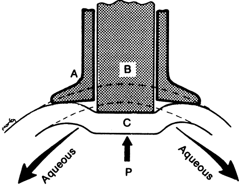
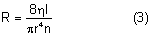
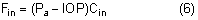
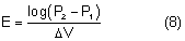
After being produced by the ciliary processes in the posterior chamber, the aqueous circulates throughout the eye, coming into contact with the vitreous, lens, iris, and corneal endothelium. Aqueous drainage occurs by two pathways at the iridocorneal angle. Trabecular or conventional outflow, which predominates in humans, involves passage of aqueous through the trabecular meshwork and into Schlemm's canal, collector channels, and episcleral veins. A small portion of aqueous exits the eye by the uveoscleral or unconventional pathway across the anterior ciliary muscle and iris root, into the suprachoroidal space, and out through the emissarial channels of the sclera.

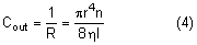
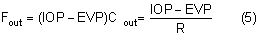
The episcleral veins have an internal pressure that reflects the central venous pressure. The episcleral venous pressure (EVP) is the lower limit of intraocular pressure in an intact eye if uveoscleral outflow is ignored. An increase in the EVP makes it more difficult for the aqueous to drain and leads to increased intraocular pressure.4

CHARACTERISTICS

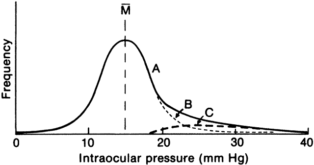
The average intraocular pressure is thought to be about 15 mmHg.2,5 Under normal conditions, intraocular pressure is distributed evenly throughout the eye; therefore, the perfusion pressure of the retina, choroid, and ciliary body must be greater than intraocular pressure before blood can flow through these structures. The intraocular tissue pressure of about 15 mmHg is higher than elsewhere in the body, where the average is about 5 mmHg.6

The distensible uveal tissue absorbs about 2 mmHg of the intraocular pressure. The potential space between the uvea and sclera, the suprachoroidal space, therefore has a pressure about 2 mmHg lower than the intraocular pressure.7 After glaucoma-filtering surgery, the intraocular pressure may become very low and the suprachoroidal space may fill with a plasma transudate (choroidal effusion) and cause detachment of the choroid and ciliary body.

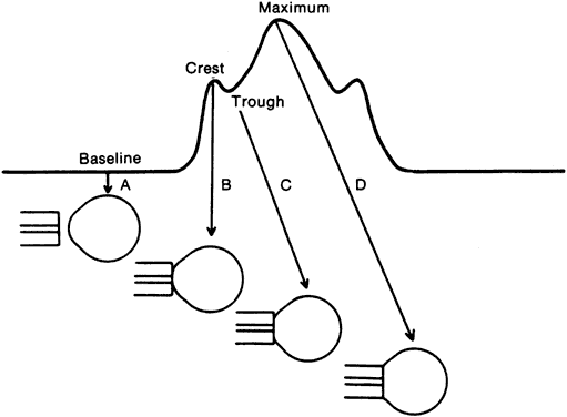
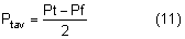
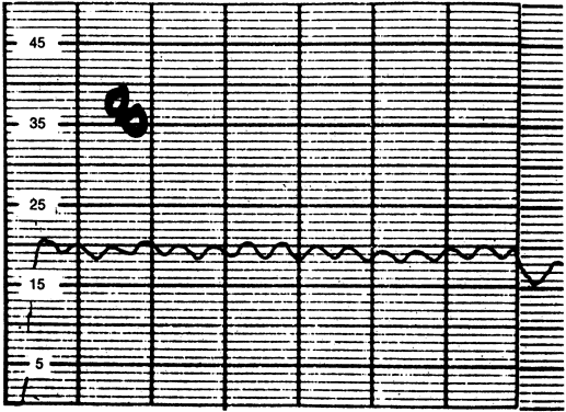
The normal intraocular pressure is pulsatile, reflecting its vascular origin (Fig. 1).8 The pulses follow the arterial pulses, and a diagnosis of cardiac arrhythmia can actually be made from a continuous measurement of the intraocular pressure.9 The amplitude of the pulse is generally 2 to 3 mmHg but may be higher if there is a large arterial pulse pressure, such as in hypertension or aortic regurgitation.8

Fig. 1. The intraocular pressure is pulsatile in nature, having a normal magnitude of 2 mmHg, as shown in this tracing made with a pneumotonometer.

The intraocular pressure is a dynamic function like heart rate and blood pressure and is influenced by many factors. A single measurement of intraocular pressure does not necessarily reflect the average pressure in that hour, day, or week.

DIURNAL VARIATION

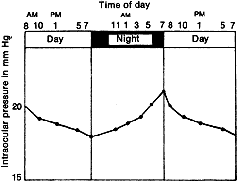
Intraocular pressure in humans follows a diurnal rhythm.4,8 Most commonly, the pressure is highest in the early morning (around 7 AM), and lowest in the evening (around 5 PM; Fig. 2) but considerable individual variation exists regarding the time of day or night when the peak and nadir occur.10–12 The intraocular pressure usually varies less than 5 mmHg from peak to nadir, although differences as large as 15 mmHg have been found. Some people with elevated intraocular pressure are found to have large diurnal peaks and troughs13 and can show variations of as much as 8 mmHg from hour to hour.14 Irregular sleep patterns alter the usual diurnal pattern of intraocular pressure.15

Fig. 2. Diurnal intraocular pressure changes. The most common pattern of daily intraocular pressure variation is for the pressure to be highest in the early morning. The magnitude of the variation is usually about 5 mmHg. Considerable individual variability occurs to time of peak and nadir and to magnitude. (Duke-Elder S: System of ophthalmology. Vol 4. The Physiology of the Eye and of Vision. London: Henry Kimpton, 1968.)

The diurnal pattern of intraocular pressure has been correlated with circadian rhythms of other functions, such as blood pressure, body temperature, and adrenal corticosteroid secretion. In many subjects, the intraocular pressure rises several hours after an increase in plasma adrenal corticosteroids.16,17 This pattern may be due to changes in both aqueous secretion and outflow resistance.18,19

The existence of diurnal variation implies that a single random intraocular pressure measurement does not adequately represent average pressure over a 24-hour period. Peak intraocular pressure can only be obtained by around the clock measurements or with measurements taken at different times of the day. In glaucoma patients, normal intraocular pressures recorded during office hours can give a false clinical impression of adequate pressure control.20–22 Phelps and colleagues argued that a single office pressure measurement was likely, though not certain, to be representative of the peak diurnal pressure.23 Zeimer, however, using Phelps and coworkers' data, calculated that the 95% confidence interval for such a prediction was ±6.6 mmHg, an uncertainty of more than 13 mmHg.12 A small (about 1.5 mmHg) seasonal variation in intraocular pressure has been described, with the highest pressures recorded in winter and the lowest in summer.23,24

INFLUENCING FACTORS

The intraocular pressure is subject to numerous short- and long-term influences (Tables 1 and 2). The intraocular pressure undergoes rhythmic oscillations other than those associated with the arterial pulse. It varies with the respiratory cycle, presumably through changes in venous pressure. Vasomotor variations are associated with waves having a periodicity of 3 to 8 per second (Traube-Hering waves). Although each of these variations produces changes of only 1 to 2 mmHg, the difference between the highest and lowest pressure over a 1-minute period can be as much as 8 mmHg.8


TABLE 7-1. Factors Raising Intraocular Pressure

  General
  Age
  Systemic
  Large increase in blood pressure
  Increased carotid blood flow
  Valsalva maneuver
  Carotid-cavernous fistula
  Plasma hypo-osmolarity
  Hyperbaria
  Local
  Increased episcleral venous pressure
  Blockage of ophthalmic vein
  Blockage of trabecular meshwork
  Co-contraction of extraocular muscles
  Restricted extraocular muscle
  Acute external pressure
  Forced blinking
  Relaxation of accommodation
  Prostaglandin release (biphasic)
  Hypersecretion of aqueous (?)
  Pharmacologic
  Intravenous ketamine
  Succinylcholine (co-contraction)
  Cycloplegic agents
  Corticosteroids (in some)


TABLE 7-2. Factors Lowering Intraocular Pressure

  General
  Exercise
  Systemic
  Large decrease blood pressure
  Decreased carotid blood flow
  Decreased central or jugular venous pressure
  Sympathetic stimulation
  Parasympathetic stimulation
  Hypothermia
  Acidosis
  Plasma hyperosmolarity
  Adrenalectomy
  General anesthesia
  Local
  Decreased episcleral venous pressure
  Decreased ophthalmic artery blood flow
  Prolonged external pressure
  Retrobulbar anesthesia
  Ocular trauma
  Intraocular surgery
  Retinal detachment
  Choroidal detachment
  Inflammation
  Accommodation
  Increased aqueous outflow
  Pharmacologic
  Parasympathomimetics
  Epinephrine
  α-Agonists
  α-Antagonists
  Carbonic anhydrase inhibitors
  Prostaglandin derivatives
  Cardiac glycosides
  Guanethidine
  9-Tetrahydrocannabinol
  β-Adrenergic antagonists
  Muscle relaxants
  Dopamine agonists


The intraocular pressure is relatively immune to physiologic changes in arterial blood pressure. Large swings in blood pressure cause the intraocular pressure to vary in the same direction, however.26,27 Ligating one carotid artery causes a drop in intraocular pressure on that side, presumably due to a decrease in aqueous secretion, although the effect diminishes over several days.28 External carotid compression has a similar result, and measurements of the magnitude of the pressure drop and the rate of recovery have been used to estimate carotid blood flow.29 Interestingly, when one carotid artery is ligated or compressed, the intraocular pressure in the contralateral eye rises, possibly due to the increased blood flow and pressure in the contralateral carotid artery.28

Obstruction of the venous return from the eye or head raises the venous pressure in the episcleral system and thus raises the intraocular pressure. The intraocular pressure rises about 0.8 mmHg for each 1 mmHg rise in EVP.30 Conditions raising EVP include external pressure on the jugular vein, compression of the superior vena cava by a tumor, cavernous sinus thrombosis, arteriovenous fistula in the cavernous sinus (transmitting arterial pressure directly to the venous system) and any change in the orbital apex (e.g., neoplastic, inflammatory, dysthyroid) that compromises the venous drainage of the eye. A tight collar or necktie may also compress the jugular vein and raise the intraocular pressure.

The Valsalva maneuver, in which expiration is forced against a closed glottis producing no air flow, is a related condition that increases venous pressure and hence intraocular pressure.31 Increased intrathoracic pressure collapses the superior vena cava, obstructing venous inflow from the head. Coughing and straining with defecation produce similar changes.

Changes in position affect the intraocular pressure. There is a 2- to 4-mmHg increase in intraocular pressure when changing from the sitting or upright position to the supine position.32–34 Placing the head below the level of the heart in Trendelenburg position may cause a considerable temporary increase in the intraocular pressure.34 Inverted posture may rapidly lead to intraocular pressures of more than 30 mmHg.36,37 These changes parallel an increase in the venous pressure in the head but whether this is the mechanism for the increased intraocular pressure is not known. Some patients with glaucoma may have a much larger change in intraocular pressure with position than normal individuals.38

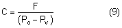
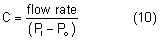
External pressure on the eye increases the intraocular pressure, at least initially. The eye cannot expand significantly to accommodate the fluid displaced by indenting the globe and the intraocular pressure rises. The external pressure, however, accelerates the rate of aqueous outflow and the displaced volume eventually leaves the eye.1,4 If the external pressure is released, the decreased intraocular volume yields an intraocular pressure that is lower than the initial intraocular pressure before applying external pressure. This sequence of events forms the basis by which repeated tonometry decreases intraocular pressure and is why some surgeons massage the eye before intraocular surgery.39,40 Measurement of outflow facility in an eye during tonography is also based on the decrease in intraocular pressure after external pressure.

Forced eyelid closure is a form of external pressure on the eye. Blinking causes a pressure rise of 5 to 10 mmHg, and forced blinking has resulted in recorded intraocular pressures as high as 90 mmHg.39 Voluntary lid fissure widening, as is common during tonometry, is associated with an increased intraocular pressure of about 2 mmHg, whereas pressure on the eyelid caused by holding the eyelids open may result in much larger increases.41

Attempts to move the globe in a direction opposite to a mechanically restricted extraocular muscle raises the intraocular pressure. It is thought that this pressure rise is due to compression of the eye between the restricted muscle and the contracting muscle. This occurs, for example, when upgaze is attempted and the inferior rectus muscle is entrapped by an orbital floor fracture. With dysthyroid involvement of the inferior rectus muscle, the intraocular pressure on attempted upgaze may rise 3 to 10 mmHg.42,43 Intraocular pressure in normal individuals does not rise more than 2 mmHg with upgaze. Therefore, this phenomenon can be a helpful diagnostic sign when recognized. If unrecognized in a thyroid patient, it can cause an overestimation of resting intraocular pressure.

A similar increase in intraocular pressure is seen when the extraocular muscles co-contract, as in Duane's syndrome, or after aberrant regeneration of a previously injured third nerve.4 Intraocular pressure may increase after intravenous succinylcholine, a muscle-depolarizing agent commonly used by anesthesiologists to paralyze the skeletal muscles. Succinylcholine is generally thought to raise intraocular pressure by causing tonic extraocular muscle contraction; therefore, clinicians have avoided its use in patients with open eye injuries to avoid extrusion of global contents.44,45 Kelly and coworkers,46 however, have shown that succinylcholine raises intraocular pressure even when the extraocular muscles are detached from the globe. They attribute the ocular hypertensive effect of succinylcholine mainly to its cycloplegic action and to a lesser degree on its effect on EVP and choroidal blood volume. Nondepolarizing muscle relaxants, such as pancuronium or atracurium, do not raise the intraocular pressure.47,48

Exercise can decrease the intraocular pressure, perhaps because of the acidosis produced by short-term physical exertion, although this mechanism has been disputed.49,50 Even brief physical exertion can cause a mild lowering of intraocular pressure, possibly through sympathetic constriction of choroidal vessels and reduction in uveal volume.51 Continuous strenuous exercise can significantly reduce intraocular pressure, probably through increased plasma osmolarity.52 Physical exertion of longer duration appears to have less effect on the intraocular pressure but exercise conditioning has been reported to lead to a small decrease in baseline intraocular pressure.53 This effect may be more pronounced in patients with higher baseline intraocular pressures.54

Blunt ocular trauma usually has a biphasic effect on intraocular pressure: there is initially a transient rise, followed by a more prolonged decline until the injury resolves.1 After trauma, inflammatory debris or swelling of the trabecular beams may obstruct aqueous outflow. Prostaglandins may also play a role. In rabbits, topical application of prostaglandin-E2 causes a rise in intraocular pressure, which is prevented by pretreatment with prostaglandin synthetase inhibitors such as aspirin and indomethacin.55 The role of humoral factors is supported by the observation of consensual responses in the contralateral eye after ocular trauma and topical prostaglandin application.56 The late, more prolonged decrease in intraocular pressure after trauma is probably due to the decrease in aqueous secretion usually seen in inflammatory conditions.

The level of accommodation has a small effect on the intraocular pressure. Accommodative effort decreases the intraocular pressure, whereas relaxation of accommodation returns the intraocular pressure to baseline values.57,58 These changes parallel the larger effects of parasympathomimetic and parasympatholytic agents.

The intraocular pressure is depressed by general anesthesia, irrespective of the type of anesthetic.47,59 Possible mechanisms include muscle relaxation, decreased blood pressure, increased blood carbon dioxide levels, and a direct central effect of the anesthetic agent.59,60 One important exception is ketamine, which may transiently raise intraocular pressure when given intravenously.44 Retrobulbar anesthesia causes a considerable drop in intraocular pressure due to inhibition of the ciliary body and loss of extraocular muscle tone.1 A large volume of anesthetic instilled into the retro- or peribulbar space may initially compress the globe and raise intraocular pressure, however.

Changes in plasma osmolality profoundly affect the intraocular pressure.4 Water passes freely across the blood-aqueous and blood-vitreous interfaces in either direction, but solute molecules such as ions exchange at much slower rates. If the total concentration of solute molecules in the blood exceeds the concentration in the aqueous and vitreous, water from the vitreous and probably aqueous is drawn into the plasma. This loss of water from the eye decreases the intraocular pressure. Interestingly, the empirically measured water loss is less than osmolality calculations predict, suggesting that there are limiting factors in the eye in addition to compensatory mechanisms.4 After several hours, the concentration of solute increases in the intraocular fluids, and the intraocular pressure returns toward baseline. It may even exceed baseline, resulting in a “rebound” phenomenon.61

The ocular response to plasma hyperosmolality is used clinically in situations such as acute angle closure glaucoma, in which the intraocular pressure has risen to dangerous levels. Hyperosmolar agents are given to raise plasma osmolality and lower intraocular pressure. An effective hyperosmolar agent must penetrate the eye relatively slowly. If osmotic equilibrium is obtained too quickly, the intraocular pressure effect is short-lived.5,45 Useful and relatively nontoxic agents include intravenous urea and mannitol, and oral glycerol and isosorbide. Oral ethanol can also raise plasma osmolality and reduce intraocular pressure indirectly by reducing the production of antidiuretic hormone.62

Conversely, if the concentration of solute in the plasma is lower than that of the intraocular fluids, water enters the eye from the plasma and raises intraocular pressure.4,63 This pressure increase may be considerable in glaucoma patients and forms the basis for the water-provocative test.5 In this test, a patient rapidly drinks a liter of water, lowering the plasma osmolality. Patients exhibiting an intraocular pressure increase of more than 8 mmHg are thought to have glaucoma. The water provocative test, however, has a high false-positive rate and limited diagnostic value.5 Nevertheless, the sensitivity of the intraocular pressure to hypo-osmolality in glaucoma patients should be considered, particularly when there are alterations in systemic fluids and electrolytes. Intraocular pressure, for example, may increase significantly during hemodialysis. Whether or this effect is mediated by changes in plasma osmolality remains controversial.64,65

A drop in body temperature causes a decrease in the intraocular pressure, probably by inhibition of aqueous secretion. A relatively large change in temperature (more than 1°) is necessary to produce significant pressure lowering, however; it is therefore unlikely that temperature plays a major role in physiologic variations of intraocular pressure.66

Finally, blood pH affects intraocular pressure, with systemic acidosis lowering intraocular pressure. In addition to inhibiting carbonic anhydrase in the ciliary body, oral and parenteral carbonic anhydrase inhibitors lower intraocular pressure to some extent by producing a systemic metabolic acidosis.67

Back to Top
PHARMACOLOGY
The most commonly used drugs that affect intraocular pressure include parasympathomimetics, sympathomimetics, carbonic anhydrase inhibitors, prostaglandins, osmotic agents, and corticosteroids.

PARASYMPATHETIC DRUGS

Parasympathetic drugs, commonly known as miotics, cause constriction of the pupillary sphincter (miosis) and ciliary muscles. Contraction of the ciliary muscles, which insert on the scleral spur, causes a mechanical alteration in the trabecular meshwork, resulting in an increase in aqueous outflow facility and decrease in intraocular pressure.68 The decrease in intraocular pressure is independent of miosis and seems to be greater when the baseline pressure is higher. Ciliary muscle contraction also results in a significant reduction of uveoscleral outflow.45 In addition, miotics cause forward movement of the lens, anterior chamber shallowing, iris and ciliary body vasodilation, breakdown of the blood-aqueous barrier, and cataract formation.

The drugs in this group share the same qualitative effects; they differ mainly in their potency. Pilocarpine, the most commonly used miotic, acts directly on the ciliary muscle. Carbachol, which is more potent than pilocarpine, acts directly on the muscle and also inhibits acetylcholinesterase, the enzyme responsible for the catabolism of the neurotransmitter acetylcholine. Drugs acting as pure acetylcholinesterase inhibitors include physostigmine (eserine), echothiophate, demecarium bromide, and isoflurophate. The cholinesterase inhibitors are generally more potent than pilocarpine and carbachol.45 In eyes with closed anterior chamber angles, miotics are ineffective in increasing outflow facility. They may even cause a paradoxical rise in intraocular pressure by decreasing uveoscleral outflow.

Parasympatholytic drugs have the opposite effect of their parasympathetic counterparts. Parasympatholytic agents inhibit ciliary muscle contraction, reduce trabecular outflow facility, and increase uveoscleral outflow. They are used to dilate the pupil for fundus examination, prevent accommodation, reduce inflammation, and prevent ciliary spasm. Drugs in this class include atropine, homatropine, scopolamine, cyclopentolate, and tropicamide. Some patients, including those with open-angle glaucoma, are more sensitive than others to the effects of parasympatholytic agents and may respond to such an agent with pressure rises that can exceed 8 mmHg.69 These drugs are competitive inhibitors of the parasympathomimetics. If used concomitantly, these two types of drugs will usually cancel each other's effects.45 In eyes with narrow angles, parasympatholytic drugs may precipitate angle closure as part of their pupil dilation effect. This is an effect distinct from their brief effect on the ciliary muscle. Systemic agents with anticholinergic action, such as phenothiazines and antihistamines, can elevate intraocular pressure either by directly inhibiting aqueous outflow or by inducing angle closure.

SYMPATHOMIMETIC DRUGS

The effects of adrenergic drugs on the intraocular pressure are complex and seemingly paradoxical. Topical adrenergic agonists such as epinephrine cause a decrease in intraocular pressure. Surprisingly, β-adrenergic blocking agents such as propranolol and timolol also reduce the intraocular pressure. Sympathomimetic pharmacology has not been fully elucidated, but the paradox may result from differing sites of action for each drug. The effect of epinephrine, an alpha and beta agonist, is largely due to an increase in outflow facility, and is probably controlled by adrenergic receptors serving outflow pathways.70 Some evidence suggests that the uveoscleral outflow pathway is primarily affected.71 A study showed that dipivefrin, another α- and β-agonist, reduces blood flow in the ciliary body.72 β-Blockers such as timolol appear to exert their hypotensive effect by reducing aqueous production, perhaps by inhibiting tonic α-adrenergic-controlled formation of aqueous.45,73

Two kinds of α-adrenergic receptors exist: α1 and α2. Stimulation of ocular α1-receptors leads to elevation of intraocular pressure, mydriasis, eyelid retraction, and vasoconstriction. Ocular α2-receptors mediate a reduction in intraocular pressure, probably by inhibiting adenylate cyclase. This causes a decrease in cyclic adenosine monophosphate and subsequently a decrease in norepinephrine release and decrease in β2 stimulation, resulting in a reduction in aqueous production.74

Clonidine, apraclonidine, and brimonidine are α-adrenergic agonists that lower intraocular pressure in humans.75 Clonidine acts centrally on α-adrenergic receptors in the brainstem, reducing sympathetic outflow. This leads to decreased vascular resistance, hypotension, bradycardia, and sedation.76 In the eye, clonidine causes decreased aqueous production and conjunctival vasoconstriction. Apraclonidine, a more polar analogue of clonidine, crosses the blood-brain barrier less readily and has fewer systemic effects than clonidine.74 This drug decreases aqueous production, decreases EVP, and increases trabecular outflow facility.77 Brimonidine is more selective for α2-receptors than apraclonidine.78 Brimonidine has been shown to decrease aqueous production and increase uveoscleral outflow without affecting EVP.79 Interestingly, brimonidine has been shown to have neuroprotective activity in an animal model of optic nerve injury.80 The clinical implications of this finding are unknown at this time.

CARBONIC ANHYDRASE INHIBITORS

Carbonic anhydrase inhibitors are sulfonamide derivatives that compete for the active site of carbonic anhydrase. Inhibition of this enzyme leads to decreased bicarbonate production and sodium co-transport, thereby reducing aqueous production. Carbonic anhydrase inhibitors also induce ciliary body vasoconstriction and disrupt normal intraocular buffering systems.81 Orally administered agents (acetazolamide, methazolamide, ethoxzolamide, dichlorphenamide) cause a systemic acidosis, which contributes to decreased intraocular pressure. Dorzolamide, a topical carbonic anhydrase in-hibitor, does not alter systemic acid-base balance and has somewhat less effect on lowering pressure.82

PROSTAGLANDINS

Prostaglandins are fatty acid derivatives produced throughout the body, including ocular tissue. Initial studies using rabbits showed that prostaglandins given intracamerally or topically caused conjunctival hyperemia, miosis, iris vasodilation, blood-aqueous barrier breakdown, and increased intraocular pressure.54 In contrast, subsequent experiments in other animals with lower dosages showed marked ocular hypotension.83 Prostaglandin compounds initially tested in humans showed significant external side effects. The phenyl-substituted prostaglandin-F2α analog PhXA41 (latanoprost) was found to have relatively few side effects while significantly lowering intraocular pressure.84

Latanoprost increases uveoscleral outflow with minimal effect on outflow facility and aqueous production.85 The mechanism by which latanoprost increases uveoscleral outflow may involve activation of enzymes that degrade ciliary muscle extracellular matrix.86,87 Despite having few systemic side effects, latanoprost causes increased iris pigmentation by promoting increased melanin production in iris melanocytes. The long-term significance of this unique ocular side effect awaits further study.

Other prostaglandin analogues with ocular hypotensive effects are currently under investigation.88

OSMOTIC AGENTS

Urea and mannitol are intravenous osmotic agents used to raise plasma osmolality and reduce intraocular pressure relatively rapidly.5,44 Glycerol and isosorbide can be given orally. Glycerol should be used with great caution in diabetic patients because it may precipitate ketoacidosis. Ethyl alcohol may be used when no other agents are available. All osmotic agents can cause significant hemodynamic alterations in patients with compromised cardiac and renal function.

CORTICOSTEROIDS

Prolonged administration of topical and systemic corticosteroids can produce a significant intraocular pressure increase in susceptible individuals.89,90 About 26% of the normal population have a modest increase of intraocular pressure of up to 8 mmHg after 4 to 6 weeks of topical use, whereas 4% have a greater than 8-mmHg rise. More than 90% of primary open-angle glaucoma patients in one series had their intraocular pressure elevated by topical corticosteroids.90 In some otherwise normal patients, a syndrome indistinguishable from primary open-angle glaucoma can be produced complete with optic nerve damage after prolonged use of topical corticosteroids.90 Systemic corticosteroids take longer to raise intraocular pressure, probably because of less ocular penetration than topical preparations.91

Studies by both Becker92 and Armaly93 strongly suggest that the susceptibility to the intraocular pressure-raising effects of steroids is inherited and may be closely linked to the gene(s) for primary open-angle glaucoma. Intraocular pressure response to steroids may be a risk factor for future development of primary open-angle glaucoma.94 Relatives of primary open-angle glaucoma patients have a higher percentage of steroid response than the general population.92,93 Furthermore, the pituitary-adrenal axis, conjunctival fibroblasts, and peripheral blood lymphocytes of steroid responders were more sensitive to the effects of corticosteroids than those of nonresponders.95 These studies suggest a genetically determined hypersensitivity to corticosteroids on the part of primary open-angle glaucoma patients, their relatives, and possibly some people with a genetic propensity for glaucoma but no expressed manifestations of the disease. This hypothesis has been challenged by Schwartz and coworkers.96

Studies have shown that the increased intraocular pressure induced by corticosteroids is due to a decreased outflow facility.97 In rabbits, the decreased outflow may be due to a buildup of mucopolysaccharides in the trabecular meshwork.98 The exact mechanism for this pressure rise in humans is not known, however.

Back to Top
CONTROL OF INTRAOCULAR PRESSURE
The intraocular pressure in normal individuals varies within a somewhat limited range. Changes in intraocular pressure produce compensatory changes in aqueous secretion and outflow, tending to bring the intraocular pressure back toward baseline. As intraocular pressure rises, aqueous inflow decreases. This is called pseudofacility.99 Although these homeostatic mechanisms could be purely local, many studies have implicated a central control mechanism.

Afferent fibers in the long posterior and short ciliary nerves that respond to changes in intraocular pressure have been identified in animals; however, no clear relation between discharge frequency and intraocular pressure has been found.100,101 Specialized nerve endings in the scleral spur have been discovered that closely resemble mechano- or baroreceptors elsewhere in the body. These nerve endings may respond to changes in ciliary muscle tone or intraocular pressure.102 Stimulation of some parts of the diencephalon causes intraocular pressure to increase, whereas stimulation of other areas causes the pressure to decrease.103,104 Changes in intraocular pressure can also be produced by administration of certain drugs into the cerebrospinal fluid. These drugs, however, may act by altering blood pressure and body temperature, which are also known to influence intraocular pressure. If the optic nerve is cut, the intraocular pressure fails to respond in the usual way to intravenous hyperosmotic agents, suggesting that the optic nerve contains fibers controlling intraocular pressure.105,106 Sectioning the optic nerve, however, may reduce the vascular supply to the eye, which may also explain these findings.107

When the cervical sympathetic ganglion is stimulated, the intraocular pressure decreases because of diminished aqueous secretion. It gradually returns toward normal, although it never regains baseline levels. Surprisingly, when the cervical sympathetic ganglion is removed, the intraocular pressure drops initially and returns to normal in 3 to 4 days. This effect appears to be due to an increase in outflow facility. If catecholamines are given topically or intracamerally, there is a dramatic decrease in intraocular pressure known as “denervation hypersensitivity.”108,109 Holland and coworkers used the drug 6-hydroxy-dopamine to produce a chemical sympathectomy. After the administration of hydroxydopamine, even small doses of topical catecholamines dramatically reduced the intraocular pressure.110

Stimulation of the parasympathetic system, which supplies the eye by the third cranial nerve, also causes a decrease in intraocular pressure through an increase in aqueous outflow facility.56,111 Conversely, a decrease in parasympathetic tone decreases the facility of outflow.

Regulation of intraocular pressure, however, probably involves more sophisticated mechanisms than the opposing actions of the sympathetic and parasympathetic systems. Work using immunohistochemical techniques has demonstrated the presence of several neurotransmitters and neuropeptides in the anterior segment.112 Vasointestinal peptide, neuropeptide Y, substance P, calcitonin gene-related peptide, and cholecystokinin have been found in ciliary body and trabecular meshwork.

In summary, many aspects of the central nervous system influence the intraocular pressure. Pressure-sensitive afferent fibers have been identified, and the intraocular pressure is affected by sympathetic, parasympathetic, and diencephalic influences. Innervation of the eye also involves a diversity of biologically active neuropeptides, whose exact function is the subject of continuing investigation. Studies suggest the existence of a control center that maintains pressure homeostasis within the eye, but the nature and location of such a center have yet to be determined.

Back to Top
POPULATION DATA
Several descriptive studies of intraocular pressure in a presumed normal population have been published. The number of eyes tested ranges from about 100 to almost 20,000. The statistics are remarkably similar, despite differing ethnic groups and methods of measurement. The largest study, by Leydhecker, used the Schiøtz tonometer and found a mean intraocular pressure of 15.5 mmHg, with a standard deviation of 2.57 mmHg.8 Studies using the more accurate Goldmann applanation tonometer have found almost the same mean and scatter as the Leydhecker study.8,113

Although there is excellent agreement regarding the mean intraocular pressure, determining the lower and particularly the upper limits of normal is much more difficult. The classic method for determining the upper limit of normal is to assume that the values for intraocular pressure are distributed in a “normal” (gaussian) fashion. Under this assumption, the normal values cluster symmetrically on either side of the mean. If one arbitrarily chooses a point 2 standard deviations above the mean as the upper limit of normal (about 20.5 mmHg), 95% of the population will have values between 10.5 and 20.5 mmHg and 2.5% of the population will be included as abnormal. Alternatively, if one chooses 2.5 standard deviations above the mean as the upper limit of normal (about 24 mmHg), then less than 1% of the normal population be included as abnormal.

Unfortunately, the distribution of intraocular pressure in the population does not fit a bell-shaped gaussian curve.2,8 Instead, there is a skew to the right (Fig. 3), meaning that the number of people who have intraocular pressures higher than the mean is greater than the number having pressures lower than the mean. This situation arises with many physiologic parameters, including blood pressure and fasting blood sugar. Gaussian statistics cannot be accurately applied to a parameter that does not have a gaussian distribution.

Fig. 3. Distribution of intraocular pressure in population. Line A indicates the frequency distribution of intraocular pressures in the population. Notice that the distribution is skewed (tailed) to the right. Theoretically, the tail could be made up of two subpopulations. Dotted line B represents the “normal” population completing a gaussian distribution. Line C represents the abnormal population, which added to the normal population produces the tail.

It has been assumed that two different populations explain the skewed distribution (see Fig. 3). The normal population has a true bell-shaped distribution of intraocular pressure. Superimposed on this normal curve is the population with glaucoma, with high pressures eplaining the long tail on the right side of the distribution curve. Because the upper end of the normal population and the lower end of the glaucoma population overlap, it is impossible to separate the glaucomatous individual from the high-normal individual using pressure criteria alone.

Armaly114,115 proposed that if the normal population were divided into several subpopulations based on gender and age, the distribution curve of each of these subpopulations would be gaussian in nature. In a study of more than 2000 individuals, he found a slight increase of mean intraocular pressure in each decade above the age of 40. In addition, in people 40 years of age and older, women had slightly higher pressures than men.

The notion that age has an independent effect on intraocular pressure has been challenged. Large cross-sectional studies of almost 200,000 Japanese subjects by Shiose actually show a decrease in intraocular pressure with age. Shiose suggested that the Japanese data may be reconciled with Western data by postulating that age normally leads to a reduction in intraocular pressure, but this effect is overcome in Western populations by the increased prevalence of systolic hypertension and obesity, both of which are associated with increased intraocular pressure.116 This issue can only be resolved by long-term observations of individuals and not by cross-sectional methods.

In the population as a whole, the right and left eyes have equal pressures. The difference between the two eyes rarely exceeds 4 mmHg. An abnormality should be suspected whenever a greater difference is found consistently.8

Armaly's studies on various populations in Iowa suggest that the level of intraocular pressure is inherited as a polygenic multifactorial trait.117 The effects of a common environment are less clear; two studies comparing the intraocular pressure of spouses had contradictory results.117,118

Back to Top
ABNORMAL INTRAOCULAR PRESSURES

GLAUCOMA

Glaucoma is a disorder usually associated with elevated intraocular pressure, in which the optic nerve undergoes a characteristic pattern of damage (cupping) leading to loss of visual function.

Not too long ago, all people with intraocular pressures higher than 2 standard deviations above the mean (22 mmHg) were considered to have glaucoma. This definition has proved inadequate. Longitudinal studies by Armaly119 and Perkins120 have shown that only a small percentage of individuals with pressures higher than 22 mmHg develop the full clinical picture of glaucoma, at least over a 7- to 10-year period. A significant proportion of patients with typical glaucomatous optic nerve damage have intraocular pressures well below 22 mmHg, so-called “normal” or “low-tension” glaucoma. These observations imply that the level of intraocular pressure that damages the optic nerve differs between individuals. A cutoff of 21 mmHg for “normal” is arbitrary because it is a combination of optic nerve susceptibility and intraocular pressure level that produces glaucoma. Glaucoma is primarily an optic neuropathy, not an intraocular pressure abnormality.

Intraocular pressure, therefore, does not define glaucoma. It is a strong risk factor for glaucoma and the only risk factor amenable to treatment. This reality has dictated our therapeutic focus on controlling intraocular pressure by medical and surgical means. Unfortunately, it has also limited our thinking about glaucoma. Strictly speaking, the typical clinical features of glaucoma—optic nerve damage and visual field loss—do not constitute a disease. They merely represent the end stage of several multifactorial processes whose final common pathway involves anatomic and functional deficits of the optic nerve. Deeper understanding of the genetic and biochemical factors involved in the pathogenesis of glaucoma may provide future treatment options that do not involve lowering intraocular pressure.

A complete discussion of glaucoma is beyond the scope of this chapter. It is sufficient to say that a pressure higher than 22 mmHg should be considered suspect but not necessarily abnormal. The clinical management of a patient who has elevated intraocular pressure should depend on many factors, including age, race, family history of glaucoma, anterior chamber angle status, optic nerve appearance, visual field, and other concomitant ocular and systemic diseases.

HYPOTONY

Although controversial, 8 mmHg has been suggested by many authorities as the level below which hypotony can be diagnosed.1 Under normal conditions, intraocular pressure cannot fall below EVP. A reduction in intraocular pressure can be due to failure of aqueous secretion secondary to inflammation, poor ciliary body perfusion, destruction of the ciliary body by disease, cryotherapy, or laser energy. Profound hypotony can result from conditions that create a new outflow path for aqueous, thereby bypassing EVP. This can occur after traumatic ocular injuries such as cyclodialysis and ruptured globe, perforation of the cornea from infection, or an overfunctioning glaucoma operation. In these situations, intraocular pressure can approach atmospheric pressure despite normal aqueous production by the ciliary body.

Back to Top
MEASUREMENT OF INTRAOCULAR PRESSURE
Three methods of measuring intraocular pressure are available: manometry, applanation, and indentation. Manometry is the only direct measurement of intraocular pressure: the other two are indirect methods and are called tonometry. Some devices use a combination of applanation and indentation tonometry. The instruments for performing tonometry are tonometers. Their purpose is to get an accurate measurement of intraocular pressure with the least disturbance to the eye. Each technique has its advantages and disadvantages, and none is ideal.

Some general principles apply to any method of measuring intraocular pressure. Extraneous factors that can influence the pressure reading must be avoided. The fingers of the examiner can press on the eyelid or eye while holding the lids apart, raising the intraocular pressure. Squeezing of the eyelids, eye movements, a tight collar or necktie, and repetitive measurements, particularly with Schiotz tonometry, can affect the intraocular pressure reading.

MANOMETRY

Intraocular pressure is higher than atmospheric pressure; therefore, if a small hollow needle is inserted into the anterior chamber, aqueous humor flows out through the needle. If the needle is attached to a reservoir of fluid that is raised just high enough to prevent any loss of aqueous, the height of the column of fluid, usually calibrated in centimeters of water or millimeters of mercury, reflects the intraocular pressure (Fig. 4). Movement of the fluid column caused by changes in intraocular pressure can be detected by an electronic strain gauge.

Fig. 4. Manometry. A small, hollow needle is inserted into the anterior chamber. A reservoir of isotonic solution is raised until no aqueous humor leaves the eye, yet no reservoir fluid is added to the eye. That height is the intraocular pressure in centimeters of water and can be converted to millimeters of mercury by a simple formula.

Manometry is most used as a laboratory technique in performing continuous pressure measurements over time, evaluating the effect of physiologic and pharmacologic manipulations on pressure, and studying aqueous humor dynamics in postmortem eyes.4,121 The ethical use of manometry in living human eyes is restricted to eyes undergoing enucleation or intraocular surgery.122 This places constraints on the length of time manometry may be performed. In addition, these eyes may harbor significant ocular pathology affecting intraocular pressure. Intraoperative data obtained from diseased eyes may not apply to normal eyes.

APPLANATION TONOMETRY

The pressure inside a flexible sphere with thin walls can be closely approximated by knowing the force necessary to just flatten (applanate) a given area of the sphere. These parameters are related by the formula, pressure = force/area (Imbert-Fick law). One can either measure the force necessary to flatten a fixed area or measure the area flattened by a fixed force. Both methods have been used in designing tonometers (Fig. 5).1,4

Fig. 5. Comparison of fixed-area and fixed-force applanation tonometers. In fixed-area applanation, a constant area of applanation a is used for every measurement. i) A greater force of applanation (F) is required to applanate this area in an eye with higher intraocular pressure (P). ii) A smaller force (f) is required to applanate the same area in an eye with lower intraocular pressure (p). In fixed-force applanation, a constant applanating force (F, the weight of the tonometer) is used for every measurement. iii) A smaller area of applanation (a) is achieved with this force in an eye with higher intraocular pressure (P). iv) A larger area of applanation (A) is obtained with the same force in an eye with lower intraocular pressure (p).

Fixed-Force Tonometers

Maklakoff in 1855 developed the first practical applanation tonometer.1 This instrument was a metal cylinder of known weight with a flat bottom. A dye was smeared on the anesthetized cornea and with the patient supine, the cylinder was allowed to rest on the cornea. The applanated area of cornea transferred the dye to the tonometer, and the area of applanation could be calculated from the diameter of the circle of dye on the tonometer bottom. The pressure could be derived from the Imbert-Fick formula because the applanating force (the weight of the tonometer) was known.

The Maklakoff tonometer has several disadvantages. The relatively large applanation area and heavy weight of this tonometer raises the intraocular pressure above its resting state (Po) to a higher level (Pt) during measurement. The Maklakoff tonometer records this higher pressure (Pt) rather than the “undisturbed” intraocular pressure (Po). It is possible to calibrate the tonometer in such a way as to derive Po from Pt but it must be assumed that most eyes respond in the same way to tonometer placement. Also, the slightest movement of the eye or examiner smears the ink spot, making it larger than the actual area of flattening. This leads to an underestimate of the intraocular pressure.1,2,4

Posner has revived this kind of tonometer for clinical use (Fig. 6).123 His version is plastic, which reduces its weight and the discrepancy between Po and Pt. Ink is smeared on the tonometer footplate. When the tonometer rests briefly on the eye, the ink from the footplate is transferred to the flattened area of the cornea. The area on the footplate that is devoid of ink corresponds to the area of applanation. The footplate is then pressed onto a sheet of paper, and the diameter of the inkless area may be converted to the intraocular pressure. The Posner-Inglima tonometer has the advantage of being inexpensive and disposable. It can be used at home or whenever cleaning and sterilization of a tonometer are impractical.

Fig. 6. Posner-Inglima fixed-force applanation tonometer. (A) This tonometer is lowered onto the eye. (B) The cornea is flattened by the fixed weight of the plastic instrument. (C) The area of flattening is seen on the footplate as the light central portion where the ink originally smeared on the footplate has transferred to the flattened cornea. (D) The footplate is then pressed onto a sheet of paper. The remaining ink is transferred to the paper. The diameter of the flattened area can be measured from the clear central portion. The diameter can then be used to calculate the area applanated and the intraocular pressure. In actual use, the instrument is supplied with a scale that allows reading of the intraocular pressure directly from the diameter.

Fixed-Area Tonometers

GOLDMANN APPLANATION TONOMETRY.

In 1888, Fick devised a tonometer that maintained a fixed area of applanation.124 The intraocular pressure was determined by measuring an adjustable force necessary to flatten this predetermined area of the corneal surface. In the 1950s, Goldmann modernized this concept and developed the tonometer that has become the standard against which all others are judged.125

The applanating surface of the Goldmann tonometer has a diameter of 3.06 mm placed in the center of a plastic cylinder of 7 mm total diameter. The plastic cylinder is attached to an arm pushed forward through a spring-loaded knob. The amount of force on the cylinder is precisely controlled and can be read from a scale on the knob. The device is mounted on a slit-lamp biomicroscope (Fig. 7).1,4

Fig. 7. Goldmann applanation cylinder. This conical, plastic device has a total diameter of 7 mm and an applanating surface of 3.06 mm in diameter. A doubling prism inside the cylinder allows viewing of the applanating area through a slit-lamp biomicroscope has two rings (mires).

To determine the intraocular pressure, topical anesthetic and fluorescein dye are placed in the eye. The dye mixes with the tears and fluoresces a brilliant yellowish green when activated by cobalt blue light. When the corneal surface is flattened by the plastic cylinder, the tear layer is squeezed out from the applanated surface, and the tear meniscus is seen through the clear center of the plastic cylinder as a ring of fluorescence. The force knob is adjusted until the applanated area is exactly 3.06 mm in diameter. The Goldmann tonometer uses an ingenious optical method to allow this adjustment to be precise: two prisms are positioned in the plastic cylinder, apex to apex, in such a way that the fluorescent ring of the tear meniscus is seen as two half rings (mires), one above and one below. The orientation and power of the prisms are such that the two mires are optically separated by exactly 3.06 mm. The force knob is turned until the inside edges of the end of each split ring just touch, forming a “lazy S” pattern, which is the measurement endpoint (Fig. 8). Typically, the pulse pressure causes an oscillation of the mires. The force knob is adjusted until these oscillations are centered about the endpoint. When the endpoint is reached, the applanated area has a diameter of 3.06 mm.

Fig. 8. Goldmann applanation mires. i) The tear meniscus encircling the applanated surface of the cornea (a) is stained by fluorescein dye, appearing bright green under cobalt-blue light. Prisms in the applanating cylinder convert the image of the circular meniscus into two half rings (mires) separated by 3.06 mm. ii) Too little fluorescein dye produces thin mires, which can lead to an overestimation of the intraocular pressure. iii) Too much fluorescein dye produces thick mires, which yield an underestimation of the intraocular pressure.

The Goldmann tonometer closely measures the true intraocular pressure. During applanation, there is little displacement of intraocular fluid because a small amount of force is applied over a small area of the cornea. Typically, 0.5 μl are displaced with 1 to 2 g of applanation force at physiologic intraocular pressure. This small amount of fluid displacement results in an intraocular pressure during applanation (Pt), which is only 3% higher than the undisturbed intraocular pressure (Po). This difference can be ignored for all practical purposes.

During applanation, corneal rigidity pushes back against the tonometer head. This force adds to the measured intraocular pressure and causes the intraocular pressure to be overestimated. The surface tension of the tears, however, creates a capillary attraction that pulls the tonometer surface toward the cornea, lowering the force required to applanate the cornea and causing the intraocular pressure to be underestimated. With an applanation diameter of 3.06 mm, the forces of corneal rigidity and tear surface tension cancel out. Furthermore, this diameter allows the applanating force in grams to be converted to intraocular pressure in millimeters of mercury by multiplying the indicated gram force by 10.1,4,126

High corneal astigmatism (more than 3 diopters) can introduce significant errors in Goldmann applanation tonometry, unless a modified measurement technique is used. Schmidt127 averaged readings taken with the tonometer head oriented over the flat and steep meridians. A more accurate method involves rotating the tonometer prism to a 43° angle from the major axis of astigmatism, measured in minus cylinder. This position is indicated by a red line on the prism holder. If this is not done, a 2- to 3-mmHg error may be induced. Alternatively, readings taken with the prism oriented horizontally and vertically can be averaged.128

Goldmann assumed an average corneal thickness of 0.52 mm in designing his tonometer. Excessively thin or thick corneas yield under- or overestimations of pressure, respectively, which can lead to a diagnosis of low-tension glaucoma or ocular hypertension.129–138 Corneas that are edematous or scarred have an abnormal elasticity, usually leading to an underestimate of true intraocular pressure. Too much fluorescein produces wider mires and result in an underestimate of the pressure. Finding the endpoint may be difficult in a patient with an irregular cornea (which produces irregular mires)4 or wide pulse pressure. Because the optical endpoint is subjective, readings taken by different examiners can differ by 2 mmHg. The sources of error with Goldmann applanation tonometry are reviewed by Whitacre and Stein.139

Despite the limitations, the Goldmann applanation tonometer is considered to be the “gold standard” for clinical use. Unfortunately, it cannot be used without a slit lamp, is not portable, and requires the patient to be in a sitting position. Draeger and Perkins140,141 have each used the Goldmann principle to design tonometers that are handheld, portable, battery powered, and usable in supine as well as sitting positions. This brings accurate applanation tonometry to screening clinics, the bedside, and the operating room. These tonometers are moderately expensive and share the disadvantage of being inaccurate on scarred, edematous, or irregular corneas. Spread of infectious agents is possible through applanation tonometry unless the applanating prisms are sterilized before each use.

NONCONTACT AIR PUFF TONOMETER. The noncontact air puff tonometer works on the same basic principle as the Goldmann (Fig. 9). A puff of air—the force of which increases linearly over several milliseconds—is directed at the cornea. The air puff is designed so that it hits the cornea with a known and reproducible area. The air pulse then progressively flattens the cornea and finally produces a slight concavity. The moment of applanation is determined by an optical sensor positioned so that an oblique light is reflected into the sensor only when the cornea is flat and acting as a plane mirror. At that precise moment, the sensor sends an electrical impulse to the air pulse generator, shutting it off. A microcomputer monitors the force of the air puff and records the force being generated at the moment of applanation. The computer determines the intraocular pressure from the force and the known area and displays it in digital form. The most recent models have sophisticated automatic monitoring, alignment, and measurement systems.142

Fig. 9. Principle of air-puff tonometer. A puff of air of known area is generated against cornea (B). At the moment of corneal applanation a light (T), which is usually reflected from the normal cornea into space, suddenly is reflected (R) into an optical sensor (A). When the sensor is activated by the reflected light, the air generator is switched off. The level of force at which the generator stops is recorded, and a computer calculates and displays the intraocular pressure. (D) Optical detector. (Adapted from B. Grolman, American Optical Corp.)

Correlation between the most recent noncontact tonometers and the Goldmann readings is close in normal eyes.143–146 Some patients find the air puff startling and mildly uncomfortable; however, painless measurements can be taken without anesthesia. This facilitates use in screening programs by nonmedical personnel and in children.147–149 Furthermore, the air puff tonometer does not touch the eye, and transmission of infectious agents from eye to eye is unlikely.

The air puff tonometer is a complex instrument and subject to a higher incidence of breakdown than the simpler Goldmann device. Like the Goldmann, it is inaccurate in scarred, edematous, or irregular corneas. Most air puff tonometers are large, not very portable, and must be used with the patient in the sitting position. Finally, they are expensive and require regular calibration.150

Indentation Tonometry

The basic principle behind indentation tonometry is simple: a known force will indent a fluid or gas filled object to a greater degree if the internal pressure is low, compared with when the internal pressure is high. The force can be supplied by digital pressure or a known weight.

Palpation

Palpation of the globe is the simplest, least expensive, and least accurate method of estimating intraocular pressure. It is useful when other methods are unavailable or subject to gross error. Palpation may be the only feasible technique in patients who are unwilling or unable to undergo other methods of intraocular pressure measurement.

The patient is asked to close his or her eyes and look down. Redundant skin of the upper eyelid is displaced, and the central meridian of the globe is balloted alternately with the tips of each index finger. By comparing tactile estimations of intraocular pressure to formal pressure measurements, the examiner's sense of touch can be “calibrated” to a limited extent. Although palpation correlates poorly with Goldmann applanation readings, palpation may have a limited role in screening for marked elevations of intraocular pressure.151 Palpation is best avoided in eyes with significant trauma.

Schiøtz Tonometer

Schiøtz developed an excellent tonometer in 1905 and continued to refine it through 1927. His refined tonometer became the most widely used in the world and because of its simplicity, reliability, and relative accuracy, it is the only indentation tonometer in widespread use today. The tonometer has been modified only slightly since Schiøtz's time.1

In the Schiøtz tonometer, gravity provides a known force on a weighted metal plunger. The plunger rides inside a metal cylinder attached to a footplate curved to match the average human corneal curvature (Fig. 10). The top of the plunger rides along a curved lever that attaches to a pointer, which in turn rides along a scale. For each 0.05 mm that the plunger sinks below the level of the footplate, the pointer moves up 1 scale unit. Thus, the lower the intraocular pressure, the farther into the cornea the plunger sinks and the higher the scale reading. Scale readings can be converted to millimeters of mercury by conversion tables, based on the amount of weight placed on the plunger. Before each measurement, the tonometer is placed on a solid steel block, which should result in no plunger depression and a scale reading of zero.

Fig. 10. Schiøtz tonometer. Arrow points to scale. Each scale marking indicates 0.05 mm of plunger movement.

The scale measuring the amount of indentation is linear. The relation between the amount of indentation and intraocular pressure is not linear but about logarithmic, so that the higher intraocular pressures are compressed toward the lower end of the scale.152 Below the scale reading of 3, it is not possible to get an accurate pressure reading other than to know that the pressure is elevated above the “normal” range.2,4 Therefore, additional weights of 2, 4.5, and 9.5 g, respectively, may be added to the plunger to give effective plunger weights of 7.5, 10, and 15 g, respectively. The heavier weights cause the plunger to sink deeper for a given intraocular pressure and to give a higher scale reading. In effect, the heavier weights expand the lower end of the scale (Table 3).


TABLE 7-3. Schiøtz Scale Readings: Intraocular Pressure (PO) Conversion Table (from 1955 revision): Assumes Average Ocular Rigidity (PO in mmHg)


 Plunger Load
Scale Reading5.5 g7.5 g10 g15 g
3.024.435.850.681.8
3.522.433.046.976.2
4.020.630.443.471.0
4.518.928.040.266.2
5.017.325.837.261.8
5.515.923.834.457.6
6.014.621.931.853.6
6.513.420.129.449.9
7.012.218.527.246.5
7.511.217.025.143.2
8.010.215.623.140.2
8.59.414.321.338.1
9.08.513.119.634.6
9.57.812.018.032.0
10.07.110.916.529.6
(Kolker AE, Hetherington J Jr: Becker-Shaffer's Diagnosis and Therapy of the Glaucomas. 4th ed. St. Louis: CV Mosby, 1976.)


Conversion tables to obtain Po (resting intraocular pressure) from Pt (pressure with the tonometer on the eye) were developed from studies done on cadaver eyes by Friedenwald.152 These values were basically confirmed by McBain153,154 in studies using an adjustable manometer. The scale reading of the tonometer with each plunger weight was recorded. The volume of aqueous humor displaced by the weight of the tonometer was also measured. These observations were recorded and plotted on a logarithmic scale to yield the Friedenwald nomogram (Fig. 11).

Fig. 11. Scleral rigidity from Friedenwald nomogram. Solid line (A) joins Po (determined by applanation tonometry) and Pt1 (Schiøtz scale reading with 5.5-g weight). Theoretically, the same line could be obtained by joining scale readings with any two Schiøtz weights indicated by dots. Slope of line is scleral rigidity, which can be obtained directly from nomogram by drawing parallel line through 20 on the pressure scale (dotted line B to scleral rigidity scale on top of nomogram). In this case, scleral rigidity is 0.022.

SCLERAL RIGIDITY

When a significant external force is applied to the eye (Fig. 12), the intraocular pressure is raised, blood is squeezed out of internal blood vessels, the corneal and scleral coats are distended, and the volume of the eye expands slightly. The relative resistance an eye offers to expansion for a given rise in intraocular pressure is known as scleral rigidity. The reciprocal of rigidity is elasticity.

Fig. 12. Schiøtz tonometer on cornea. Footplate (A) rests on corneal surface supporting weight of tonometer, which raises intraocular pressure to Pt. The plunger (B) sinks into the cornea (C), displacing a volume of aqueous humor until the elasticity of the cornea and the intraocular pressure (P) push back with enough force to prevent further sinking of the plunger.

Scleral rigidity varies from individual to individual. Friedenwald's tables for conversion of Schiøtz scale readings to intraocular pressure are calculated based on an average scleral rigidity.155 In myopic eyes, scleral rigidity is lower than average, and the Schiøtz plunger sinks deeper into the cornea, compared with an eye with average scleral rigidity at the same intraocular pressure (Table 4). The scale reading will be higher and the intraocular pressure will be underestimated. A diagnosis of glaucoma may be missed. Conversely, hyperopes and patients with scarred corneas have higher scleral rigidity, resulting in overestimation of their intraocular pressures.155


TABLE 7-4. Effect of Scleral Rigidity on Intraocular Pressure Measurement by Schiøtz Tonometry and Outflow Facility*


 APOC
Average rigidity (0.0215)19.519.00.18
Low rigidity (0.0135)23.523.00.38
High rigidity (0.0315)15.515.00.11

*Using a weight of 5.5 g, a scale reading of 4.5 on the Schi<aso>tz tonometer would indicate a pressure of 19 mmHg, using the 1955 revised conversion table (see Table 3). This table assumes an average ocular rigidity. Note that if the patient has a low ocular rigidity, the figure of 19 mmHg significantly underestimates the true intraocular pressure of 23.5 indicated in column A. The opposite is true for a patient with high ocular rigidity. An even larger error in outflow facility is seen with changes in the scleral (ocular) rigidity.
A = applanation reading (mmHg); PO = intraocular pressure by Schi<aso>tz corrected for ocular rigidity at scale reading of 4.5 (mmHg); C = outflow facility corrected for ocular rigidity with final Schi<aso>tz scale reading 6.5.
(Kolker AE, Hetherington J Jr: Becker-Shaffer's Diagnosis and Therapy of Glaucomas. 4th ed. St. Louis, CV Mosby, 1976.)


Scleral rigidity can be calculated from the Friedenwald nomogram as follows (see Fig. 11): Two different weights are used to obtain two tonometer scale readings (two different values for Pt). A more accurate method is to use the applanation tonometry value as Po and one Schiøtz reading as Pt. The slope of the line formed by joining these two points gives the scleral rigidity (mmHg change in pressure per cubic mm change in volume). In clinical practice, scleral rigidity measurements are not made as often as they should be. This introduces significant errors and makes dependence on Schiøtz tonometry hazardous.

Errors of Schiøtz Tonometry

In addition to the potentially large error that an abnormal scleral rigidity can produce in Schiøtz tonometry, other errors may arise. The footplate must be the right curvature and size, and the total instrument must be the correct weight for the Friedenwald tables and nomogram to apply.2 Poor attention to detail in manufacture can make a tonometer totally unreliable. The American Academy of Ophthalmology has established a Committee on Tonometer Standardization, which has set rigid criteria for Schiøtz tonometers. A tonometer certified by this committee or by a laboratory committed to the same standards can be expected to perform reliably.

Dirty tonometers are also potentially inaccurate. If tears inside the barrel are allowed to dry, the friction between the barrel and plunger is increased. A common error is to autoclave the tonometer without first carefully cleaning it. The secretions in the barrel harden, causing the plunger to stick. If the tonometer is not placed perpendicular to the corneal surface, additional friction is produced between plunger and barrel. A cornea that is scarred, edematous, or of abnormal curvature gives inaccurate readings with the Schiøtz.

A corneal abrasion can be caused by the plunger if the eye or tonometer moves during measurement. The Schiøtz tonometer must be used in the supine position or in the sitting position with the head back far enough to be horizontal. An initial blink or avoidance reaction may occur as the patient sees the tonometer descending toward his or her eye.

Despite the many potential sources of error, the Schiøtz tonometer has been a remarkably useful instrument for the past 75 years. Compared with most other methods of tonometry, it is inexpensive, simple, portable, and easily sterilized. Patients and their families can be taught how to use it for home tonometry. Although largely replaced in the United States by Goldmann or other types of tonometry, the Schiøtz tonometer still has a place in clinical practice, particularly if checked for accuracy against Goldmann tonometry in each patient.

COMBINATION APPLANATION-INDENTATION TONOMETRY

The McKay-Marg, Tono-Pen (Intermedics Intraocular, Pasadena, CA), and pneumotonometer instruments have properties of both applanation and indentation tonometers. The original McKay-Marg tonometer is no longer manufactured and has been supplanted by a handheld and battery-operated version, the Tono-Pen.

Mackay-Marg Tonometer

The Mackay-Marg tonometer uses a microplunger that protrudes a small amount from a flat footplate of a tubular handpiece (Fig. 13). The microplunger is connected to a sensitive transducer, which converts plunger displacement into an electrical signal that is recorded on a paper chart, much like an electrocardiogram.4,156

Fig. 13. Mackay-Marg tonometer handpiece.

The shape of the tracing reflects the stages of applanating the cornea (Fig. 14). As the plunger is pressed into the cornea, both intraocular pressure and corneal elasticity push back, as with the Schiotz tonometer. During this phase of measurement, a rising trace is recorded. When the plunger no longer protrudes from the footplate, the footplate has applanated the cornea. The force of corneal elasticity is taken off the plunger, resulting in a slight drop in the trace. As the plunger and footplate are pressed farther into the cornea, the unit raises intraocular pressure by displacing aqueous humor, and a rise in the pressure is again recorded. The true intraocular pressure, Po, representing the point of applanation, is the bottom of the trough in the pressure recording. Note that there is no disturbance of the intraocular pressure until this point is passed.3,157,158

Fig. 14. Schematic drawing of Mackay-Marg handpiece. The plunger of the Mackay-Marg handpiece is connected to a strain gauge, which measures force. When the tip first contacts the cornea, the force increases (B) from baseline (A). When the cornea is flattened, the baseplate takes up some of the force from the plunger, causing a slight, momentary drop in the force registered by the strain gauge (C). As the handpiece is pressed further against the eye, the force of the plunger continues to increase (D). The point of applanation is indicated on the tracing at the trough (C). (Hilton GF, Shaffer R: Electronic application tonometry. Am J Ophthalmol 62: 840, 1966.)

The Mackay-Marg tonometer is the most accurate tonometer in scarred or edematous corneas because the intraocular pressure reading is independent of corneal elasticity.159 This has been confirmed manometrically. The end point is measured electrically; therefore, the reading is unaffected by optical factors such as an irregular cornea or high astigmatism. Several readings may be required to obtain a good tracing, however, and the interpretation of the readings may be difficult.2

The values from Mackay-Marg tonometry correlate well with those from Goldmann tonometry.6 The mean values from the Mackay-Marg tonometer, however, may be 1.5 to 3 mmHg higher than those from the Goldmann tonometer on normal corneas.2 One study160 has seriously questioned the reliability of individual readings but most studies indicate a reasonable degree of reliability.2,4 Phelps and Phelps pointed out that even with Goldmann tonometry there may be significant variation from reading to reading in the same patient within a short time.161

Tono-Pen

The Tono-Pen is a handheld, battery-operated version of the Mackay-Marg tonometer (Fig. 15). The tip is covered by a disposable latex cover and applied perpendicularly to gently indent an anesthetized cornea. Each measurement requires several applanations. An acceptable applanation is indicated by an audible click after contact with the cornea. A microprocessor averages several acceptable waveforms and gives a digital readout of intraocular pressure on a liquid crystal display, with an estimate of the variability between the component readings.

Fig. 15. The Tono-Pen.

Studies comparing the Tono-Pen with the Goldmann applanation tonometer revealed a reasonable correlation between the two methods.162–164 Other investigators have confirmed this for the range 10 to 20 mmHg but report that at lower intraocular pressures (less than 9 mmHg), the Tono-Pen tends to give higher readings than the Goldmann tonometer. Conversely, at higher intraocular pressures (more than 21 mmHg) the Tono-Pen tends to underestimate the Goldmann reading.165–167 Unlike Goldmann applanation or the original McKay-Marg, the Tono-Pen does not give any indication of the value around which intraocular pressure varies due to pulse and respiration.

The Tono-Pen, like the Mackay-Marg tonometer, is especially useful for obtaining intraocular pressures from scarred, edematous, irregular, or transplanted corneas. It can measure intraocular pressure through a bandage contact lens and with the patient in any position.168,169 Its portability and ease of use are important advantages in screening situations, which must be weighed against its relatively high cost and imperfect accuracy.

Pneumatic Tonometer

First developed by Durham and coworkers170 and later refined by Langham and colleagues,171 the pneumatic tonometer allows a continuous intraocular pressure reading. This tonometer displays the intraocular pulse and makes a permanent record on a moving paper chart.

The pneumatic tonometer uses a silicone membrane 5 mm in diameter as the applanating surface (Fig. 16). The membrane is fixed to a light plastic tip attached to a plastic piston. The piston rides on a nearly frictionless “air bearing” and is driven against the eye by a carefully regulated gas flow. As the membrane is pressed against the cornea, the gas pressure in the handpiece increases until both the cornea and membrane are flat. At this point, the pressure in the handpiece equals the intraocular pressure and is measured by a transducer. No pressure from the hand holding the probe is transmitted to the eye because the pressure needed to push the tip against the eye is generated by the gas flow, and the piston rides on a frictionless bearing.171 Although the pneumatic tonometer was designed as an applanation device, it may display some of the properties of an indentation tonometer by deforming the cornea and displacing a significant amount of intraocular fluid.5,172

Fig. 16. Schematic drawing of pneumotonometer plunger. The plunger (B) is pushed forward by gas pressure and rides on an air bearing. When the cornea (A) is flattened, so is the membrane on the tip of the plunger. The pressure in the tip then equals the intraocular pressure, and that pressure is transmitted to the area indicated by Pm, sensed and electronically displayed. Pe, pressure in eye (i.e., intraocular pressure). (West C, Capella JA, Kaufman H: Measurement of intraocular pressure with pneumatic applanation tonometer. Am J Ophthalmol 74:507, 1972.)

The tip of the pneumatic tonometer is held against the cornea for at least 5 to 10 seconds. This produces a pulsatile record on the graph paper, reflecting the pulsatile nature of the intraocular pressure. The pneumatic tonometer correlates fairly well with Goldmann tonometry in normal eyes.173 On the average, the intraocular pressure is overestimated by 2 to 4 mmHg with the pneumatic tonometer. Although more accurate than Goldmann tonometry in diseased corneas, it is less accurate than the Mackay-Marg tonometer.173 There is also a suggestion that it begins to underestimate intraocular pressure at high pressure levels.

The pneumatic tonometer is a complicated instrument that is expensive to purchase and maintain because of the gas canisters required for its operation. Its correlation with Goldmann tonometry has been variable. It can be used with the patient in any position, however, and can be operated by a technician. It also provides a permanent graphic record and the opportunity to study ocular pulse characteristics. Some units are also equipped for tonography.

Back to Top
QUANTITATIVE RELATIONS
The formula used to model the relations among intraocular pressure, intraocular volume, and aqueous flow rate is derived from Poiseuille's law, which describes the flow through a system of straight, rigid tubes.1,4 This equation,

states that flow is proportional to the pressure difference across the tube or tubes, the number (n) of tubes, and the fourth power of the radius (r) of tubes connecting the two ends of the system. Flow is inversely proportional to the length of tubes (l) and the viscosity (η) of the fluid in the system. In a fixed system such as the eye, r, n, η, and l are constants and equation 1 simplifies to

where R is a resistance constant given by,

This resistance constant can also be written as its reciprocal, the coefficient of outflow facility (C),

The values derived from the assumption that the living eye behaves like a system of rigid tubes approximate what occurs in vivo, and this model has been helpful in understanding some of the basic mechanisms of aqueous humor dynamics.

In the eye, P1 equals the intraocular pressure and P2 is the EVP. So the basic equation for aqueous outflow becomes

Assuming EVP remains constant, aqueous flow increases as intraocular pressure increases. Under steady-state conditions, inflow and outflow are equal. The equation for inflow (Fin) is

where Pa is ciliary artery pressure and Cin is the facility of inflow.1,2,4

The uveoscleral outflow is independent of intraocular pressure.174,175 Under normal conditions, uveoscleral outflow comprises less than 10% of the total outflow and is usually ignored. The true outflow is represented by the equation

where U is the uveoscleral outflow rate.

Changes in the volume of the aqueous or vitreous causes changes in the intraocular pressure because the intraocular fluid is contained within tissues that are not very expandable. The relation between intraocular fluid volume change and intraocular pressure change can be derived experimentally. Known volumes of fluid are injected in enucleated human cadaver eyes and the resulting pressures are measured manometrically. Multiple measurements with different injected volumes can be used to develop a relation between pressure change and volume change.

Plotting the log of intraocular pressure versus volume change yields a straight line whose slope gives a measure of scleral rigidity. This relation is known as Friedenwald's formula,

where E is the ocular rigidity coefficient, P1 is the starting pressure, P2 is the pressure after the ocular volume is changed, and ΔV is the change in volume.2,4

Friedenwald2,4 arrived at a value of 0.0215 for average ocular rigidity, the value used for Schiøtz tonometer tables and tonography. Unfortunately, ocular rigidity is not a constant and appears to decrease as intraocular pressure increases.1,176 Furthermore, the relations do not hold in the living eye because intravascular blood volume within the eye varies with intraocular pressure, and living tissue has viscoelastic properties. The Friedenwald relations work fairly well for clinical purposes, however, and reveal much about aqueous dynamics.

The Friedenwald nomogram may be used to arrive at a value for ocular rigidity. Friedenwald equated the distortion produced by the indentation of the Schiøtz tonometer with a change in volume produced by fluid injection. Tonometry with different weights should produce different but characteristic distortions and volume changes. These changes should appear along a straight line on the log scale. The slope of this line is the scleral rigidity and can be found by drawing a line parallel to the line formed by the different tonometry scale readings through the arbitrary reference point for the nomogram (20 mmHg; see Fig. 11). The Friedenwald theory, data, and nomograms may also be used to estimate the facility of outflow.152

MEASURING THE FACILITY OF OUTFLOW

Recalling the equation for aqueous flow and solving for outflow facility,

Outflow facility is given as flow per minute per millimeter of mercury pressure gradient between the anterior chamber and episcleral veins. The normal human outflow facility is about 0.30 μl/min/mmHg.4 If the values for C, Po, and Pv are known, the flow rate F can be calculated.

The C value quantitatively characterizes the aqueous drainage system, and its measurement is of interest in evaluating clinical states such as glaucoma. In addition, measurement of outflow facility can provide valuable information concerning the mechanism of action of pressure-lowering drugs and diverse physiologic factors that affect intraocular pressure.4

The C value can be determined by turnover of fluorescein or other tracers. These methods are somewhat tedious and subject to considerable errors. The other methods of determining C are perfusion, tonography, and suction cup. Only the latter two have practical clinical utility.

PERFUSION

In the perfusion method, useful in the laboratory, the living or enucleated eye is cannulated and known volumes of fluid can be injected under a known pressure (Pi).177 Outflow facility is simply calculated from the equation

where Po is the undisturbed intraocular pressure. In the enucleated eye, Po is 0 mmHg. Perfusion studies, in vivo and in vitro, are independent of ocular rigidity and corneal curvature. The average human value for C obtained in vitro by this method is 0.28 μl/min/mmHg. Despite artifacts caused by cannulation, deepening of the anterior chamber by extra fluid injected, postmortem tissue changes, lack of blood flow, and alteration of aqueous secretion, the in vitro value correlates well with the values obtained in vivo by other techniques.4,5

TONOGRAPHY

Tonography is based on the observation that pressing on the eye causes an initial increase in intraocular pressure, then a decrease. The Schiøtz tonometer is used both to cause the initial rise to Pt and to measure the subsequent fall in pressure. The rate of decline in intraocular pressure is a measure of the ease or difficulty with which fluid can be forced out of the eye by the weight of the tonometer. This process is similar to forcing air out of an air mattress. Pressing on the mattress indents and deforms the mattress, displacing some of the air. The pressure inside the mattress is raised temporarily. This increases the rate of air loss through the open valve of the air mattress. By measuring the decreasing pressure in the air mattress and knowing the volume displaced by pressing on the mattress, it is possible to calculate the volume of air lost and more importantly, how easily the open valve allows air to escape. In tonography, the volume of fluid lost as the Schiøtz tonometer presses on the eye for a given time period is measured by how far the plunger sinks into the cornea; the pressure change is inferred from the Friedenwald relations. With a few assumptions and a formula, the outflow facility can be estimated.

That intraocular pressure seems to decrease after repeated Schiøtz tonometry had been long known when Grant178 first described clinical tonography in 1950. Tonography quantifies the change in pressure over time and became clinically feasible by the development of a Schiøtz tonometer that records the position of the plunger electronically. The recording is usually made over 4 minutes, a period found to give repeatable readings.178

When the tonometer is placed on the eye, the intraocular pressure is raised from Po to Pt. The degree of pressure rise depends on the plunger weight. As the tonometer stays on the eye, aqueous is forced out of the eye. The pressure declines to final value (Pf) at the end of the 4 minutes (and would continue to decline at a decreasing rate until a steady state was reached). The average pressure during tonography (Ptav) is assumed to be

It is assumed that the Ptav provides the pressure gradient that forces the fluid out of the eye. The volume change (ΔV) is assumed to be equivalent to the increased indentation of the tonometer plunger during the measurement. Note that the change in corneal indentation of the tonometer plunger gives both change in volume and—by reference to the Friedenwald data—change in pressure.5,177–179

Although tables have been constructed that allow easy calculation of outflow facility, these tables are based on the formula

The assumptions inherent in this formula (none of which are true) are that the ocular rigidity is average; that placing the tonometer on the eye does not alter intraocular blood volume, EVP, or aqueous secretion rate; and that the facility of outflow is not affected by the tonometer itself. The true topographic outflow facility should consider these factors, leading to the formula

where ΔVS is the change in the distention of the ocular coats, ΔVC the change in corneal indentation, ΔVB the change in ocular blood volume, FtT the change in aqueous secretion (Ft) over the time (T) of the tonogram, and Pvt the EVP with the tonometer on the eye.

The intraocular blood volume changed when the tonometer rests on the eye and squeezes blood out of the eye. This can cause large errors in the value of C but no practical way has been found to measure this in the living human eye.180 EVP is raised an average of 1.25 mm with the tonometer on the eye.2 Tonographic tables contain a correction factor for this, but it is not known how variable this factor is from eye to eye.181 A significant variation in ocular rigidity results in a dramatic error in calculated outflow facility (see Table 4). A technique called constant-pressure tonography has been developed that eliminates the problem of scleral rigidity.2 This method appears to be reliable but remains largely a laboratory procedure.

Pseudofacility

Aqueous secretion is decreased in the living eye when intraocular pressure is increased.99 This decrease in aqueous secretion contributes to the decreasing intraocular pressure during tonography and is included in the calculated C value. In other words, the decreased pressure and ocular fluid volume after tonography are due to not only fluid forced out of the eye through the drainage system but to fluid that is prevented from entering the eye as aqueous secretion decreases. Therefore, the facility of outflow consists of two parts: the facility of the trabecular meshwork/Schlemm's canal drainage system (true facility) and the facility due to decreased aqueous secretion (pseudofacility). This is represented as

The pseudofacility has been calculated to be about 0.06 μl/min/mmHg in human eyes, about 20% of the normal human C value.99,182 Because the pseudofacility does not change when the trabecular outflow decreases, the pseudofacility becomes a greater proportion of the total facility as the true facility decreases. Pseudofacility may make up as much as 80% to 90% of the total facility in a glaucomatous patient whose true outflow facility is near zero.180

Tonography has all the potential errors of Schiøtz tonometry. The 1955 tables appear reasonably accurate but are based on a relatively few enucleated eyes. Tonography is also subject to many artifacts related to the individual instrument, line-voltage fluctuations, operator, and patient. The instrument must be standardized and carefully calibrated to ensure good readings. Potential operator errors include finger pressure on the eye while retracting the lid, movement of the tonometer, failure to keep the tonometer vertical, use of the wrong plunger weight, and use of the wrong table for calculations. Squeezing of the eyelids, blinking, the Valsalva maneuver, coughing, and ocular movement all produce artifacts in the tonogram record. The tonogram also reflects changes in blood pressure and cardiac arrhythmias.2

Despite these potential errors, tonography correlates well with other methods of measuring outflow.183 Perhaps many of the errors cancel each other out. The average outflow facility is 0.28 μl/min/mmHg, with a standard deviation of ±0.05. About 97.5% of the population have values higher than 0.18 and 99.9% have values higher than 0.13. Most glaucoma patients have C values less than 0.17.5 Basic influences on the outflow facility are discussed in other chapters.

At one time, tonography was considered to be important for the diagnosis of glaucoma. Subsequent studies failed to substantiate any predictive role for tonography in glaucoma, however. Furthermore, tonography correlates well with intraocular pressure. That is, a high intraocular pressure makes a low outflow facility likely because hypersecretion of aqueous is exceedingly rare. The role of tonography as a useful clinical tool has diminished considerably in the past two decades, and it remains a limited adjunct in the diagnosis of borderline glaucoma.5 Tonography has been advocated as an aid in the diagnosis of carotid insufficiency.28,184 The most significant role for tonography has been in evaluating drug mechanisms and as an experimental tool to study influences on aqueous humor dynamics.

SUCTION CUP

A suction cup can be applied to the perilimbal area, theoretically occluding the episcleral veins and thus trabecular outflow from the eye.3,182 The intraocular pressure rises abruptly during application of the suction cup. By analyzing the intraocular pressure rise and its subsequent decay, values for aqueous secretion and outflow facility can be calculated. The higher the pressure rise per minute of suction cup application, the higher the aqueous secretory rate. After the suction cup has been released, the rate of return to normal intraocular pressure gives information regarding outflow: the slower the return to baseline intraocular pressure, the lower the outflow facility.

In addition to inherent limitations similar to those of tonography, the suction cup method changes the outflow facility and secretory rate.1,3 Although the resultant values are similar to those of tonography, variability is much greater and large discrepancies occasionally are seen between this method and tonography.2,185

Back to Top
REFERENCES

1. Duke-Elder S: System of Ophthalmology. The Physiology of the Eye and of Vision. Vol 4, p 157. St. Louis, CV Mosby, 1968

2. Moses R, Hart W: Adler's Physiology of the Eye. 8th ed., p 223. St. Louis, CV Mosby, 1987

3. Krupin T, Wax M, Moolchandani J: Aqueous production. Trans Ophthalmol Soc UK 105:156, 1986

4. Davson H: The Eye. Vegetative Physiology and Biochemistry. Vol 1, 3rd ed., p 147. San Diego, Academic Press, 1962

5. Hoskins HD, Kass MA: Becker-Shaffer's Diagnosis and Therapy of the Glaucomas. 6th ed., p 79. St. Louis, CV Mosby, 1989

6. Mountcastle V: Medical Physiology. Vol 2, 14th ed. St. Louis, CV Mosby, 1980

7. Moses RA. Detachment of the ciliary body: Anatomical and physical considerations. Invest Ophthalmol 4:935, 1965

8. Leydhecker W: The intraocular pressure: Clinical aspects. Ann Ophthalmol 8:389, 1976

9. Lichter PR, Bergstrom TJ: Premature ventricular systole: Detection by applanation tonometry. Am J Ophthalmol 81:797, 1976

10. Drance SM: The significance of the diurnal tension variations in normal and glaucomatous eyes. Arch Ophthalmol 64:494, 1960

11. De Venecia G, Davis MD: Diurnal variation of intraocular pressure in the normal eye. Arch Ophthalmol 69:752, 1963

12. Zeimer RC: Circadian variations in intraocular pressure. In Ritch R, Shields MB, Krupin T (eds): The Glaucomas. St. Louis, CV Mosby, 1989

13. Henkind P, Leitman M, Weitzman E: Diurnal curve in man: New observations. Invest Ophthalmol 12:705, 1973

14. Kitazawa Y, Horie T: Diurnal variation of intraocular pressure in open angle glaucoma. Am J Ophthalmol 79:557, 1975

15. Frampton P, Da Rin D, Brown B: Diurnal variation of intraocular pressure and the overriding effects of sleep. Am J Optom Physiol Opt 64:54, 1987

16. Schwartz B: Physiological effects of corticosteroids on the eye. Int Ophthalmol Clin 6:753, 1966

17. Weitzman ED, Henkind P, Leitman M et al: Correlative 24-hour relationships between intraocular pressure and plasma cortisol in normal subjects and patients with glaucoma. Br J Ophthalmol 59:566, 1975

18. Ericson LA: Twenty-four hour hourly variations of the aqueous flow. Acta Ophthalmol 50:1, 1958

19. Horwich H, Breinin GM: Phasic variations in tonography. Arch Ophthalmol 51:687, 1954

20. Drance SM: Diurnal variation of intraocular pressure untreated glaucoma: Significance in patients with chronic simple glaucoma. Arch Ophthalmol 70:302, 1963

21. Alpar JJ: The use of home tonometry in the diagnosis and treatment of glaucoma. Glaucoma 5:130, 1983

22. Wilensky JT, Geiser DK, Mori MT et al: Self-tonometry to manage glaucoma patients with apparently controlled intraocular pressure. Arch Ophthalmol 105:1072, 1987

23. Phelps CD, Woolson RF, Kolker AE et al: Diurnal variation in intraocular pressure. Am J Ophthalmol 77:367, 1970

24. Blumenthal M, Blumenthal R, Peritz E et al: Seasonal variation in intraocular pressure. Am J Ophthalmol 69:608, 1970

25. Shiose Y: Statistical analysis of systemic effect on intraocular pressure. Glaucoma 6:231, 1984

26. Kaskel D, Baumgart W, Metzler U, Fink H: Blood pressure, blood flow and intraocular pressure. Ophthalmic Res 6:338, 1974

27. Bulpitt CJ, Hodes C, Everitt MG: Intraocular pressure and systemic blood pressure in the elderly. Br J Ophthalmol 49:717, 1975

28. Bills A: Effects of cervical sympathetic tone on blood pressure and uveal blood flow after carotid occlusion. Exp Eye Res 2:203, 1963

29. Solis C, Zylberglajt F, Barrios RR: Results of carotid compression topographic test in carotid and vertebrobasiIar occlusion. Am J Ophthalmol 74:1113, 1972

30. Macri FJ: Interdependence of venous and eye pressure. Arch Ophthalmol 65:442, 1961

31. Rosen DA, Johnston VC: Ocular pressure patterns in the Valsalva maneuver. Arch Ophthalmol 62:810, 1959

32. Galin MA, Mclvor JW, Magruder GB: Influence of position on intraocular pressure. Am J Ophthalmol 55:720, 1963

33. Anderson DR, Grant WM: The influence of position on intraocular pressure. Invest Ophthalmol 12:204, 1973

34. Krieglstein GK, Langham ME: Influence of body position on intraocular pressure of normal and glaucomatous eyes. Ophthalmologica 171:132, 1975

35. Tarkkanen A, Leikola J: Postural variations of the intraocular pressure as measured with the Mackay-Marg tonometer. Acta Ophthalmol 45:569, 1967

36. Friberg TR, Sanborn G, Weinreb RN: Intraocular and episcleral venous pressure increase during inverted posture. Am J Ophthalmol 103:523, 1987

37. Weinreb RN, Cook J, Friberg TR: Effects of inverted body position on intraocular pressure. Am J Ophthalmol 98:784, 1984

38. Tsukahara S, Sasaki T: Postural changes of intraocular pressure in normal persons and in patients with primary open angle glaucoma and low-tension glaucoma. Br J Ophthalmol 68:389, 1984

39. Coleman DJ, Trokel S: Direct recorded intraocular pressure variations in a human subject. Arch Ophthalmol 82:637, 1969

40. Moses RA: Repeated applanation tonometry. Ophthalmologica 142:663, 1961

41. Moses RA, Carniglia PE, Grodzki WJ Jr et al: Proptosis and increase of intraocular pressure in voluntary lid fissure widening. Invest Ophthalmol Vis Sci 25:989, 1984

42. Zappia RJ, Winkelman JZ, Gay AJ: Intraocular pressure changes in normal subjects and the adhesive muscle syndrome. Am J Ophthalmol 71:880, 1971

43. Nardi M, Bartolomei MP, Romani A et al: Intraocular pressure changes in secondary positions of gaze in normal subjects and in restrictive ocular motility disorders. Graefes Arch Clin Exp Ophthalmol 226:8, 1988

44. Cunningham AJ, Barry P: Intraocular pressure: Physiology and implications for anesthetic management. Can Anesth Soc J 33:195, 1986

45. Havener WH: Ocular Pharmacology. 5th ed. St. Louis, CV Mosby, 1983

46. Kelly RE, Dinner M, Turner LS et al: Succinylcholine increases intraocular pressure in the human eye with the extraocular muscles detached. Anesthesiology 79:948, 1993

47. Duncalf D: Anesthesia and intraocular pressure. Trans Am Acad Ophthalmol Otolaryngol 79:562, 1975

48. Badrinath SK, Vazeery A, McCarthy RJ et al: The effect of different methods of inducing anesthesia on intraocular pressure. Anesthesiology 65:431, 1986

49. Stewart RH, LeBlanc R, Becker B: Effects of exercise or aqueous dynamics. Am J Ophthalmol 69:245, 1970

50. Shapiro A, Shoenfeld Y, Shapiro Y: The effect of standardized submaximal work load on intraocular pressure Br J Ophthalmol 62:679, 1978

51. Orgul S, Flammer J: Moderate exertion lasting only seconds reduces intraocular pressure. Graefe's Arch Clin Exp Ophthalmol 232:262, 1994

52. Ashkenazi I, Melamed S, Blumenthal M: The effect of continuous strenous exercise on intraocular pressure. Invest Ophthalmol Vis Sci 33:2874, 1992

53. Passo MS, Goldberg L, Elliot DL et al: Exercise conditioning and intraocular pressure. Am J Ophthalmol 103:754, 1987

54. Era P, Parssinen O, Kallinen M et al: Effect of bicycle ergometer test on intraocular pressure in elderly athletes and controls. Acta Ophthalmol 71:301, 1993

55. Green K, Kim K. Pattern of ocular response to topical and systematic prostaglandin. Invest Ophthalmol 14:36, 1975

56. Chiang TS, Thomas RP: Consensual ocular hypertensive response to prostaglandin E2. Invest Ophthalmol 11:845, 1972

57. Armaly MF, Jepson NC: Accommodation and the dynamics of the steady-state intraocular pressure. Invest Ophthalmol 1:480, 1962

58. Armaly MF, Burian HM: Changes in the tonogram during accommodation. Arch Ophthalmol 60:60, 1958

59. Kornblueth W, Aladbimoff IC, Magora F et al: Influence of general anesthesia on intraocular pressure in man. Arch Ophthalmol 61:84, 1959

60. Samual JR, Beaugie A: Effect of carbon dioxide on the intraocular pressure in man during general anesthesia. Br J Ophthalmol 58:62, 1974

61. Robbins RM, Obstbaum SA, Galin MA: Intraocular pressure rebound. Br J Ophthalmol 55:177, 1971

62. Houle RE, Grant WM: Alcohol, vasopressin, and intraocular pressure. Invest Ophthalmol 6:145, 1967

63. Casey WJ: Intraocular pressure and facility in monkey after water drinking. Arch Ophthalmol 74:841, 1965

64. Burn RA: Intraocular pressure during hemodialysis. Br J Ophthalmol 57:511, 1973

65. Ramsell JT, Ellis PP, Paterson CA: Intraocular change during hemodialysis. Am J Ophthalmol 72:926, 1971

66. Pollack IP, Becker B: The effect of hypothermia on aqueous humor dynamics. IV. Carotid artery ligation and blood flow. Am J Ophthalmol 51:1039, 1961

67. Bietti G, Virno M, Pecori-Giraldi J et al: Acetazolamide, metabolic acidosis and intraocular pressure. Am J Ophthalmol 80:360, 1975

68. Kaufman PL, Barany EH: Loss of acute pilocarpine effect on outflow facility following surgical disinsertion and retrodisplacement of the ciliary muscle from the scleral spur in the cynomolgus monkey. Invest Ophthalmol 15:793, 1976

69. Harris LS: Cycloplegic-induced intraocular pressure elevations. Arch Ophthalmol 79:242, 1968

70. Langham ME, Palewicz K: The pupillary, the intraocular pressure and the vasomotor responses to noradrenaline in rabbits. J Physiol 267:339, 1977

71. Shenker HI, Yablonski ME, Podos SM et al: Fluorophotometric study of epinephrine and timolol in human subjects. Arch Ophthalmol 99:1212, 1981

72. Michelson G, Groh MJM: Dipivefrin reduces blood flow in the ciliary body in humans. Ophthalmology 101:659, 1994

73. Coakes RL, Brubaker RF: The mechanism of timolol in lowering intraocular pressure. Arch Ophthalmol 96:2045, 1978

74. Novack GD, Robin AL, Derick RJ: New medical treatments for glaucoma. Int Ophthalmol Clin 33:183, 1993

75. Greenfield DS, Liebmann JM, Ritch R: Advances in glaucoma therapy: Brimonidine tartrate. Semin Ophthalmol 12:127, 1997

76. Hoffman BB, Lefkowitz RJ: Catecholamines, sympathomimetic drugs, and adrenergic receptor antagonists. In Hardman JG, Gilman AG, Limbird LE (eds): Goodman and Gilman's The Pharmacological Basis of Therapeutics. 9th ed. New York, McGraw-Hill, 1996

77. Toris CB, Tafoya ME, Camras CB et al: Effects of apraclonidine on aqueous humor dynamics in human eyes. Ophthalmology 102:456, 1995

78. Burke J, Manlapaz C, Kharlamb A et al: Therapeutic use of α2-adrenoreceptor agonists in glaucoma. In Lanier S, Limbird L (eds): Alpha-Adrenergic Receptors: Structure, Function and Therapeutic Implications, p 179. Reading, UK, Harwood Academic Pub, 1996

79. Toris CB, Gleason ML, Camras CB et al: Effects of brimonidine on aqueous humor dynamics in human eyes. Arch Ophthalmol 113:1514, 1995

80. Yoles E, Muler S, Schwartz M et al: Injury-induced secondary degeneration of rat optic nerve can be attenuated by alpha2-adrenoceptor agonists AGN 191103 and brimonidine. Invest Ophthalmol Vis Sci 37:S114, 1996

81. Macri FJ, Cervaro SJ: A possible vascular mechanism for the inhibition of aqueous humor formation by ouabain and acetazolamide. Exp Eye Res 20:563, 1975

82. Donohue EK, Wilensky JT: Dorzolamide: A review. Semin Ophthalmol 12:119, 1997

83. Stern FA, Bito LZ. Comparison of the hypotensive and other ocular effects of PGs E2 and F2α on cat and rhesus monkey eyes. Invest Opthalmol Vis Sci 22:588, 1982

84. Alm A, Villumsen J, Tornquist P et al: Intraocular pressure-reducing effect of PhXA41 in patients with increased eye pressure. A one-month study. Ophthalmology 100:1312, 1993

85. Toris CB, Camras CB, Tablonski ME: Effects of PhXA41, a new prostaglandin F2α analog, on aqueous humor dynamics in human eyes. Ophthalmol 100:1297, 1993

86. Lindsey JD, Hoang DT, Weinreb RN: Induction of c-fos by prostaglandin F2α in human ciliary smooth muscle cells. Invest Ophthalmol Vis Sci 35:242, 1994

87. Lindsey JD, Kashiwagi K, Boyle D et al: Prostaglandins increase proMMP-1 and proMMP-3 secretion by human ciliary smooth muscle cells. Curr Eye Res 15:869, 1996

88. Wyse TB, Talluto DM, Krupin T et al: Topical prostaglandins for glaucoma therapy. J Glaucoma 6:180, 1997

89. Armaly MF: Effect of corticosteroids on intraocular pressure and fluid dynamics. 1. The effect of dexamethasone in the normal eye. II. The effect of dexamethasone in the glaucomatous eye. Arch Ophthalmol 70:482, 1963

90. Becker B: The intraocular pressure response to topical corticosteroids. Invest Ophthalmol 4:198, 1965

91. Covell LL: Glaucoma induced by systemic steroid therapy. Am J Ophthalmol 45:108, 1958

92. Becker B, Hahn KA: Topical corticosteroids and heredity in primary open angle glaucoma. Am J Ophthalmol 57:543, 1964

93. Armaly MF: Inheritance of dexamethasone hypertension and glaucoma. Arch Ophthalmol 77:747, 1967

94. Lewis JM, Priddy T, Judd J et al: Intraocular pressure response to topical dexamethasone as a predictor for the development of primary open angle glaucoma. Am J Ophthalmol 106:607, 1988

95. Palmberg PF, Rachlin D, Becker B: Differential sensitivity at the cellular level in primary open angle glaucoma: Prednisolone and ouabain. Invest Ophthalmol 15:403,1976

96. Schwartz JT, Renling FH, Feinleib M et al: Twin study on ocular pressure after topical dexamethasone. Am J Ophthalmol 76:126, 1973

97. Armaly MF: Aqueous outflow facility in monkeys and the effect of topic corticoids. Invest Ophthalmol 3:534, 1964

98. Lorenzetti OJ: Effects of corticosteroids on ocular dynamics in rabbits. J Pharmacol Exp Ther 175:763, 1970

99. Kupfer C: Clinical significance of pseudofacility. Am J Ophthalmol 75:193, 1973

100. Gallego R, Belmonte C: Nervous efferent activity in the ciliary nerves related to intraocular pressure change. Exp Eye Res 19:331, 1974

101. Perkins ES: Sensory mechanisms and intraocular pressure. Exp Eye Res 1:160, 1961

102. Tamm E, Flugel C, Stefani FH et al: Mechanoreceptive or possible baroreceptive nerve endings in the human scleral spur region. Invest Ophthalmol Vis Sci 34:993, 1993

103. Von Sallman L, Lowenstein O: Responses of intraocular pressure, blood pressure and cutaneous vessels to electric stimulation of the diencephalon. Am J Ophthalmol 39:11, 1955

104. Gloster J, Greaves DP: Effect of diencephalic stimulation upon intraocular pressure. Br J Ophthalmol 4:513, 1957

105. Krupin T, Posos SM, Lehman RA et al: Effects of optic nerve transection on intraocular pressure in monkeys. Arch Ophthalmol 84:668, 1970

106. Podos SM, Krupin T, Becker B: Mechanism of intraocular pressure response after optic nerve transection. Am J Ophthalmol 72:79, 1971

107. Serafano DM, Brubaker RF: Intraocular pessure after optic nerve transection. Invest Ophthalmol Vis Sci 17:68, 1978

108. Sears M, Neufeld A: Adrenergic modulation of the outflow of aqueous humor. Invest Ophthalmol 14:83, 1975

109. Langham ME, Rosenthal AR: Role of cervical and sympathetic nerve in regulating intraocular pressure and circulation. Am J Physiol 210:786, 1966

110. Holland MG, Wei C, Gupla S: Review and evaluation of 6-hydroxydopamine: Chemical sympathectomy for the treatment of glaucoma. Ann Ophthalmol 5:539, 1973

111. Armaly MF: Studies on intraocular effects of the orbital parasympathetic pathway: Effect on intraocular pressure. Arch Ophthalmol 62:117, 1959

112. Denis P, Nordmann JP, Elena PP et al: Central nervous system control of intraocular pressure. Fundam Clin Pharmacol 8:230, 1994

113. Davanger M, Holter O: The statistical distribution of intraocular pressure in the population. Acta Ophthalmol 43:314, 1965

114. Armaly MF: On the distribution of applanation pressure. Arch Ophthalmol 73:11, 1965

115. Armaly MF: Age and sex correction of applanation pressure. Arch Ophthalmol 78:480, 1967

116. Shiose Y: Intraocular pressure: New perspectives. Surv Ophthalmol 34:413, 1990

117. Armaly MF: The genetic determination of ocular pressure in the normal eye. Arch Ophthalmol 78:187, 1967

118. Leven RZ, Workman PL, Broder SW et al: Heritability of ocular pressure. Arch Ophthalmol 84:730, 1970

119. Armaly MF: Ocular pressure and visual fields: A ten year follow-up study. Arch Ophthalmol 81:25, 1969

120. Perkins ES: The Bedford glaucoma survey, I. Long term follow-up of borderline cases. II. Rescreening of normal population. Br J Ophthalmol 57:179, 1973

121. Ellingsen BA, Grant WM: The relationship of pressure and aqueous outflow in enucleated human eyes. Invest Ophthalmol 10:430, 1971

122. Blumenthal M, Cahane M, Ashkenazi I: Direct intraoperative continuous monitoring of intraocular pressure. Ophthalmic Surg 23:132, 1992

123. Posner A: A disposable applanation tonometer. Eye Ear Nose Throat Mon 44:70, 1965

124. Fick A: Ueber messung des druckes im auge. Ariv Fur Die Gesammte Physiologie Des Menschen & Der Thiere 42:86, 1888

125. Goldman H: Applanation tonometry. In Newell FW (ed): Conference on Glaucoma, Transaction of the Second Conference. Princeton, NJ, Josiah Macy, Jr. Foundation, 1957

126. Moses RA: The Goldmann applanation tonometer. Am J Ophthalmol 46:865, 1958

127. Schmidt T: Zur Applanationstonometrie an der Spalt-lampe. Ophthalmologica 133:337, 1957

128. Holladay JT, Allison ME, Prager TC: Goldmann applanation tonometry in patients with regular corneal astigmatism. Am J Ophthalmol 96:90, 1983

129. Tomlinson A, Leighton DA: Ocular dimensions in low tension glaucoma. Br J Ophthalmol 56:97, 1972

130. Ehlers N, Hansen FK: Central corneal thickness in low-tension glaucoma. Acta Ophthalmol 52:740, 1974

131. Hansen FK, Ehlers N: Elevated tonometer readings caused by a thick cornea. Acta Ophthalmol 49:775, 1971

132. Johnson M, Kass MA, Moses RA et al: Increased corneal thickness simulating elevated intraocular pressure. Arch Ophthalmol 96:664, 1978

133. Whitacre MM, Stein RA, Hassanein K: The effect of corneal thickness on applanation tonometry. Am J Ophthalmol 115:592, 1993

134. Argus WA: Ocular hypertension and central corneal thickness. Ophthalmology 102:1810, 1995

135. Wolfs RC, Klaver CC, Vingerling JR et al: Distribution of central corneal thickness and its association with intraocular pressure: The Rotterdam study. Am J Ophthalmol 123:767, 1997

136. Shah S, Bates AC, Chatterjee A et al: Relationship of corneal thickness and measured intraocular pressure drop. Invest Ophthalmol Vis Sci 38:S248, 1997

137. Bron AM, Creuzot-Garcher CP, Boutillon S et al: Corneal thickness and intraocular pressure meaning. Invest Ophthalmol Vis Sci 38:S1056, 1997

138. Herndon LW, Choudhri SA, Cox T et al: Central corneal thickness in normal, glaucomatous, and ocular hypertensive eyes. Arch Ophthalmol 115:1137, 1997

139. Whitacre MM, Stein R: Sources of error with use of Goldmann-type tonometers. Surv Ophthalmol 38:1, 1993

140. Draeger J: Principle and clinic application of a portable applanation tonometer. Invest Ophthalmology 6:132, 1967

141. Perkins ES: Hand-held applanation tonometer. Br J Ophthalmology 49:591, 1965

142. Popovich KS, Shields MB: A comparison of intraoocular pressure measurements with the XPERT noncontact tonometer and Goldmanmn applanation tonometry. J Glaucoma 6:44, 1997

143. Moseley MJ, Thompson JR, Deutsch et al: Comparison of the Keeler Pulsair 2000 non-contact tonometer with Goldmann applanation. Eye 7:127, 1993

144. Hansen MK: Clinical comparison of the XPERT non-contact tonometer and the conventional Goldmann applanation tonometer. Acta Ophthalmol Scand 73:176, 1995

145. Kretz G, Demailly P: X-PERT NCT advanced logic tonometer valuation. Int Ophthalmol 16:287, 1992

146. Hollo G, Follmann P, Pap G: A clinical evaluation of EXPERT NCT for glaucoma screening by optometrists. Int Ophthalmol 16:291, 1992

147. Forbes M, Pico G Jr, Crolman B: A non-contact applanation tonometer. Arch Ophthalmol 91:134, 1974

148. Evans K, Wishart PK: Intraocular pressure measurement in the children using the Keeler Pulsair tonometer. Ophthal Physiol Opt 12:287, 1992

149. Pensiero S, Da Pozzo S, Perissutti P et al: Normal intraocular pressure in children. J Pediatr Ophthalmol Strabismus 29:79, 1992

150. Atkinson PL, Wishart PK, James JN et al: Deterioration in the accuracy of the Pulsair non-contact tonometer with use: Need for regular calibration. Eye 6:530, 1992

151. Baum J, Chaturvedi N, Netland PA et al: Assessment of intraocular pressure by palpation. Am J Ophthalmol 119:650, 1995

152. Friedenwald JS: Tonometer calibration. Trans Am Acad Ophthalmol Otolaryngol 61:108, 1957

153. McBain EH: Volume of indentation and Po determination. Arch Ophthalmol 63:936, 1960

154. McBain EH: Tonometer calibration: Determination of Pt formula by use of strain gauge and recording potentiometer on enucleate human eyes. Arch Ophthalmol 57:520, 1957

155. McBain EH: Tonometer calibration. II. Ocular rigidity. Arch Ophthalmol 60:1080, 1958

156. Mackay RS, Marg E: Fast automatic electronic tonometers based on an exact theory. Acta Ophthalmol 37:495, 1959

157. Moses RA, Marg E, Oeschli R: Evaluation of the basic validity and clinical usefulness of the Mackay-Marg tonometer. Invest Ophthalmol 1:78, 1962

158. Moses RA: The Mackay-Marg tonometer. Trans Am Acad Ophthalmol Otolaryngol 66:88, 1962

159. Kaufman HE, Wind CA, Waltman SE: Validity of Mackay-Marg electronic applanation tonometer in patients with scarred and irregular corneas. Am J Ophthalmol 69:1003, 1970

160. Petersen WC, Schlegel WA: Mackay-Marg tonometry by technicians. Am J Ophthalmol 76:933, 1973

161. Phelps CD, Phelps CK: Measurement of intraocular pressure: A study of its reproducibility. Graefes Arch Clin Exp Ophthalmol 198:39, 1976

162. Minckler DS, Baerveldt G, Heuer DK et al: Clinical evaluation of the Oculab Tono-Pen. Am J Ophthalmol 104:168, 1987

163. Stamper RL, Gardner ID, Bron AJ: Evaluation of a new, portable tonometer with digital display. Ophthalmol 94:133, 1987

164. Christoffersen T, Fors T, Ringberg U et al: Tonometry in the general practice setting: Tono-Pen compared to Goldmann applanation tonometry. Acta Ophthalmol 71:103, 1993

165. Kao SF, Lichter PR, Bergstrom TJ et al: Clinical comparison of the Oculab Tono-Pen to the Goldmann applanation tonometer. Ophthalmology 94:1541, 1987

166. Frenkel RE, Hong YJ, Shin DH: Comparison of the Tono-Pen to the Goldmann applanation tonometer. Arch Ophthalmol 106:750, 1988

167. Kooner KS, Cooksey JC, Barron JB et al: Tonometry comparison: Goldmann versus Tono-pen. Ann Ophthalmol 24:29, 1992

168. Panek WC, Boothe WA, Lee DA et al: Intraocular pressure measurement with the Tono-Pen through soft contact lenses. Am J Ophthalmology 109:62, 1990

169. Mark LK, Asbell PA, Torres MA et al: Accuracy of intraocular pressure measurements with two different tonometers through bandage contact lenses. Cornea 11:277, 1992

170. Durham DG, Bigliano RP, Masino JA: Pneumatic applanation tonometer. Trans Am Acad Ophthalmol Otolaryngol 69:1029, 1965

171. Langham ME, Leydhecker W, Krieglstein G et al: Pneumotonographic studies on normal and glaucomatous eyes. Adv Ophthalmol 32:108, 1976

172. Moses RA, Grodzki WJ: The pneumatonograph. Arch Ophthalmol 97:547, 1979

173. West CE, Capella JA, Kaufman HE: Measurement of intraocular pressure with pneumatic applanation tonometer. Am J Ophthalmol 74:505, 1972

174. Bill A, Barany EH: Gross facility, facility of conventional routes and pseudofacility of aqueous humor outflow in the cynomolgous monkey. Arch Ophthalmol 75:665, 1966

175. Bill A. Conventional and uveoscleral drainage of aqueous humor in cynomolgous monkey at normal and high intraocular pressure. Exp Eye Res 5:45, 1966

176. Gloster J, Perkins ES: Distensibility of the human eye. Br J Ophthalmol 43:97, 1959

177. Hetland-Eriksen J, Odberg T: Experimental tonography on enucleated human eyes. 1. The validity of Grant's tonography formula. Invest Ophthalmol 14:199, 1975

178. Grant WM: A topographic method for measuring the facility and rate of aqueous flow in human eyes. Arch Ophthalmol 44:204, 1950

179. Hetland-Eriksen J, Odberg T: Experimental tonography on enucleated human eyes. Invest Ophthalmol 14:944, 1975

180. Goldmann H: Summary in Glaucoma Symposium: Tutzing Castle, 1966, p 256. Basel: Karger, 1967

181. Moses RA, Becker B: Clinical tonography: The scleral rigidity correction. Am J Ophthalmol 45:196 1958

182. Brubaker RF, Kupfer C: Determination of pseudofacility in the eye of the rhesus monkey. Arch Ophthalmol 75:693, 1966

183. Kronfeld PC: Some basic statistics of clinical tonography. Invest Ophthalmol 7:319, 1968

184. Horwen I, Nornes H, Syrdalen P et al: Dynamic tonometry in carotid occlusive disease. Acta Ophthalmol 49:913, 1971

185. Langham ME: Evaluation of the pressure cup technique for the measurement of aqueous humor formation. Invest Ophthalmol 1:484, 1962

Back to Top