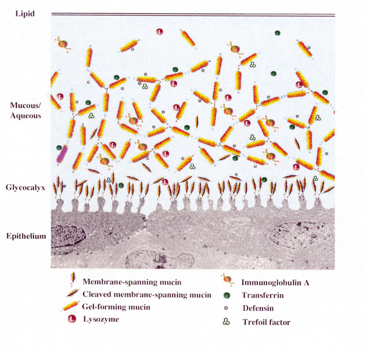
Chapter 3
The Tear Film
ERIN PETERS and KATHRYN COLBY
Main Menu   Table Of Contents

Search

INTRODUCTION
TEAR FILM STRUCTURE
LACRIMAL FUNCTIONAL UNIT
CLINICAL EVALUATION OF THE TEAR FILM
DRY EYE
CONCLUSIONS
REFERENCES

INTRODUCTION
The tear film is an exceedingly complex structure, the intricacies of which are not yet completely understood. There has been tremendous growth in our understanding of tear film biology over the last decade. Gone are the days when we viewed the tear film as a simple structure composed of segregated layers of mucus, water and electrolytes, and lipid. Now we know that many of the tear film components interact to create a hydrated gel that allows the tear film to accomplish its multiple functions. This chapter reviews the current understanding of the structure of the tear film and will introduce the concept of the integrated lacrimal functional unit as a key component of the healthy ocular surface. Currently available techniques to evaluate tear film function and stability are discussed. Finally, current theories of dry eye disease are reviewed to add insight into the biology of the normal tear film.
Back to Top
TEAR FILM STRUCTURE
The tear film is responsible for providing a smooth refractive surface for clear vision, maintaining the health of corneal and conjunctival epithelia, and acting as the first line of defense against microbial infections. Tear film composition is dynamic and in a constant state of flux, responding to environmental conditions in order to maintain ocular surface homeostasis. Traditionally, the tear film was described as being composed of three separate and distinct layers: mucin, aqueous, and lipid. However, new studies suggest that mixing between the mucin and aqueous layer occurs, creating a gradient of decreasing mucin concentration into the aqueous layer. This aqueous-mucin layer forms a hydrated gel (Fig. 1) with complex biology, which is then covered by the lipid layer, which has its own highly ordered structure. For the sake of simplicity, the mucin, aqueous, and lipid layers are considered separately here.

Fig. 1. Diagram of the tear film hydrated gel. Membrane-associated mucins on the microplicae of the epithelium form the glycocalyx. Secretory mucins admix with the aqueous layer containing antimicrobial factors, such as lysozyme, and immunoglobulins secreted by the lacrimal gland. The anterior lipid layer provides stability by interacting with the mucin-aqueous phase, and is itself composed of polar and nonpolar phases. (From Gipson IK, Argueso P. The role of mucins in the function of the corneal and conjunctival epithelia. Int Rev Cytol 231:1-49, 2003, with permission.)

MUCIN LAYER

Ocular mucus is composed of mucin, immunoglobulins, urea, salts, glucose, leukocytes, cellular debris, and enzymes.1 Mucins are high molecular weight glycoproteins that are heavily glycosylated: 50% to 80% of their mass can be attributed to their carbohydrate side chains.2 Tandem repeats of amino acids rich in serine and threonine found in their protein backbone serve as sites for O-type glycosylation.3,4 Heavy glycosylation imparts an overall negative charge to the glycoprotein, making the mucins highly hydrophilic and able to admix with the aqueous layer and maintain water on the surface of the eye.

Mucins are classified as either membrane-associated or secretory. Secretory mucins are further divided into two groups: large gel-forming mucins or small soluble mucins.

Membrane-associated mucins form the glycocalyx, a dense barrier to pathogen penetrance, at the epithelial cell–tear film interface.2 The membrane-associated mucins contain a hydrophobic transmembrane domain that anchors the mucin on the apical surface of the epithelial cells, a short cytoplasmic tail that extends into the cytoplasm, and an extracellular domain that reaches into the tear film.5

Estimated to extend 200 to 500 nm from the cell surface, the extracellular, highly glycosylated tandem repeat domains, also called ectodomains, function as a disadhesive preventing cell–cell and cell–matrix interactions.2,6 This property provides a lubricating surface that allows lid epithelia to glide over the corneal and conjunctival epithelia without adherence.7

The cytoplasmic tail of the membrane-associated mucin is thought to affect epithelial activity by interacting with cytoplasmic proteins and facilitating signal transduction.2 The short cytoplasmic domains are also reported to be associated with the actin cytoskeleton,8,9 which helps support the microplicae structure.2 The presence of epidermal growth factor (EGF)-like domains on the membrane associated mucins suggests a potential role in the regulation of epithelial cell growth.9–14

Soluble forms of the membrane-associated mucins have also been identified in the tear film, although the exact mechanisms for this occurrence are still unknown.2,13,15 Possible mechanisms include: cleavage and release of the extracellular domain into the tear film in a process termed ectodomain shedding; posttranslational processing of the mucin into two subunits where one subunit remains anchored in the plasma membrane, and the other soluble subunit is packaged in secretory granules and released into the tear film; or as some data suggest, mucin shedding from the cell surface over time leaves the oldest cells without microplicae and membrane associated mucins. The oldest cells lose their disadhesive character with the loss of the mucin, which results in the adherence of the old cells to the mucus of the tear film and their removal via the nasolacrimal duct.

Secreted mucins move easily over the mucins composing the glycocalyx because of the repulsive forces between them, which result from their anionic character.7 Secretory mucins act as a “cleaning crew,” moving through the tear fluid and collecting debris that can then be removed via the nasolacrimal duct during blinking.7 The secreted mucins are classified as either gel-forming or soluble. The large gel-forming mucins are probably the largest glycoproteins known based on their high molecular weight and are considered gel forming because they are responsible for the rheological properties of mucus.2,16 The small soluble mucins lack cysteine-rich D domains and are present as monomeric species.2

The majority of ocular mucins are secreted by the conjunctival goblet cells;1 however, ocular mucins are also produced by the stratified squamous epithelium of the cornea and conjunctiva, and new evidence suggests that the lacrimal gland also contributes to mucin production.17–19 Corneal and conjunctival stratified squamous cells contain the membrane-associated mucins MUC1, MUC4, and MUC16.20–22 MUC1 is a likely candidate for the glycocalyx as it is present in the apical cell membranes of the superficial ocular surface cells.23,24 Soluble forms of MUC1, MUC4, and MUC16 have also been detected in the tears2 and MUC4 has been detected in the lacrimal gland.18 MUC2, a gel-forming secretory mucin, and MUC7, a soluble secretory mucin, have been identified in tears and are both present in the conjunctiva, although the exact cellular source in the conjunctiva is unknown.17,19 MUC7 is also secreted by the lacrimal gland.19 MUC5AC, a large gel-forming mucin, is expressed by the goblet cells of the conjunctival epithelium and has been identified in tears in some individuals.25 MUC5AC is the major mucin present at the ocular surface providing the scaffolding of the mucus layer.2,17,20 Mucin secretion by the corneal and conjunctival stratified squamous epithelial cells is not as well studied as mucin secretion by conjunctival goblet cells, which is discussed later in the section on the lacrimal functional unit.

AQUEOUS LAYER

The middle aqueous layer of the tear film consists of water, electrolytes, proteins, peptide growth factors, immunoglobulins, cytokines, vitamins, antimicrobials, and hormones secreted by the lacrimal glands (Table 1).26,27

TABLE 1. The normal tear film has numerous constituents

Tear Constituents
Water
Electrolytes
Na+
K+
Mg2+
Ca2+
Cl-
HCO3-
PO43-
Proteins
Albumin
β-Lysin
Ceruloplasmin
Complement
Defensins
Group II phospholipase A2
Histamine
Interferon
Lactoferrin
Lipocalin
Lysozyme
Matrix metalloproteinases
Plasminogen activator
Prostaglandins
Proteases
Transferrin
Peptide growth factors
Epidermal growth factor (EGF)
Hepatocyte growth factor (HGF)
Transforming growth factor-β (TGF-β)
Immunoglobulins
IgA
IgG
IgD
IgE
IgM
sIgA (2 molecules of IgA joined by SC)
Cytokines
IL-1α
IL-1β
IL-6
Vitamins
Lipids
Mucins


Electrolytes present in the tear film include sodium, potassium, magnesium, calcium, chloride, bicarbonate, and phosphate ions.27 The electrolytes are responsible for the osmolarity of tears, acting as a buffer to maintain a constant pH and contribute to maintaining epithelial integrity of the ocular surface.28,29 An increase in osmolarity of the aqueous layer is a global feature of dry eye syndrome and damages the ocular surface directly and indirectly by triggering inflammation.27,30

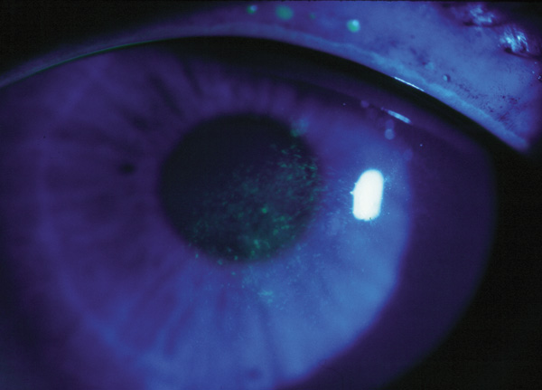
Proteins found in human tears are species-specific. More than 60 proteins have been identified in human tears including albumin, immunoglobulins, metal-carrying proteins, complement, histamine, plasminogen activator, prostaglandins, proteases, and antimicrobials.31 Considering that the thin nonkeratinzed epithelium and abundant blood supply of the conjunctiva make the conjunctiva an ideal entrance for infectious agents, it is imperative that the ocular surface have a strong defense system to protect against invading microorganisms. The primary defense system of the ocular surface is composed of the nonspecific immunity conferred by lysozyme, lactoferrin, β-lysin, complement, defensins, and group II phospholipase A2 and the specific immunity of antibodies, such as secretory immunoglobulin A (sIgA).27 In aqueous-deficient dry eye syndrome, the concentration of lysozyme, lactoferrin, lipocalin, and sIgA are reduced, compromising the integrity of the defense system, which may make the ocular surface more susceptible to infection, in addition to the symptoms of dry eye.32,33

Lysozyme, lactoferrin, and lipocalin are regulated antimicrobial proteins, secreted in response to an intracellular stimulus with a rate of secretion that approximately matches the rate of tear flow. Therefore, their concentration remains relatively constant with various flow rates.34–36 Lysozyme, an antimicrobial enzyme, lyses bacterial cell walls in the same manner as penicillin. It is one of the most important protein components in the tear film and acts synergistically with β-lysin, an enzyme that attacks bacterial cell membranes. Lactoferrin, a metal-binding protein, also has antimicrobial properties and may enhance antibody activity against certain microorganisms. Lipocalin, a lipid-binding protein, scavenges potentially harmful hydrophobic molecules and has recently been shown to inhibit bacterial and fungal infections through sequestering microbial siderophores.37 Further evidence suggests that lipocalin may contribute to a stable tear film by interacting with meibomian lipids and delivering them to the aqueous layer, and when complexed with other tear components, may be responsible for the high viscosity and low surface tension of tears.38–40Immunoglobulins are constitutively produced and transported to the tear film from the conjunctiva. Thus, reflex tearing reduces the concentration of immunoglobulins and a reduction in tear flow increases their concentration.36 An example of one such immunoglobulin is sIgA. sIgA is formed by two molecules of immunoglobulin A (IgA) produced by plasma cells in the adenoid layer of the conjunctiva being joined by secretory component (SC), a protein molecule produced by the conjunctival epithelium. sIgA is produced at a rate dependent on its rate of synthesis, which is regulated by its local endocrine environment.34,35,41

In nonstimulated tears, a small proportion of proteins are derived through the leakage of plasma components through ocular surface capillaries.27 These components include albumin; IgG; ceruloplasmin, a copper-carrying protein with oxidizing potential; transferrin, an iron-carrying protein; and monomeric IgA.27 Their concentrations increase in dry eye syndrome resulting from decreased tear volume and leakage from chronically inflamed surface capillaries.27 Peptide growth factors (such as EGF, transforming growth factor-β (TGF-β) and hepatocyte growth factor [HGF]), and vitamin A act via autocrine and paracrine mechanisms to regulate epithelial proliferation, motility, and differentiation. Peptide growth factors are also involved in corneal wound healing and immune modulation.42,43 Evidence suggests that the concentration of EGF is decreased in dry eye syndrome, and it seems reasonable to suppose that the other growth factors secreted by the lacrimal glands are similarly affected.27,44

LIPID LAYER

The anterior layer of the tear film is composed of meibomian oil secreted by the meibomian glands and is the major barrier to evaporation from the ocular surface.45,46 The lipid layer is also responsible for providing stability to the tear film through interaction with the aqueous-mucin phase, providing a smooth optical surface for the cornea, and acting as a barrier against foreign particles.38 McCulley and Shine47 have proposed that the anterior lipid layer is composed of two phases: a polar surfactant phase overlaid by a nonpolar phase. The polar lipid phase, primarily composed of phospholipids and glycolipids, is multifunctional. The highly structured polar lipid layer acts as a surfactant between the hydrophilic aqueous mucin layer and the thick, nonpolar lipid layer; facilitates interaction with the aqueous–mucin layer; provides a barrier; and offers a structural component on which the nonpolar phase depends.47,48 The nonpolar phase (mainly composed of wax, cholesterol esters, and triglycerides) provides the air-tear film interface and is responsible for retarding evaporation.47

A normal tear film lipid layer is able to reduce evaporation by approximately 90% to 95%.49–51 The rate of evaporation is affected by the thickness of the lipid layer, and it has been postulated that a decrease in thickness may cause evaporative dry eye.45 In fact, mild to moderate dry eye states exhibit a lack of confluence in the tear film lipid layer.52 Lipid composition may also affect evaporation because it has been reported that the phospholipid content of meibomian oil is decreased in patients with dry eye and meibomian lipid composition is altered in anterior and posterior blepharitis, although the exact mechanisms have yet to be elucidated.48,53–58 The melting range of meibomian oil is dependent on lipid composition and may be lowered by the presence of branched and unsaturated fatty acids and alcohols.38 The low melting range of meibomian oil (19.5°C to 32.0°C), attributed to the variety of lipids present in the meibomian oil, facilitates meibomian delivery and contributes to tear film stability as the surface corneal temperature (32°C) is close to the upper limit of the melting range, allowing the lipid to exist in a relatively solid state.45

Meibomian oil secretion is a continuous process, occurring 24 hours per day during waking and sleeping hours, and is aided by blink action.45 The rate of secretion is controlled by neural, hormonal, and vascular influences.38 The lid margin reservoir of oil in an adult male has been estimated to contain approximately 300 μg of meibomian oil, which is more than adequate to refresh the lipid layer after each blink considering that the preocular tear film holds 9 μg of lipid.59,60 The casual oil level (or resting level) in the marginal lid reservoir is highest just after waking and lower late morning and at the end of the day, suggesting that oil retained during sleep is discharged when blinking is resumed after waking.59,61 Forced blinking, as well as deliberate expression of meibomian oil, has been shown to increase the thickness of the tear film lipid layer;62,63 however, evidence also shows that oil delivery can be maintained in the absence of blinking, implying that blinking is not essential for delivery. Studies show a rise in the casual level of lipid on the lid margin with age in both genders; however, lower rises in levels in women before menopause suggests a hormonal influence on meibomian secretion.23

Back to Top
LACRIMAL FUNCTIONAL UNIT
In order to maintain ocular surface homeostasis, the appropriate release in quantity and composition of tear film components must occur in response to various stimuli. Secretion of tear film components is coordinated and controlled by the lacrimal functional unit described by Stern et al.64 The components of the functional unit act in tandem to maintain the integrity of the normal ocular surface; when any component of the unit is compromised, normal lacrimal support of the ocular surface is impaired, resulting in ocular pathology.64

The lacrimal functional unit is composed of the ocular surface tissues (cornea and conjunctiva, including goblet cells and meibomian glands), the lacrimal glands (main and accessory [Wolfring and Krause]), and their interconnecting sensory (CN V) and autonomic (CN VII) innervation.64 Innervation occurs from the parasympathetic system (acetylcholine and vasoactive intestinal peptide [VIP] containing nerve terminals), sympathetic nerves (neuropeptide Y and norepinephrine), and sensory fibers from the trigeminal nerve (substance P and calcitonin gene-related peptide).65–67

Tear film secretion from the lacrimal functional unit is reflexive.68 The cornea has 60 times more nerve endings than dental pulp.69 The sensory nerves of the highly innervated cornea and conjunctiva form the afferent limb of a simple reflex arc that conducts stimuli from the external environment back to the central nervous system.70,71 The efferent limb of the reflex arc is composed of the sympathetic and parasympathetic nerves that innervate the ocular surface epithelia and tear-producing glands. These nerves are responsible for stimulating the conjunctival epithelial goblet cells, lacrimal glands, and meibomian glands to secrete their respective components (mucus, aqueous, and lipid, respectively) onto the ocular surface to provide protection from the original stimulus.71

GOBLET CELLS

In addition to the epithelial cells, goblet cells are the second cell type found in the conjunctival epithelium and are the main source of mucus secretion. Found singly or in clusters, goblet cells are interspersed among the stratified squamous cells of the conjunctival epithelium. Goblet cells contain secretory granules in the apical portion of the cell that contain the mucins and other glycoproteins that are secreted onto the mucus layer of the tear film. MUC5AC, the large gel-forming mucin that is the major mucin present at the ocular surface, has been identified to be associated with the goblet cells, but other mucins, such as MUC2 and MUC7, may also be associated.

Conjunctival goblet cells are secretory apocrine glands, meaning all or most of the contents of the secretory granules in each cell are secreted in response to a given stimulus. Thus, controlling the number of cells responding to the stimulus regulates secretion.71 Direct and indirect neural control of mucin secretion has been described.72 Activation of sensory nerves innervating the stratified squamous cells of the cornea and conjunctiva induces goblet cell secretion by stimulating a local reflex arc.65 Both sympathetic and parasympathetic nerves innervate the conjunctival goblet cells. However, only parasympathetic neurotransmitters, acetylcholine and VIP, have been shown to activate mucus synthesis and secretion by the goblet cells.71 Muscarinic receptors present on conjunctival goblet cells bind cholinergic agonists leading to activation of a specific signaling pathway, which results in an increase in intracellular Ca2+ concentration and goblet cell secretion.73–75 Although sympathetic nerves have not been shown to stimulate goblet cell secretion, α-adrenergic receptors are present on immature goblet cells and may participate by regulating mucin synthesis, goblet cell proliferation, or other processes.71,76

Purinergic agonists also stimulate goblet cell secretion by activating the P2Y2 subtype of purinergic receptors.73,74 The presence of growth factor receptors in conjunctival goblet cells and the prevalence of growth factors, such as EGF, in the tears and conjunctiva suggests a role for growth factor regulation of the goblet cells, although the exact mechanism is still unknown.71 Sex hormones are also known to influence goblet cell density.77

LACRIMAL GLAND

In a normal individual, when afferent nerves of the ocular surface are stimulated, a reflex results in immediate blinking, withdrawal of the head, and secretion of reflex tears from the main lacrimal gland.64 Ocular surface irritation caused by blinking and environmental factors such as low humidity environments, wind exposure, bacteria, viruses, and even contact lens use results in chronic stimulation of the nerves of the ocular surface and increased secretory activity in the main and accessory lacrimal glands.64

The lacrimal gland is densely innervated with parasympathetic nerves, and to a lesser extent, with sympathetic and sensory nerves.78,79 Parasympathetic nerves release the neurotransmitters acetylcholine and VIP in response to corneal and conjuctival stimulation.35 Acetylcholine binds to muscarinic receptors on the basolateral membranes of lacrimal gland acinar cells, activating a signal transduction cascade that results in an increase of intracellular Ca2+ concentration and secretion of proteins, water, and electrolytes from the lacrimal gland.71 Cholinergic stimulation also induces increased concentrations of lactoferrin and EGF in glandular tissues and tears,80,81 possibly via a protein kinase C (PKC) pathway.71 VIP released by the parasympathetic nerves binds to two types of VIP receptors, VIPR1 and VIPR2, and activates a cyclic adenosine monophosphate (cAMP)-dependent signaling pathway resulting in secretion of proteins, water, and electrolytes from the lacrimal gland.71 Norepinephrine released from sympathetic nerves binds to both α1-and α-adrenergic receptors.71 When bound to α1-adrenergic, a signaling pathway involving Ca2+ and PKC is activated leading to secretion. α-Adrenergic agonists stimulate secretion through a cAMP-dependent signaling pathway.

Hormone action is also part of the control system to regulate the health of the ocular surface whereby certain stimuli at the ocular surface are communicated to the lacrimal gland.77 The lacrimal gland has a two-way communication system with endocrine organs, although the method of communication is unknown, which can relay information when a certain response is needed from the endocrine system.77 Androgens have recently been shown to influence the expression of over 2,000 genes in the mouse lacrimal gland82 as well as modulating the lacrimal gland anatomy. Androgen action on the lacrimal gland may be regulated by neurotransmitters such as VIP, β-adrenergic agonists, or cholinergic agonists, and cytokines.77 Androgens have also been shown to regulate the ocular secretory immune system in experimental animals through stimulating the synthesis and secretion of SC, the protein molecule required for formation of sIgA, by the lacrimal gland and increasing the concentration of sIgA in lacrimal tissue.77

The effect of estrogen on the lacrimal gland is controversial and has produced conflicting results, although estrogens do seem to promote inflammation.77 An epidemiologic study of 25,665 postmenopausal women found that estrogen replacement therapy increased the prevalence of dry eye signs and symptoms.83

MEIBOMIAN GLANDS

Human meibomian glands are embedded in the tarsal plate with 20 to 25 and 30 to 40 individual glands present in a single row along the lower and upper eyelid, respectively.84 Meibomian glands are holocrine glands that discharge the entire content of their acinar cells in the process of secretion.45 Multiple acini comprise the meibomian gland, and it is these cells that are responsible for synthesizing the meibomian gland lipids, both polar and nonpolar, that are excreted as meibum onto the ocular surface.47,84 Meibomian gland function is subject to vascular, neuronal, and hormonal influences. Similar to lacrimal glands, meibomian glands have a rich blood supply and are innervated through trigeminal afferents and parasympathetic and sympathetic autonomic efferents.45 Therefore, meibomian gland function may be under neuronal control through direct innervation of the meibomian gland acini or through indirect regulation of the vasculature to control the synthesis and/or excretion of meibomian gland lipids.47 VIP nerves are in direct contact with meibomian gland acinar cells.47 The meibomian glands also possess androgen and estrogen receptors, suggesting a regulatory role for the sex hormones.85–90 In fact, androgens regulate gene expression and lipid production in the meibomian gland, and it has been suggested that androgen deficiency and estrogens may contribute to meibomian gland dysfunction and evaporative dry eye.77

Back to Top
CLINICAL EVALUATION OF THE TEAR FILM
This section reviews the currently available modalities used to examine the structure and function of the tear film in health and disease. Many of the tests are complementary. A number are useful primarily in research settings at the present time. One of the difficulties in studying the tear film, and also in caring for patients with disorders of the tear film, is the lack of tests that give consistent results across different examiners and that are readily available and affordable for use in clinical settings. Advances in this area will improve our understanding of the biology of the tear film and the changes that accompany its disorders.

TEAR SECRETION

Several measures of tear secretion currently exist, although there is tremendous variability in the ways each are performed.

Schirmer I

The Schirmer I test is used to evaluate aqueous-deficient dry eye by measuring reflex tear secretion in response to conjunctival stimulation. However, other stimuli, such as sensory and psychological, are likely to be involved in reflex tear secretion, not just conjunctival stimuli.91 The test is performed without anesthesia by placing a strip of Whatman #41 paper at the junction of the middle and lateral thirds of the lower eyelid of each eye to minimize irritation to the cornea. The patient is told to look forward and blink normally while the strip is held in place for 5 minutes. The amount of wetting is recorded in millimeters.30 Anything less than 10 mm is considered diagnostic of aqueous tear deficiency. When topical anesthetic is applied before performing the Schirmer I test, the basal rate of tear secretion can be measured. For the Schirmer I test with anesthetic, less than 5 mm is diagnostic of aqueous tear deficiency and 5 to 10 mm is suggestive.

Schirmer II

The Schirmer II test measures reflex tear secretion in response to nasal stimulation. The Whatman #41 paper is placed in the lower eyelid in the same manner as the Schirmer I test, followed by stimulation of the nasal mucosa with a cotton-tipped applicator. Wetting of less than 15 mm after 5 minutes is associated with a defect in reflex secretion. This test is uncomfortable for the patient because of the vigorous stimulation of the nasal mucosa. Variability in the results of Schirmer tests require that consistent results be achieved before diagnoses are made.

Phenol Red Thread Test

Phenol red, a pH-sensitive dye, can be used to indicate tear volume by a Schirmer-like test. The end of a cotton thread dyed with phenol red is placed in the lower eyelid. The wetted length on the thread is measured over a period of 15 seconds, which is easily viewed by the color change of yellow to red in the presence of the near neutral tears. Anything less than 6 mm is diagnostic of dry eye.

Meniscometry

Tear meniscus volume is reduced in aqueous-deficient dry eye as indicated by a reduced height and radius of curvature.92 Dry eye patients have significantly smaller menisci than normal eyes; patients with dry eye with punctal plugs have significantly larger menisci than normal eyes.93 Meniscometry is a noninvasive way to assess tear volume indirectly by measuring the tear meniscus radius and it correlates well with the Schirmer test.93 A videomeniscometer records images of the ocular surface with a digital video recorder and transfers them to a computer, where image analysis software is used to calculate the radius of curvature of the meniscus applying the concave mirror formula.93 Tear kinetics can also be studied with the videomeniscometer.93

TEAR FILM STABILITY

Tear Break-Up Time

Tear break-up time assesses tear film stability and can be measured either invasively with the instillation of fluorescein or noninvasively using a keratometer or xeroscope. Fluorescein instillation is provocative in that it shortens the normal break-up time, but it still remains the standard to assess tear film stability.94 Tear break-up is best observed with use of a blue exciter and yellow barrier filter, while the patient refrains from blinking.92 The break-up time is recorded in seconds as the interval between the patient's last blink to the first appearance of a dark spot on the flourescein-stained tear film. Appearance of a dry spot in less than or equal to 7 seconds is considered abnormal and associated with an unstable tear film. Tear break-up time varies between individuals and changes within the same person throughout the day. Tear break-up time is reduced in all forms of dry eye.

The measure of the noninvasive break-up time (NIBUT) uses a grid or other pattern directed on the precorneal tear film to allow for observation of image distortion. The time from opening the eyes to the first sign of image distortion is measured in seconds. The normal range for the NIBUT is 40 to 60 seconds.

Ocular Protection Index

The ocular protection index (OPI) is a quantitative means of assessing the severity of a patient's dry eye. OPI equals break-up time (BUT)/interblink interval (IBI). The ocular surface is protected when the BUT matches or exceeds the IBI (OPI ≥ 1). The ocular surface is unprotected when the BUT is less than the IBI (OPI < 1).95

Tear Film Stability Analysis System

Videokeratography is another noninvasive method of evaluating tear film stability. Consecutive images of the corneal surface are captured every second for 10 seconds. The color change on the topographical map reflects fluctuations of the tear film over the 10-second test period. Thus, the tear film stability analysis system is two dimensional, reflecting break-up time and break-up area parameters.96 The advantages of videokeratography are that it is comprehensive and data can be easily stored for future use and comparison purposes.96 The disadvantage of this system is the cost and availability of the videokeratography unit.

TEAR TURNOVER

Tear turnover has been shown to be reduced in various forms of dry eye.97 It can be assessed by measuring the time it takes to clear 5 μL of sterile fluorescein instilled in the conjunctival sac. A standard Schirmer strip is placed over the lateral lower lid margin for a period of 1 minute at 10-, 20-, and 30-minute intervals after fluorescein instillation. The strips are then examined for the presence of fluorescein under a blue light. Fluorescence is absent in samples from normal individuals by 20 minutes. Reduced tear turnover may perpetuate ocular surface pathology by retaining inflammatory mediators in the tears.97

OCULAR SURFACE EVALUATION

Fluorescein

Fluorescein is a commonly used method to demonstrate ocular surface damage as manifested as corneal staining. Fluorescein is a vital dye that is well tolerated. Fluorescein-impregnated strips are wet with a sterile drop of nonpreserved saline solution and applied lightly to each lower tarsal plate. Alternatively, fluorescein solution may be used. The orange dye fluoresces green when excited by light passed through the standard blue exciter filter of the slit lamp and is best viewed in conjunction with a yellow barrier filter, such as a Wratten #12. Punctate staining (Fig. 2) is graded for each of five areas of the cornea (Fig. 3) on a scale of zero to three (the van Bjisterveld grading system); a score greater than three after summation of grades for all five areas of the eye indicates an unstable tear film.30 Other grading systems, such as the Oxford Scale,98 also exist to rate ocular surface damage.

Fig. 2. Fluorescein staining indicating superficial punctate keratopathy (SPK).

Fig. 3. The cornea is divided into five areas in order to grade fluorescein uptake. Each area is graded on a scale of 0 to 3, after which the grades are added together to give the final score. Scores greater than three indicate an unstable tear film. (From Lemp MA. Report of the National Eye Institute/Industry Workshop on clinical trials in dry eyes. CLAO J 21:221, 1995 with permission.)

Rose Bengal

Rose bengal staining also indicates ocular surface damage but causes pain upon instillation, particularly in dry eye patients. Rose bengal has long been considered a supravital dye, taken up by dead and degenerate cells. New studies suggest that although cells may not need to be degenerate to take up the stain, they may suffer from altered expression of membrane mucin.99,100

Lissamine Green

Lissamine green stains in a similar pattern to rose bengal. It is viewed in white light and is best observed over the white of the sclera instead of the cornea because of the dark background of the iris. The advantage of lissamine green staining is that it does not sting upon instillation and is well tolerated similar to fluorescein.

TEAR FILM OSMOLARITY

Tear Film Osmolarity

To evaluate tear film osmolarity, nanoliter samples are taken from the lower tear meniscus and measured with a Clifton depression of freezing point osmometer or an Advanced Instruments machine.30 Tear film osmolarity is not able to differentiate between tear-deficient and evaporative dry eye because an increase in osmolarity may be caused by either increased tear evaporation or a decrease in tear secretion. The test also requires expensive equipment and skilled technicians, but it is an extremely effective and sensitive tool for accurately diagnosing dry eye.

Tear Ferning

Tear ferning is dependent on the ratio of Na+ and K+ to Ca2+ and Mg2+.30 Differences in electrolyte concentrations may be related to different ferning patterns.101 Tears are collected with a glass capillary tube, placed on a glass slide, and left to dry at room temperature. After crystallization, the samples are observed in white light or by polarized microscopy and classified into four grades. Dry eye patients exhibit less ferning than normal patients.102

Tear Evaporation

Tests for tear evaporation are difficult to perform and are not able to differentiate between particular types of dry eye, but patients with dry eye do show an increased rate of evaporation compared to normals (0.43 versus 0.14 μL/min) as measured in an evaporation chamber.63 The evaporation chamber is designed to fit tightly around the eye and monitor changes in humidity.63 Humidity, temperature, and exposed surface area are all measured in order to calculate the rate of evaporation.63

LIPID LAYER EVALUATION

Interferometry

Interferometry is used to determine lipid layer thickness and fluidity93 by observing interference patterns generated by light reflected from the surface of the lipid layer and from the interface between the lipid and aqueous layer of the tear film. Studies have shown greater tear film stability with a thicker lipid layer.93

Meibometry

Meibometry assesses the baseline level of meibomian lipids by using a laser device.93 The optical density of a lipid sample taken from the eye with translucent plastic tape is read in a laser meibometer, and once corrected for the standards (meibometer reading with blank tape and without tape), the baseline lipid level is expressed as arbitrary optical density units. Presently, laser meibometry is the only method to quantitatively evaluate tear lipids.93

Back to Top
DRY EYE
One can often use disease processes to understand underlying physiology of a system. Dry eye disorders have provided important insights into the basic biology of the tear film.

The National Eye Institute/Industry Workshop in December 1993 and 1994 developed a global definition of dry eye: Dry eye is a disorder of the tear film caused by tear deficiency or excessive tear evaporation that causes damage to the interpalpebral ocular surface and is associated with symptoms of ocular discomfort.30 From this definition, two major classes of dry eye were established: tear deficient and evaporative (Fig. 4). At the time, it was recognized that the definition was limited in describing all possible causes and features of dry eye and may need to be modified in specific situations. For example, the definition does not include reference to dry eye after LASIK, because of allergy, or ocular surface-related problems103 and does not account for ocular surface damage outside the interpalpebral region or the possible absence of symptoms in the presence of dry eye signs in some patients.30 However, the common feature of all dry eye is tear film instability, a result of disease or dysfunction of one or more components of the lacrimal functional unit.104 Age, hormonal deficiencies, medications, surgery, and systemic autoimmune disease are all capable of causing dysfunction of the lacrimal functional unit that is associated with ocular surface inflammation, and ultimately, the signs and symptoms of dry eye.104

Fig. 4. National Eye Institute (NEI) classification scheme for dry eye. (From Lemp MA. Report of the National Eye Institute/Industry Workshop on clinical trials in dry eyes. CLAO J 21:221, 1995 with permission.)

TEAR-DEFICIENT DRY EYE

Tear-deficient dry eye is the largest category of dry eye. It occurs because of a disorder in lacrimal gland function, resulting in either reduced aqueous tear production and tear flow, or a failure to transfer lacrimal fluid into the conjunctival sac.30,105 Reasons for the lacrimal gland dysfunction include lacrimal gland disease, scarring of the lacrimal gland ducts, or abnormal stimulation of the lacrimal glands.27 Patients with aqueous tear deficiency may develop keratoconjunctivitis sicca (KCS), an ocular surface disease characterized by squamous metaplasia, corneal epitheliopathy, and filamentary keratitis (abnormal adherence of epithelial and mucous filaments).105 The subjective symptoms of dry eye include: heaviness of the lids, foreign body sensation, burning, stinging, itching, photophobia, dryness, soreness, and ocular fatigue.30,105 Tear-deficient dry eye is subdivided into two categories: Sjögren syndrome tear deficiency and non-Sjögren tear deficiency depending upon whether there are associated systemic signs and symptoms.

Sjögren Syndrome

Sjögren syndrome is an autoimmune exocrinopathy that is characterized by chronic inflammatory lymphocytic infiltration and immune-mediated destruction of the lacrimal and salivary glands.27,30 The mechanism for lymphocytic infiltration is unclear; however, epithelial cells of the lacrimal gland are believed to initiate and perpetuate the autoimmune reactivity by either producing immunologically active mediators or acting as antigen presenting cells.106–108 It has been hypothesized that the impaired secretory function of the lacrimal gland because of lymphocytic infiltration may be a result of a functional blockage in the neural pathway caused by inflammation. Inflammation could block the release of neurotransmitters from the nerve ends or impact the cellular response to neuromediators.69

A hormonal influence has been indicated in Sjögren syndrome because women are more afflicted with autoimmune disorders than men.77 A recent study showed that women with premature ovarian failure, a possible autoimmune disorder, were more likely to exhibit signs of ocular surface damage and dry eye symptoms than age-matched controls, although no differences were observed in tear production.109 Women suffering from premature ovarian failure are androgen deficient.109 It has also been shown that women with primary and secondary Sjögren syndrome are androgen deficient.110 Topical or systemic administration of androgens in patients with Sjögren syndrome alleviates dry eye signs and symptoms and stimulates tear flow suggesting that correction of the androgen defect in Sjögren syndrome may have a therapeutic effect on the lacrimal gland.77 This effect may extend to other autoimmune diseases as well.77

Sjögren syndrome may be primary or secondary. Primary Sjögren syndrome is aqueous dry eye in combination with dry mouth, the presence of autoantibodies, and a positive focus score on minor salivary gland biopsy.111,112 It has been estimated that primary Sjögren syndrome is a disease with a prevalence not exceeding 0.6% of the general population.113 Sjögren syndrome is secondary when it occurs in association with other autoimmune connective tissue disorders, rheumatoid arthritis being the most common.30

Non-Sjögren Syndromes

In the absence of an autoimmune disease, ocular symptoms of dry eye with evidence of lacrimal dysfunction may be diagnosed as non-Sjögren dry eye. Non-Sjögren tear deficiencies include primary and secondary lacrimal deficiencies and congenital alacrima.

The most common form of non-Sjögren dry eye is primary lacrimal deficiency (PLD), although the etiology of this disorder is unknown.105 Obata et al114 reported that the lacrimal gland dysfunction may be a result of age-related changes in the lacrimal gland including lobular and diffuse fibrosis and atrophy, as well as periductal fibrosis. Sullivan77 proposed that PLD may occur as a result of a decline in serum androgen levels experienced with menopause, pregnancy, lactation, or the use of estrogen-containing oral contraceptives in women. However, this causation is gender-specific because men treated with antiandrogen therapy do not experience an effect on aqueous tear production.77 Also, an immune mechanism cannot be excluded as lacrimal gland destruction has been reported by a round-cell infiltration associated with acinar loss and ductal pathology.115,116Congenital alacrima is an uncommon condition in children resulting from the absence or hypoplasia of the lacrimal gland, or abnormalities of the innervation of the lacrimal gland.117 The most common condition associated with alacrima is the Riley-Day syndrome, in which decreased tear production may be caused by abnormal parasympathetic innervation of the lacrimal gland.105 Patients with this condition produce a reduced amount of tears when crying, reflex lacrimation is absent, and corneal sensation is decreased. These symptoms make the patients more prone to corneal ulceration and perforation.105,118,119

Secondary lacrimal deficiency may be caused by numerous conditions, including lacrimal gland infiltration by sarcoid granulomata, lymphomas, and neurofibromas; lacrimal gland inflammation associated with viral infections such as human T-cell leukemia/lymphoma virus type 1 (HTLV-1), human immunodeficiency virus type 1 (HIV-1), hepatits C, and Epstein-Barr virus; graft-versus-host disease; vitamin A deficiency through xerophthalmia, a mucin deficiency, and a protein deficiency; lacrimal gland ablation; sensory denervation; refractive surgery; and seventh-nerve palsy affecting the greater superficial petrosal nerve or the nervus intermedius damages caused by interference with the secretomotor fibers of the lacrimal gland.27

Aqueous deficiency also occurs when the secretory ducts and orifices of the orbital and accessory lacrimal glands are scarred as a result of cicatrizing conjunctival diseases. The most common cicatrizing diseases are: trachoma, ocular cicatricial pemphigoid, erythema multiforme and Stevens-Johnson symdrome, and chemical and thermal burns.105

Systemic medications can also increase the risk of developing dry eye in patients with marginal tear production. Common medications that decrease tear production include diuretics and those with antimuscarinic effects because they inhibit the cholinergic signal transduction pathway.27 Examples of such medications include antihistamines, antihypertensives, and antipsychotic drugs.27

EVAPORATIVE DRY EYE

Evaporative dry eye occurs when lacrimal function is normal and the volume of lacrimal fluid is sufficient to cover the ocular surface, but another tear abnormality exists that leads to increased tear evaporation.30 A study has shown that the tear evaporation rate is positively correlated with tear osmolarity and inversely related to tear stability.52 Hyperosmolarity damages the ocular surface directly120–122 and indirectly by triggering inflammation.123 Hyperosmolarity activates inflammatory pathways such as stress-activated protein kinases, MAP kinase p38, c-Jun NH2-terminal kinase (JNK), and the transcriptional regulator NF-κB,124–126 which then induce the release of inflammatory mediators including interleukin (IL)-8 and matrix metalloproteinases MMP-9 and MMP-13, which have been implicated in the pathogenesis of dry eye.127–129

Meibomian Gland Disease

Obstructive meibomian gland dysfunction (MGD) is the most common cause of evaporative dry eye.45 Expression of meibomian oil maintains the thickness of the lipid layer and reduces tear evaporation; when expression of oil is reduced by disease, evaporation increases leading to hyperosmolar conditions at the ocular surface.130–132Hyperkeratinization of the terminal ductules of meibomian glands is a key event in meibomian gland dysfunction and most likely results from inflammation in and around the distal parts of the glands.45 The increase in incidence of MGD with age may be attributed to a drop in androgen levels, which promotes a proinflammatory environment.45

Blepharitis

In squamous blepharitis, desquamated cells derived from the lid margin may deliver skin lipid to the tear film causing tear instability and tear evaporation, resulting in punctate keratitis.30 Blepharitis aggravates dry eye syndrome by acting as a source of antigenic and proinflammatory substances, and adversely influencing lipid production.57 Conjunctival impression cytology demonstrates epithelial impairment, rupture of intercellular junctions, loss of goblet cells, and-deficient mucin secretion in patients with blepharitis.133

Blink Disorders

Neuronally controlled lid blinking mechanically spreads the tear film over the ocular surface while also removing and draining old tears via the nasolacrimal ducts into the nose.134 Thus, disorders that affect the frequency of blinking will not only influence the integrity of the tear film but will also affect the removal of tears from the ocular surface. Delayed tear clearance caused by decreased blinking may promote ocular surface inflammation by prolonging exposure to noxious substances present in the tears. In addition, decreased blinking will increase the rate of evaporation from the eye by prolonging interblink periods and may decrease lipid layer thickness by reducing meibomian oil secretion.27 Epidemiologic studies have found that the prevalence of dry eye is higher in visual display terminal users as a function of their decreased blink rate and increased aperture size that occurs with a forward-looking gaze.135 Decreased blink frequency also occurs in Parkinson's disease and occasionally with the use of antipsychotic medicines.

Disorders of Lid Aperture and Lid/Globe Congruity

The width of the lid aperture determines the size of the ocular surface area that is exposed and subject to evaporation. Patients with proptosis of any cause will experience greater rates of evaporation from the ocular surface, which may result in surface drying. Poor lid/globe congruity affects tear film resurfacing between blinks.

Contact Lens Use

Contact lens wear disrupts the stability of the tear film leading to increased evaporation from the ocular surface. Sensory loss as a result of prolonged contact lens wear may also decrease lacrimal secretion by interfering with the afferent reflex arc.

INFLAMMATORY DRY EYE

Inflammation has been observed in all stages of dry eye.106,136 Increased levels of inflammatory cytokines, especially IL-6, have been observed in the lacrimal glands, conjunctival epithelium, and/or tear fluid of patients with dry eye.64,106,137,138 Chronic immune-mediated inflammatory processes are involved in the pathogenesis of dry eye, gradually leading to dysfunction and destruction of the lacrimal glands and impairment of the conjunctival epithelium. Recent research suggests that ocular surface inflammation occurs with changes in tear film composition and stability due to dysfunction of the lacrimal functional unit, and it has been proposed that dryness is the downstream consequence of inflammation.68

Lacrimal functional unit impairment promotes inflammation by several mechanisms: decreased secretion of anti-inflammatory factors, such as lactoferrin, by a dysfunctional lacrimal gland; increased production of proinflammatory cytokines (IL-1), tumor necrosis factor-α (TNF-α), and proteolytic enzymes by stressed ocular surface and glandular epithelial cells, as well as by the inflammatory cells infiltrating those tissues; activation of latent inactive cytokines and proteases normally present in the tear film that serve as an early defense mechanism after ocular surface infection and wounding; and increased tear film osmolarity.104 Stimulation of inflamed lacrimal glands and conjunctiva could deliver proinflammatory tears to the ocular surface, worsening the condition and contributing to a viscous cycle of dry eye.139 In fact, excessive nervous stimulation aimed at triggering the mechanisms of regulation and repair may lead to neurogenic inflammation, activation of T cells, and the subsequent release of inflammatory cytokines into the lacrimal glands, tear film, and conjunctiva.64

Hormonal regulation of the lacrimal functional unit appears to modulate inflammation. Androgen binding to receptors in the lacrimal gland leads to altered expression of cytokines and protooncogenes.140 Anti-inflammatory cytokines, such as transforming growth factor-β (TGF-β), accumulate as a result of androgen action, whereas the reduction of androgen levels leads to the release of pro-inflammatory cytokines, such as IL-1β, IL-2, interferon-γ; , and TNF-α. Decreases in the level of circulating anti-inflammatory androgens that occur with aging and autoimmune disorders have been implicated in promoting inflammation of the lacrimal gland.104 In addition, the increased incidence of dry eye in women,141 particularly in older, postmenopausal women,142 may be a result of the loss of an anti-inflammatory environment when circulating levels of androgens drop below a threshold level.143,144

Initial studies also indicate that diet may be a factor in the development of dry eye. A study of 32,470 women found that a higher intake of ω-3 fatty acids contributed to a decreased risk of developing dry eye syndrome.145 This result could potentially be explained by the anti-inflammatory and immune modulating effects of ω-3 fatty acids.

Back to Top
CONCLUSIONS
The recent decade has seen an explosion in our understanding of the tear film. More work remains to be done, especially regarding the role of hormonal, inflammatory and nutritional influences on the physiology of the tear film. Better techniques to evaluate the tear film need to be developed to further our understanding of this complex structure whose proper functioning is essential to good vision.
Back to Top
REFERENCES

1. Davidson HJ, Kuonen VJ: The tear film and ocular mucins. Vet Ophthalmol 7:71, 2004

2. Gipson IK, Hori Y, Argüeso P: Character of ocular surface mucins and their alteration in dry eye disease. Ocular Surface 2:131, 2004

3. Gendler SJ, Spicer AP: Epithelial mucin genes. Annu Rev Physiol 57:607, 1995

4. Moniaux N, Escande F, Porchet N, et al: Structural organization and classification of the human mucin genes. Front Biosci 6:D1192, 2001

5. Argueso P, Gipson IK: Epithelial mucins of the ocular surface: structure, biosynthesis and function. Exp Eye Res 73:281, 2001

6. Komatsu M, Carraway CA, Fregien NL, et al: Reversible disruption of cell-matrix and cell-cell interactions by overexpression of sialomucin complex. J Biol Chem 272:33245, 1997

7. Gipson IK: Distribution of mucins at the ocular surface. Exp Eye Res 78:379, 2004

8. Parry G, Beck JC, Moss L, et al: Determination of apical membrane polarity in mammary epithelial cell cultures: the role of cell–cell, cell–substratum, and membrane–cytoskeleton interactions. Exp Cell Res 188:302, 1990

9. Williams SJ, McGuckin MA, Gotley DC, et al: Two novel mucin genes down-regulated in colorectal cancer identified by differential display. Cancer Res 59:4083, 1999

10. Pratt WS, Crawley S, Hicks J, et al: Multiple transcripts of MUC3: Evidence for two genes, MUC3A and MUC3B. Biochem Biophys Res Commun 275:916, 2000

11. Gum JR Jr, Crawley SC, Hicks JW, et al: MUC17, a novel membrane-tethered mucin. Biochem Biophys Res Commun 291:466, 2002

12. Carraway KL, Price-Schiavi SA, Komatsu M, et al: Multiple facets of sialomucin complex/MUC4, a membrane mucin and erbB2 ligand, in tumors and tissues (Y2K update). Front Biosci 5:D95, 2000

13. Carraway KL, Ramsauer VP, Haq B, et al: Cell signaling through membrane mucins. Bioessays 25:66, 2003

14. Williams SJ, Wreschner DH, Tran M, et al: MUC13, a novel human cell surface mucin expressed by epithelial and hemopoietic cells. J Biol Chem 276:18327, 2001

15. Higuchi T, Orita T, Nakanishi S, et al: Molecular cloning, genomic structure and expression analysis of MUC20, a novel mucin protein, up-regulated in injured kidney. J Biol Chem 279:1968, 2004

16. Sellers LA, Allen A, Morris ER, et al: The rheology of pig small intestinal and colonic mucus: Weakening of gel structure by non-mucin components. Biochim Biophys Acta 1115:174, 1991

17. Dartt DA: Control of mucin production by ocular surface epithelial cells. Exp Eye Res 78:173, 2004

18. Carraway KL, Carvajal ME, Li P, et al: ErbB2 and its ligand MUC4 (sialomucin complex) in rat lacrimal gland. Adv Exp Med Biol 506:289, 2002

19. Jumblatt MM, McKenzie RW, Steele PS, et al: MUC7 expression in the human lacrimal gland and conjunctiva. Cornea 22:41, 2003

20. Inatomi T, Spurr-Michaud S, Tisdale AS, et al: Expression of secretory mucin genes by human conjunctival epithelia. Invest Ophthalmol Vis Sci 37:1684, 1996

21. Pflugfelder SC, Liu Z, Monroy D, et al: Detection of sialomucin complex (MUC4) in human ocular surface epithelium and tear fluid. Invest Ophthalmol Vis Sci 41:1316, 2000

22. Argueso P, Spurr-Michaud S, Russo CL, et al: MUC16 mucin is expressed by the human ocular surface epithelia and carries the H185 carbohydrate epitope. Invest Ophthalmol Vis Sci 44:2487, 2003

23. Bron AJ: The Doyne Lecture: Reflections on the tears. Eye 11:583, 1997

24. Inatomi T, Spurr-Michaud S, Tisdale AS, et al: Human corneal and conjunctival epithelia express MUC1 mucin. Invest Ophthalmol Vis Sci 36:1818, 1995

25. Argueso P, Balaram M, Spurr-Michaud S, et al: Decreased levels of the goblet cell mucin MUC5AC in tears of patients with Sjogren syndrome. Invest Ophthalmol Vis Sci 43:1004, 2002

26. Holly FJ, Lemp MA: Tear physiology and dry eyes: review. Surv Ophthalmol 22:69, 1997

27. Johnson ME, Murphy PJ: Changes in the tear film and ocular surface from dry eye syndrome. Prog Ret Eye Res 23:449, 2004

28. Carney LG, Hill RM: Human tear pH. Diurnal variations. Arch Ophthalmol 94:821, 1976

29. Bachman WG, Wilson G: Essential ions for maintenance of the corneal epithelial surface. Invest Ophthalmol Vis Sci 26:1484, 1985

30. Lemp MA: Report of the National Eye Institute/Industry Workshop on clinical trials in dry eyes. CLAO J 21:221, 1995

31. Gachon AM, Verrelle P, Betail G, et al: Immunological and electrophoretic studies of human tear proteins. Exp Eye Res 29:539, 1979

32. Janssen PT, van Bijsterveld OP: Tear fluid proteins in Sjogren's syndrome. Scan J Rheumatol 61:224, 1986

33. Mackie IA, Seal DV: Diagnostic implications of tear protein profiles. Br J Ophthalmol 68:321, 1984

34. Burgess TL, Kelly RB: Constitutive and regulated secretion of proteins. Annu Rev Cell Biol 3:243, 1987

35. Dartt DA: Signal transduction and control of lacrimal gland protein secretion: A review. Curr Eye Res 8:619, 1989

36. Fullard RJ, Snyder C: Protein levels in nonstimulated and stimulated tears of normal human subjects. Invest Ophthalmol Vis Sci 31:1119, 1990

37. Fluckinger M, Haas H, Merschak P, et al: Human tear lipocalin exhibits antimicrobial activity by scavenging microbial siderophores. Antimicrob Agents Chemother 48:3367, 2004

38. Bron AJ, Tiffany JM, Gouveia SM, et al: Functional aspects of the tear film lipid layer. Exp Eye Res 78:367, 2004

39. Pandit JC, Nagyová B, Bron AJ, et al: Physical properties of stimulated and unstimulated tears. Exp Eye Res 68:247, 1999

40. Tiffany JM, Nagyová B: The role of lipocalin in determining the physical properties of tears. Adv Exp Med Biol 506:581, 2002

41. Sullivan DA, Hann LE: Hormonal influence on the secretory immune system of the eye: endocrine impact on the lacrimal gland accumulation and secretion of IgA and IgG. J Steroid Biochem 34:253, 1989

42. Sommer A: Effects of vitamin A deficiency on the ocular surface. Ophthalmology 90:592, 1983

43. Schultz G, Khaw PT, Oxford K, et al: Growth factors and ocular wound healing. Eye 8:184, 1994

44. Ohashi Y, Ishida N, Nakata K, et al: Abnormal protein profiles in tears with dry eye syndrome. Am J Ophthalmol 136:291, 2003

45. Bron AJ, Tiffany JM: The contribution of meibomian disease to dry eye. Ocular Surface 2:149, 2004

46. McCulley JP, Shine WE: Meibomian gland function and the tear lipid layer. Ocular Surface 1:97, 2003

47. McCulley JP, Shine WE: The lipid layer of tears: dependent on meibomian gland function. Exp Eye Res 78:361, 2004

48. Shine WE, McCulley JP: Polar lipids in human meibomian gland secretions. Curr Eye Res 26:89, 2003

49. Mathers WD, Lane JA: Meibomian gland lipids, evaporation, and tear film stability. Adv Exp Med Biol 438:349, 1998

50. Tsubota K, Yamada M: Tear evaporation from the ocular surface. Invest Ophthalmol Vis Sci 33:2942, 1992

51. Hisatake K: Evaporation of water in a vessel. J Appl Phys 73:7395, 1993

52. Craig JP, Tomlinson A: Importance of the lipid layer in human tear film stability and evaporation. Optom Vis Sci 74:8, 1997

53. Dougherty JM, McCulley JP: Analysis of the free fatty acid component of meibomian secretions in chronic blepharitis. Invest Ophthalmol Vis Sci 27:52, 1986

54. Dougherty JM, McCulley JP: Bacterial lipases and chronic blepharitis. Invest Ophthalmol Vis Sci 27:486, 1986

55. Shine WE, McCulley JP: The role of cholesterol in chronic blepharitis. Invest Ophthalmol Vis Sci 32: 2272, 1991

56. Shine WE, Silvany R, McCulley JP: Relation of cholesterol-stimulated Staphylococcus aureus growth to chronic blepharitis. Invest Ophthalmol Vis Sci 34:2291, 1993

57. Shine WE, McCulley JP: Keratoconjunctivitis sicca associated with meibomian secretion polar lipid abnormality. Arch Ophthalmol 116:849, 1998

58. McCulley JP, Shine WE: Eyelid disorders: The meibomian gland, blepharitis and contact lenses. Eye Contact Lens 29:S93, 2003

59. Chew CK, Hykin PG, Jansweijer C, et al: The casual level of meibomian lipids in humans. Curr Eye Res 12:255, 1993

60. Chew CK, Jansweijer C, Tiffany JM, et al: An instrument for quantifying meibomian lipid on the lid margin: The meibometer. Curr Eye Res 12:247, 1993

61. Bron AJ: Control of meibomian secretion and its role in ocular surface disease. Castroviejo Lecture, American Academy of Ophthalmology, Chicago, November 1993

62. Korb D, Greiner JV: Increase in tear film lipid layer thickness following treatment of meibomian gland dysfunction. In Sullivan DA (ed): Lacrimal Gland, Tear Film, and Dry Eye Syndromes. Basic Science and Clinical Relevance,. New York: Plenum Press, 1994, 293–298

63. Mathers WD, Binarao F, Petroll M: Ocular water evaporation and the dry eye. A new measuring device. Cornea 12:335, 1993

64. Stern ME, Beuerman RW, Fox RI, et al: The pathology of dry eye: The interaction between the ocular surface and the lacrimal glands. Cornea 17:584, 1998

65. Dartt DA, McCarthy DM, Mercer HJ, et al: Localization of nerves adjacent to goblet cells in rat conjunctiva. Curr Eye Res 14:993, 1995

66. Seifert P, Spitznas M: Immunocytochemical and ultrastructural evaluation of the distribution of nervous tissue and neuropeptides in the meibomian gland. Graefes Arch Clin Exp Ophthalmol 234:648, 1996

67. Seifert P, Stuppi S, Spitznas M, et al: Differential distribution of neuronal markers and neuropeptides in the human lacrimal gland. Graefes Arch Clin Exp Ophthalmol 234:232, 1996

68. Stern ME, Pflugfelder SC: Inflammation in dry eye. Ocular Surface 2:124, 2004

69. Baudouin C: The pathology of dry eye. Surv Ophthalmol ;45:S211, 2001

70. Acosta MC, Tan ME, Belmonte C, et al: Sensations evoked by selective mechanical, chemical, and thermal stimulation of the conjunctiva and cornea. Invest Ophthalmol Vis Sci 42:2063, 2001

71. Dartt DA: Dysfunctional neural regulation of lacrimal gland secretion and its role in the pathogenesis of dry eye syndromes. Ocular Surface 2:76, 2004

72. Corfield AP, Carrington SD, Hicks SJ, et al: Ocular mucins: purification, metabolism, and functions. Prog Ret Eye Res 16:627, 1997

73. Jumblatt JE, Jumblatt MM: Detection and quantification of conjunctival mucins. Adv Exp Med Biol 438:239, 1998

74. Jumblatt JE, Jumblatt MM: Regulation of ocular mucin secretion by P2Y2 nucleotide receptors in rabbit and human conjunctiva. Exp Eye Res 67:341, 1998

75. Dartt DA, Rios JR, Kanno H, et al: Regulation of conjunctival goblet cell secretion by Ca2+ and protein kinase C. Exp Eye Res 71:619, 2000

76. Rios JD, Forde K, Diebold Y, et al: Development of conjunctival goblet cells and their neuroreceptor subtype expression. Inv Ophthalmol Vis Sci 41:2127, 2000

77. Sullivan DA: Tearful relationships? Sex, hormones, the lacrimal gland, and aqueous-deficient dry eye. Ocular Surface 2:92, 2004

78. Dartt DA, Kessler TL, Chung EH, et al: Vasoactive intestinal peptide-stimulated glycoconjugate secretion from conjunctival goblet cells. Exp Eye Res 63:27, 1996

79. Nikkinen A, Uusitalo H, Lehtosalo , et al: Distribution of adrenergic nerves in the lacrimal glands of guinea pig and rat. Exp Eye Res 40:751, 1985

80. Yoshino K, Monroy D, Pflugfelder SC: Cholinergic stimulation of lactoferrin and epidermal growth factor secretion by the human lacrimal gland. Cornea 15:617, 1996

81. Zoukhri D, Hodges RR, Dicker DM, et al: Role of protein kinase C in cholinergic stimulation of lacrimal gland protein secretion. FEBS Lett 351:67, 1994

82. Sullivan DA, Suzuki T, Schirra F, et al: Sex and sex steroid influence on gene expression in mouse lacrimal and meibomian glands [Abstract]. International Congress on Eye Research, Sydney, Australia, August 2004

83. Schaumberg DA, Buring JE, Sullivan DA, et al: Hormone replacement therapy and the prevalence of dry eye syndrome. JAMA 286:2114, 2001

84. Bron AJ, Tiffany JM: The meibomian glands and tear film lipids. In Sullivan DA, Dartt DA, Meneray MA (eds): Advances in Experimental Medicine and Biology; Lacrimal Gland, Tear Film, and Dry Eye Syndromes 2: Basic Science and Clinical Relevance. New York: Plenum, 1998, 281–295

85. Sullivan BD, Evans JE, Krenzer KL, et al: Impact of anti-androgen treatment on the fatty acid profile of neutral lipids in human meibomian gland secretions. J Clin Endocrinol Metab 85:4866, 2000

86. Sullivan BD, Evans JE, Krenzer KL, et al: Impact of androgen deficiency on the lipid profiles in human meibomian gland secretions. Adv Exp Med Biol 506:449, 2002

87. Sullivan DA, Sullivan BD, Ullman MD: Androgen influence on the meibomian gland. Invest Ophthalmol Vis Sci 41:3732, 2000

88. Krenzer KL, Dana MR, Ullman MD, et al: Effect of androgen deficiency on the human meibomian gland and ocular surface. J Clin Endocrinol Metab 85:4874, 2000

89. Sullivan BD, Evans JE, Cermak JM, et al: Complete androgen insensitivity syndrome: effect on human meibomian gland secretions. Arch Ophthalmol 120:1689, 2002

90. Sullivan DA, Sullivan BD, Evans JE, et al: Androgen deficiency, meibomian gland dysfunction, and evaporative dry eye. Ann NY Acad Sci 966:211, 2002

91. Gupta A, Heigles TJ, Pflugfelder SC: Nasolacrimal stimulation of aqueous tear production. Invest Ophthalmol Vis Sci 37:851, 1996

92. Bron AJ: Diagnosis of dry eye. Surv Ophthalmol 45:S221, 2001

93. Yokoi N, Komuro A: Non-invasive methods of assessing the tear film. Exp Eye Res 78:399, 2004

94. Mengher LS, Bron AJ, Tonge SR, et al: A non-invasive instrument for clinical assessment of the pre-corneal tear film stability. Curr Eye Res 4:1, 1985

95. Ousler GW, Emory TB, Welch D, et al: Factors that influence the inter-blink interval (IBI) as measured by the ocular protection index (OPI). Invest Ophthalmol Vis Sci 56:B31, 2002

96. Goto T, Zheng X, Klyce SD, et al: A new method for tear film stability analysis using videokeratography. Am J Ophthalmol 135:607, 2003

97. Pflugfelder SC, Tseng SC, Sanabria O, et al: Evaluation of subjective assessments and objective diagnostic tests for diagnosing tear-film disorders known to cause ocular irritation. Cornea 17:38, 1998

98. Bron AJ, Evans VE, Smith JA: Grading of corneal and conjunctival staining in the context of other dry eye tests. Cornea 22:640, 2003

99. Danjo Y, Watanabe H, Tisdale AD, et al: Alteration of mucin in human conjunctival epithelia in dry eye. Invest Ophthalmol Vis Sci 39:2602, 1998

100. Watanabe H, Tanaka M: Rose bengal staining and expression of mucin-like glycoprotein in cornea epithelium. Invest Ophthalmol Vis Sci 37:S357, 1996

101. Barabino S, Chen W, Dana MR: Tear film and ocular surface tests in animal models of dry eye: Uses and limitations. Exp Eye Res 79:613, 2004

102. Guillon J: Current clinical techniques to study the tear film and tear secretions. In Korb, Craig J, Doughty M, et al (eds): The Tear Film: Structure, Function and Clinical Examination,. Oxford: Butterworth Heinemann, 2002, 51–81

103. Doughty M: Introduction. In Korb DR, Craig J, Doughty M, et al (eds): The Tear Film: Structure, Function and Clinical Examination,. Oxford: Butterworth Heinemann, 2002, 1– 3

104. Pflugfelder SC: Anti-inflammatory therapy for dry eye. Am J Ophthalmol 137:337, 2004

105. Murillo-Lopez F, Pflugfelder SC: Disorders of tear production and the lacrimal system. In Krachmer JH, Mannis MJ, Holland EJ (eds): Cornea: Cornea and External Disease, Clinical Diagnosis and Management. St. Louis: Mosby-Year Book, Inc, 1997, 663–686

106. Brignole F, Pisella PJ, Goldschild M, et al: Flow cytometric analysis of inflammatory markers in conjunctival epithelial cells of patients with dry eyes. Invest Ophthalmol Vis Sci 41:1356, 2000

107. Fox PC, Speight PM: Current concepts of autoimmune exocrinopathy: Immunologic mechanisms in the salivary pathology of Sjögrens syndrome. Crit Rev Oral Biol Med 7:144, 1996

108. Tsubota K, Fukagawa K, Fujihara T, et al: Regulation of human leukocyte antigen expression in human conjunctival epithelium. Invest Ophthalmol Vis Sci 40:28, 1999

109. Smith JA, Vitale S, Reed GF, et al: Dry eye signs and symptoms in women with premature ovarian failure. Arch Ophthalmol 122:273, 2004

110. Sullivan DA, Bélanger A, Cermak JM, et al: Are women with Sjogren's syndrome androgen deficient? J Rheumatology 20:2413, 2003

111. Vitali C, Bombardieri S., Haralampos M, et al: Preliminary criteria for the classification of Sjogren's syndrome. Results of a prospective action supported by the European Community. Arthritis Rheum 36:340, 1993

112. Fox RI, Robinson CA, Curd JG, et al: Sjogren's syndrome: Proposed criteria for classification. Arthritis Rheum 29:577, 1986

113. Fox RI, Satio I: Criteria for diagnosis of Sjogren's syndrome. Rheum Dis Clin North Am 20:391, 1994

114. Obata H, Yamamoto S, Horiuchi H, et al: Histopathologic study of human lacrimal gland. Ophthalmology 102:678, 1995

115. Murray SB, Lee WR, Williamson J: Ageing changes in the lacrimal gland: A histological study. Clin Exp Gerontol 3:1, 1981

116. Nasu M, Matsubara O, Yamamoto H: Post-mortem prevalence of lymphocytic infiltration of the lachrymal gland: A comparative study in autoimmune and non-autoimmune diseases. J Pathol 143:11, 1984

117. Moore BD: Lacrimal system abnormalities. Optom Vis Sci 71:182, 1994

118. Kroop IG: The production of tears in familial dysautonomia: preliminary report. J Pediatr 48:328, 1956

119. Liebman SD: Ocular manifestations of Riley-Day syndrome. Arch Ophthalmol 56:719, 1956

120. Gilbard JP, Carter JB, Sang DN, et al: Morphologic effect of hyperosmolarity on rabbit corneal epithelium. Ophthalmology 91:1205, 1984

121. Gilbard JP, Rossi SR, Gray KL, et al: Tear film osmolarity and ocular surface disease in two rabbit models for keratoconjunctivitis sicca. Invest Ophthalmol Vis Sci 29:374, 1988

122. Huang AJ, Tseng SC, Kenyon KR: Paracellular permeability of corneal and conjunctival epithelia. Invest Ophthalmol Vis Sci 30:684, 1989

123. Ciprandi G, Buscaglia S, Pesce G, et al: Effects of conjunctival hyperosmolar challenge in allergic subjects and normal controls. Int Arch Allergy Immunol 104:92, 1994

124. Duzgun SA, Rasque H, Kito H, et al: Mitogen-activated protein phosphorylation in endothelial cells exposed to hyperosmolar conditions. J Cell Biochem 76:567, 2000

125. Bode JG, Gatsios P, Ludwig S, et al: The mitogen-activated protein (MAP) kinase p38 and its upstream activator MAP kinase kinase 6 are involved in the activation of signal transducer and activator of transcription by hyperosmolarity. J Biol Chem 274:30222, 1999

126. Kyriakis JM, Avruch J: Protein kinase cascades activated by stress and inflammatory cytokines. Bioessays 18:567, 1996

127. Pflugfelder SC, Stern ME, Beuerman RW: Dysfunction of the integrated functional unit and its impact on tear film stability and composition. In Pflugfelder SC, Stern ME, Beuerman RW (eds): Dry Eye and the Ocular Surface—A Unified Approach, New York: Marcel Dekker, 2004, 63–88.

128. Yeh S, Pflugfelder SC, Stern ME: Pathological effects of lacrimal keratoconjunctivitis on the ocular surface. In Pflugfelder SC, Stern ME, Beuerman RW (eds): Dry Eye and the Ocular Surface—A Unified Approach,. New York: Marcel Dekker, 2004, 191–203

129. Li DQ, Lokeshwar BL, Solomon A, et al: Regulation of MMP-9 production by human corneal epithelial cells. Exp Eye Res 73:449, 2001

130. Mathers WD, Lane JA, Sutphin JE, et al: Model for ocular tear film function. Cornea 15:110, 1996

131. Yokoi N, Takehisa Y, Kinoshita S: Correlation of tear lipid layer interference patterns with the diagnosis and severity of dry eye. Am J Ophthalmol 122:818, 1996

132. Foulks F, Bron AJ: A clinical description of meibomian gland dysfunction. Ocular Surface 1:107, 2003

133. Lee SH, Tseng SC: Rose bengal staining and cytologic characteristics associated with lipid tear deficiency. Am J Ophthalmol 124:736, 1997

134. Doane EMG: Blinking and the mechanics of the lacrimal drainage system. Ophthalmology 88:844, 1981

135. Hikichi T, Yoshida A, Fukui Y, et al: Prevalence of dry eye in Japanese eye centers. Graefes Arch Clin Exp Ophthalmol 233:555, 1995

136. Barabino S, Rolando M, Camicione P, et al: Systemic linoleic and gamma-linolenic acid therapy in dry eye syndrome with an inflammatory component. Cornea 22:97, 2003

137. Bourcier T, De Saint-Jean M, Brignole F, et al: Expression of CD40 and CD40 ligand in the human conjunctival epithelium. Invest Ophthalmol Vis Sci 41:120, 2000

138. Pflugfelder SC, Jones D, Ji Z, et al: Altered cytokine balance in the tear fluid and conjunctiva of patients with Sjogren's syndrome keratoconjunctivits sicca. Curr Eye Res 19:201, 1999

139. Calonge M: The treatment of dry eye. Surv Ophthalmol 45:S227, 2001

140. Dogru M, Tsubota K: New insights into the diagnosis and treatment of dry eye. Ocular Surface 2:59, 2004

141. McCarty CA, Bansal AK, Livingston PM, et al: The epidemiology of dry eye in Melbourne, Australia. Ophthalmology 105:1114, 1998

142. Schaumberg DA, Sullivan DA, Buring JE, et al: Prevalence of dry eye syndrome among US women. Am J Ophthalmol 136:318, 2003

143. Azzarolo AM, Wood RL, Mircheff A, et al: Androgen influence on lacrimal gland apoptosis, necrosis, and lymphocytic infiltration. Invest Ophthalmol Vis Sci 40:592, 1999

144. Sullivan DA, Krenzer KL, Sullivan BD, et al: Does androgen insufficiency cause lacrimal gland inflammation and aqueous tear deficiency? Invest Ophthalmol Vis Sci 40:1261, 1999

145. Trivedi KA, Dana MR, Gilbard JP, et al: Dietary omega-3 fatty acid intake and risk of clinically diagnosed dry eye syndrome in women. Invest Ophthalmol Vis Sci 811:B786, 2003

Back to Top