Chapter 1
Eyebrows, Eyelids, and Face: Structure and Function
MICHAEL S. McCRACKEN, JONATHAN D. DEL PRADO, and DON O. KIKKAWA
Main Menu   Table Of Contents

Search

EMBRYOLOGY
EYEBROWS AND FOREHEAD
EYELIDS
FACE
REFERENCES

The eyelids are essential to protect the eye and maintain the ocular surface. They also make an important contribution to the overall appearance of the face. Understanding of eyelid function is dependent on knowledge of eyelid development and on an appreciation of the complex interaction between the forehead, eyelids, and face.
Back to Top
EMBRYOLOGY
The neural crest and mesoderm give rise to the mesenchymal structures of the head. Laminae of mesoderm develop beneath the skin at the second branchial arch and engulf the head and neck. In the developing superficial muscle plane of the head, the temporal lamina will give rise to the frontalis muscle, whereas the orbicularis oculi, corrugator superciliaris, and procerus muscle arise from the infraorbital lamina.

In the development of the eyelids, a complex inductive interaction occurs between the surface ectoderm and mesoderm. During the 8- to 12-mm embryonic stage (4 to 5 weeks), a mesenchymal condensation (the frontonasal process) located superior to the optic cup gives rise to the primitive upper lid. A similar condensation inferior to the optic cup (the maxillary process) gives rise to the primitive lower eyelid.1

Sevel2 defined five stages of eyelid development. The first stage, the stage of lid folds, occurs at 6 to 8 weeks. During this stage, the upper lid folds develop from the frontonasal process, and the lower lid develops from the maxillary process. Two layers of epithelium line the anterior surface of the eyelid fold, and a single layer lines the posterior surface. This posterior layer becomes the palpebral conjunctiva, whereas the anterior layers become the eyelid skin. The second stage, known as the stage of eyelid fusion, occurs from 8 weeks to 5 months. During this stage, the two superficial layers of each lid fuse to one another via desmosome formation,3 rendering a small cavity of conjunctiva. This allows for the stage of development of specialized structures of the lids from 8 weeks to 7 months. The orbicularis oculi develops from the second branchial arch, and by the end of this stage its three segments are apparent. The lacrimal puncta and canaliculi develop from superficial epithelium. The rapid growth of the maxilla relative to the frontal bone positions the inferior punctum lateral to its superior counterpart.4 The glands of Zeis and Moll arise as outgrowths from their respective ciliary epithelial cells, whereas the meibomian glands develop from downgrowth of basal cells in the inner edge of the adhering lid margins.5 The tarsal plates, levator palpebrae superioris, and orbital septum also arise during this phase. From 5 months to 7 months, the stage of eyelid separation occurs, with the lids separating in the nasal to temporal direction. This separation has been attributed to the keratinization of the epithelium of the upper and lower lid margins2; however, this separation might also be the result of sebum production by the meibomian glands.6 The fifth and final stage is the stage of eyelid maturation. This stage is marked by maturation of the motor nerves to the orbicularis oculi and levator palpebrae superioris muscles and by the degeneration of lanugo hair.

Back to Top
EYEBROWS AND FOREHEAD

STRUCTURE

Located at the junction of the forehead and the upper eyelid, the eyebrow is a transverse elevation of hair that starts medially just inferior to the orbital margin and ends laterally above the orbital margin. The eyebrows are formed by the transverse elevation of the superciliary ridge of the frontal bone. The eyebrow layer consists of the skin, subcutaneous connective tissue, a muscular layer, a submuscular areolar layer, and the pericranium. The superciliary ridge is more prominent in males and often absent or less prominent in females. Because it extends only over the medial half to two thirds of the orbit, the lateral brow lacks this extra bony support. Laterally, the brow is supported by fascial attachments to the temporalis fascia. The eyebrow skin is thick and mobile and contains sebaceous glands. The subcutaneous tissue layer, like that of the scalp, consists of more fibrous tissue than fat. It is also correspondingly as thick as the eyebrow skin. The muscular layer is composed of the vertical fibers of the frontalis, the horizontal fibers of the orbicularis oculi, and the oblique fibers of the corrugator supercilii (Fig. 1).

Fig. 1. Superficial facial muscles and cutaneous landmarks (right upper lid orbicularis removed to illustrate deeper structures).

The corrugator supercilii muscle originates from the frontal bone near the superomedial orbital margin and inserts at the muscle and skin behind and immediately superior to the middle third of the eyebrow. The frontalis muscle and the orbicularis interdigitate in the eyebrow, a unique feature in the superficial muscle plane of the face. The corrugator motor nerve is usually seen traversing the orbital margin between the arcus marginalis and the supraorbital ridge on the posterior fat pad wall. It supplies the muscle on its lateral side temporal to the supraorbital nerve. Branches of the facial nerve provide motor innervation to the frontalis muscle. The supraorbital artery pierces the frontalis muscle 3 cm above the supraorbital ridge, whereas the supraorbital vein lies horizontally on the undersurface of the orbital orbicularis. The supraorbital vein is continuous with the angular, frontal, and superior orbital veins, as well as the deep preauricular plexus.

The procerus muscle arises from the nasal bone and the upper nasal cartilage. It travels superiorly to insert on the medial forehead skin.

The depressor supercilii muscle is distinct from the orbicularis oculi and corrugator muscles.7,8 Its origin is the frontal potion of the maxillary bone. It travels superiorly and inserts on the skin superior to the medial canthal tendon.7

The galea aponeurosis joins the frontalis muscle anteriorly and splits around the frontalis muscle into a superficial and deep galea. The thinner superficial layer continues inferiorly as the anterior muscle sheath of the frontalis and orbicularis muscle, and the deep galea becomes the posterior muscle sheath of these two muscles. This deep galea layer divides inferiorly and encompasses the eyebrow fat pad. The eyebrow fat pad complex has also been referred to as the retro-orbicularis oculi fat (ROOF). The ROOF is bounded anteriorly by the posterior frontalis-orbicularis muscle fascia. Posteriorly, the fascial boundary of the ROOF is dense and fibrous and extends inferiorly to the eyelid as the orbital septum9 (Fig. 2). Eyebrow fat contains fibrous septa with interseptate fat-filled spaces. This fat pad contains the branches of the facial nerve. The ptosis surgeon must be cognizant of the ROOF, because the lower edge of the brow fat pad occasionally droops inferiorly into the eyelid and this eyebrow fat pad may be mistaken for the preaponeurotic fat pad. This error in judgment will cause the orbital septum to be misidentified as the levator aponeurosis and mistakenly advanced, resulting in marked lagophthalmos. Normally, the eyebrow fat pad is above the superior orbital rim, but hereditary and involutional changes can cause its descent. The eyebrow fat pad is continuous with the sub-superficial muscular aponeurotic system (SMAS) fat in the malar region and the postorbicularis layer in the lower eyelid.10 (see Fig. 2).

Fig. 2. Sagittal section of ocular and facial anatomy.

Although each individual has distinctive general form, size, direction of hair growth, and eyebrow color, the two eyebrows are mirror images of each other. Furthermore, there are differences between genders, with the eyebrow assuming a more arched appearance in females. The eyebrow hair in the older adult male is heavier and bushier than in the older adult female.

There are three types of hair in the eyebrow: (1) fine vellus hair; (2) the slightly larger and lightly pigmented hair; and (3) the large terminal hair, also known as the supercilia. The fine vellus hairs form an effective moisture barrier to keep sweat from running downward into the eye. The supercilia give the eyebrow its apparent color and configuration. The individual supercilia are relatively wide and may reach a length of 8 to 10 cm in later life. The supercilia are too large and too widely spaced to make a good moisture barrier.

The follicle of each supercilium is surrounded by a rich vascular network and abundant sensory nerve endings. The skin of the eyebrow and glabellar region contains numerous sebaceous glands. Eccrine sweat glands are sparse, except for in the tail of the brow. This predominance of sebaceous glands causes a greasy skin texture of the eyebrow and its skin. Along with the fine vellus hair in the eyebrow, this greasy texture produces an efficient barrier to fluids running down from the forehead. The fluid flow is redirected medially and laterally, away from the eye. Eyebrow cilia grow in different directions depending on their position.11 The upper rows grow down and laterally at an angle of less than 30 degrees from the vertical, whereas the lower most cilia grow up and laterally, also at an angle of less than 30 degrees. An abrupt reversal occurs when these cilia meet in the midline of the eyebrow. However, this reversal does not occur at the medial end of the eyebrow, where the eyebrow sweeps superolaterally. Shaving or cutting the eyebrow hair does not affect its subsequent growth. In fact, full regrowth of a shaven eyebrow occurs within 6 months.12

FUNCTION

The eyebrow, together with the eyelid, serves several diverse and complex functions in the visual system. Its primary function is the protection and maintenance of the anterior structures of the eye. Because of its position and curvature, the eyebrow shields the eyes from bright light coming from directly above, and it is an effective barrier to liquids running from the forehead into the eye. The large hairs of the eyebrow have abundant sensory innervation and are very sensitive to tactile stimulation. Accordingly, stimulation of the supercilia results in reflex blinking of both eyelids.

The eyebrows can be elevated, depressed, or drawn together. Their position is critical to facial configuration and expression. Maximal elevation of both medial and lateral portions of the eyebrows gives rise to the look of surprise. Depression of the medial portion of the eyebrow depicts anger or concern. Elevation of only one eyebrow portrays a quizzical or questioning expression. These expressions serve as nonverbal forms of communication to convey emotion.

Eyebrow elevation helps clear the visual axis and is a natural compensatory response to the forehead sagging and dermatochalasis that occur with aging. In maximal upgaze, the eyebrows also elevate to provide maximal clearance of the visual axis, in coordination with lid, globe, and forehead elevation.

Eyebrow position is functionally dependent on an interplay of elevators and depressors.13 The main elevator is the frontalis muscle. The frontalis muscle elevates the forehead and eyebrows primarily medial to the conjoined fascia of the superficial temporalis fascia and the temporalis fascia proper. It also serves as an accessory elevator of the upper eyelid. Maximal action of the frontalis will result in an additional 3 to 5 mm of elevation of the upper eyelid.14 The depressors of the eyebrow are the corrugator supercilii, which draws the eyebrow inferomedially causing vertical glabellar folds, the procerus, which also depresses the medial brow and gives rise to the horizontal creases, and the depressor supercilii muscle, which depresses the medial brow.7,8 The orbicularis oculi depresses the eyebrow both medially and laterally. Eyebrow depressors are used during forceful eyelid closure. They are also active during visual concentration. Over time, gravity also asserts a downward force on the eyebrows and facial tissues (Fig. 3).

Fig. 3. Brow muscle force vectors. Open arrows depict upward muscular force vectors and solid arrows depict downward muscular force and gravity.

AGING AND DISEASE STATES

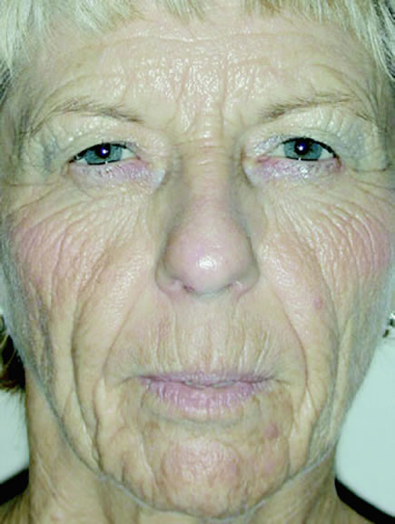
In aging, several changes are noted in the eyebrows, eyelids, and face. Among the common complaints of aging individuals are facial wrinkles known as rhytids and pigmentary abnormalities (dyschromia). These age-related facial changes are believed to be caused by three main factors: (1) photo-induced aging or ultraviolet (UV) damage, (2) mechanical influences such as gravity and muscle contraction, and (3) chronologic or intrinsic aging changes.

Photo-induced aging or UV damage comes from habitual sunlight exposure to skin. Eighty percent of UV-related photo-induced aging occurs before the age of 20, and photo-induced aging alone accounts for more than 90% of age-related skin changes.15 Skin changes are characterized clinically by elastosis, irregular pigmentation, roughness or dryness, teleangiectasia, atrophy, deep wrinkling, and a variety of neoplasms.16

Mechanical influences from gravity and contraction of the muscles of facial expression cause a decrease in skin elasticity resulting from constant stretch and tension.

Chronologic skin aging is the histologic and physiologic changes seen in sun-protected skin of most older individuals. Clinical changes seen in the skin are laxity, dryness, and fine wrinkling. Histologic changes are also noted in the aging skin. In the epidermis, there is a flattened dermal-epidermal junction, giving the appearance of atrophy and cellular heterogeneity. The melanocyte density declines, and the melanocytes tend to cluster focally, producing lentigines. The Langerhans cells also decrease in number with advancing age. Changes in the dermis include attenuation in number and diameter of elastic fibers in the papillary dermis, an increase in number and thickness of elastic fibers in the reticular fibers, and a coarsening of collagen fibers with an increase in density of the collagen network.17

Eyebrow ptosis may occur in young or aged individuals. It might be genetic or result from a combination of gravity, loss of tissue elasticity, decreased subcutaneous tissue, and progressive bony resumption. Clinical observations show that the lateral eyebrow segment usually becomes ptotic earlier because it receives less support from the deeper structures than the medial eyebrow. The eyebrow fat pad complex promotes mobility and gravitational descent of the eyebrow, particularly the lateral eyebrow segment.13 The three forces acting on the lateral eyebrow are (1) frontalis muscle resting tone, which elevates the eyebrow segment medial to the temporal fusion line of the skull; (2) gravity, which causes the soft tissue mass lateral to the temporal line to descend over the temporal fascia plane; and (3) corrugator supercilii muscle activity in conjunction with lateral orbicularis oculi muscle action, which counteract frontalis muscle activity to cause depression of the brow.13 These forces produce downward displacement of the lateral eyebrow. In acquired eyebrow ptosis, there are usually accompanying deep forehead wrinkles as a result of compensatory frontalis overaction. This frontalis contraction, however, does not extend far laterally because of lack of lateral frontalis fibers. Repetitive frontalis muscle contraction is noted in aging patients with symptoms of visual obstruction and a feeling of eyelid heaviness or fullness. Visual obstruction may be caused by brow ptosis, blepharoptosis, dermatochalasis, or any combination of the three.

Synophrys is a condition in which hypertrophy of the eyebrows result in fusion at the midline glabellar area. Synophrys can occur as an isolated finding or may be associated with a generalized hypertrichosis. A congenital absence of eyebrow is usually associated with generalized alopecia, whereas isolated absence is extremely rare.

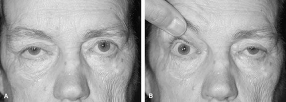
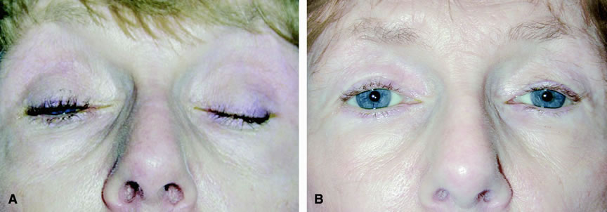
The eyebrow is a helpful indicator of certain localized and systemic disease conditions. Paralysis of the levator palpebrae muscle causes upper eyelid ptosis. As a result, the ipsilateral frontalis muscle elevates the brow in an effort to lift the eyelid. The brow can provide as much as an additional 3 to 5 mm of eyelid elevation.13 In peripheral facial nerve palsy, the ipsilateral frontalis muscle loses muscle tone and the eyebrow assumes a lower position. Hence, eyebrow position may be used to distinguish a third nerve palsy from a seventh nerve palsy.

Back to Top
EYELIDS

STRUCTURE

Although relatively small, the eyelids have a complex structure to allow their various essential functions. The upper and lower lid margins each consist of both anterior and posterior lamellae. The anterior lamella is comprised of skin, muscle, and glands. The posterior lamella is composed of the tarsal plate, conjunctiva, and glands (see Fig. 2).

The eyelid skin is amongst the thinnest and most mobile skin in the body. The eyelid skin receives its sensory innervation from the first and second divisions of the trigeminal nerve. Melanocytes in the epidermis allow for increased pigment production resulting from sun exposure or inflammatory states. Underneath the skin is loose areolar tissue with very little subcutaneous fat. This loose connective tissue plane provides a potential space for considerable accumulation of fluid.

The largest muscle of the eyelid is the orbicularis oculi, which lies just deep to the eyelid skin and anterior to the tarsal plate and septum. It circumferentially surrounds the palpebral fissure. The orbicularis oculi is composed of overlapping myofibrils of varying length. The orbicularis oculi is considerably larger than the remainder of the eyelid, allowing rapid eyelid closure. Functionally, the orbicularis is divided into three concentric components. From outermost to innermost, these are the orbital, preseptal, and pretarsal portions. The deep heads of the pretarsal orbicularis oculi join together near the common canaliculus to form Horner's muscle, which inserts on the posterior lacrimal crest. The orbicularis oculi serves to protract, or close, the upper and lower lids and also assists in the function of the lacrimal pump system. The muscle of Riolan is a small aggregate of orbicularis fibers separated from the orbicularis by the eyelash bulbs. The muscle of Riolan is visible through the thin lid margin skin as the gray line.18 It is thought to rotate the lashes during eyelid closure, and may also help expel glandular contents.

The upper eyelid retractors consist of the levator palpebrae superioris and Mueller's muscle (see Fig. 2). The levator shares a common sheath with the superior rectus. Both are innervated by the superior division of the oculomotor nerve. The levator originates from the lesser wing of the sphenoid bone, in the superomedial orbital apex. It travels anteriorly and slightly superiorly until Whitnall's ligament, where it is redirected inferiorly and anteriorly. The distal 14 to 20 mm of the muscle are tendinous and are referred to as the levator aponeurosis. The aponeurosis sends attachments through the orbicularis onto the upper eyelid dermis and also inserts onto the anterior tarsal plate. The tarsal plate of each lid is composed of dense connective tissue and provides support for the lid. Normal levator excursion from downgaze to upgaze should measure approximately 14 to 17 mm. Levator force generation may also be measured to determine the force produced by contraction of the muscle. This is done by attaching a force transducer to the eyelashes.19

Mueller's muscle originates from the undersurface of the levator and inserts into the superior border of the upper lid tarsus. It is composed of sympathetically innervated smooth muscle fibers and contains α2-adrenergic receptors. Temporally, the muscle sends extensions between the two lobes of the lacrimal gland.20 Mueller's muscle provides 2 mm of additional elevation of the upper eyelid. It may be stimulated by sympathetic responses, such as fear, or pharmacologically by the instillation of phenylephrine hydrochloride (Neo-Synephrine) drops.

The lower lid retractors consist of the capsulopalpebral fascia and the inferior tarsal muscle (see Fig. 2). The capsulopalpebral head of the inferior rectus originates from the sheath of the inferior rectus muscle. This fascia splits to accommodate the inferior oblique muscle. The anterior portion is the capsulopalpebral fascia, and the posterior aspect is known as the inferior tarsal muscle. The inferior tarsal muscle is composed of sympathetically innervated smooth muscle. The muscles fuse superiorly and insert together onto the Tenon's fascia and inferior tarsal border. The capsulopalpebral fascia also inserts into the skin, orbicularis, and septum. Lockwood's ligament is a thickening of the capsulopalpebral fascia, which supports the globe.

The orbital septum arises from the arcus marginalis and is found deep to the orbicularis oculi. In the Caucasian, or occidental upper lid, it inserts into the levator aponeurosis 2 to 5 mm above the superior tarsal border. In the Asian upper lid, the septum inserts onto the levator aponeurosis more inferiorly (Fig. 4). In the lower lid, the septum fuses with the lower lid retractors 4 to 5 mm from the inferior tarsal border. The complex then inserts into the inferior border of the lower lid tarsus.21,22 The postorbicularis fascia lies just anterior to the orbital septum.23 The septum serves as a barrier to the posterior extension of preseptal fluid and as a partial barrier to the spread of preseptal infection.

Fig. 4. A. Sagittal section of Asian eyelid anatomy.B. Sagittal section of Caucasian eyelid anatomy.

The lid crease is determined by the insertion of projections of the levator palpebrae superioris into the dermis. In the Occidental eyelid, the levator aponeurosis and septum fuse above the tarsus. In the Asian eyelid, however, the septum extends inferiorly to fuse with the levator aponeurosis just below the upper tarsal border (see Fig. 4). This inferior extent of the septum causes any levator aponeurotic extensions to occur more inferiorly. This inferior extent of the septum also allows the anterior and inferior extension of the preaponeurotic fat. Other unique features of the Asian eyelid include vertically narrow palpebral fissures, lash ptosis, a full appearance to the upper lids and sulci, and, commonly, epicanthal folds.21

Two noteworthy features of the skin inferior to the lower eyelid are the nasojugal fold medially and the malar fold laterally. These folds are the result of fibrous attachments between the skin and the periosteum (see Fig. 4). They serve as a barrier to the inferior migration of fluid.

Various glands in the eyelid and adnexa contribute to the ocular tear film. The meibomian glands, located in the upper and lower lid tarsal plates, exit the tarsus on the lid margin posterior to the gray line. Low-grade infection of these glands results in blepharitis. Occlusion of their orifices can lead to hordeola, or styes. Furthermore, they may be the site of sebaceous cell carcinoma. The meibomian glands, along with the glands of Zeiss (located in the tarsal conjunctiva) produce the exterior, lipid layer of the precorneal tear film. The central of the three tear film layers is the aqueous layer. It is produced by the lacrimal gland in the lacrimal gland fossa of the superotemporal orbit and by the accessory glands of Krause and Wolfring. These accessory glands are located in the conjunctival fornices and palpebral conjunctiva near the upper tarsal border, respectively. The innermost component of the tear film, the mucin component, is formed by the goblet cells. These cells are distributed throughout the conjunctival epithelium but are most dense in the tarsal and inferonasal aspects. The mucin component binds to the microvilli of the corneal epithelium and increases the wetability of the ocular surface.

The conjunctiva is a continuous lining extending from the mucocutaneous junction of the lid margin, across the fornix, and up to the corneal limbus. The palpebral conjunctiva is tightly bound anteriorly to the tarsus. The conjunctiva serves an immune function. In addition to acting as a physical barrier, it houses immune cells and its substantia propria is highly vascular. Its continuous, smooth surface allows movement of the globe without excessive redundant tissue. Furthermore, the conjunctival attachments to the medial and lateral canthal tendons, the trochlea, and the suspensory ligaments of the lacrimal gland contribute to forniceal suspension.

The palpebral fissure of the average adult measures approximately 10 to 12 mm vertically and 30 mm horizontally. In normal adults, the upper eyelid is 1.5 to 2 mm below the upper limbus, and the lower eyelid sits at the level of the inferior limbus.3 The lateral canthal angle is typically 2 mm higher than the medial canthal angle.

The eyelid margins are the surfaces of the lids that oppose each other during closure. The anterior most distinguishing feature of the eyelid margin is the lashes. The upper lid contains five or six rows of cilia, whereas the lower lid contains three to four.24 Cilia serve a protective function for the ocular surface. Loss of lashes, or madarosis, may be an indication of an eyelid neoplasm. Whitening of lashes, or poliosis, may be a sign of blepharitis, medicamentosa, or Vogt-Koyanagi-Harada syndrome. Posterior to the cilia is the gray line, described previously. Near the posterior extent of the eyelid margin are the meibomian gland orifices. The meibomian glands arise in the dense fibrous connective tissue of the tarsus and exit the lid at the margin. The upper lid tarsus is 8 to 12 mm high, whereas the lower lid tarsus is only 3 to 4 mm high. The tarsal plates of the upper and lower lids contain approximately 30 to 40 and 20 to 30 meibomian glands, respectively.24 Distichiasis is the abnormal origin of metaplastic eyelashes from the meibomian glands. This may be present congenitally or in association with Stevens Johnson syndrome, ocular cicatricial pemphigoid, chemical injury, or other trauma. In contrast, trichiasis is the inward misdirection of eyelashes with normal origin. Poliosis refers to loss of eyelash pigmentation.

The eyelids receive a dual blood supply, from the internal and external carotid arteries, with multiple anastomoses. Although the orbital lymphatics were described only recently,25 the lymphatic drainage of the eyelid is well known. The medial aspect of the lids and conjunctiva drains to the submandibular nodes, whereas the lateral aspect of the lids and conjunctiva drains to the preauricular nodes.

FUNCTION

The eyelids also contribute significantly to the facial features. They are important in the expression of emotion, as well as in facial recognition, and they indicate states of attention and emotion. As a result of intricate neurologic control mechanisms, the eyelids move in concert both with one another and with the globes.

Eyelid opening is produced primarily by the levator palpebrae superioris, which is innervated by the superior division of the oculomotor nerve and which elevates the upper eyelid. The levator muscles of both eyes share a common brainstem nucleus. Furthermore, some nerve fibers may innervate the levator of both upper eyelids. Both levators have been shown to obey Hering's law26–28 (Fig. 5A and B). As such, in patients with unilateral ptosis, the contralateral lid will demonstrate compensatory elevation because of increased stimulation of the ptotic lid. This concept is crucial in ptosis surgery, because the contralateral lid may demonstrate postoperative ptosis after correction of the previously ptotic lid.

Fig. 5. A. Patient with right upper lid ptosis.B. Same patient with manual elevation of ptotic right upper lid now manifesting left upper lid ptosis (Hering's law).

The sympathetically innervated Mueller's muscle also contributes up to 2 mm of upper eyelid elevation. It is responsible for the “wide eyed” appearance associated with sympathetic states such as surprise. Like the levator, it sends extensions temporally between the two lobes of the lacrimal gland. This may account for the temporal flare seen in thyroid eye disease.

Eyelid closure is attained by the action of the three segments of the orbicularis oculi. During closure, the upper lid moves down and the lower lid moves both up and nasally.29 In normal patients, Sherrington's law of reciprocal innervation has been demonstrated to apply to the eyelid protractors and retractors. A decrease in levator activity has been demonstrated during downgaze.30 This is consistent with observations of downward movement of the upper lid during downgaze in patients with facial palsy.29,31

Blinking is the most common form of eyelid closure, although most blinks do not result in complete eyelid closure. Blink rates decrease with visual attention and increase with stress and decreased attention.32

Contraction of the orbicularis oculi is responsible for the generation of a blink. However, the levator is inhibited during a blink, as expected because of Sherrington's law.33 Three types of blink are generally described: spontaneous, reflex, and voluntary. Spontaneous blinks are the baseline involuntary blinks that have no external stimulus. The rate of spontaneous blinking varies among individuals and may be influenced by the environment. The average rate is roughly 15 spontaneous blinks per minute.34 This type of blink has the smallest electromyogram (EMG) amplitude.35

Reflex blinks, resulting from contraction of the pretarsal orbicularis, may be elicited by corneal tactile sensation. The corneal reflex blink pathway begins with efferent fibers from the first division of the trigeminal nerve, which pass through the brainstem and activate the seventh nerve. Cutaneous stimulation, auditory stimuli, and bright visual stimuli may also trigger reflex blinks. The afferent limbs for these stimuli are the trigeminal, auditory, and optic nerves, respectively. Interestingly, reflex blinks have been noted in blind patients.36

Voluntary blinks involve recruitment of the orbital portion of the orbicularis muscle. Not surprisingly, this type of blink demonstrates the greatest EMG amplitude.35 The initiation of a voluntary blink requires a higher potential amplitude than the initiation of other types of blink. Winking (voluntary closure of one eye) is a form of voluntary blink. Interestingly, a suppression of the visual obstruction produced by voluntary blinks has been demonstrated.37 Typically, there is no alteration in eyelid closure after upper eyelid blepharoplasty, even with the excision of a skin and orbicularis flap.38

Horizontal eye movements are associated with horizontal lower lid movement and a much smaller degree of horizontal upper lid movement.32 Vertical eye movements induce vertical movement in the upper lid. In upgaze, the upper lid moves upward as a result of co-contraction of the levator along with the superior rectus. The lower lid also moves up during upgaze, most likely resulting from the intimate relationship of the inferior rectus and the lower lid retractors. The upper and lower lids also move down during downgaze. This is most likely because of inhibition of the levator rather than gravity, because it is observed when the head of a patient with a seventh nerve palsy is inverted. Downward movement of the lower lid during downgaze is most likely the result of tension on the lower lid retractors resulting from inferior rectus contraction.

Bell's phenomenon is the upward rotation of the globe during eye closure. This phenomenon is commonly observed during forced eyelid closure and serves to protect the cornea from exposure in cases of lagophthalmos. However, there may be a downward movement of the globe during voluntary and reflex blinks.39

Concentration on a visual task is associated with palpebral fissure widening in normal patients. This response involves roughly 2 mm of upper lid elevation. The lower lid is displaced laterally and approximately 0.5 mm superiorly.32

AGING AND DISEASE STATES

The general effects of aging on the skin and musculature have been described previously. The skin laxity associated with aging causes dermatochalasis of the eyelids. Advanced upper eyelid dermatochalasis, known as pseudoblepharoptosis, may obstruct the visual axis. Atrophy of the orbital septum produces prolapse of the orbital fat of the upper and lower lids. Descent of the globe within the orbit from loss of support by Lockwood's ligament may contribute to prolapse of the lower lid fat pads40 (Figs. 6 and 7). Dermatochalasis and fat prolapse of the upper and lower lids are commonly treated by blepharoplasty. Skin wrinkles may also be treated by injection of collagen or fat, botulinum toxin injections, laser resurfacing, and chemical peels.

Fig. 6. Schematic of facial aging changes: young right face; aged left face.

Fig. 7. Patient with actinic changes and facial rhytids (see Figure 6 for schematic details).

With age, the levator aponeurosis may become infiltrated with fat and disinsert from the tarsus. Contact lens wear, orbital edema, and intraocular surgery may accelerate this process, which results in involutional ptosis.41 This causes elevation of the lid crease and may cause compensatory brow arching.42 Involutional ptosis is distinguished from congenital ptosis by demonstration of good levator function in the former.

With aging, horizontal lid laxity may cause ectropion or entropion. Loss of the attachments of the orbitomalar ligament to the inferior orbital rim periosteum may also contribute to ectropion.10 Lid retractor disinsertion, involutional enophthalmos, and overriding of the pretarsal orbicularis oculi by the preseptal orbicularis oculi may also contribute to entropion.

Aging affects the lid margin, causing telangiectasias, lid thickening, hyperkeratinization, and meibomian gland orifice changes (including pouting, narrowing, and obliteration). Although normal adults show a decrease in production of meibomian gland secretions, they do not demonstrate an alteration in the composition of these secretions.43

Aging also results in a decrease in amplitude and peak velocity of the closure phase of both spontaneous and voluntary blinks. This is in part because of narrowing of the palpebral fissure with age. However, it may also be partially because of a reduction in dopamine levels with age.33 This theory is supported by the increased blink rate in patients with schizophrenia (who demonstrate elevated dopamine levels) and the decreased rate in Parkinson's patients (who demonstrate low dopamine levels).33

Older patients demonstrate a lower threshold to generate reflex blinks, longer duration of reflex blinks, and increased reflex blink latency (time between a stimulus and the beginning of the blink). This is likely the result of the age-related decrease in dopamine.34 A similar increase in blink duration and excitability is seen in patients suffering from Parkinson's disease, who also have decreased dopamine levels.34 The increase in excitability may predispose older adults to blepharospasm.44,45

Horner's syndrome is a clinical triad of unilateral miosis, ptosis, and anhidrosis. One distinguishing clinical feature is the reverse ptosis (elevation) of the ipsilateral lower lid (Fig. 8). Horner's syndrome is caused by a lesion somewhere in the sympathetic pathway. The ptosis and reverse ptosis are attributed to decreased sympathetic innervation of Mueller's muscle and the inferior tarsal muscle.

Fig. 8. Patient with right Horner's syndrome. Note right upper lid ptosis, reverse ptosis (elevation) of right lower lid, and miosis.

Blepharospasm is the idiopathic involuntary closure of both eyelids. Although its etiology is unknown, sympathetic dysfunction has been implicated.46 Benign essential blepharospasm consists of idiopathic forceful contraction of the orbicularis oculi muscles. In addition to abnormal orbicularis contraction, there may also be an element of alteration in levator function.47 Spasms may be exacerbated by stress, fatigue, visual attention, and caffeine. Females outnumber males with the disorder, and the average age of onset is in the sixth decade. It is bilateral and may be visually incapacitating. Of note, benign essential blepharospasm extinguishes with sleep.

This disorder must be differentiated from hemifacial spasm, which is unilateral spasm that continues during sleep. In 90% of patients with hemifacial spasm, compression of the seventh nerve by an aberrant artery is identified as the etiology. However, imaging must be obtained to rule out compression of the facial nerve by a cerebellopontine angle tumor. The differential diagnosis of benign essential blepharospasm also includes Parkinson's disease, tardive dyskinesia, myokymia, and tics. Although tardive dyskinesia has been commonly described as a result of neuroleptics, it has also been reported after antihistamines, decongestants, and tricyclic antidepressants.48

First-line therapy of benign essential blepharospasm is medical, with local injections of botulinum toxin. Eight types of toxin (A to G) have been identified. Botulinum A toxin, or Botox, is the most potent form and is the subtype most commonly used for therapeutic purposes. Recently, botulinum B toxin, a less potent form of the toxin, has been used. The greatest advantage of botulinum B toxin is its efficacy in patients with tachyphylaxis to botulinum A. Botulinum toxin acts at the neuromuscular junction to inhibit presynaptic acetylcholine release (Fig. 9). If medical therapy fails, then surgical therapy, consisting of seventh nerve ablation or orbicularis myectomy, may also be instituted.

Fig. 9. Schematic of the neuromuscular junction showing the effect of botulinum toxin.

Patients with blepharospasm may suffer from an inability to initiate eyelid opening in the absence of visible orbicularis contraction and in the absence of paralysis. This condition is known as apraxia of eyelid opening. Such patients are unable to initiate upper lid elevation after reflex blinks (Fig. 10A and B). The condition is distinguished from blepharospasm by the absence of lowering of the eyebrows below the orbital rim, or Charcot's sign.49 The site of the lesion is unknown, although the basal ganglia have been implicated.50 Peripherally, the deficit in eyelid opening is most likely because of involuntary inhibition of the levator palpebrae.51 Although apraxia of eyelid opening is most commonly seen in association with blepharospasm, it may also be seen in association with extrapyramidal disorders such as Parkinson's disease, Huntington's disease, progressive supranuclear palsy, and Shy-Drager syndrome. Examination should rule out involutional ptosis, neurogenic ptosis, and ptosis from Botox injections.51

Fig. 10. A. Patient with benign essential blepharospasm and apraxia of eyelid opening.B. After bilateral frontalis sling with silicone rod.

Botulinum toxin injections into the orbicularis oculi may benefit apraxia of eyelid opening if there is an element of persistent orbicularis contraction. This has been demonstrated in patients with52 and without49,52 benign essential blepharospasm. If chronic low-grade orbicularis contraction has caused disinsertion of the levator aponeurosis, surgical reinsertion of the aponeurosis to the tarsus may be effective. Frontalis suspension is effective therapy for apraxia of eyelid opening, and it may also reduce spasm in these patients.53

In patients with Parkinson's disease, a slight delay in lid opening after a blink has been attributed to persistence of orbicularis oculi function. Parkinson's patients may also show no habituation of reflex blinking elicited by glabellar tapping.54

Seventh nerve palsies, as described in the face section later, may cause deficiencies in eyelid closure. The resulting lagophthalmos may lead to corneal exposure and ulceration. Temporary tarsorrhaphy is a short-term treatment option. Long-term treatment may consist of lateral tarsorrhaphy or, more commonly, internal or external upper lid gold weight placement. Gold weight placement reduces corneal exposure by passively increasing upper lid excursion. Although gold weight placement may nearly double the amount of upper eyelid excursion during spontaneous blinking, but it does not change the blink rate.55

Marcus-Gunn jaw winking is a unilateral elevation of a ptotic eyelid that coincides with mouth opening or lateral movement of the jaw. It is the result of congenital aberrant innervation of the levator muscle by trigeminal nerve fibers. A number of surgical procedures for correction have been described.56–59

Back to Top
FACE

STRUCTURE

Although a description of facial anatomy, physiology, and pathophysiology is not included in most textbooks in ophthalmology, the expanding surgical domain of ophthalmic facial plastic surgery requires a thorough understanding of full facial structure and function. The eyelids and eyebrows do not function independently from the rest of the face, and, thus, it is of utmost importance to understand them in the context of the overall facial continuum.

The key soft tissue structures of the face are its musculature and supporting ligaments. The muscles of facial expression are comprised of a layered anatomic arrangement and have been well documented.60 The most superficial muscles are the orbicularis oculi, the zygomaticus minor, and the depressor anguli oris, whereas the deepest layer consists of the mentalis, the levator anguli oris, and the buccinator (see Fig. 1).

Most of the facial muscles are linked in a connective tissue matrix called the SMAS. First described by Mitz and Peyronie in 1976, it is a fibromuscular network that envelops the superficial facial muscles and acts as a distributor of facial muscular contractions.61

Several ligamentous structures support the SMAS and facial soft tissues.62 These include the orbitomalar ligament,10 zygomatic ligaments,63 masseteric cutaneous ligaments,64 and mandibular ligaments62 (Fig. 11). The deeper muscles facial of expression, the zygomaticus major and levator labii superioris, also penetrate the SMAS and provide fixation.

Fig. 11. Ligamentous soft tissue support of the face.

Anatomically, the SMAS divides the facial fat into distinct pads. Submuscular fat in certain anatomic regions has been specifically named. The ROOF is described in the eyebrow section. The suborbicularis oculi fat (SOOF) is a collection of fat between the periosteum and the orbicularis oris muscle.14,65 The ROOF and SOOF actually represent a continuous adipose layer, and these names distinguish different locations within the same layer rather than separate entities.10

The facial nerve provides motor innervation to the muscles of facial expression. An understanding of its anatomy is essential to avoid complications during facial surgery. Each lower motor neuron is primarily innervated by the contralateral upper motor neuron but also receives innervation from the ipsilateral upper motor neuron. The facial nerve exits the temporal bone via the stylomastoid foramen, entering the parotid gland. The nerve then fans out to form the pes anserinus, named after its resemblance to a goose's foot. Although many patterns of branching and anastomoses exist, five main branches run anteriorly across the face. From superior to inferior, these are the temporal, zygomatic, buccal, mandibular, and cervical. Another branch, the posterior auricular, wraps behind the ear (Fig. 12). The temporal branch and mandibular branches are the two most commonly injured during facial surgery. The temporal branch exits the parotid gland and travels in the layer of the SMAS. The mandibular branch travels deep to the platysma. Dissection in the aforementioned areas should proceed in a manner to protect these branches of the facial nerve.66

Fig. 12. Branches of the facial nerve.

FUNCTION

The muscles of facial expression give character and diversity to the human face. They also perform other functions, such as closing of the eyes and moving the cheeks and lips during mastication and speech. It is important to note that these muscles act synergistically, rather than independently, in part by the anatomic linkage of the SMAS. The muscles of facial expression in the brow and eyelid area have been discussed previously.

In the lip and nasal musculature, the orbicularis oris muscle acts as a sphincter and is essential for oral competence, speech, and social expression. Its actions include closing, pursing, and protruding the lips. The contraction of the orbicularis oris muscle is opposed superiorly by the action of the lip elevators. This muscle group includes the nasalis, the levator labii, and the zygomaticus minor muscles. The lip depressors include the orbicularis oris, the mentalis, and the depressor anguli oris muscles.

The muscles of the mouth are considered to be the most important muscles in facial expression. The multiple functions of the lips, such as oral competence, speech, and social expression, require a complex set of motions between the circular contraction of the orbicularis oris and the radial action of the lip elevators and depressors.

The main muscles used in smiling are the risorius, the zygomaticus major, and the orbicularis oris muscle. The risorius pulls the angle of the mouth laterally, and the zygomaticus major muscle pulls the corner of the mouth up and laterally. The buccinator muscle compresses the cheek to avoid injury during jaw closure.

AGING AND DISEASE STATES

The skin of the face demonstrates the same pathologic changes described for the brow skin. Superficial changes include nasal root rhytids, malar festoons, cheek rhytids, ptosis of the nasal tip, cheek sagging, deepening of the nasolabial fold, smokers lines (circumferential rhytids around the mouth) and jowl formation, and banding of the platysma67 (see Figs. 6 and 7). Bulging of the lower eyelid fat combines with descent of the SOOF and cheek fat pad to produce a double convexity deformity68 (Fig. 13).

Fig. 13. Profile of patient with unmasking of inferior orbital rim. Note double convexity deformity of orbital fat and malar fat.

Midfacial ptosis is most likely caused by attenuation of the orbitomalar ligament rather than by sagging of the orbicularis oculi, zygomaticus major, zygomaticus minor, or the SOOF.64,69 Lower facial aging, such as sagging of the platysma, is most likely the result of SMAS ptosis.10,64

Treatment modalities for facial aging changes include injection of botulinum toxin for dynamic wrinkles; chemical peels and laser resurfacing for static wrinkles; and SOOF lifting, SMAS lifting, and deep plane face lifting for soft tissue descent. Enhanced results of laser resurfacing therapy have been demonstrated in patients undergoing preoperative botulinum toxin injections to limit facial mobility in the early postoperative period.70

Seventh nerve palsy and paralysis may cause difficulties with many activities such as eating, drinking, swallowing, and expressing emotions.71 Seventh nerve palsy may be caused by central etiologies such as cortical ischemia, tumors, extrapyramidal lesions, or by brain stem lesions (such as multiple sclerosis, tumor, or ischemia). Peripheral etiologies include Guillain-Barré syndrome, otitis media, Ramsey Hunt syndrome, Lyme disease, sarcoidosis, nasopharyngeal carcinoma, diabetes mellitus, human immunodeficiency virus (HIV), and trauma. Facial nerve anatomy was described previously. Upper motor neuron lesions affect contralateral lower facial function (with less impact on upper facial function), whereas lower motor neuron lesions affect ipsilateral upper and lower facial nerve function. Bell's palsy, a diagnosis of exclusion, is idiopathic and affects upper and lower facial function. It is usually self-limited, with up to 86% of patients demonstrating complete recovery.72 Complete facial paralysis, pain in locations outside the ear, and hypertension are risk factors associated with incomplete recovery.72 Oral steroid therapy may be associated with more complete recovery.73 Jaw winking and crocodile tears (lacrimation with eating) are signs of chronic palsy.

As the boundaries of ophthalmic surgery continue to expand, the ophthalmologist must understand the interactions of the eyelids with the eyebrows and the face. This knowledge will aid the ophthalmologist in the treatment of eyelid and other disorders.

Back to Top
REFERENCES

1. Ozanics V, Jakobiec F. Prenatal development of the eye and its adnexae. In Jakobiec F (ed). Ocular Anatomy, Embryology, and Teratology. New York: Harper and Row, 1982

2. Sevel D. A reappraisal of the development of the eyelids. Eye 1988;2:123

3. Dutton J. The eyelids and anterior orbit. In Atlas of Clinical and Surgical Orbital Anatomy. Philadelphia: WB Saunders, 1984

4. Sevel D. Insufflation treatment of occluded nasolacrimal apparatus. Ophthalmology 1982;89:329

5. Tarbet K, Lemke B. Anatomy of the eyelids and lacrimal drainage system. In Albert DM, Jakobiec FA (eds). Principles and Practice of Ophthalmology. Philadelphia: WB Saunders, 2000

6. Anderson H, Ehlers N, Mattheissen M. Histochemistry and development of the human eyelids. Acta Ophthalmol 1965;43:642

7. Cook BE, Lucarelli MJ, Lemke BN. Depressor supercilii muscle: Anatomy, histology, and cosmetic implications. Ophthal Plast Reconstr Surg 2001;17:404

8. Knize DM. Muscles that act on glabellar skin: A closer look. Plast Reconstr Surg 2000;105:350

9. May JW, Fearon J, Zingarelli P. Retro-orbicularis oculi fat (ROOF) resection in aesthetic blepharoplasty: A 6-year study in 63 patients. Plast Reconst Surg 1991;7:104

10. Kikkawa DO, Lemke BN, Dortzbach RK. Relations of the superficial musculoaponeurotic system to the orbit and characterization of the orbitomalar ligament. Ophthal Plast Reconstr Surg 1996;12:77

11. Lemke BN, Stasior OG. Eyebrow incision making. Adv Ophthalmic Plast Reconstr Surg 1983;2:19

12. Fezza JP, Klippenstein, KA, Wesley, RA. Cilia regrowth of shaven eyebrows. Arch Facial Plastic Surg 1999;1:223

13. Knize D. An anatomically based study of the mechanism of eyebrow ptosis. Plast Reconstr Surg 1996;97:1321

14. Lemke BN, Stasior OG. The anatomy of eyebrow ptosis. Arch Ophthalmol 1982;100:981

15. Leyden JJ. Clinical features of ageing skin. Br J Dermatol 1990;122(Suppl 35):1

16. Yaar, M, Gilchrest, BA. Cellular and molecular mechanisms of cutaneous aging. J Dermatol Surg Oncol 1990;16:915

17. Kurban RS, Bhawan J. Histologic changes in skin associated with aging. J Dermatol Surg Oncol 1990;16:908

18. Wulc AE, Dryden RM, Khatchaturian T. Where is the gray line? Arch Ophthalmol 1987;105:1092

19. Freuh BR, Musch DC. Evaluation of levator muscle integrity in ptosis with levator force measurement. Ophthalmology 1996;103:244

20. Morton AD, Elner VM, Lemke BN et al. Lateral extensions of the Mueller muscle. Arch Ophthalmol 1996;114:1486

21. Kikkawa DO, Kim JW. Asian blepharoplasty. Int Ophthalmol Clin 1997;37:193

22. Meyer D, Linberg JV, Wobig JL et al. Anatomy of the orbital septum and associated eyelid connective tissues: Implications for ptosis surgery. Ophthal Plast Reconstr Surg 1991;7:104

23. Putterman AM, Urist MJ. Surgical anatomy of the orbital septum. Ann Ophthalmol 1974;6:290

24. Montagna W, Ford DM. Histology and cytochemistry of human skin. Arch Derm 1969;100:328

25. Gausas RE, Gonnering RS, Lemke BN et al. Identification of human orbital lymphatics. Ophthal Plast Reconstr Surg 1999;15:252

26. Gay AJ, Salmon ML, Windsor CE. Hering's law, the levators, and their relationship in diseased states. Arch Ophthal 1967;77:157

27. Averbuch-Heller L, Poonyathalang A, von Maydell D et al. Hering's law for eyelids: Still valid. Neurology 1995;45:1781

28. Gupta JS, Jain IS, Kumar K. Lid-retraction secondary to contralateral ptosis. Br J Ophthal 1964;48:626

29. Gordon G. Observations upon movements of the eyelids. Br J Ophthalmol 1951;35:339

30. Bjork A. Electromyographic studies on the coordination of antagonistic muscles in cases of abducens and facial palsy. Br J Ophthalmol 1954;38:605

31. Sewall EC. Decompression of the facial nerve: Physiology of the seventh and ninth nerves and movements of the lid in facial paralysis. Arch Otolaryngol 1933;18:746

32. Kennard DW, Ponder E, Kennedy WP. On the act of blinking. Q J Exp Physiol 1928;18:89

33. Sun WS, Baker RS, Chuke JC et al. Age-related changes in human blinks: Passive and active changes in eyelid kinematics. Invest Ophthalmol Vis Sci 1997;38:92

34. Peshori KR, Schicatano EJ, Gopalaswamy R et al: Aging of the trigeminal blink system. Exp Brain Res 2001;136:351

35. Kaneko K, Sakamoto K. Evaluation of three types of blinks with the use of electro-oculogram and electromyogram. Percept Motor Skills 1999;88:1037

36. Hall A. The origin and purposes of blinking. Br J Ophthalmol 1945;29:445

37. Riggs LA, Volkmann FC, Moore RK. Suppression of the blackout due to blinks. Vis Res 1981;21:1075

38. Abell KM, Cowen DE, Baker RS et al. Eyelid kinematics following blepharoplasty. Ophthal Plast Reconstr Surg 1999;15:236

39. Collewijn H, Van Der Steen J, Steinman RM. Human eye movements associated with blinks and prolonged eyelid closure. J Neurophysiol 1985;54:11

40. Camirand A, Doucet J, Harris J. Anatomy, pathophysiology, and prevention of senile enophthalmia and associated herniated lower eyelid fat pads. Plastic Reconstr Surg 1997;100:1535

41. Wounters RJ, van den Bosch WA, Mulder PGH et al. Upper eyelid motility in blepharoptosis and in the aging eyelid. Invest Ophthalmol Vis Sci 2001;42:620

42. Van den Bosch WA, Leenders I, Mulder P. Topographic anatomy of the eyelids, and the effects of sex and age. Br J Ophthalmol 1999;83:347

43. Hykin PG, Bron AJ. Age related morphological changes in lid margin and meibomian gland anatomy. Cornea 1992;11:334

44. Chuke JC, Baker RS, Porter JD. Bell's palsy-associated blepharospasm relieved by aiding eyelid closure. Ann Neurol 1996;39:263

45. Baker RS, Sun W, Hasan SA et al. Blink adaptation and loss of inhibition trigger blepharospasm subsequent to Bell's palsy. Invest Ophthalmol Vis Sci 1995;37:S154

46. McCann JD, Gauthier M, Morschbacher R et al. A novel mechanism for benign essential blepharospasm. Ophthal Plast Reconstr Surg 1999;15:384

47. Aramideh M, Ongerboer de Visser BW, Koelman JHTM et al Clinical and electromyographic features of levator palpebrae superioris muscle dysfunction in involuntary eyelid closure. Move Disord 1994;9:395

48. Mauriello JA, Carbonaro P, Dhillon S et al. Drug-associated facial dyskinesias—a study of 238 patients. J Neuro-Ophthalmol 1998;18:153

49. Katz B, Rosenberg JH. Botulinum therapy for apraxia of eyelid opening. Am J Ophthalmol 1987;103:718

50. Boghen D. Apraxia of lid opening: A review. Neurology 1997;48:1491

51. Jordan DR, Anderson RL, Digre KB. Apraxia of lid opening in blepharospasm. Ophthalmic Surg 1990;21:331

52. Aramideh M, Ongeboer De Visser BW, Koelman JHTM et al. Motor persistence of orbicularis oculi muscle in eyelid-opening disorders. Neurology 1995;45:897

53. De Groot V, De Wilde F, Smet L et al. Frontalis suspension combined with blepharoplasty as an effective treatment for blepharospasm associated with apraxia of eyelid opening. Ophthal Plast Reconstr Surg 2000;16:34

54. Loeffler JK, Slatt B, Hoyt WF. Motor abnormalities of the eyelids in Parkinson's disease: Electromyographic observations. Arch Ophthalmol 1966;76:178

55. Abell KM, Baker RS, Cowne DE, Porter JD. Efficacy of gold weight implants in facial nerve palsy: Quantitative alterations in blinking. Vis Res 1998;38:3019

56. Beard C. A new treatment for severe unilateral ptosis with jaw-winking. Am J Ophthalmol 1965;59:252

57. Callahan A. Correction of unilateral blepharoptosis with bilateral eyelid suspension. Am J Ophthalmol 1972;74:321

58. Putterman AM. Jaw-winking blepharoptosis treated by the Fasanella-Servat procedure. Am J Ophthalmol 1973;75:1017

59. Bullock JD. Marcus-Gunn jaw-winking ptosis: Classification and surgical management. J Pediatr Ophthalmol Strabismus 1980;17:375

60. Freilinger G, Gruber H, Happak W et al. Surgical anatomy of the mimic muscle system and the facial nerve: Importance for reconstructive and aesthetic surgery. Plastic Reconstr Surg 1988;80:686

61. Mitz V, Peyronie M. The superficial musculoaponeurotic system (SMAS) in the parotid and cheek area. Plast Reconstr Surg 1976;58:80

62. Moss CJ, Mendelson BC, Taylor GI. Surgical anatomy of the ligamentous attachments in the temple and periorbital regions. Plast Reconstr Surg 2000;105:1475

63. Furnas DW. The retaining ligaments of the cheek. Plast Reconstr Surg 1989;83:11

64. Lucarelli MJ. The anatomy of midfacial ptosis. Ophthal Plast Reconstr Surg 2000;16:7

65. Aiache AE, Ramirez OH. The suborbicularis oculi fat pads: An anatomic and clinical study. Plast Reconstr Surg 1995;95:37

66. Wobig J, Wilson M. Eyelid and facial anatomy. In Putterman A (ed). Cosmetic Oculoplastic Surgery. 3rd ed. New York: WB Saunders, 1999:51–56

67. Zimbler MS, Kokoska MS, Thomas JR. Anatomy and pathophysiology of facial aging. Facial Plast Surg Clin North Am 2001;9:179

68. Hoenig, JA, Shorr N, Shorr J. The suborbicularis oculi fat in aesthetic and reconstructive surgery. Int Ophthalmol Clin 1997;37:179

69. Hamra ST. Suborbicularis oculi fat pads. Plast Reconstr Surg 1996;97:484

70. Zimbler MS, Holds JB, Kokoska MS et al. Effect of botulinum toxin pretreatment on laser resurfacing results. Arch Facial Plast Surg 2001;3:165

71. Wachtman GS, Cohn JF, VanSwearingen JM et al. Automated tracking of facial features in patients with facial neuromuscular dysfunction. Plast Reconstr Surg 2001;107:1124

72. Katusic SK, Beard CM, Wiederholt WC et al. Incidence, clinical features, and prognosis in Bell's palsy, Rochester, Minnesota, 1968-1982. Ann Neurol 1986;20:622

73. Austin JR, Peskind SP, Austin SG et al. Idiopathic facial nerve paralysis: A randomized double-blind controlled study of placebo versus prednisone. Laryngoscope 1993;103:1326

Back to Top