|
|
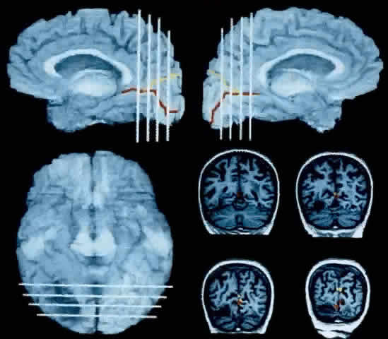
| Fig. 8. The brain of a 72-year-old woman with prosopagnosia, achromatopsia, alexia, and left homonymous hemianopia. The mesial surfaces of the right and left hemispheres, respectively, are reconstructed in the upper right and upper left panels from raw MRI data (Brainvox technique102). The ventral surface is in the lower left panel. The white lines through the reconstructed brain show the relative position of the four coronal slices shown in the lower right panel. The calcarine fissure and parieto-occipital fissure are traced in red and yellow, respectively, which automatically transfers to the coronal MRI slices. The patient had bilateral lesions that affected the fusiform gyrus and undercut the most posterior segment of the lingual gyrus. The larger lesion in the right hemisphere affected the optic radiations, causing the equivalent of a V1 scotoma in the left visual hemifield. The lesion of the left hemisphere does not reach the surface of the brain. It lies beneath the calcarine fissure and can be only in the coronal sections. Such a lesion would cause damage in a possible human homologue of the monkey's area V4 complex or disrupt connections to and from such an area. (Rizzo M, Smith V, Pokorny J, Damasio AR: Color perception profiles in central achromatopsia. Neurology 43:995, 1993) |