Chapter 40
Congenital Malformations of the Eye
Brian P. Brooks and Elias I. Traboulsi
Main Menu   Table Of Contents

Search

INTRODUCTION
DISORDERS THAT AFFECT THE ENTIRE EYE
DISORDERS THAT AFFECT THE ORBITS, EYELIDS, AND ADNEXA
MALFORMATIONS OF THE ANTERIOR SEGMENT
REFERENCES

INTRODUCTION
The purpose of this chapter is to give an overview of congenital malformations of the eye and the ocular adnexa, with emphasis on molecular and developmental mechanisms. Detailed reviews of ocular development, teratology, and many of the specific disorders in this chapter can be found elsewhere in this text.

Ocular malformations may have genetic and nongenetic causes (e.g., environmental.) Those with genetic causes may be familial or sporadic. They may be isolated or part of a broader syndrome. In many cases, the exact cause for a given malformation is not known. Depending on the population studied, the method of ascertainment, and the definition of “ocular malformation,” various published studies have found a birth prevalence of ocular malformations of 0.04 to 6.8 per 10,000 live births.1–7

The past decade has witnessed a tremendous increase in our understanding of the molecular and developmental causes of many important ophthalmic malformations (Table 1). Many of the mutations that cause ocular malformations are in transcription factors. Transcription factors are proteins that bind to specific DNA sequences (or to proteins that bind to DNA) to regulate expression of specific genes. A complex pattern of gene expression in different parts of the embryo during different times of gestation is required for normal organogenesis and cellular differentiation. Disruption of this process through the alteration of the function of any important transcription factor can have serious consequences. Development can also be affected by changes in cell-to-cell signaling, cell migration, cell death, and cell adhesion.


TABLE 1. Examples of Developmental Eye Diseases Caused by Mutations in Transcription Factors (transcription factors: see text for details and references)


Human GeneAssociated Disease(s)
SOX2Anophthalmia
CHX10Microphthalmia, cataracts & iris abnormalities
PAX6Aniridia, Peters anomaly, anterior segment dysgenesis, foveal hypoplasia, autosomal dominant keratitis, congenital cataract
PAX2Renal-coloboma syndrome
PITX2Axenfeld-Rieger syndrome, iridogoniodysgenesis, Peters anomaly
PITX3Anterior segment mesenchymal dysgenesis & cataracts, congenital cataracts
FOXC1Anterior segment dysgenesis
FOXC2Lymphedema-distichiasis, familial distichiasis
FOXL2Blepharophimosis syndrome
ARIXCongenital fibrosis of extraocular muscles, type 2
CRXCone-rod dystrophy 2; Leber congenital amaurosis; retinitis pigmentosa


This chapter describes malformations on an anatomic basis. However, current classifications and nosology of congenital ocular malformations are imperfect. For example, some abnormalities are difficult to classify because they affect multiple tissues (e.g., aniridia). Sometimes the same word (e.g., coloboma) is used in the literature to describe several distinct clinical and developmental entities. A primary defect in one structure may cause secondary malformations in another (e.g., the small orbits and short palpebral fissures in anophthalmic patients). A given genetic disorder may have a spectrum of presentations (phenotypic heterogeneity). Lastly, different genetic mutations can cause the same clinical phenotype (genetic heterogeneity). We attempt to clarify these sometimes confusing issues on a case-by-case basis.

Because the disorders covered in this chapter are largely developmental, we review the essential embryology behind each class of disease. A detailed discussion of ocular embryology is presented elsewhere in this text. Although we present development in a compartmentalized fashion, it is important to recognize that multiple, interdependent events are occurring simultaneously during embryogenesis. As such, insults or changes in gene expression at one time point may have multiple effects at later time points. For example, maternal alcohol use during the period of gastrulation—days before any ocular structures are visible morphologically—may result in ocular defects ranging from microphthalmia to coloboma to Peters anomaly.8

Throughout this chapter, we refer to “Online Mendelian Inheritance in Man” or OMIM. OMIM is a comprehensive catalog of inherited diseases available free of charge at www.ncbi.nlm.nih.gov/omim. When possible, we will provide the OMIM number for individual disorders.

Back to Top
DISORDERS THAT AFFECT THE ENTIRE EYE

DEVELOPMENTAL BIOLOGY91

Eye development becomes evident at approximately 22 days gestation, when the optic sulci appear as bilateral grooves in the neural ectoderm of the embryo. At approximately 25 days gestation, as the rostral neural tube fuses, the neural ectoderm evaginates further to form the optic vesicles. The optic vesicles are attached to the developing brain via the optic stalks. As the optic vesicles grow laterally and approach the surface ectoderm, they invaginate to form a bilayered optic cup. The inner and outer layers of the optic cup proceed to form the neural retina and the retinal pigmented epithelium, respectively. The invaginated optic cup forms a somewhat spherical structure that is open anteriorly and inferiorly. This inferior “seam” is alternatively called the optic fissure, the choroidal fissure, or the embryonic fissure, and it transmits the hyaloid vascular system beginning at approximately 5 weeks gestation. At 6 weeks gestation, the optic fissure “zippers” shut, beginning equatorially and proceeding anteriorly and posteriorly.

ANOPHTHALMIA

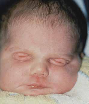
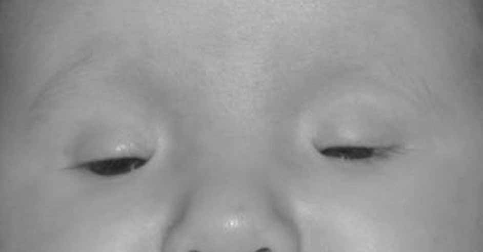
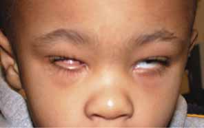
Anophthalmia refers to the complete absence of ocular tissue, presumably caused by abnormalities of optic vesicle formation or maturation. Patients may have unilateral or bilateral anophthalmia, and generally have short palpebral fissures and small orbits. In many cases when there is no clinically apparent ophthalmic tissue, remnants of lens epithelium, neuroretinal, and fibrovascular, choroid-like tissue can be detected on histologic sectioning. Terms such as “true anophthalmia,” “extreme microphthalmia,”10 and “clinical anophthalmia”11 have been used in the literature to describe what may be, in truth, a phenotypic continuum between anophthalmia and microphthalmia (see later).

Anophthalmia may be isolated or associated with a broader syndrome (Table 2).12–19 Isolated anophthalmia is generally an autosomal recessive condition.20 Driggers et al. reported a child with isolated bilateral anophthalmia and an apparent balanced chromosomal translocation {46,XX,t(3;11)(q27;p11.2)}.21 Fantes et al. found that this child had, in fact, a submicroscopic deletion that eliminated the SOX2 gene on chromosome 3, and went on to show nonsense mutations in SOX2 in four of 11 subjects with bilateral anophthalmia.22 SOX2 lies within an intron of a nonexpressed gene, SOX2OT, and is expressed in neuroectoderm early in development.23 SOX2 protein interacts cooperatively with PAX6 in the induction of lens development and delta-crystallin expression.24 Hagstrom, Traboulsi et al. found a null mutation in SOX2 in a girl with bilateral clinical anophthalmia and absence of the optic nerves, chiasm, and optic tracts (unpublished data)


TABLE 2. Clinical Syndromes Associated with Anophthalmia


SyndromeOMIM #InheritanceCharacteristics in Addition to Anophthalmia
Fryns60076AROrofacial clefting, uterine abnormalities, ear abnormalities, neural tube defects
Oculocerbral-cutaneous164180ADOrbital cysts, focal dermal hypoplasia, cerebral malformations, cleft lip/palate
Lenz microphthalmia309800X-linkedMicrophthalmos, mental retardation, distal limb abnormalities, microcephaly, orofacial clefting, tooth & skeletal anomalies, hearing loss, GU malformations, imperforate anus
Matthew-Wood601186SporadicPulmonary hypoplasia
Waardenburg206920ARSyndactyly/other distal limb abnormalities, mental retardation, skeletal anomalies
14q22–23 del607932SporadicPolydactyly, pituitary hypoplasia, ?SIX6 hemizygosity
 605856 Growth & mental retardation, callosal agenesis, heminasal hypoplasia, atypical clefting, external ear abnormalities,
 60092SporadicEsophageal atresia

Syndromes associated with anophthalmia. Clinically, phenotypic variability exists. Abbreviations: OMIM, online Mendelian inheritance in man; AR, autosomal recessive. References and specific cases may be found in OMIM.


Reconstructive surgery can be helpful in patients with congenital anophthalmia.25 Orbital expansion can be achieved with spherical implants, orbital osteotomies, bone grafts, and/or orbital expanders. Conjunctival sac reconstruction is achieved using serial expanders and buccal mucous membrane grafts. Lid reconstruction sometimes requires tissue flaps and skin grafts.

MICROPHTHALMIA

A microphthalmic eye is one with axial length less than two standard deviations below the age-adjusted mean (ie, <19 mm in a 1-year-old child and <21 mm in an adult.) Microphthalmia encompasses a range of phenotypes. Specific terms, outlined later, are applied to subgroups of microphthalmic eyes with certain characteristics. Authors of scientific communications may not use the same classification scheme or may use the more general term “microphthalmia” when a more specific term could apply. Although we will present these terms as distinct entities, remember that they likely represent points along a single, phenotypic continuum.

Simple Microphthalmia/Nanophthalmos

Simple microphthalmia refers to a small but structurally normal eye.26 The term “nanophthalmos” is used synonymously, but especially when such eyes develop angle-closure glaucoma caused by crowding from the normal-size (ergo, disproportionately large) lens or uveal effusions. Weiss et al. noted that, in general, the posterior segment is proportionately shorter than the anterior segment in such patients.26 Simple microphthalmia/nanophthalmos can be inherited as part of a broader syndrome (Table 3)27–29 or as an individual trait. The sclera of nanophthalmic eyes consists of irregularly arranged collagen lamellae, absence of normal elastic fibers, and abnormal glycogen-like deposits.30 These changes likely result in the increased thickness and decreased elasticity of the nanophthalmic sclera—changes that result in decreased blood flow through the vortex veins and/or decreased transscleral flow of protein. This alteration in ocular fluid dynamics increases the risk of uveal effusion and choroidal detachment, especially after surgery.31–33


TABLE 3. Syndromes With Simple Microphthalmos/Nanophthalmos as a Phenotype


SyndromeOMIM#InheritanceLocus (Gene)Characteristics in Addition to Simple Microphthalmia
Kenney-Caffey127000AD vs X-linkedUnknownShort stature, dense tubular bones, narrow marrow cavities, hypocalcemia/phosphatemia
Mucolipidosis IIIa 252600AR4q21-q23 (N-acetyl-glucosamine–1-phosphotransferase)Short stature, cloudy corneas, retinopathy, bone abnormalities, aortic insufficiency, mental retardation, skin thickening
 252605   
Oculodentodigital Syndrome164200AD6q21–23.2 (Connexin 43)Characteristic facies, microcephaly, syndactyly, hypo/aplasia middle phalanges, dental abnormalities, glaucoma, cataract, optic atrophy

AD, autosomal dominant; AR, autosomal recessive. Clinically, phenotypic variability exists.
aMucolipidosis type III includes three biochemical complementation groups. The ophthalmic literature does not specifically differentiate these, but the “pseudo-Hurler” phenotype is consistent.


The precise embryologic mechanism for simple microphthalmia/nanophthalmos is unclear. Based on finding this phenotype in patients with fetal alcohol syndrome, achondroplasia, and myotonic dystrophy, Weiss et al. postulated that decreased size of the optic cup, altered vitreous proteoglycans, low intraocular pressure, and abnormal release of growth factors may be important in the pathogenesis of simple microphthalmos.26 These authors point out that given many of these patients have normal corneal diameters and that the area primarily affected is the posterior segment, the pathogenesis in many of these conditions may be related to postnatal, rather than prenatal, changes in ocular development and growth.

In 1998, Othman et al. described a pedigree with autosomal dominant nanophthalmos that localizes to a 14.7cM region on chromosome 11 (NNO1).34 Affected family members had high hyperopia (range of +7.25 to +13 D) and short axial lengths (range of 17.55–19.28 mm). Twelve of 22 affected family members had a history of angle-closure glaucoma or had occludable angles. Although OMIM lists a second nanophthalmos locus, NNO2, on 15q12-q15, the phenotype described in this pedigree is more consistent with a colobomatous, complex microphthalmia (see later).35

Complex Microphthalmia

In cases in which the eye is not only small but also abnormal in other respects, the term “complex microphthalmia” is applied. Examples of associated findings are microcornea, sclerocornea, corneal opacities, aniridia, Peters anomaly and cataract. Complex microphthalmia can be associated with a syndrome (Table 4)19,35–61 or can occur as an isolated trait. Complex microphthalmia encompasses a diversity of phenotypes. Patients in the same family with the same underlying disorder may have phenotypes of varying severity. For example, patients with the Lenz microphthalmia syndrome (OMIM 309800) may have anophthalmia, colobomatous microphthalmia, or noncolobomatous microphthalmia.62


TABLE 4. Syndromes with Complex Microphthalmia as a Phenotype


SyndromeOMIM #InheritLocus (Gene)Features in Addition to Complex Microphthalmia
Aicardi syndrome304050XLDXp22Agenesis of corpus collasum; chorioretinal abnormality; infantile spasms; microcephaly, CL/CP; rib/vertebrae abnormalities; brain abnormalities; neoplasia; precocious puberty
Arhinia, choanal atresia, & microphthalmia603457AD?UnknownComplete absence of nose, choanal atresia, cleft palate
BOFS (Branchio-oculo-facial syndrome)113620ADUnknownGrowth def. (pre/postnatal); microcephaly; micrognathia; coloboma; ear abnormalities; cataract; CL/CP; dental anomalies; branchial anomalies
CATM156850AD16p13.3Congenital cataract
CHARGE214700AD, sporadUnknownColoboma, hear anomaly, choanal atresia, retardation of growth & development, genital & ear abnormalities
COFS (Cerebro-oculo-facio-skeletal syndrome)214150AR19q13.2-q13.3 (XPD); 13q33; 10q11 (ERCC6)Neurogenic arthrogryposis; cataract; microcephaly; brain malformation; blepharophimosis; hirsutism; kyphoscoliosis; osteoporosis; renal defects; large pinna; wide-spaced nipples; verticle talus; flexion contractures
Coloboma-obesity-hypogenitalism-mental retardation syndrome601794ADUnknownCataract, obesity, hypogonadism, MR
Dextrocardia with unusual facies221950ARUnknownDextrocardia, dysmorphic facies, folding on foot, normal growth
Facio-oculo-auriculo-vertebral dysplasia (Goldenhar)164210Sporad AD14q32Unilateral deformity of external ear w/ small ipsilateral half of face; epibulbar dermoid; vertebral anomalies; CL/CP; deafness; CN VII palsy; epibulbar dermoids; lid colobomas; Duane's syndrome
Facio-thoraco-genital syndrome227320ARUnknownAnteverted nares; long philtrum; thin upper lip; pectus excavatum; micrognathia; genital abnl.; wide great toes; thumbs; hypoplastic nails
Focal Dermal Hypoplasia (Goltz-Gorlin syndrome)305600XLD (lethal in males)Xp22Atrophy & linear pigmentation of skin; herniation of fat through dermal defects; multiple papillomas of mucous membranes; colobomas; digital anomalies; hypoplastic teeth; MR; striated bones
Frontonasal dysplasia sequence136760SporadUnknownHypertelorism; lateral displacement of inner canthi; widow's peak; deficit in midline frontal bone; nasal tip abnormalities; absence of corpus callosum
GOMBO syndrome233270AR3p or 22q ?Microcephaly; brachydactyly w/ clinodactyly 5, delayed growth at puberty; MR; oligophrenia (affected w/ 46, X,Y, ish der(3),t(3,22)(p25;q13))
Hallermann-Streiff syndrome (Francois dyscephalic syndrome)234100Sp. Cataracts; coloboma; small, pinched nose; hypotrichosis; postnatal growth def.; brachycephaly; malar hypoplasia; dental abnl.; atrophy of skin; wormian bones; thin, gracile long bones
Kapur-Toriello syndrome244300ARUnknownColoboma, MR, congenital heart defect, CL/CP, malrotation of intestine; displaced kidneys
Lenz microphthalmia syndrome309800X-linkedXq27-q28 Xp11.4-p21.2MR, distal limb abnormalities, microcephaly, oro-facial clefting, tooth & skeletal anomalies, hearing loss, GU malformations, imperforate anus
Macrosomia w/ microphthalmia, lethal248110ARUnknownMacrosomia, median clefting, early infant death from overwhelming infection
Meckel syndrome, type 1–324900AR17q22-q23, 8q24, 11q13Renal cysts; developmental anomalies of the CNS (esp. encephalocele); hepatic duct dyplasia & cysts; polydactyly
Microphthalmia w/ cyst, limb anomalies, bilateral facial clefts607597?UnknownFeatures similar to Waardenburg ophthalmo-acromelic syndrome (206920), cerebro-oculonasal syndrome (605627), and craniotelencephalic dysplasia (218670), but not complete for any.
MIDAS syndrome309801X-linkedXp22.31Dermal atrophy, linear skin defects; sclerocornea, orbital cysts, presumed lethal in hemizygous males
MMEP601349AD (?)6q21 (SNX3 {sorting nexin 3}, 1 case)Microcephaly, ectrodactyly of the lower limbs, prognathism
Nanophthalmos 2 (NNO2)605738AD15q12-q15Coloboma, microcornea. Variable expressivity w/ some showing only Peters anomaly, optic nerve agenesis
Oculodentodigital Syndrome164200AD6q21–23.2 (Connexin 43)Characteristic facies, microcephaly, syndactyly, hypo/aplasia middle phalanges, dental abnormalities, glaucoma, cataract, optic atrophy
Oculo-dento-osseous dysplasia257850ARUnknownMicrocornea; dysplastic iris; PHPV; long, narrow nose w/ hypoplastic nasal alae; malformed teeth w/ abnormal enamel; 4–5 syndactyly (fingers); widened long bones
Oculo-facio-cardio-dental syndrome300166XLD ? male lethalUnknownCongenital cataract; long, narrow face; CL/CP; ASD/VSD; canine radiculomegaly & other dental anomalies
Osteoporosis-Pseudoglioma Syndrome (OPPG)259770AR11q13.4 (Low density lipoprotein receptor-related protein 5)Osteogenesis imperfecta; retinal dysplasia, PHPV; corneal opacity; secondary glaucoma; pseudoglioma; hypotonia; ligamentous laxity
Steinfeld Syndrome184705ADUnknownHoloprosencephaly; ulnar/radial hypoplasia w/ phocomelia; renal dysplasia; CL/CP (median); Tetralogy of Fallot; vertebral/rib anomalies
Solitary median maxillary central incisor (SMM1)147250AD7q36 (SHH, sonic hedgehog)Single central incisor; holoprosencephaly; short stature w/ growth hormone def.; coloboma; ectodermal dysplasia
Walker-Warburg syndrome236670AR9q34.1 (protein Omannosyltrans-ferase)Hydrocephalus; agyria; retinal dysplasia/congenital retinal detachment; encephalocele; AC abnormalities; cataract; glaucoma; coloboma; Peters' anomaly; PHPV; CL/CP; imperforate anus; GU anomalies; lissencephaly; seizures
Warburg Micro syndrome 1 (WARBM1)600118AR?UnknownMicrocornea; cataract; abnormal ERG; optic atrophy; microcephaly; MR; hypogenitalism;

Sp, sporadic; XLD, X-linked dominant; AR, autosomal-recessive; AD, autosomal-dominant; CL, cleft lip; CP, cleft palate; ASD, atrial-septal defect; VSD, ventriculo-septal defect; MR, mental retardation; AC, anterior chamber; PHPV, persistent hyperplastic primary vitreous; GU, genitourinary.


Numerous chromosomal abnormalities have been associated with microphthalmia (Table 5).63 Warburg and Friedrich provided a comprehensive review of these disorders in 1987.64 Presumably, any chromosomal disorder that significantly affects telencephalon or cranial neural crest development could result in microphthalmia.


TABLE 5. Chromosomal Abnormalities Associated With Microphthalmia


Chromosomal AbnormalityOther Features
Duplication 3q syndrome (3q21->ter dup)Mental & growth deficiency; broad nasal root; hypertrichosis; craniosynostosis; anteverted nares; cardiac defects; chest deformities; genital abnormalities; umbilical hernia
4p- (Wolf-Hirschhorn syndrome)Growth deficiency; microcephaly, ocular hypertelorism with broad or beaked nose; Peters anomaly; cranial asymmetry; low-set, simple ears with preauricular dimple; MR; seizures; CL and/or CP; anterior segment dysgenesis (occasional)
Duplication 4p syndromeMR; seizures; growth deficiency; obesity; microcephaly; characteristic faces; genital abnormalities; kyphoscoliosis
Trisomy 9 mosaic syndromeJoint contractures; congenital heart defects; low-set, malformed ears; prenatal growth deficiency; MR; sloping forehead; micrognathia; fleshy nasal tip with slit-like nostrils; narrow chest; kyphoscoliosis; hypoplastic toes
Duplication 10q syndromePtosis, short palpebral fissures, camptodactyly, MR, prenatal growth deficiency, microcephaly, heart & renal malformations
13q-, 13 ringMicrocephaly with MR (brain abnormalities; high nasal bridge; bilateral retinoblastoma; thumb hypoplasia; cardiac defects; hypospadias, cryptorchidism
Trisomy 13 (Patau syndrome)Holoprosencephaly; moderate microcephaly; sloping forehead; coloboma; retinal dysplasia; CL and/or CP; posterior scalp defects; abnormal external ears w/ deafness; polydactyly; narrow hyperconvex fingernails; cardiac defects; genital abnormalities; single palmar crease; prominent heel
18q-Midface hypoplasia, prominent antihelix, whorl digital pattern, small stature, MR, hypotonia, nystagmus, conductive deafness, microcephaly, midface hypoplasia, vertical talus, genital abnormalities
Trisomy 18 (Edward's syndrome)Clenched hand; short sternum; low-arch dermatoglyphics; polyhydramnios; single umbilical artery; small placenta; feeble fetal activity; MR; hypertonicity; hypoplasia of skeletal muscle, subcutaneous, adipose tissue; prominent occiput; low-set malformed auricles; micrognathia; absent distal finger creases; cardiac defects
Triploidy syndromeLarge placenta w/ hydatidiform changes, growth deficiency, syndactyly of 3rd/4th fingers, congenital heart defects, brain anomalies/holoprosencephaly

Abnormalities associated with microphthalmia. Only the more common chromosomal abnormalities are listed. The reader is referred to Warburg and Friedrich (1987) for a more complete discussion.64 MR, mental retardation; CL, cleft lip; CP, cleft palate.


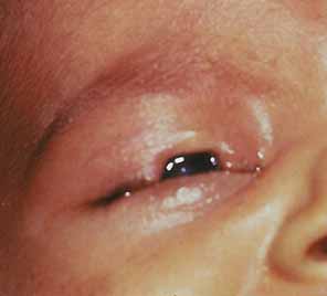
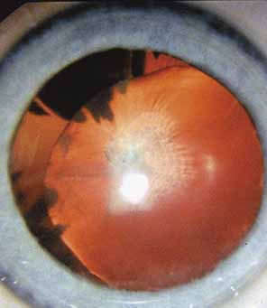
Complex microphthalmia is often associated with uveal colobomas. In the most common use of the term, “coloboma” refers to a defect in the iris, retina/uvea, and/or optic nerve as a result of faulty closure of the embryonic fissure at the 7- to 20-mm stage of development. Colobomas tend to be inferonasal, which is the normal position of the embryonic fissure (Fig. 1). The term “coloboma” has also been applied to other anomalies of the optic nerve, the macula, and the uveal tract that are not related to faulty closure of the embryonic fissure. This loose use of terminology creates unnecessary confusion when describing a syndrome. Hornby et al. found in their series that 72 of 185 eyes with coloboma also were microphthalmic and 71 of those 72 had microcornea.65 Thus, not every patient with microphthalmia has coloboma and not every patient with coloboma has microphthalmia. Microphthalmia has been postulated to arise from faulty development of the secondary vitreous and, hence, not enough pressure needed for prenatal eye growth.66

Fig. 1 “Typical” inferonasal coloboma of the optic nerve head, retina, and choroid.

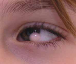
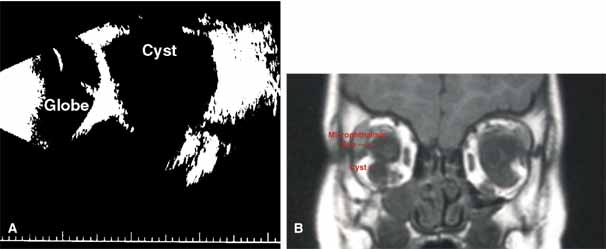
“Microphthalmia with cyst” refers to cases of colobomatous microphthalmia with a posterior eye wall defect through which a cyst lined with neuroectodermally derived tissue protrudes into the orbit. These cysts may be small and only detected by imaging (Fig. 2); sometimes, they may be large enough to cause progressive proptosis. Most cases are isolated, but familial occurrences have been reported.67–69 Inheritance is likely autosomal recessive. Depending on the size of the cyst, its appearance, and the visual potential of the microphthalmic eye, these cysts may be managed by observation, excision with or without enucleation, and/or aspiration.70 Aspirated cysts tend to reaccumulate fluid. Socket expansion improves cosmesis.

Fig. 2 A. B-scan ultrasonogram shows microphthalmia with cyst. B. MRI demonstrates cyst located on the inferior aspect of a microphthalmic eye.

Complex microphthalmia can also be seen as part of Peters anomaly and persistent hyperplastic primary vitreous (PHPV). These disorders are covered in the anterior segment and retina/vitreous sections of this chapter.

Not all cases of complex microphthalmia are genetically determined. Intrauterine infection and maternal exposure to alcohol or other teratogens can also cause complex microphthalmia.

Percin et al. described mutations in the homeobox gene CHX10 (14q24.3) in two families with autosomal recessive, nonsyndromic microphthalmia, iris abnormalities, and coloboma.71 Like other homeobox proteins, CHX10 is a transcription factor that binds to specific DNA sequences in the regulatory regions of other genes and affects their transcription during development. Chx10 mutations were previously found in the ocular retardation mouse model, which is characterized by microphthalmia, a thin hypocellular retina, and optic nerve aplasia.72

One form of autosomal recessive complex microphthalmia was found by Bessant et al. to map to 14q32.73 Phenotypic features included sclerocornea (8/8 patients) with secondary corneal vascularization (6/8 patients) and staphyloma formation (3/8 patients), elevated intraocular pressure (5/8 patients), and short axial length (5/5 measured).74 These authors ruled out CHX10 and OTX2 as candidate genes in this pedigree.

Lehman et al. reported a large Mexican American pedigree with isolated X-linked recessive colobomatous microphthalmia that maps to the proximal p or q arm of the X chromosome.75

Microphthalmia can usually be diagnosed by inspection and/or palpation of the eye through the eyelids. The presence of a bulge in the lower lid usually indicates microphthalmos with cyst (Fig. 3). Axial length measurements may aid in the diagnosis of simple microphthalmia/nanophthalmos. Refraction and amblyopia treatment may be helpful in cases in which the macula is well developed. In cases in which no useful vision is present, scleral shells may improved cosmesis. Oculoplastics procedures to enlarge the palpebral fissures and the socket are sometimes helpful.

Fig. 3 Patient with bilateral microphthalmia. Bulge in right lower lid indicates microphthalmia with cyst.

CONGENITAL CYSTIC EYE

Congenital cystic eye results from failure of the optic vesicle to invaginate, producing a cystic structure that may contain elements of dysplastic retina or lens material.76 These cysts do not contain an epithelial lining, may not be evident at birth, and may grow with time. Approximately 30 cases have been described in the literature. Most cases are unilateral, but bilateral developmental eye anomalies have been described.77–80 Orbital imaging can aid in the diagnosis and preoperative planning.81

Oculocerebrocutaneous syndrome (Delleman syndrome, OMIM 164180)82 is a sporadic condition associated with orbital cysts, focal dermal hypoplasia, periorbital skin appendages, and cerebral malformations. Anophthalmia may also occur in patients with this syndrome.

PERSISTENT HYPERPLASIA PRIMARY VITREOUS (PHPV) AND PERSISTENCE OF FETAL VASCULATURE (PFV)

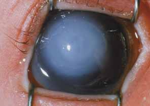
Persistent hyperplastic primary vitreous (PHPV) or persistence of fetal vasculature (PFV) is a complex malformation of the eye characterized by the presence of remnants of the hyaloid and of the tunica vasculosa lentis systems of blood vessels, together with proliferation of fibrovascular tissue behind the lens, and variable degrees of posterior pole and/or anterior retinal dysplasia.83,84 The eye in PHPV is generally microphthalmic but can be enlarged with accompanying myopia.85 Although PHPV primarily affects the posterior part of the eye, the ciliary body, iris, and lens are involved to a variable extent. In the typical case, ciliary processes are elongated and converge to a plaque of retrolental fibrovascular tissue that may involve the lens capsule proper (Fig. 4). The lens may be completely clear or may be cataractous. The fibrovascular plaque may adhere to the posterior lens capsule and vessels can invade the lens—a finding pathognomonic of PHPV. The retrolental membrane may contain adipose tissue, cartilage, and smooth muscle tissue. This is thought to be the result of metaplastic changes in tissues of mesenchymal origin. In a series of 47 eyes, Font et al. found adipose tissue in 10 and cartilage in one.86 The peripheral retina may also be drawn anteriorly into the retrolental membrane. In what has been referred to as “posterior PHPV,” the retina around the disc is pulled up into the posterior vitreous and is thrown into folds that may involve the macular and lead to poor vision. The retina is generally normal in structure, but dysplastic changes have been described. Vitreoretinal traction may lead to retinal breaks and detachment. The fellow eye in PHPV is usually normal but may have a Mittendorf dot or, rarely, one of other abnormalities.

Fig. 4 PHPV. There are numerous elongated ciliary processes drawn to a small lens with an opacified and wrinkled posterior capsule.

PHPV is unilateral in approximately 90% of cases. The globe varies in size from normal to moderately decreased, being slightly smaller than normal in the majority of cases. The cornea is clear. The anterior chamber is shallow in smaller eyes because of anterior displacement of the iris/lens diaphragm; this predisposes patients with PHPV to angle-closure glaucoma, which usually develops later in life. Lens extraction prevents secondary angle-closure glaucoma. The iris may be normal but frequently shows small notches at the pupillary margin, where iridohyaloidal vessels were located in the developing eye and failed to regress as the iris matures and the tunica vasculosa lentis resorbs. Patent iridohyaloidal vessels may be observed to course over the anterior iris surface, the pupillary margin, and the posterior iris surface to anastomose with vessels in the retrolental membrane. These iridohyaloidal vessels are characteristic of PHPV.

Clinically, patients with PHPV present with a small eye since birth, a white pupillary reflex from a cataract or retrolental membrane, and/or strabismus because of poor vision. Retinoblastoma should be ruled out using clinical examination, ultrasonography, and computed tomography.87 There is no intraocular calcification in PHPV.88 Retinoblastoma generally occurs in normal-size eyes except in patients with 13q deletions and microphthalmia. PHPV and retinoblastoma have been rarely reported together in a microphthalmic eye.89

PHPV and its variants are not very rare. They are a common cause of unilateral congenital cataracts. PHPV has also been reported in patients with recessive oculodento-osseous dysplasia,47 in one patient with protein C deficiency,90 in the oculopalatocerebral syndrome, and in two generations of one family with Rieger anomaly.91

PHPV may be the result of defective formation of the secondary vitreous, which is derived from the inner retinal cells starting in the ninth week of gestation. The secondary vitreous compresses the regressing primary vitreous, which is probably derived from mesenchyme and contains the hyaloid system of blood vessels that anastomose with the tunica vasculosa lentis anteriorly. The globe remains small because its growth depends partly on the expansion of the secondary vitreous.92 The cause of PHPV has not been identified yet. Familial occurrences of PHPV have been reported in dizygotic twins, in two brothers, and in a mother and son.93

Visual prognosis in PHPV is generally guarded. Early cataract surgery may result in relatively good visual results in selected patients.94,95 Eyes with anterior PHPV have a better vision potential than those with posterior pole dysplasia. An anterior approach to lens extraction and retrolental membrane removal is recommended in mild anterior cases. A combined lensectomy-vitrectomy using a pars-plana surgical approach should be considered in patients with significant mid-vitreal or posterior vitreal components to the PHPV. Tractional and rhegmatogenous retinal detachments have been reported in patients with PHPV and are treated surgically as needed.

SYNOPHTHALMIA/CYCLOPIA

Cyclopia refers to a single, midline eye; synophthalmia refers to an apparent midline fusion of two eyes. These conditions are points along a phenotypic continuum. Clinical findings vary but include a proboscis above the eye(s) and forebrain malformations. Many fetuses with cyclopia or synophthalmia have chromosomal abnormalities and are spontaneously aborted.96

Formation of a cyclopic or synophalmic globe may occur by one of two embryologic mechanism.97 At the trilaminar embryo stage, a field of bilobed, midline ectodermal tissue is “fated” to become the eyes. Failure of this bilobed area to separate completely into two fields could result in synophthalmia or cyclopia. Alternatively, previously separated globes could fuse as a result of faulty midline development.

The latter mechanism is likely responsible for the cyclopia and synophthalmia seen in holoprosencephaly. Holoprosencephaly is a genetically and phenotypically heterogeneous group of disorders that manifest with midline abnormalities.98,99 Classically, this includes complete (alobar) or partial (semilobar) “fusion” of the normal, bilateral cerebral hemispheres into a single hemisphere. Mutations in several genes important in forebrain development have been identified in families with holoprosencephaly, including sonic hedgehog (SHH, 7q36), ZIC2 (13q32), SIX3 (2p21), and transforming growth factor beta-induced factor (TGIF, 18p11.3).100–103

ANIRIDIA

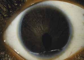
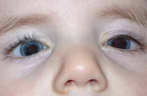
Classic aniridia is a sporadic or autosomal dominant, bilateral, pan-ocular condition caused by haploinsufficiency of the PAX6 homeobox gene on 11p13.104,105 Contrary to the connotation of “aniridia,” most aniridic patients have at least some rudimentary iris tissue. “Aniridia” is also used to describe absence of iris tissue in some syndromes that are possibly unrelated to PAX6 haploinsufficiency (see later).

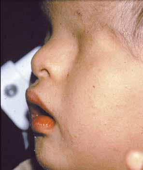
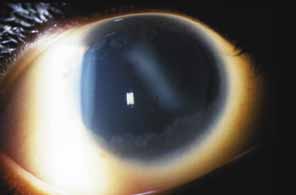
Aniridia affects multiple tissues in the eye and has a broad range of clinical presentations. Iris abnormalities range from almost complete absence to radial iris clefts to iris hypoplasia (Fig. 5).106–109 Foveal hypoplasia is characteristic, but of varying severity, which include optic nerve hypoplasia, corneal pannus, and/or a superficial corneal dystrophy (Fig. 6), microcornea, Peters anomaly, congenital cataracts, ectopia lentis, and glaucoma.110,111–114 Visual potential varies with the severity of the aniridic phenotype. Vision-limiting factors include optic nerve and foveal hypoplasia, cataracts, and—occasionally—the keratopathy that arises in older patients with aniridia. The keratopathy/corneal pannus is presumably caused by insufficient/absent limbal stem cells. Nystagmus develops in aniridic patients, presumably caused by congenital poor visual acuity. Iris vascular anomalies and leakage have been diagnosed on fluorescein angiography of the anterior segment.109

Fig. 5 Aniridia with significant remnants of iris tissue in both eyes.

Fig. 6 Keratopathy of aniridia. Note superficial epithelial haze, most prominent in inferior and temporal aspects of the cornea.

Shaw et al. estimated the prevalence of aniridia in the lower peninsula of Michigan in 1960 to be approximately 1 in 64,000.115 Approximately two-thirds of patients have at least one other affected family member; the remaining one-third are sporadic.

Pathologic studies of aniridic eyes are sometimes complicated by the presence of advanced glaucoma and/or a history of multiple surgical interventions. In 1983, Margo described pathologic features of seven eyes from seven children 6 months to 14 years of age.116 None of these eyes had advanced glaucoma or previous surgery. Congenital anomalies included iris and ciliary body hypoplasia, attenuation of Bowman membrane, and (in two eyes from children with 11p deletions) anomalous anterior chamber angles. Other abnormalities included corneal pannus, cataracts, and peripheral anterior synechiae. Resolution was insufficient to comment on retinal or optic nerve anomalies.

Elsas et al. classified aniridia into four types based on clinical criteria.107 Type I aniridia—what we call classic aniridia—is autosomal-dominant and is characterized by near-absence of iris tissue, foveal hypoplasia, nystagmus, corneal pannus, optic nerve hypoplasia, secondary glaucoma, and (sometimes) dislocation of the lens. Type II aniridia patients have varying iris abnormalities, no nystagmus, and relatively preserved visual acuity. Patients with type III aniridia have classic aniridia and mental retardation. Type IV aniridia patients also have genitourinary anomalies, an increased risk of Wilmss tumor, and mental retardation (WAGR syndrome, discussed later). This system was devised before our detailed knowledge of the cytogenetic and molecular causes of aniridia. Therefore, it does not take into account more recent findings of the phenotypic, cytogenetic, and mutational heterogeneity of this disorder.

Grant and Walton noted progressive angle changes in aniridic patients.117 Although the trabecular meshwork of 25 aniridic patients who did not develop glaucoma appeared normal on gonioscopy, the angles of 31 aniridic patients with glaucoma showed progressive obstruction of the trabecular meshwork by iris tissue. These authors recommended prophylactic goniotomy in aniridic patients who show progressive angle changes before glaucoma develops. This recommendation has not been studied in a controlled fashion.

Cytogenetic studies have played a pivotal role in understanding the molecular basis of aniridia. WAGR syndrome (OMIM 194072)—an acronym for Wilms tumor, aniridia, genitourinary abnormalities, and mental retardation—is associated with deletions of chromosome 11p13 (reviewed by Mannens et al.).118 By comparing the phenotype of patients with the size of the deletion, as determined using both cytogenetic and molecular markers, the critical region for aniridia and for Wilms tumor were identified. Other cytogenetic abnormalities such as paracentric inversions, balanced translocation, and unbalanced translocation involving 11p13 have been reported in subjects with aniridia, both with or without systemic findings.119,120 Not all chromosomal aberrations directly disrupt the PAX6 gene, suggesting that local chromatin structure or cis-acting elements are important for PAX6 expression.121 Crolla and van Heyningen have shown that some patients with deletion of both the Wilms tumor gene (WT1) and PAX6 may not be cytogenetically visible and can only be detected with fluorescent in-situ hybridization (FISH).120

Because the PAX6 gene and the Wilms tumor gene (WT1) are on the same 700-kilobase region of 11p13, chromosomal changes that cause aniridia may also affect the WT1 locus. In general, the increased risk of Wilms tumor occurs in sporadic aniridia cases. The exception to this rule is families in which an affected parent has a deletion of WT1 and passes his/her chromosomal abnormality onto their child. In these cases, autosomal-dominant inheritance is present. Not everyone with a WT1 deletion develops Wilms tumor. Gronskov et al. found two out of five subjects with WT1 deletions that developed Wilms tumor.122 Muto et al. found 13 (45%) of 29 patients with deletions in the Wilms tumor critical region developed Wilms tumor.123 Therefore, cytogenetic studies focused on 11p13 are reasonable in both sporadic aniridia and familial aniridia, when the number of affected individuals in the family is small.

In 1989 Mannens et al. mapped the aniridia locus to markers on 11p13.124 Further analysis of an aniridia pedigree originally mapping to chromosome 2 (AN1 locus)125 showed that the true linkage in this family was also to 11p13 (the AN2 locus.) In 1991, Ton et al. discovered that a homeobox transcription factor gene, now know as PAX6, was either totally or partially deleted in two patients with aniridia.104 Since then, numerous reports of mutations in PAX6 have been reported in patients with aniridia.105 A detailed database of PAX6 mutations can be found online at http://pax6.hgu.mrc.ac.uk/.

The PAX6 gene covers approximately 25 kb of DNA, consists of 14 exons, and codes for a 422-amino acid transcription factor.105 This 422-amino acid protein contains two distinct motifs that bind to a range of DNA target sequences—a paired domain and a homeodomain.126,127 (The terms “paired domain” and “homeodomain” are applied to these portions of the protein because they strongly resemble similarly named genes important in body segmentation in Drosophila). DNA binding is modified by alternative splicing of one portion of the paired domain, which affects the target sequence of DNA recognized. Translation begins at exon 4 and one exon (5a) is alternatively spliced. PAX6 protein is highly conserved across vertebrates. PAX6 is expressed in all layers of the neuroectoderm that forms the developing eye, as well as in the surface ectodermal cells that become the lens.128

Nearly all patients with classic aniridia have mutations predicted to cause loss of function of one PAX6 allele (see http://pax6.hgu.mrc.ac.uk/). In general, the ocular phenotype of these patients is indistinguishable from patients with complete deletion of the gene, implying that haploinsufficiency is the mechanism of disease. Patients with PAX6 missense mutations can have aniridia or a variety of other ocular phenotypes—corneal pannus/keratopathy, isolated foveal hypoplasia, congenital cataracts, Peters anomaly, anterior segment dysgenesis, uveal ectropion, optic nerve hypoplasia, optic nerve coloboma/morning glory discs, and persistent hyperplastic primary vitreous.114,129–134,135 For example, Kivlin et al. reported on what appeared to be a new, autosomal-dominant keratopathy (ADK) associated with ocular irritation and photophobia, and anterior stromal vascularization and inflammation.136 Pearce et al. suggested that ADK was an aniridia variant based on the presence of iris abnormalities and macular hypoplasia in a four-generation family with ADK.110 Mirzayans et al. subsequently found splice-site mutations in PAX6 in a family with ADK.137

The precise mechanism behind this phenotypic variability is unclear. Presumably, other gene products and environmental factors regulate or interact with PAX6 during ocular development; variations/abnormalities in these proteins lead to the variability in the aniridia phenotype. A few putative target genes activated by PAX6 have been identified, including lens crystallins, cell adhesion molecules like L1 and NCAM, glucagon, and insulin.138–142

Heterozygous small eye (Sey) mice have disrupted expression of one allele of Pax6 and are considered a model of human aniridia.143–145 The phenotype is variable, as in humans, and includes iris abnormalities, cataract, Peters anomaly, microphthalmia, and keratopathy.146

Treatment of aniridia centers around the treatment of its individual ophthalmic manifestations. As always, aggressive treatment of amblyopia in children and optimal refraction in all patients should be pursued. Cataracts should be removed if visually significant during the amylogenic period or later in life if visual deterioration appears to be linked to cataract progression. Some success has been reported with sulcus-based intraocular lenses and with black diaphragm aniridia lenses in these patients.147,148 Glaucoma is common and intraocular pressure should always be checked. It can be quite difficult to manage and often requires surgery. Numerous methods, including trabeculotomy, seton implantation, and ciliary body ablation, have had some success.149–152 As previously mentioned, some advocate prophylactic goniotomy in patients with progressive angle changes.153 No clear consensus exists on how to approach this difficult clinical problem.

The corneal pannus and ocular surface problems associated with aniridia can also cause visual deterioration. Penetrating keratoplasty is not usually successful because it does not treat the presumed, underlying limbal stem cell deficiency.154,155 Holland et al. have reported success with keratolimbal allografts in patients with aniridia.156 In their study, 74% of eyes had an improved ocular surface, mean visual acuity increased from 20/1000 to 20/165, and 70% of those undergoing secondary penetrating keratoplasty had successful grafts. However, continued use of immunosuppressive drugs appears to be required for optimal results.

Syndromes With Associated Aniridia

Warburg et al. described a girl with aniridia, mental retardation, gonadal streaks, gonadoblastoma, hearing loss, kidney malrotation, and dysmorphic facies that had an interstitial deletion between bands 11 and 13 on 11p.157 As discussed previously, the constellation of Wilms tumor, aniridia, genitourinary anomalies, and mental retardation (WAGR) is caused by deletions in this region of chromosome 11. Identification of patients with such cytogenetic abnormalities was critical in the isolation of PAX6 as the gene for human aniridia.

Pupil abnormalities reminiscent of aniridia are also seen with cerebellar ataxia and mental retardation in a condition often referred to as Gillespie syndrome. (The original patients described by Gillespie, however, have some features such as congenital cataracts that are not part of what is now referred to as “Gillespie syndrome”).158 The pupil in these patients appears constantly dilated and often has iris strands adherent to the lens. Unlike classic aniridia, patients with Gillespie syndrome generally do not have cataracts, corneal abnormalities, or foveal hypoplasia. Visual acuity has not been consistently recorded in the literature, but may be reduced.159 The nystagmus seen in these patients appears to be more related to cerebellar and other CNS pathology than to sensory issues. Although inheritance is generally thought to be autosomal recessive, Nelson et al.. have questioned whether some autosomal dominant cases might exist.159 This same group has found extensive white matter and cerebral abnormalities in one child with Gillespie syndrome, suggesting that the brain pathology may extend beyond the cerebellum. Glaser et al. did not find mutations in the PAX6 gene and the phenotype did not segregate with genetic markers on 11p in families with Gillespie syndrome.160

Mirkinson and Mirkinson described a family with aniridia and hypoplastic/aplastic patellae.161 The proband's grandmother had glaucoma, but it is unclear if this was of childhood onset. No other stigmata of classic, PAX6-associated aniridia are mentioned. No mutations have been reported in this pedigree.

Hamming et al. reported a mother and her two children with variable iris abnormalities, ptosis, nystagmus, foveal hypoplasia, corneal pannus, and cataracts.162 The mother had unspecified cardiac anomalies, recurrent miscarriages, and alopecia. One of the children was moderately mentally retarded and both children were obese. These authors suggest that this family has a rare, autosomal-dominant aniridia variant. Although chromosomal studies were reportedly normal, PAX6 mutation testing was not performed.

Although Levin et al. reported a child with aniridia, congenital glaucoma, hydrocephalus, and a ring chromosome 6 {46 XY, r(6) (p24→q26)}, the “aniridic” iris abnormalities were unilateral.163 The contralateral eye had findings more consistent with Axenfeld-Rieger syndrome. Ring chromosomes are formed when the long (q) and short (p) arms of a chromosome fuse together. Such joining is invariably associated with the loss of genetic material from one or both chromosomal arms. In this case, the patient was missing genetic material distal to p24 on the short arm and to q26 on the long arm of chromosome 6. This patient's ocular findings might be explained by a deletion of the FOXC1 gene on 6p25 (see discussion of Axenfeld-Rieger syndrome).

Walker and Dyson report on an association of classic aniridia with mental retardation.164 Although most patients with classic aniridia are intellectually normal, PAX6 is expressed in the developing brain and it is conceivable that in some individuals it may contribute to a mental handicap.

Back to Top
DISORDERS THAT AFFECT THE ORBITS, EYELIDS, AND ADNEXA

DEVELOPMENTAL BIOLOGY

The bones, cartilage, fat, and connective tissue of the orbit arise from periocular mesenchyme in the frontonasal and maxillary processes that appear by the fourth week of gestation. The bones of the orbit are membranous, with the exception of the sphenoid, which forms from a cartilaginous precursor. Ossification begins in the fifth month and fusion of the bones occurs in the sixth month of gestation. Eyelid formation begins between the fourth and fifth weeks of gestation with the proliferation of ectodermal cells in the area of the future outer canthus. During the second month of gestation, the ectoderm of the future lids becomes invested with periocular mesenchyme, which leads to the formation of the upper and lower eyelid folds. These folds grow closer together and fuse at the eighth week of gestation, beginning nasally and proceeding temporally. The orbicularis forms from a condensation of mesenchyme at approximately 10 weeks gestation. The adnexal structures form between the third and sixth months of gestation. The eyelids reopen between the fifth month and seventh month of gestation. The lacrimal glands begin forming between the sixth and seventh weeks of gestation as cords of epithelial cells from the conjunctiva invaginate supertemporally and are surrounded by periocular mesenchyme, which becomes the lacrimal acini.

TERMINOLOGY

Several specific terms are used to describe abnormalities in the appearance or the position of the orbits, eyes, and eyelids. Hypertelorism refers to an increased distance between the orbits, often measured clinically as an increase in the interpupillary distance (IPD), although true hypertelorism refers to an increased separation between the two orbits and is a radiological term). Hypertelorism should be distinguished from telecanthus, which connotes an increased distance between the inner canthi but not an increase in the IPD or separation between the orbits. Hypertelorism occurs in a variety of craniofacial syndromes such as frontonasal dysplasia and Greig cephalopolysyndactyly (Fig. 7). Similarly, hypotelorism refers to closely set orbits, as in holoprosencephaly. Epicanthus refers to a medial fold of skin that may partially obscure the inner canthus. Epicanthus tarsalis is used to describe a fold of skin between the superior orbital rim and the nose; epicanthus palpebralis is used to describe a fold of skin equally distributed between the superior and inferior medial canthus; and epicanthus inversus refers to a fold of skin extending inferolaterally from the nose to the inferior orbital rim. Epicanthal folds are common in babies and may give the mistaken impression of esotropia (pseudoesotropia). Epicanthus inversus occurs in the blepharophimosis syndrome (see later).

Fig. 7 Typical facial features of infant with Greig syndrome. Note prominent forehead, telecanthus, and hypertelorism. The child also had digital anomalies characteristic of this syndrome.

CRYPTOPHTALMOS

Cryptophthalmos refers to a failure of the eyelids to form correctly during embryonic development, resulting in covering of the globe by a continuous layer of skin (Fig. 8). Although the lens and posterior structures are sometimes intact, the underlying cornea and anterior segment structures may not be present or may be malformed. Typically, no conjunctival fornices are present and microphthalmia may be present. Cryptophthalmos may be unilateral or bilateral, isolated, or syndromic. The phenotypic continuum in this disorder ranges from a complete, uninterrupted layer of skin from brow to cheek, through partial formation of lids and adnexal structures, to adherence of lids directly to the globe. Thomas et al. reviewed isolated and syndromic cryptophthalmos in 1986.165 Of 127 cases in the literature at that time, 27 were isolated and 97 had other congenital malformations. The most common syndromic cause of cryptophthalmos is Fraser syndrome, an autosomal-recessive condition (see later). Some pedigrees of isolated cryptophthalmos show autosomal dominant inheritance, preservation of some adnexal structures, and microphthalmia (Fig. 9).165,166

Fig. 8 Recessive cryptophthalmos. Skin of forehead continues over globe and onto cheek (courtesy of Irene H. Maumenee, MD).

Fig. 9 Dominant cryptophthalmos. There is a very tall upper lid whose center is adherent to the underlying cornea. (Reprinted with permission from Saal et al, Am J Med Genet 43:785–788, 1992.)

Fraser syndrome is an autosomal-recessive condition characterized by cryptophthalmos, cutaneous syndactyly, genitourinary abnormalities, craniofacial dysmorphism, abnormalities of the larynx, orofacial clefting, mental retardation, and musculoskeletal abnormalities.167 Approximately 12% of patients with Fraser syndrome do not have cryptophthalmos. Homozygous mutations in the FRAS1 gene on 4q21 were reported in subjects with Fraser syndrome by McGregor et al..168 However, not all subjects with Fraser syndrome map to 4q21, implying genetic heterogeneity. The FRAS1 gene contains 75 exons and codes for a 4007-amino acid protein that is expressed in the basement membrane of corneal, skin, kidney, and gut epithelia.169 Mice homozygous for a nonsense mutation in the homologous gene, Fras1, develop hemorrhagic blistering during development, as well as postnatal cryptophthalmos, syndactyly, and renal anomalies.

The surgical management of cryptophthalmos is complex and challenging.170 Reconstruction is made difficult in many cases by the absence of normal corneal and anterior segment tissue and the absence of conjunctival fornices. Before any surgery is attempted, visual potential should be determined. Diagnostic imaging, visual evoked responses, and electroretinography may be helpful. Some success has been achieved with amniotic membrane grafts in the reconstruction of conjunctival fornices in patients with partial cryptophthalmos.171 Advances in keratoprostheses may some day help these patients.

CONGENITAL ECTROPION

Congenital ectropion is a rare condition in which the eyelid is outwardly rotated away from the globe. Some authors make a clear distinction between congenital ectropion and congenital eversion of the eyelids, stating that the former is a surgical disease and the latter can be managed conservatively.172–174 Other authors feel the congenital eversion of the upper lids is best managed surgically.175–177

Congenital ectropion may be more common in blacks178 and is associated with Down syndrome.172,179,180 Dermatologic conditions that shorten the anterior lamellae—such as congenital nonbullous ichthyosiform erythroderma (OMIM 242100), harlequin ichthyosis, and lamellar ichthyosis (OMIM 242300)—may cause ectropion.181,182,183 The relative shortage of malar and eyelid skin, coupled with eyelid abnormalities such as coloboma, may lead to ectropion in mandibulofacial dysostosis (Treacher-Collins syndrome, OMIM 154500.) The lymphedema-distichiasis syndrome (OMIM 153400) may present with congenital ectropion, probably because of tarsal abnormalities.184,185 The blepharocheilodontic syndrome is an autosomal-dominant condition (OMIM 119580) characterized by facial clefting, ectropion, oligodontia, euryblepharon, and lagophthalmos.186

Management of congenital ectropion depends on its severity, ie, the amount of corneal exposure and ocular irritation caused. Sometimes, lubrication may be sufficient. In cases in which the anterior lamella is shortened, horizontal tightening coupled with vertical lengthening (e.g., use of skin grafts) may be helpful.187 Yen et al reported the use of lateral tarsal strips, lower eyelid retractor disinsertion, myocutaneous advancement of the cheek and eyelids, and lateral tarsorrhaphy in a case of blepharocheilodontic syndrome.188

CONGENITAL ENTROPION

Congenital entropion is inward rotation of an eyelid toward the globe. Eyelashes directed toward the cornea can lead to irritation, epithelial defects, and secondary scarring and infection.189,190 Congenital entropion may occur alone or in association with epiblepharon—a redundant fold of skin and orbicularis that rotates the lid inward. Congenital “kinking” of the tarsus or disinsertion of lid retractors are proposed mechanisms. Congenital entropion may be sporadic or an autosomal-dominant trait;191 it has been reported in trisomy 13,192 Duane retraction syndrome,193 ectrodactyly-ectodermal dysplasia-clefting syndrome (OMIM 129900, 604292, 602077),194 and the Fukuyama congenital muscular dystrophy (OMIM 607440).195 In a retrospective review of 25 cases of congenital tarsal kinking, Sires found that the average age at diagnosis was 7.2 weeks, and ulcers (mostly sterile) were present in one-half of cases.196 Four of 15 children developed amblyopia, but not necessarily from corneal scarring.

If corneal injury cannot be prevented with conservative measures such as lubrication or lid taping, then surgery is indicated. Advancement of eyelid retractors with excision of a small ellipse of skin is helpful in cases caused by congenital retractor disinsertion. A variety of techniques have been proposed for patients with tarsal kinking and hypoplasia, including excision of eyelid skin and orbicularis,189 lid support sutures,197 advancement of otherwise-normal lid retractors,198 stretching of the tarsal plate followd by temporary lid sutures,199 and full-thickness sutures.200

EYELID COLOBOMA

An eyelid coloboma is a congenital, full-thickness defect of the lid (Fig. 10). Although we prefer to reserve the term “coloboma” for defects of embryonic fissure closure, the term “eyelid coloboma” is well entrenched in the literature and is also appropriate for this condition.

Fig. 10 Typical upper eyelid coloboma. This particular patient did not have Goldenhar syndrome or any other malformation.

Eyelid colobomas may occur in one or more lids. They may be part of a genetic syndrome or as a result of a fetal disruption of lid tissues (e.g., by an amniotic band.) They may result from a failure of the mesodermal folds of the lids to meet or from premature separation of the lids during development.

Colobomas of the upper lid are associated with Goldenhar syndrome (OMIM 164210), also known as hemifacial microsomia or oculoauriculovertebral dysplasia. Goldenhar syndrome is characterized by eyelid coloboma, epibulbar dermoids, unilateral ear anomalies and deafness with a small ipsilateral face, and vertebral anomalies. Cardiac and central nervous system anomalies have also been reported. Inheritance is usually sporadic, but some pedigrees with autosomal-dominant inheritance have been reported.201,202 Linkage to 14q32 has been reported in one pedigree.203 Eyelid colobomas in Goldenhar syndrome tend to involve the middle and/or nasal third of the upper lid. In general, upper lid colobomas are more likely to result in corneal exposure than lower lid colobomas.

Colobomas of the lower lid are associated with Treacher Collins-Franceschetti (TCF) syndrome (OMIM 154500), also known as mandibulofacial dysostosis. TCF syndrome is autosomal-dominant with variable expressivity and is characterized by antimongoloid slant to the palpebral fissures, eyelid colobomas, micrognathia, macrostomia, microtia/other ear anomalies, hypoplastic zygomatic arches, and conductive hearing loss.204

The Burn-McKeown syndrome is characterized by bilateral choanal atresia, cardiac defects, abnormalities of the external ear (often with hearing deficits), and a characteristic face, which includes coloboma of the lower lids.205

Kinsey and Streeten reported a case of upper eyelid coloboma and symblepharon, along with corneal ectasia, microphthalmia, and other complex eye malformations in a child with median cleft syndrome/frontonasal dysplasia.206 In this case, it is unclear if the observed defects were genetic or teratogenic, because the mother received diazepam during her pregnancy.

Leventer et al. reported a congenital intraocular teratoma associated with eyelid coloboma.207 The eyelid coloboma may have been caused by interruption of eyelid development by the growing tumor.

The treatment and the timing of treatment of a congenital eyelid defect depend on the position and size of the defect. In cases in which corneal exposure and secondary infection are a concern, early closure is indicated. However, conservative management of sizable defects has been reported.208 Delaying surgery may reduce the risk of anesthesia and lessen the amylogenic effect of the eyelid closure that results from some repair procedures. In general, defects involving less than one-third of the lid can be closed directly.209 Defects up to half the lid may require a relaxing lateral cantholysis or semicircular flap to release tension on the wound. Larger defects may need more extensive use of flaps and grafts and will likely involve multiple operations. Patients with eyelid coloboma in conjunction with a syndrome (e.g., TCF) may require extensive craniofacial reconstruction as well.210,211

BLEPHAROPHIMOSIS, EPICANTHUS IVERSUS, AND PTOSIS SYNDROME (BPES)

BPES is an autosomal-dominant disorder characterized by small horizontal palpebral fissures (blepharophimosis), folds of skin extending from a flat nasal bridge inferolaterally (epicanthus inversus), and congenital ptosis (Fig. 11). Although the distance between the medial canthi is usually increased (telecanthus), the interpupillary distance is usually normal. BPES patients have been divided into two subgroups, type I and type II, based on the presence or absence of premature ovarian failure in females, respectively. A third putative type of BPES was found to be allelic to Sathre-Chotzen syndrome, with mutations in the TWIST gene.212

Fig. 11 A 4-month-old baby with blepharophimosis syndrome.

A series of BPES patients with chromosomal abnormalities around 3q21–23 suggested that the gene for this disorder was in that region.213–219 In 2001, Crisponi et al. described mutations in a transcription factor gene, FOXL2, on 3q23 in several pedigrees with BPES.220 FOXL2 (like FOXC1 in Rieger syndrome and FOXC2 in lymphedema-distichiasis, discussed later), is a member of the forkhead family of transcription factors and is expressed in the periocular mesenchyme that forms the lids during development and in the follicular cells of adult ovaries. Mutations predicted to cause truncation of the FOXL2 gene are seen in families with type I BPES. In contrast, in-frame duplications or insertions that extend the FOXL2 protein are seen in families with type II BPES.221 These type II alleles may be hypomorphic (ie, they retain some, but not all, of the normal function of that allele), whereas the type I alleles are likely to cause loss-of-function.

The major concern in children with BPES is amblyopia and head posture from ptosis.222 Because the epicanthus inversus and telecanthus do not generally improve with age, corrective surgery may be undertaken in a staged fashion or as a single procedure.223

The term blepharophimosis can also be used as a descriptive term to describe small palpebral fissures. This trait can be seen in a variety of syndromes such as Ohdo syndrome (OMIM 249620, blepharophimosis, ptosis, cardiac malformations, mental retardation, and hypoplastic teeth) and van den Ende-Gupta syndrome (OMIM 600920, blepharophimosis, arachnodactyly, congenital contractures). A more extensive list is available on OMIM and is beyond the scope of this chapter.

DISTICHIASIS

Distichiasis refers to the presence of a second row of eyelashes that arise from the meibomian glands (Fig. 12). (This is in distinction to trichiasis in which eyelashes arise from their normal location but are misdirected). Distichiasis can be inherited either alone (OMIM 12630) or, more commonly, as part of the lymphedema-distichiasis (LD) syndrome (OMIM 153400).184,224 The aberrant lashes in distichiasis may cause ocular irritation and photophobia.

Fig. 12 Distichiasis. Several lashes are arising from meibomian gland orifices and are rubbing against the globe.

The LD syndrome is an autosomal-dominant disorder characterized by congenital distichiasis and peripheral lymphedema that usually begins at approximately the age of puberty. Other reported associated features include lower eyelid ectropion, congenital ptosis, photophobia, cleft palate, webbing of the neck, vertebral anomalies, extradural cysts, varicose veins, renal abnormalities, and congenital heart disease.225–230 Fang et al. recently described mutations in the forkhead transcription factor, FOXC2, in families with LD.231 Numerous additional mutation—all predicted to cause haploinsufficiency of FOXC2—have been reported in LD families.185,232–235 Brooks et al. recently reported a family presenting with distichiasis and no lymphedema who had a truncating mutation in FOXC2.236 This pedigree suggests that LD syndrome and simple distichiasis may be points along a phenotypic continuum, rather than separate genetic defects. Ophthalmologists should also be aware that LD syndrome might present as distichiasis alone and should counsel their patients accordingly.

Ocular irritation and photophobia may be treated conservatively with lubrication. Severe cases may cause corneal scarring and may be treated with cryotherapy, lid-splitting procedures, and/or tarsal excision.237–241

CHORISTOMAS

Choristomas are benign, congenital tumors consisting of normal tissue in an abnormal location.242 Epibulbar and orbital choristomas are the most common epibulbar and orbital tumors in children. Choristomas are divided into four histologic types: (1) dermoids, which contain collagenous connective tissue surrounded by epidermis; (2) lipodermoids, which are similar to dermoids but also contain fatty tissue; (3) single-tissue choristomas, which contain either dermis-like or meso/ectodermal tissue of one type; and (4) complex choristomas, which contain tissues of several types. A teratoma is a choristoma that contains tissue from all three embryonic layers, ie, ectoderm, mesoderm, and endoderm. A teratoid tumor contains tissue from two embryonic layers. Dermoid cysts frequently contain a keratinized epithelium with adnexal structures.

Epibulbar choristomas vary in appearance and visual significance. Their color may be white, yellow, or pink (Fig. 13). They may be unilateral or bilateral and range in size from relatively small and flat lesions to large, bulky masses that fill the interpalpebral space and displace the globe. Epibulbar choristomas can cause astigmatism that, if untreated, can lead to amblyopia. The appearance of the tumor may be a cosmetic problem that affects a child's psychosocial development. Treatment involves dissection and excision, along with treatment of refractive error and amblyopia.243 Excision of any choristoma that involves the cornea will leave a residual opacity. It is therefore important to warn parents that surgery will not completely remove the opacity but will remove the mass. Occasionally, if the choristoma creates an opacity that extends into the visual axis, a lamellar keratoplasty may be indicated.244

Fig. 13 Inferotemporal limbal dermoid.

Conjunctival or episcleral osseous choristomas contain compact bone.245 They may be freely mobile or may adhere to sclera or other extraocular muscles. In one case, an osseous choristoma mimicked extraocular extension of retinoblastoma.246

Choristomas may also occur in the orbit, with a predilection for the supratemporal quadrant and the nasal area above the lacrimal sac. Orbital choristomas may present as a bulging mass or as proptosis in childhood. Sometimes they are asymptomatic and present, after years of growth, in adulthood. The spontaneous rupture of an orbital choristoma may cause acute proptosis and inflammation, mimicking an orbital cellulitis. Orbital imaging with CT or MRI is helpful in diagnosis and surgical excision.

Choristomas may be associated with other ophthalmic abnormalities, especially in syndromic children. As previously mentioned, epibulbar dermoids may be seen in Goldenhar syndrome (OMIM 164210). Epibulbar choristomas are also seen in nevus sebaceous of Jadassohn (OMIM 163200), a sporadic disorder characterizes by seizures, multiple skin lesions with malignant potential, arachnoid cysts, and mental retardation. These patients may also have posterior choristomas that contain cartilage and appear as flat, yellow lesions in the fundus.247–249 Encephalocraniocutaneous lipomatosis (ECCL) is characterized by multiple systemic choristomas and hamartomas (abnormal tissue found in its normal location), mental retardation, and seizures. Epibulbar choristomas are common in ECCL.250

Pedigrees primarily involving ocular or periocular choristomas have been reported. Plewes and Jacobson described a family with presumably dominant frontonasal dermoids that extended into the intracranial cavity.251 Mattos et al. reported a three-generation Peruvian family with dominant limbal choristomas in a ring configuration and no other systemic abnormalities.252 X-linked recessive corneal dermoids were observed in a Puerto Rican family and tentatively mapped to Xq24-qter.253,254

Back to Top
MALFORMATIONS OF THE ANTERIOR SEGMENT

DEVELOPMENTAL BIOLOGY

At approximately day 27 of gestation, the surface ectoderm over the developing optic vesicle begins to thicken in response to inductive signals from the optic vesicle and anterior neuroectoderm.97 This thickening, called the lens placode, is facilitated by adhesions, but not direct cell-to-cell contact, between the optic vesicle and the surface ectoderm. As the surface ectoderm in this area is mitotically active, these adhesions cause crowding of the surface ectoderm cells, which elongate and begin to invaginate to form a lens vesicle. PAX6 expression in the optic vesicle and the surface ectoderm is required for proper lens formation.255 Bone morphogenetic protein–4 (BMP–4) is also important in lens induction.256 The area of contact between the optic vesicle and the surface epithelium determines the size of the lens.

As the lens vesicle continues to expand, it begins to “pinch off” from the surface ectoderm, with complete separation at approximately 33 days gestation. The presence of a somewhat spherical lens vesicle separates the eye into future anterior and posterior segments. Failure of the lens vesicle to completely separate from the surface ectoderm can lead to anomalies such as anterior lenticonus (seen in Alport syndrome, OMIM 301050, 203780, 104200), anterior polar cataracts, and Peters anomaly. The lens vesicle initially consists of a monolayer of epithelial cells with their apices directed inward. The basement membrane of this epithelium forms the lens capsule. The surface ectoderm, which reforms a continuous layer after the separation of the lens vesicle, becomes the future corneal epithelium.

At 37 days of gestation, the posterior lens epithelial cells elongate to fill the lens vesicle, lose their nuclei and most organelles, and form the primary lens fibers. The primitive lens filled with primary lens fibers forms the embryonic lens nucleus. The posterior portions of the primary lens fibers detach from the posterior capsule, leaving room for migration of anterior, mitotically active epithelial cells. As these cells pass the equator of the lens, they become more elongated and eventually lose their nuclei and most organelles. These cells form the secondary lens fibers that migrate anteriorly and posteriorly around the embryonic nucleus and form interdigitations visible as the two Y-sutures of the lens.

After lens vesicle separation, the anterior margins of the optic cup advance beneath the ectoderm. Neural crest cells migrate between the surface ectoderm and optic cup, filling the anterior chamber. This neural crest will form the future corneal stroma, corneal stroma, iris stroma, ciliary body, and iridocorneal angle structures. By the sixth month of gestation, Descemet membrane and the corneal endothelium are functioning and the cornea is transparent. Mesenchymal tissue around the lens forms the tunica vasculosa lentis, which anastomoses with the hyaloid artery system.

The anterior and posterior iris epithelium as well as the bilayered ciliary body epithelium form from the nonpigmented and pigmented cells of the optic cup. These cells are arranged apex-to-apex. The iris stroma and ciliary muscles form from neural crest.

The iridocorneal angle begins to form during week 15 of gestation. The iris and ciliary body move posteriorly and the mesenchyme in the anterior segment undergoes rearrangement, including significant atrophy. Spaces continuous with the anterior chamber appear in the mesenchyme, forming an early trabecular meshwork. Schlemm canal is present by week 16 of gestation. Abnormalities in the remodeling of the mesenchyme of the anterior segment are postulated to be the cause of congenital glaucoma and of iridogoniodysgenesis. The trabecular meshwork continues to mature postnatally.

ANTERIOR SEGMENT DYSGENESIS

Abnormalities during development of the anterior segment can result in a variety of phenotypes. Historically, these have been given descriptive names (e.g., iridogoniodysgenesis, iris hypoplasia) or eponyms (e.g., Axenfeld-Rieger anomaly, Peters anomaly) Molecular genetic discoveries have shown that phenotypes that were considered separate disorders were, in fact, caused by mutations in the same gene. Similarly, mutations in distinct classes of genes were found to cause identical phenotypes. The phenotype in any individual is clearly influenced by a variety of heretofore-uncharacterized genetic and environmental factors during development.

Peters Anomaly

Peters anomaly (OMIM 604229, 603807) refers to a central corneal opacity related to a congenital absence of Descemet membrane and endothelium sometimes accompanied by iridocorneal adhesions and/or lenticulo-corneal adhesions. Peters anomaly may be unilateral or bilateral, and, although most often sporadic, autosomal-recessive and autosomal-dominant pedigrees have been reported.257–262 The severity of Peters anomaly ranges from a mild central corneal opacity to dense opacification with extensive irido-corneal and lenticulo-corneal adhesions and cataract formation (Fig. 14). Glaucoma may be present at birth or develop later in childhood in some cases.263,264 Peters anomaly may occur in the presence of other eye malformations such as sclerocornea (which may also include areas where Descemet membrane is absent), microphthalmia, uveal coloboma, and persistent fetal vasculature (PFV.)265–268 The developmental mechanisms leading to Peters anomaly may include incomplete separation of the lens vesicle from the surface ectoderm and/or abnormal migration and differentiation of neural crest in the anterior segment.

Fig. 14 Moderately severe corneal opacification in a patient with Peters anomaly.

The genetics of Peters anomaly are complex. Although mutations in genes such as PAX6,114,269 PITX2,270 CYP1B1,271 and FOXC1272 have been reported in some pedigrees, the underlying genetic cause is unclear in many cases.273 Approximately 40% of mice lacking on active copy of the forkhead transcription factor FoxE3 have Peters anomaly.274 Patients with Peters anomaly and several different chromosomal abnormalities (e.g., interstitial deletion 2q14q21,275 4p- [Wolf-Hirschorn syndrome],275 partial trisomy 5p,276 trisomy 9,277 11q-,278 and 21 ring279) have been reported. David et al. reported a family with an apparent balanced chromosomal translocation, t(1;7) (q41;p21), and Peters anomaly.280 This translocation disrupts the histone deacetylase 9 (HDAC9) and transforming growth factor beta 2 (TGFβ2) genes, which they suggest may be involved in the pathogenesis of Peters. Teratogens such as alcohol and isotretinoin and intrauterine infections may also produce Peters anomaly.281–283

Up to 60% of patients with Peters anomaly have some form of systemic abnormalities or developmental delay.266,284 Traboulsi and Maumenee define the “Peters plus” syndrome as one that includes midline defects, cleft lip and/or palate, ear abnormalities, midline abnormalities, mental retardation/developmental delay, cardiac abnormalities, and/or genitourinary abnormalities. The “Peters plus” syndrome is likely caused by a host of different genetic mutations and environmental teratogenic agents.

The Krause-Kivlin syndrome (OMIM 261540)—sometimes also referred to as “Peters plus” syndrome285—consists of Peters anomaly, short-limb dwarfism, with variable mental retardation, a “cupid-bow” upper lip, and frequent cleft lip with or without cleft palate.286 It likely represents a distinct, autosomal-recessive syndrome.287–289

The visual prognosis for patients with Peters anomaly depends on the severity of opacification in the visual axis. Mild cases may require little or no treatment. Unilateral cases are often accompanied by amblyopia in the affected eye. More severe cases may require cataract extraction, lysis of iridocorneal adhesions, and corneal transplantation. However, the risk of corneal transplant rejection in children is substantial.290–292,293 Signs of glaucoma should be monitored and treated appropriately.

Axenfeld-Rieger Syndrome

Axenfeld-Rieger syndrome (ARS) is a multisystem genetic disorder inherited in an autosomal-dominant fashion.294,295 It includes several prominent eye findings, including posterior embryotoxon, iris strands adherent to Schwalbe line, and iris hypoplasia that can lead to corectopia, polycoria, or pseudopolycoria (Fig. 15). More importantly, approximately half of ARS patients develop glaucoma.295–297 These abnormalities are likely caused by abnormalities in neural crest migration and differentiation during anterior segment development.298 Patients with the full-blown ARS—as opposed to the Axenfeld-Reiger anomaly that is limited to eye findings—can also have dental anomalies (microdontia, hypodontia) (Fig. 16), a flattened midface, and umbilical ring anomalies (Fig. 17). In some cases of ARS, patients have abnormalities in pituitary, cardiac, and limb development.

Fig. 15 Rieger anomaly. Almost 360-degree anteriorly displaced Schwalbe line. The pupil is inferiorly displaced and its edge is adherent to Schwalbe line.

Fig. 16 Absent, misshapen, and misdirected teeth in a patient with Rieger syndrome.

Fig. 17 Umbilical anomaly in a child with Rieger syndrome.

In 1996 Semina et al. reported mutations in PITX2(RIEG1), a gene on 4q25-q26 that codes for a homeobox transcription factor, in subjects with ARS.299 The mutations that were initially reported clustered around the homeobox region. Subsequently, additional mutations throughout the PITX2 sequence have been observed in subjects with various forms of anterior segment dysgenesis (ARS, iris hypoplasia, iridogoniodysgenesis, and Peters anomaly).300–307 These mutations include nonsense, missense, frameshift, and splicing mutations, as well as an in-frame duplication.298 Such mutations interfere with protein stability, protein–DNA interaction, nuclear localization, and/or transactivation.308–310 Mutations that eliminate PITX2 transactivation in vitro appear more likely to produce an ARS phenotype, whereas others that retain some transactivation ability appear more likely to produce a milder, iris hypoplasia phenotype.310 Although most mutations likely result in a loss of protein function and PITX2 haploinsufficiency, Priston et al report a V45L mutation in a syndromic ARS family that enhances in vitro transactivation.303 This observation suggests that either too much or too little PITX2 activity can produce an ARS phenotype. Lastly, Saadi et al demonstrated that mutation of the bicoid-specific lysine to glutamic acid in the PITX2 homeobox (K88E) produces a dominant-negative effect on wild-type PITX2 protein.304 PITX2 expresses multiple protein isoforms as a result of alternative splicing.311,312 A great deal of interfamilial and intrafamilial phenotypic variability exists in patients with PITX2 mutations.

ARS can also be caused by mutations in the FOXC1 gene on 6p25, a member of the forkhead family of transcription factors.272,313–318 These mutations are predicted to cause a loss of FOXC1 function and functional experiments support a variety of molecular mechanisms, including reduced protein expression, decreased DNA binding, and decreased transactivation.319 Nishimura et al have also reported pedigrees with duplications of 6p25 and an ARS-like phenotype, suggesting that either too much or too little FOXC1 activity may lead to anterior segment dysgenesis.320 As with subjects with PITX2 mutations, subjects with FOXC1 mutations have a great deal of phenotypical variability. At least two other ARS loci await cloning on 13q14 and 16q24.

The management of ARS centers on monitoring for and treatment of glaucoma. Ophthalmologists should be aware of the systemic manifestations of ARS and should refer patients appropriately.

Back to Top
REFERENCES

1. Baird PA, Anderson TW, Newcombe HB, Lowry RB: Genetic disorders in children and young adults: a population study. Am J Hum Genet 42(5):677–93, 1988

2. Campbell H, Holmes E, MacDonald S, et al: A capture-recapture model to estimate prevalence of children born in Scotland with developmental eye defects. J Cancer Epidemiol Prev 7(1):21–8, 2002

3. Morrison D, FitzPatrick D, Hanson I, et al: National study of microphthalmia, anophthalmia, and coloboma (MAC) in Scotland: investigation of genetic aetiology. J Med Genet 39(1):16–22, 2002

4. Hornby SJ, Gilbert CE, Rahi JK, et al: Regional variation in blindness in children due to microphthalmos, anophthalmos and coloboma. Ophthalmic Epidemiol 7(2):127–38, 2000

5. Busby A, Dolk H, Collin R, et al: Compiling a national register of babies born with anophthalmia/microphthalmia in England 1988–94. Arch Dis Child Fetal Neonatal Ed 79(3):F168–73, 1998

6. Bermejo E, Martinez-Frias ML: Congenital eye malformations: clinical-epidemiological analysis of 1,124,654 consecutive births in Spain. Am J Med Genet 75(5):497–504, 1998

7. Stoll C, Alembik Y, Dott B, Roth MP: Congenital eye malformations in 212,479 consecutive births. Ann Genet 40(2):122–28, 1997

8. Stromland K, Miller M, Cook C: Ocular teratology. Surv Ophthalmol 35(6):429–46, 1991

9. Chow RL, Lang RA: Early eye development in vertebrates. Annu Rev Cell Dev Biol 17:255–96, 2001

10. Brunquell PJ, Papale JH, Horton JC, et al: Sex-linked hereditary bilateral anophthalmos. Pathologic and radiologic correlation. Arch Ophthalmol 102(1):108–13, 1984

11. Kohn G, el Shawwa R, el Rayyes E: Isolated “clinical anophthalmia” in an extensively affected Arab kindred. Clin Genet 33(5):321–24, 1988

12. Fryns JP: Fryns syndrome: a variable MCA syndrome with diaphragmatic defects, coarse face, and distal limb hypoplasia. J Med Genet 24:271–74, 1987

13. Seller MJ, Davis TB, Fear CN, et al: Two sibs with anophthalmia and pulmonary hypoplasia (the Matthew-Wood syndrome). Am J Med Genet 62(3):227–29, 1996

14. Sandler D, Mancuso A, Becker T, et al: Association of anophthalmia and esophageal atresia. Am J Med Genet 59(4):484–91, 1995

15. Traboulsi EI, Nasr A, Fahd SD, et al: Waardenburg's recessive anophthalmia syndrome. Ophthal Paediat Genet 4:13–8, 1984

16. Ahmad ME, Dada R, Dada T, Kucheria K: 14q(22) deletion in a familial case of anophthalmia with polydactyly. Am J Med Genet 120A(1):117–22, 2003

17. Guion-Almeida ML, Richieri-Costa A: New syndrome of growth and mental retardation, structural anomalies of the central nervous system, and first branchial arch, anophthalmia, heminasal a/hypoplasia, and atypical clefting: report on four Brazilian patients. Am J Med Genet 87(3):237–44, 1999

18. Moog U, Kruger G, Stengel B, et al: Oculocerebrocutaneous syndrome: a case report, a follow-up, and differential diagnostic considerations. Genet Couns 7(4):257–65, 1996

19. Hermmann J, Opitz JM: The Lenz microphthalmia syndrome. Birth Defects Orig Artic Ser V(2):138–43, 1969

20. Hesselberg C: Congenital bilateral anophthalmia. Acta Ophthalmol 29:183–9, 1951

21. Driggers RW, Macri CJ, Greenwald J, et al: Isolated bilateral anophthalmia in a girl with an apparently balanced de novo translocation: 46,XX,t(3;11)(q27;p11.2). Am J Med Genet 87(3):201–2, 1999

22. Fantes J, Ragge NK, Lynch SA, et al: Mutations in SOX2 cause anophthalmia. Nat Genet 33(4):461–3, 2003

23. Wood H, Episkopou V: Comparative expression of the mouse Sox1, Sox2, and Sox3 genes from pre-gatrulation to early somite stages. Mech Dev 86(1–2):197–201, 1999

24. Kamachi Y, Uchikawa M, Tanouchi A, et al: PAX6 and SOX2 form a co-DNA-binding partner complex that regulates initiation of lens development. Genes Dev 15(10):1272–86, 2001

25. Krastinova D, Kelly MB, Mihaylova M: Surgical management of the anophthalmic orbit, part 1: congenital. Plast Reconstr Surg 108(4):817–26, 2001

26. Weiss AH, Kousseff BG, Ross EA, Longbottom J: Simple microphthalmos. Arch Ophthalmol 107(11):1625–30, 1989

27. Paznekas WA, Boyadjiev SA, Shapiro RE, et al: Connexin 43 (GJA1) mutations cause the pleiotropic phenotype of oculodentaldigital dysplasia. Am J Hum Genet 72(2):408–18, 2003

28. Traboulsi EI, Maumenee IH: Ophthalmologic findings in mucolipidosis III (pseudo-Hurler polydystrophy). Am J Ophthalmol 102(5):592–7, 1986

29. Boynton JR, Pheasant TR, Johnson BL, et al: Ocular findings in Kenny's syndrome. Arch Ophthalmol 97(5):896–900, 1979

30. Yamani A, Wood I, Sugino I, et al: Abnormal collagen fibrils in nanophthalmos: a clinical and histologic study. Am J Ophthalmol 127(1):106–8, 1999

31. Brockhurst RJ: Vortex vein decompression for nanophthalmic uveal effusion. Arch Ophthalmol 98(11):1987–90, 1980

32. Brockhurst RJ: Nanophthalmos with uveal effusion: a new clinical entity. Trans Am Ophthalmol Soc 72:371–403, 1974

33. Brockhurst RJ: Cataract surgery in nanophthalmic eyes. Arch Ophthalmol 108(7):965–7, 1990

34. Othman MI, Sullivan SA, Skuta GL, et al: Autosomal dominant nanophthalmos (NNO1) with high hyperopia and angle- closure glaucoma maps to chromosome 11. Am J Hum Genet 63(5):1411–8, 1998

35. Morle L, Bozon M, Zech JC, et al: A locus for autosomal dominant colobomatous microphthalmia maps to chromosome 15q12-q15. Am J Hum Genet 67(6):1592–7, 2000

36. al-Gazali LI, Mueller RF, Caine A, et al: Two 46,XX,t(X;Y) females with linear skin defects and congenital microphthalmia: a new syndrome at Xp22.3. J Med Genet 27(1):59–63, 1990

37. Yokoyama Y, Narahara K, Tsuji K, et al: Autosomal dominant congenital cataract and microphthalmia associated with a familial t(2;16) translocation. Hum Genet 90(1–2):177–8, 1992

38. Ng D, Hadley DW, Tifft CJ, Biesecker LG: Genetic heterogeneity of syndromic X-linked recessive microphthalmia-anophthalmia: is Lenz microphthalmia a single disorder? Am J Med Genet 110(4):308–14, 2002

39. Vervoort VS, Viljoen D, Smart R, et al: Sorting nexin 3 (SNX3) is disrupted in a patient with a translocation t(6;13)(q21;q12) and microcephaly, microphthalmia, ectrodactyly, prognathism (MMEP) phenotype. J Med Genet 39(12):893–9, 2002

40. Teebi AS, al-Saleh QA, Hassoon MM, et al: Macrosomia, microphthalmia, +/- cleft palate and early infant death: a new autosomal recessive syndrome. Clin Genet 36(3):174–7, 1989

41. Aughton DJ: Clinical anophthalmia, dextrocardia, and skeletal anomalies in an infant born to consanguineous parents. Am J Med Genet 37(2):178–81, 1990

42. Gupta PC, Peralta D, Parker M, et al: Bilateral microphthalmia with cyst, facial clefts, and limb anomalies: a new syndrome with features of Waardenburg syndrome, cerebro-oculo-nasal syndrome, and craniotelencephalic dysplasia. Am J Med Genet 117A(1):72–5, 2003

43. Gorlin RJ, Marashi AH, Obwegeser HL: Oculo-facio-cardio-dental (OFCD) syndrome. Am J Med Genet 63(1):290–2, 1996

44. Thiele H, Musil A, Nagel F, Majewski F: Familial arhinia, choanal atresia, and microphthalmia. Am J Med Genet 63(1):310–3, 1996

45. Verloes A, Temple IK, Bonnet S, Bottani A: Coloboma, mental retardation, hypogonadism, and obesity: critical review of the so-called Biemond syndrome type 2, updated nosology, and delineation of three “new” syndromes. Am J Med Genet 69(4):370–9, 1997

46. Warburg M: Hydrocephaly, congenital retinal nonattachment, and congenital falciform fold. Am J Ophthalmol 85(1):88–94, 1978

47. Traboulsi EI, Faris BM, Der Kaloustian VM: Persistent hyperplastic primary vitreous and recessive oculo-dento- osseous dysplasia. Am J Med Genet 24(1):95–100, 1986

48. Verloes A, Delfortrie J, Lambotte C: GOMBO syndrome of growth retardation, ocular abnormalities, microcephaly, brachydactyly, and oligophrenia: a possible “new” recessively inherited MCA/MR syndrome. Am J Med Genet 32(1):15–8, 1989

49. Goltz RW: Focal dermal hypoplasia: an update. Arch Dermatol 128:1108–11, 1992

50. Cohen MM Jr: Hallermann-Streiff syndrome: a review. Am J Med Genet 41(4):488–99, 1991

51. Meira LB, Graham JM, Jr. , Greenberg CR, et al: Manitoba aboriginal kindred with original cerebro-oculo-facio-skeletal syndrome has a mutation in the Cockayne syndrome group B (CSB) gene. Am J Hum Genet 66:1221–8, 2000

52. Graham JM, Jr. , Anyane-Yeboa K, Raams A, et al: Cerebro-oculo-facio-skeletal syndrome with a nucleotide excision-repair defect and a mutated XPD gene, with prenatal diagnosis in a triplet pregnancy. Am J Hum Genet 69:291–300, 2001

53. Artman HG, Boyden E: Microphthalmia with single central incisor and hypopituitarism. J Med Genet 27(3):192–3, 1990

54. Nothen MM, Knopfle G, Fodisch HJ, Zerres K: Steinfeld syndrome: report of a second family and further delineation of a rare autosomal dominant disorder. Am J Med Genet 46(4):467–70, 1993

55. Kapur S, Toriello HV: Apparently new MCA/MR syndrome in sibs with cleft lip and palate and other facial, eye, heart, and intestinal anomalies. Am J Med Genet 41(4):423–5, 1991

56. Wilf-Miron R, Goodman RM: Facio-thoraco-genital syndrome: a newly recognized birth defect syndrome. J Craniofac Genet Dev Biol 7(1):19–22, 1987

57. Carney SH, Brodsky MC, Good WV, et al: Aicardi syndrome: more than meets the eye. Surv Ophthalmol 37(6):419–24, 1993

58. Tellier AL, Cormier-Daire V, Abadie V, et al: CHARGE syndrome: report of 47 cases and review. Am J Med Genet 76(5):402–9, 1998

59. Gong Y, Slee RB, Fukai N, et al: LDL receptor-related protein 5 (LRP5) affects bone accrual and eye development. Cell 107(4):513–23, 2001

60. Salonen R, Paavola P: Meckel syndrome. J Med Genet 35(6):497–501, 1998

61. Schrander-Stumpel CT, de Die-Smulders CE, Hennekam RC, et al: Oculoauriculovertebral spectrum and cerebral anomalies. J Med Genet 29(5):326–31, 1992

62. Lenz W: Recessiv geschlechtsgebundene mikrophthalmologie mit multi-plen misbildungen. Z Kinderheilkd 77:384–90, 1955

63. Jones KL: Smith's Recognizable Patterns of Human Malformation. Philadelphia: WB Saunders Company, 1997.

64. Warburg M, Friedrich U: Coloboma and microphthalmos in chromosomal aberrations. Chromosomal aberrations and neural crest cell developmental field. Ophthalmic Paediatr Genet 8(2):105–18, 1987

65. Hornby SJ, Adolph S, Gilbert CE, et al: Visual acuity in children with coloboma: clinical features and a new phenotypic classification system. Ophthalmology 107(3):511–20, 2000

66. Coulombre A: The role of intraocular pressure in the development of the chick eye. J Exp Zool 133:211–25, 1956

67. Porges Y, Gershoni-Baruch R, Leibu R, et al: Hereditary microphthalmia with colobomatous cyst. Am J Ophthalmol 114(1):30–4, 1992

68. Makley TA, Jr. , Battles M: Microphthalmos with cyst. Report of two cases in the same family. Surv Ophthalmol 13(4):200–6, 1969

69. Leatherbarrow B, Kwartz J, Noble JL: Microphthalmos with cyst in monozygous twins. J Pediatr Ophthalmol Strabismus 27(6):294–8, 1990

70. McLean CJ, Ragge NK, Jones RB, Collin JR: The management of orbital cysts associated with congenital microphthalmos and anophthalmos. Br J Ophthalmol 87(7):860–3, 2003

71. Ferda Percin E, Ploder LA, Yu JJ, et al: Human microphthalmia associated with mutations in the retinal homeobox gene CHX10. Nat Genet 25(4):397–401, 2000

72. Burmeister M, Novak J, Liang MY, et al: Ocular retardation mouse caused by Chx10 homeobox null allele: impaired retinal progenitor proliferation and bipolar cell differentiation. Nat Genet 12(4):376–84, 1996

73. Bessant DA, Khaliq S, Hameed A, et al. A locus for autosomal recessive congenital microphthalmia maps to chromosome 14q32. Am J Hum Genet 62(5):1113–6, 1998

74. Bessant DA, Anwar K, Khaliq S, et al: Phenotype of autosomal recessive congenital microphthalmia mapping to chromosome 14q32. Br J Ophthalmol 83(8):919–22, 1999

75. Lehman DM, Sponsel WE, Stratton RF, et al: Genetic mapping of a novel X-linked recessive colobomatous microphthalmia. Am J Med Genet 101(2):114–9, 2001

76. Hayashi N, Repka MX, Ueno H, et al: Congenital cystic eye: report of two cases and review of the literature. Surv Ophthalmol 44(2):173–9, 1999

77. Pasquale LR, Romayananda N, Kubacki J, et al: Congenital cystic eye with multiple ocular and intracranial anomalies. Arch Ophthalmol 109(7):985–7, 1991

78. Sacks JG, Lindenberg R: Efferent nerve fibers in the anterior visual pathways in bilateral congenital cystic eyeballs. Am J Ophthalmol 68(4):691–5, 1969

79. Waring GO, 3rd , Roth AM, Rodrigues MM: Clinicopathologic correlation of microphthalmos with cyst. Am J Ophthalmol 82(5):714–21, 1976

80. Goldberg SH, Farber MG, Bullock JD, et al: Bilateral congenital ocular cysts. Ophthalmic Paediatr Genet 12(1):31–8, 1991

81. Mansour AM, Li HK: Congenital cystic eye. Ophthal Plast Reconstr Surg 12(2):104–7, 1996

82. Hoo JJ, Kapp-Simon K, Rollnick B, Chao M: Oculocerebrocutaneous (Delleman) syndrome: a pleotropic disorder affecting ectodermal tissues with unilateral predominance. Am J Med Genet 40(3):290–3, 1991

83. Haddad R, Font RL, Reeser F: Persistent hyperplastic primary vitreous. A clinicopathologic study of 62 cases and review of the literature. Surv Ophthalmol 23(2):123–34, 1978

84. Goldberg MF: Persistent fetal vasculature (PFV): an integrated interpretation of signs and symptoms associated with persistent hyperplastic primary vitreous (PHPV). LIV Edward Jackson Memorial Lecture. Am J Ophthalmol 124(5):587–626, 1997

85. Cheung JC, Summers CG, Young TL: Myopia predicts better outcome in persistent hyperplastic primary vitreous. J Pediatr Ophthalmol Strabismus 34(3):170–6, 1997

86. Font RL, Yanoff M, Zimmerman LE: Intraocular adipose tissue and persistent hyperplastic primary vitreous. Arch Ophthalmol 82(1):43–50, 1969

87. Mafee MF, Goldberg MF: Persistent hyperplastic primary vitreous (PHPV): role of computed tomography and magnetic resonance. Radiol Clin North Am 25(4):683–92, 1987

88. Potter PD, Shields CL, Shields JA, Flanders AE: The role of magnetic resonance imaging in children with intraocular tumors and simulating lesions. Ophthalmology 103(11):1774–83, 1996

89. Irvine AR, Albert DM, Sang DN: Retinal neoplasia and dysplasia. II. Retinoblastoma occurring with persistence and hyperplasia of the primary vitreous. Invest Ophthalmol Vis Sci 16(5):403–7, 1977

90. Hermsen VM, Conahan JB, Koops BL, Cunningham RD: Persistent hyperplastic primary vitreous associated with protein C deficiency. Am J Ophthalmol 109(5):608–9, 1990

91. Storimans CW, Van Schooneveld MJ: Rieger's eye anomaly and persistent hyperplastic primary vitreous. Ophthalmic Paediatr Genet 10(4):257–62, 1989

92. Boeve MH, Vrensen GF, Willekens BL, et al: Early morphogenesis of persistent hyperplastic tunica vasculosa lentis and primary vitreous (PHTVL/PHPV). Scanning electron microscopic observations. Graefes Arch Clin Exp Ophthalmol 231(1):29–33, 1993

93. Lin AE, Biglan AW, Garver KL: Persistent hyperplastic primary vitreous with vertical transmission. Ophthalmic Paediatr Genet 11(2):121–2, 1990

94. Pollard ZF: Persistent hyperplastic primary vitreous: diagnosis, treatment and results. Trans Am Ophthalmol Soc 95:487–549, 1997

95. Anteby I, Cohen E, Karshai I, BenEzra D: Unilateral persistent hyperplastic primary vitreous: course and outcome. J Aapos 6(2):92–9, 2002

96. Howard RO: Chromosomal abnormalities associated with cyclopia and synophthalmia. Trans Am Ophthalmol Soc 75:505–38, 1977

97. Cook C, Sulik K, Wright KW: Embryology. In Wright KW, Spiegel PH, (eds): Pediatric Ophthalmology and Strabismus, 2nd ed. New York : Spinger-Verlag, 2003

98. Muenke M: Holoprosencephaly as a genetic model for normal craniofacial development. Dev Biol 5:293–301, 1994

99. Wallis D, Muenke M: Mutations in holoprosencephaly. Hum Mutat 2000 ;16:99–108, 2000

100. Belloni E, Muenke M, Roessler E, et al: Identification of Sonic hedgehog as a candidate gene responsible for holoprosencephaly. Nat Genet 14(3):353–6, 1996

101. Wallis DE, Roessler E, Hehr U, et al: Mutations in the homeodomain of the human SIX3 gene cause holoprosencephaly. Nat Genet 22(2):196–8, 1999

102. Brown SA, Warburton D, Brown LY, et al: Holoprosencephaly due to mutations in ZIC2, a homologue of Drosophila odd-paired. Nat Genet 20(2):180–3, 1998

103. Gripp KW, Wotton D, Edwards MC, et al: Mutations in TGIF cause holoprosencephaly and link NODAL signalling to human neural axis determination. Nat Genet 25(2):205–8, 2000

104. Ton CC, Hirvonen H, Miwa H, et al: Positional cloning and characterization of a paired box- and homeobox-containing gene from the aniridia region. Cell 67(6):1059–74, 1991

105. Glaser T, Walton DS, Maas RL: Genomic structure, evolutionary conservation and aniridia mutations in the human PAX6 gene. Nat Genet 2(3):232–9, 1992

106. Traboulsi EI, Jurdi-Nuwayhid F, Torbey NS, Frangieh GT: Aniridia, atypical iris defects, optic pit and the morning glory disc anomaly in a family. Ophthalmic Paediatr Genet 7(2):131–5, 1986

107. Elsas FJ, Maumenee IH, Kenyon KR, Yoder F: Familial aniridia with preserved ocular function. Am J Ophthalmol 83(5):718–24, 1977

108. Hittner HM, Riccardi VM, Francke U: Aniridia caused by a heritable chromosome 11. Ophtalmology 86(6):1173–83, 1979

109. Hittner HM, Riccardi VM, Ferrell RE, et al: Variable expressivity in autosomal dominant aniridia by clinical, electrophysiologic, and angiographic criteria. Am J Ophthalmol 89(4):531–9, 1980

110. Pearce WG, Mielke BW, Hassard DTR, et al: Autosomal dominant keratitis: a possible aniridia variant. Can J Ophthalmol 30(3):131–7, 1995

111. David R, MacBeath L, Jenkins T: Aniridia associated with microcornea and subluxated lenses. Br J Ophthalmol 62(2):118–21, 1978

112. Mackman G, Brightbill FS, Optiz JM: Corneal changes in aniridia. Am J Ophthalmol 87(4):497–502, 1979

113. Layman PR, Anderson DR, Flynn JT: Frequent occurrence of hypoplastic optic disks in patients with aniridia. Am J Ophthalmol 77(4):513–6, 1974

114. Hanson IM, Fletcher JM, Jordan T, et al: Mutations at the PAX6 locus are found in heterogeneous anterior segment malformations including Peters' anomaly. Nat Genet 6(2):168–73, 1994

115. Shaw MW, Falls HF, Neel JV: Congenital aniridia. Am J Hum Genet 12(4):389–415, 1960

116. Margo CE: Congenital aniridia: a histopathologic study of the anterior segment in children. J Pediatr Ophthalmol Strabismus 20(5):192–8, 1983

117. Grant WM, Walton DS: Progressive changes in the angle in congenital aniridia, with development of glaucoma. Am J Ophthalmol 78(5):842–7, 1974

118. Mannens M, Hoovers JM, Bleeker-Wagemakers EM, et al: The distal region of 11p13 and associated genetic diseases. Genomics 11(2):284–93, 1991

119. Fukushima Y, Hoovers J, Mannens M, et al: Detection of a cryptic paracentric inversion within band 11p13 in familial aniridia by fluorescence in situ hybridization. Hum Genet 91(3):205–9, 1993

120. Crolla JA, van Heyningen V: Frequent chromosome aberrations revealed by molecular cytogenetic studies in patients with aniridia. Am J Hum Genet 71(5):1138–49, 2002

121. Fantes J, Redeker B, Breen M, et al: Aniridia-associated cytogenetic rearrangements suggest that a position effect may cause the mutant phenotype. Hum Mol Genet 4(3):415–22, 1995

122. Gronskov K, Olsen JH, Sand A, et al: Population-based risk estimates of Wilmss tumor in sporadic aniridia. A comprehensive mutation screening procedure of PAX6 identifies 80% of mutations in aniridia. Hum Genet 109(1):11–8, 2001

123. Muto R, Yamamori S, Ohashi H, Osawa M: Prediction by FISH analysis of the occurrence of Wilmss tumor in aniridia patients. Am J Med Genet 108(4):285–9, 2002

124. Mannens M, Bleeker-Wagemakers EM, Bliek J, et al: Autosomal dominant aniridia linked to the chromosome 11p13 markers catalase and D11S151 in a large Dutch family. Cytogenet Cell Genet 52(1–2):32–6, 1989

125. Ferrell RE, Chakravarti A, Hittner HM, Riccardi VM: Autosomal dominant aniridia: probable linkage to acid phosphatase–1 locus on chromosome 2. Proc Natl Acad Sci U S A 77(3):1580–2, 1980

126. Czerny T, Schaffner G, Busslinger M: DNA sequence recognition by Pax proteins: bipartite structure of the paired domain and its binding site. Genes Dev 7(10):2048–61, 1993

127. Xu HE, Rould MA, Xu W, et al: Crystal structure of the human Pax6 paired domain-DNA complex reveals specific roles for the linker region and carboxy-terminal subdomain in DNA binding. Genes Dev 13(10):1263–75, 1999

128. Nishina S, Kohsaka S, Yamaguchi Y, et al: PAX6 expression in the developing human eye. Br J Ophthalmol 83(6):723–7, 1999

129. Azuma N, Nishina S, Yanagisawa H, et al: PAX6 missense mutation in isolated foveal hypoplasia. Nat Genet 13(2):141–2, 1996

130. Azuma N, Hotta Y, Tanaka H, Yamada M: Missense mutations in the PAX6 gene in aniridia. Invest Ophthalmol Vis Sci 39(13):2524–8, 1998

131. Azuma N, Yamada M: Missense mutation at the C terminus of the PAX6 gene in ocular anterior segment anomalies. Invest Ophthalmol Vis Sci 39(5):828–30, 1998

132. Azuma N, Yamaguchi Y, Handa H, et al: Missense mutation in the alternative splice region of the PAX6 gene in eye anomalies. Am J Hum Genet 65(3):656–63, 1999

133. Azuma N, Yamaguchi Y, Handa H, et al: Mutations of the PAX6 gene detected in patients with a variety of optic-nerve malformations. Am J Hum Genet 72(6):1565–70, 2003

134. Hanson I, Churchill A, Love J, et al: Missense mutations in the most ancient residues of the PAX6 paired domain underlie a spectrum of human congenital eye malformations. Hum Mol Genet 8(2):165–72, 1999

135. Glaser T, Jepeal L, Edwards JG, et al: PAX6 gene dosage effect in a family with congenital cataracts, aniridia, anophthalmia and central nervous system defects. Nat Genet 7(4):463–71, 1994

136. Kivlin JD, Apple DJ, Olson RJ, Manthey R: Dominantly inherited keratitis. Arch Ophthalmol 104(11):1621–3, 1986

137. Mirzayans F, Pearce WG, MacDonald IM, Walter MA: Mutation of the PAX6 gene in patients with autosomal dominant keratitis. Am J Hum Genet 57(3):539–48, 1995

138. Richardson J, Cvekl A, Wistow G: Pax–6 is essential for lens-specific expression of zeta-crystallin. Proc Natl Acad Sci U S A 92(10):4676–80, 1995

139. Cvekl A, Sax CM, Li X, et al: Pax–6 and lens-specific transcription of the chicken delta 1-crystallin gene. Proc Natl Acad Sci U S A 92(10):4681–5, 1995

140. Chalepakis G, Wijnholds J, Giese P, et al: Characterization of Pax–6 and Hoxa–1 binding to the promoter region of the neural cell adhesion molecule L1. DNA Cell Biol 13(9):891–900, 1994

141. Holst BD, Wang Y, Jones FS, Edelman GM: A binding site for Pax proteins regulates expression of the gene for the neural cell adhesion molecule in the embryonic spinal cord. Proc Natl Acad Sci U S A 94(4):1465–70, 1997

142. Sander M, Neubuser A, Kalamaras J, et al: Genetic analysis reveals that PAX6 is required for normal transcription of pancreatic hormone genes and islet development. Genes Dev 11(13):1662–73, 1997

143. Hill RE, Favor J, Hogan BL, et al: Mouse small eye results from mutations in a paired-like homeobox-containing gene. Nature 354(6354):522–5, 1991

144. Hill RE, Favor J, Hogan BL, et al: Mouse Small eye results from mutations in a paired-like homeobox-containing gene. Nature 355(6362):750, 1992

145. van der Meer-de Jong R, Dickinson ME, Woychik RP, et al: Location of the gene involving the small eye mutation on mouse chromosome 2 suggests homology with human aniridia 2 (AN2). Genomics 7(2):270–5, 1990

146. Ramaesh T, Collinson JM, Ramaesh K, et al: Corneal abnormalities in Pax6+/- small eye mice mimic human aniridia-related keratopathy. Invest Ophthalmol Vis Sci 44(5):1871–8, 2003

147. Segal EI, Li HK: Transscleral ciliary sulcus fixation of a posterior chamber lens in an eye with congenital aniridia. J Cataract Refract Surg 23(4):595–7, 1997

148. Reinhard T, Engelhardt S, Sundmacher R: Black diaphragm aniridia intraocular lens for congenital aniridia: long-term follow-up. J Cataract Refract Surg 26(3):375–81, 2000

149. Arroyave CP, Scott IU, Gedde SJ, et al: Use of glaucoma drainage devices in the management of glaucoma associated with aniridia. Am J Ophthalmol 135(2):155–9, 2003

150. Okada K, Mishima HK, Masumoto M, et al: Results of filtering surgery in young patients with aniridia. Hiroshima J Med Sci 49(3):135–8, 2000

151. Adachi M, Dickens CJ, Hetherington J, Jr. , et al: Clinical experience of trabeculotomy for the surgical treatment of aniridic glaucoma. Ophthalmology 104(12):2121–5, 1997

152. Wiggins RE Jr. , Tomey KF: The results of glaucoma surgery in aniridia. Arch Ophthalmol 110(4):503–5, 1992

153. Chen TC, Walton DS: Goniosurgery for prevention of aniridic glaucoma. Trans Am Ophthalmol Soc 96:155–65, 1998

154. Kremer I, Rajpal RK, Rapuano CJ, et al: Results of penetrating keratoplasty in aniridia. Am J Ophthalmol 115(3):317–20, 1993

155. Gomes JA, Eagle RC Jr. , Gomes AK, et al: Recurrent keratopathy after penetrating keratoplasty for aniridia. Cornea 15(5):457–62, 1996

156. Holland EJ, Djalilian AR, Schwartz GS: Management of aniridic keratopathy with keratolimbal allograft: a limbal stem cell transplantation technique. Ophthalmology 110(1):125–30, 2003

157. Warburg M, Mikkelsen M, Andersen SR, et al: Aniridia and interstitial deletion of the short arm of chromosome 11. Metab Pediatr Ophthalmol 4(2):97–102, 1980

158. Gillespie FD: Aniridia, Cerebellar Ataxia, and Oligophrenia in Siblings. Arch Ophthalmol 73:338–41, 1965

159. Nelson J, Flaherty M, Grattan-Smith P: Gillespie syndrome: a report of two further cases. Am J Med Genet 71(2):134–8, 1997

160. Glaser T, Ton CC, Mueller R, et al: Absence of PAX6 gene mutations in Gillespie syndrome (partial aniridia, cerebellar ataxia, and mental retardation). Genomics 19(1):145–8, 1994

161. Mirkinson AE, Mirkinson NK: A familial syndrome of aniridia and absence of the patella. Birth Defects Orig Artic Ser 11(5):129–31, 1975

162. Hamming NA, Miller MT, Rabb M: Unusual variant of familial aniridia. J Pediatr Ophthalmol Strabismus 23(4):195–200, 1986

163. Levin H, Ritch R, Barathur R, et al: Aniridia, congenital glaucoma, and hydrocephalus in a male infant with ring chromosome 6. Am J Med Genet 25(2):281–7, 1986

164. Walker FA, Dyson C: Dominantly inherited aniridia associated with mental retardation and other eye abnormalities. Birth Defects Orig Artic Ser 10(7):147–9, 1974

165. Thomas IT, Frias JL, Felix V, et al: Isolated and syndromic cryptophthalmos. Am J Med Genet 25(1):85–98, 1986

166. Saal HM, Traboulsi EI, Gavaris P, et al: Dominant syndrome with isolated cryptophthalmos and ocular anomalies. Am J Med Genet 43(5):785–8, 1992

167. Slavotinek AM, Tifft CJ: Fraser syndrome and cryptophthalmos: review of the diagnostic criteria and evidence for phenotypic modules in complex malformation syndromes. J Med Genet 39(9):623–33, 2002

168. McGregor L, Makela V, Darling SM, et al: Fraser syndrome and mouse blebbed phenotype caused by mutations in FRAS1/Fras1 encoding a putative extracellular matrix protein. Nat Genet 34(2):203–8, 2003

169. Vrontou S, Petrou P, Meyer BI, et al: Fras1 deficiency results in cryptophthalmos, renal agenesis and blebbed phenotype in mice. Nat Genet 34(2):209–14. 2003

170. Ferri M, Harvey JT: Surgical correction for complete cryptophthalmos: case report and review of the literature. Can J Ophthalmol 34(4):233–6, 1999

171. Stewart JM, David S, Seiff SR: Amniotic membrane graft in the surgical management of cryptophthalmos. Ophthal Plast Reconstr Surg 18(5):378–80, 2002

172. Miller R, Martin F, Allen H: A case of congenital ectropion in Down's syndrome. Aust N Z J Ophthalmol 16(2):119–25, 1988

173. Raab EL, Saphir RL: Congenital eyelid eversion. Ophthalmic Surg 21(10):736, 1990

174. Hughes JL, O'Connor PS, Leone C: Simple treatment for congenital eyelid eversion. Ophthalmic Surg 15(7):609, 1984

175. Watts MT, Dapling RB: Congenital eversion of the upper eyelid: a case report. Ophthal Plast Reconstr Surg 11(4):293–5, 1995

176. Dawodu OA: Total eversion of the upper eyelids in a newborn. Niger Postgrad Med J 8(3):145–7. 2001

177. Bentsi-Enchill KO: Congenital total eversion of the upper eyelids. Br J Ophthalmol 65(3):209–13, 1981

178. Lu LW, Bansal RK, Katzman B: Primary congenital evershon of the upper lids. J Pediatr Ophthalmol Strabismus 16(3):149–51, 1979

179. Bleeker-Wagemakers EM, Delleman JW, Walbeek K: Congenital eversion of the eyelids in a case of Down's syndrome. Ophthalmologica 173(3–4):250–6, 1976

180. Sellar PW, Bryars JH, Archer DB: Late presentation of congenital ectropion of the eyelids in a child with Down syndrome: a case report and review of the literature. J Pediatr Ophthalmol Strabismus 29(1):64–7, 1992

181. Virolainen E, Niemi KM, Ganemo A, et al: Ultrastructural features resembling those of harlequin ichthyosis in patients with severe congenital ichthyosiform erythroderma. Br J Dermatol 145(3):480–3, 2001

182. Cruz AA, Menezes FA, Chaves R, et al: Eyelid abnormalities in lamellar ichthyoses. Ophthalmology 107(10):1895–8, 2000

183. Oestreicher JH, Nelson CC: Lamellar ichthyosis and congenital ectropion. Arch Ophthalmol 108(12):1772–3, 1990

184. Falls HF, Kertesz ED: A new syndrome combining pterygium colli with developmental anomalies of the eyelids and lymphatics of the lower extremeties. Trans Am Ophthalmol Soc 62:248–75, 1964

185. Erickson RP, Dagenais SL, Caulder MS, et al: Clinical heterogeneity in lymphoedema-distichiasis with FOXC2 truncating mutations. J Med Genet 38(11):761–6. 2001

186. Allanson JE, McGillivray BC: Familial clefting syndrome with ectropion and dental anomaly—without limb anomalies. Clin Genet 27(4):426–9, 1985

187. Heher KL, Katowitz JA: Surgical management of the patient with ocular adnexal malformations. In Traboulsi E (ed): Genetic Diseases of the Eye. New York: Oxford University Press, 1998;36

188. Yen MT, Lucci LM, Anderson RL: Management of eyelid anomalies associated with Blepharo-cheilo-dontic syndrome. Am J Ophthalmol 132(2):279–80, 2001

189. Salour H, Owji N, Razavi ME, Zeaei H: Tarsal kink syndrome associated with congenital corneal ulcer. Ophthal Plast Reconstr Surg 19(1):81–3, 2003

190. Luchs JI, Laibson PR, Stefanyszyn MA, et al: Infantile ulcerative keratitis secondary to congenital entropion. Cornea 16(1):32–4, 1997

191. Amacher AG, 3rd , Mazzoli RA, Gilbert BN, et al: Dominant familial congenital entropion with tarsal hypoplasia and atrichosis. Ophthal Plast Reconstr Surg 18(5):381–4, 2002

192. Lucci LM, Fukumoto WK, Alvarenga LS: Trisomy 13: a rare case of congenital tarsal kink. Ophthal Plast Reconstr Surg 19(5):408–10, 2003

193. Patton N, Leatherbarrow B: Primary congenital upper eyelid entropion in association with Duane's retraction syndrome. Eye 14 (Pt 3A):385–7, 2000

194. Ireland IA, Meyer DR: Ophthalmic manifestations of ectrodactyly-ectodermal dysplasia-clefting syndrome. Ophthal Plast Reconstr Surg 14(4):295–7, 1998

195. Chijiiwa T, Nishimura M, Inomata H, et al: Ocular manifestations of congenital muscular dystrophy (Fukuyama type). Ann Ophthalmol 15(10):921–8, 1983

196. Sires BS: Congenital horizontal tarsal kink: clinical characteristics from a large series. Ophthal Plast Reconstr Surg 15(5):355–9, 1999

197. Bosniak S, Hornblass A, Smith B: Re-examining the tarsal kink syndrome: considerations of its etiology and treatment. Ophthalmic Surg 16(7):437–40, 1985

198. Bartley GB, Nerad JA, Kersten RC, Maguire LJ: Congenital entropion with intact lower eyelid retractor insertion. Am J Ophthalmol 112(4):437–41, 1991

199. el-Mulki S, Lawson J, Taylor D: A new and simple procedure for correction of congenital tarsal kink. J Pediatr Ophthalmol Strabismus 35(3):172–3, 1998

200. Quickert MH, Wilkes TD, Dryden RM: Nonincisional correction of epiblepharon and congenital entropion. Arch Ophthalmol 101(5):778–81, 1983

201. Regenbogen L, Godel V, Goya V, Goodman RM: Further evidence for an autosomal dominant form of oculoauriculovertebral dysplasia. Clin Genet 21(3):161–7, 1982

202. Godel V, Regenbogen L, Goya V, Goodman RM: Autosomal dominant Goldenhar syndrome. Birth Defects Orig Artic Ser 18(6):621–8, 1982

203. Kelberman D, Tyson J, Chandler DC, et al: Hemifacial microsomia: progress in understanding the genetic basis of a complex malformation syndrome. Hum Genet 109(6):638–45, 2001

204. Dixon MJ: Treacher Collins syndrome. Hum Mol Genet 5 Spec No:1391–6, 1996

205. Wieczorek D, Teber OA, Lohmann D, Gillessen-Kaesbach G: Two brothers with Burn-McKeown syndrome. Clin Dysmorphol 12(3):171–4, 2003

206. Kinsey JA, Streeten BW: Ocular abnormalities in the median cleft face syndrome. Am J Ophthalmol 83(2):261–6, 1977

207. Leventer DB, Corona J, Linberg JV, et al: Congenital intraocular teratoma associated with eyelid coloboma. Am J Ophthalmol 132(2):277–9, 2001

208. Yeo LM, Willshaw HE: Large congenital upper lid coloboma—successful delayed conservative management. J Pediatr Ophthalmol Strabismus 34(3):190–2, 1997

209. Patipa M, Wilkins RB, Guelzow KW: Surgical management of congenital eyelid coloboma. Ophthalmic Surg 13(3):212–6, 1982

210. Fuente del Campo A, Martinez Elizondo M, Arnaud E: Treacher Collins syndrome (mandibulofacial dysostosis). Clin Plast Surg 21(4):613–23, 1994

211. Roddi R, Vaandrager JM, van der Meulen JC: Treacher Collins syndrome: early surgical treatment of orbitomalar malformations. J Craniofac Surg 6(3):211–7, 1995

212. Dollfus H, Kumaramanickavel G, Biswas P, et al: Identification of a new TWIST mutation (7p21) with variable eyelid manifestations supports locus homogeneity of BPES at 3q22. J Med Genet 38(7):470–2. 2001

213. Wolstenholme J, Brown J, Masters KG, et al: Blepharophimosis sequence and diaphragmatic hernia associated with interstitial deletion of chromosome 3 (46,XY,del(3)(q21q23)). J Med Genet 31(8):647–8, 1994

214. Moncla A, Philip N, Mattei JF: Blepharophimosis-mental retardation syndrome and terminal deletion of chromosome 3p. J Med Genet 32(3):245–6, 1995

215. Small KW, Stalvey M, Fisher L, et al: Blepharophimosis syndrome is linked to chromosome 3q. Hum Mol Genet 4(3):443–8, 1995

216. Amati P, Chomel JC, Nivelon-Chevalier A, et al: A gene for blepharophimosis-ptosis-epicanthus inversus syndrome maps to chromosome 3q23. Hum Genet 96(2):213–5, 1995

217. Harrar HS, Jeffery S, Patton MA: Linkage analysis in blepharophimosis-ptosis syndrome confirms localisation to 3q21–24. J Med Genet 32(10):774–7, 1995

218. Noda K, Mashima Y, Nakamura Y, Tanaka Y: Blepharophimosis-ptosis-epicanthus inversus syndrome associated with interstitial deletion of chromosome 3q21–23. J Pediatr Ophthalmol Strabismus 35(4):242–3, 1998

219. Costa T, Pashby R, Huggins M, Teshima IE: Deletion 3q in two patients with blepharophimosis-ptosis-epicanthus inversus syndrome (BPES). J Pediatr Ophthalmol Strabismus 35(5):271–6. 1998

220. Crisponi L, Deiana M, Loi A, et al: The putative forkhead transcription factor FOXL2 is mutated in blepharophimosis/ptosis/epicanthus inversus syndrome. Nat Genet 27(2):159–66, 2001

221. De Baere E, Dixon MJ, Small KW, et al: Spectrum of FOXL2 gene mutations in blepharophimosis-ptosis-epicanthus inversus (BPES) families demonstrates a genotype—phenotype correlation. Hum Mol Genet 10(15):1591–600, 2001

222. Beckingsale PS, Sullivan TJ, Wong VA, Oley C: Blepharophimosis: a recommendation for early surgery in patients with severe ptosis. Clin Experiment Ophthalmol 31(2):138–42, 2003

223. Karacaoglan N, Sahin U, Ercan U, Bozdogan N: One-stage repair of blepharophimosis: a new method. Plast Reconstr Surg 93(7):1406–9, 1994

224. Pico G: Congenital ectropion and distichiasis: Etiologic and hereditary factors: A report of cases and review of the literature. Trans Am Ophthalmol Soc 55:663–700, 1957

225. Brice G, Mansour S, Bell R, et al: Analysis of the phenotypic abnormalities in lymphoedema-distichiasis syndrome in 74 patients with FOXC2 mutations or linkage to 16q24. J Med Genet 39(7):478–83, 2002

226. Corbett CR, Dale RF, Coltart DJ, Kinmonth JB: Congenital heart disease in patients with primary lymphedemas. Lymphology 15(3):85–90, 1982

227. Chynn KY: Congenital spinal extradural cyst in two siblings. Am J Roentgenol Radium Ther Nucl Med 101(1):204–15, 1967

228. Robinow M, Johnson GF, Verhagen AD: Distichiasis-lymphedema. A hereditary syndrome of multiple congenital defects. Am J Dis Child 119(4):343–7, 1970

229. Schwartz JF, O'Brien MS, Hoffman JC Jr: Hereditary spinal arachnoid cysts, distichiasis, and lymphedema. Ann Neurol 7(4):340–3, 1980

230. Pap Z, Biro T, Szabo L, Papp Z: Syndrome of lymphoedema and distichiasis. Hum Genet 53(3):309–10, 1980

231. Fang J, Dagenais SL, Erickson RP, et al: Mutations in FOXC2 (MFH–1), a forkhead family transcription factor, are responsible for the hereditary lymphedema-distichiasis syndrome. Am J Hum Genet 67(6):1382–8, 2000

232. Finegold DN, Kimak MA, Lawrence EC, et al: Truncating mutations in FOXC2 cause multiple lymphedema syndromes. Hum Mol Genet 10(11):1185–9, 2000

233. Erickson RP: Lymphedema-distichiasis and FOXC2 gene mutations. Lymphology 34(1):1, 2001

234. Bell R, Brice G, Child AH, et al: Analysis of lymphoedema-distichiasis families for FOXC2 mutations reveals small insertions and deletions throughout the gene. Hum Genet 108(6):546–51, 2001

235. Traboulsi EI, Al-Khayer K, Matsumoto M, et al: Lymphedema-distichiasis syndrome and FOXC2 gene mutation. Am J Ophthalmol 134(4):592–6, 2002

236. Brooks BP, Dagenais SL, Nelson CC, et al: Mutation of the FOXC2 gene in familial distichiasis. J Aapos 2003 ;7(5):354–7, 2003

237. Bedford PG: Distichiasis and its treatment by the method of partial tarsal plate excision. J Small Anim Pract 14(1):1–5, 1973

238. Frueh BR: Treatment of distichiasis with cryotherapy. Ophthalmic Surg 12(2):100–3, 1981

239. Anderson RL, Harvey JT: Lid splitting and posterior lamella cryosurgery for congenital and acquired distichiasis. Arch Ophthalmol 99(4):631–4, 1981

240. Delaney MR, Rogers PA: A simplified cryotherapy technique for trichiasis and distichiasis. Aust J Ophthalmol 12(2):163–6, 1984

241. O'Donnell BA, Collin JR: Distichiasis: management with cryotherapy to the posterior lamella. Br J Ophthalmol 77(5):289–92, 1993

242. Mansour AM, Barber JC, Reinecke RD, Wang FM: Ocular choristomas. Surv Ophthalmol 33(5):339–58, 1989

243. Panton RW, Sugar J: Excision of limbal dermoids. Ophthalmic Surg 22(2):85–9, 1991

244. Newsom R, Ayliffe W, Dhar-Munshi S, et al: Management of corneal opacification associated with epibulbar choristomata. Br J Ophthalmol 83(12):1404–5, 1999

245. Gayre GS, Proia AD, Dutton JJ: Epibulbar osseous choristoma: case report and review of the literature. Ophthalmic Surg Lasers 33(5):410–5, 2002

246. Marback EF, Stout TJ, Rao NA: Osseous choristoma of the conjunctiva simulating extraocular extension of retinoblastoma. Am J Ophthalmol 133(6):825–7, 2002

247. Shields JA, Shields CL, Eagle RC Jr. , et al: Ophthalmic features of the organoid nevus syndrome. Trans Am Ophthalmol Soc 94:65–87, 1996

248. Shields JA, Shields CL, Eagle RC Jr. , et al: Ocular manifestations of the organoid nevus syndrome. Ophthalmology 104(3):549–57, 1997

249. Traboulsi EI, Zin A, Massicotte SJ, et al: Posterior scleral choristoma in the organoid nevus syndrome (linear nevus sebaceus of Jadassohn). Ophthalmology 106(11):2126–30, 1999

250. Almer Z, Vishnevskia-Dai V, Zadok D: Encephalocraniocutaneous lipomatosis: case report and review of the literature. Cornea 22(4):389–90, 2003

251. Plewes JL, Jacobson I: Familial frontonasal dermoid cysts: report of four cases. J Neurosurg 34:683–6, 1971

252. Mattos J, Contreras F, O'Donell FE Jr: Ring dermoid syndrome: a new syndrome of autosomal-dominantly inherited, bilateral, annular limbal dermoids with corneal and conjunctival extension. Arch Ophthalmol 98:1059–61, 1980

253. Hekind P, Marinoff G, Manas A, Freidman A: Bilateral corneal dermoids. Am J Ophthalmol 76:972–7, 1973

254. Dar P, Javed AA, Ben-Yishay M, et al: Potential mapping of corneal dermoids to Xq24-qter. J Med Genet 38:719–23, 2001

255. Collinson JM, Hill RE, West JD: Different roles for Pax6 in the optic vesicle and facial epithelium mediate early morphogenesis of the murine eye. Development 127(5):945–56, 2000

256. Furuta Y, Hogan BL: BMP4 is essential for lens induction in the mouse embryo. Genes Dev 12(23):3764–75, 1998

257. Holmstrom GE, Reardon WP, Baraitser M, et al: Heterogeneity in dominant anterior segment malformations. Br J Ophthalmol 75(10):591–7, 1991

258. Withers SJ, Gole GA, Summers KM: Autosomal dominant cataracts and Peters anomaly in a large Australian family. Clin Genet 55(4):240–7, 1999

259. DeRespinis PA, Wagner RS: Peters' anomaly in a father and son. Am J Ophthalmol 104(5):545–6, 1987

260. Tabuchi A, Matsuura M, Hirokawa M: Three siblings with Peters' anomaly. Ophthalmic Paediatr Genet 5(3):205–12, 1985

261. Boel M, Timmermans J, Emmery L, et al: Primary mesodermal dysgenesis of the cornea (Peters' anomaly) in two brothers. Hum Genet 51(2):237–40, 1979

262. Kresca LJ, Goldberg MF: Peters' anomaly: dominant inheritance in one pedigree and dextrocardia in another. J Pediatr Ophthalmol Strabismus 15(3):141–6, 1978

263. Taylor RH, Ainsworth JR, Evans AR, Levin AV: The epidemiology of pediatric glaucoma: the Toronto experience. J Aapos 3(5):308–15, 1999

264. Heath DH, Shields MB: Glaucoma and Peters' anomaly. A clinicopathologic case report. Graefes Arch Clin Exp Ophthalmol 229(3):277–80, 1991

265. Matsubara A, Ozeki H, Matsunaga N, et al: Histopathological examination of two cases of anterior staphyloma associated with Peters' anomaly and persistent hyperplastic primary vitreous. Br J Ophthalmol 85(12):1421–5, 2001

266. Traboulsi EI, Maumenee IH: Peters' anomaly and associated congenital malformations. Arch Ophthalmol 110(12):1739–42, 1992

267. Sullivan TJ, Clarke MP, Heathcote JG, et al: Multiple congenital contractures (arthrogryposis) in association with Peters' anomaly and chorioretinal colobomata. J Pediatr Ophthalmol Strabismus 29(6):370–3, 1992

268. Mayer UM: Peters' anomaly and combination with other malformations (series of 16 patients). Ophthalmic Paediatr Genet 13(2):131–5, 1992

269. Baulmann DC, Ohlmann A, Flugel-Koch C, et al: Pax6 heterozygous eyes show defects in chamber angle differentiation that are associated with a wide spectrum of other anterior eye segment abnormalities. Mech Dev 118(1–2):3–17, 2002

270. Doward W, Perveen R, Lloyd IC, et al: A mutation in the RIEG1 gene associated with Peters' anomaly. J Med Genet 36(2):152–5, 1999

271. Vincent A, Billingsley G, Priston M, et al: Phenotypic heterogeneity of CYP1B1: mutations in a patient with Peters' anomaly. J Med Genet 38(5):324–6, 2001

272. Honkanen RA, Nishimura DY, Swiderski RE, et al: A family with Axenfeld-Rieger syndrome and Peters Anomaly caused by a point mutation (Phe112Ser) in the FOXC1 gene. Am J Ophthalmol 135(3):368–75, 2003

273. Churchill AJ, Booth AP, Anwar R, Markham AF: PAX 6 is normal in most cases of Peters' anomaly. Eye 12 (Pt 2):299–303, 1998

274. Ormestad M, Blixt A, Churchill A, et al: Foxe3 haploinsufficiency in mice: a model for Peters' anomaly. Invest Ophthalmol Vis Sci 43(5):1350–7, 2002

275. Frydman M, Steinberger J, Shabtai F, et al: Interstitial deletion 2q14q21. Am J Med Genet 34(4):476–9, 1989

276. Dichtl A, Jonas JB, Naumann GO: Atypical Peters' anomaly associated with partial trisomy 5p. Am J Ophthalmol 120(4):541–2, 1995

277. Ginsberg J, Soukup S, Ballard ET: Pathologic features of the eye in trisomy 9. J Pediatr Ophthalmol Strabismus 19(4):37–41, 1982

278. Bateman JB, Maumenee IH, Sparkes RS: Peters' anomaly associated with partial deletion of the long arm of chromosome 11. Am J Ophthalmol 97(1):11–5, 1984

279. Cibis GW, Waeltermann J, Harris DJ: Peters' anomaly in association with ring 21 chromosomal abnormality. Am J Ophthalmol 100(5):733–4, 1985

280. David D, Cardoso J, Marques BB, et al: Molecular characterization of a familial translocation implicates disruption of HDAC9 and possible position effect on TGFbeta2 in the pathogenesis of Peters' anomaly. Genomics 81(5):489–503, 2003

281. Chan T, Bowell R, O'Keefe M, Lanigan B: Ocular manifestations in fetal alcohol syndrome. Br J Ophthalmol 75(9):524–6, 1991

282. Cook CS, Sulik KK: Keratolenticular dysgenesis (Peters' anomaly) as a result of acute embryonic insult during gastrulation. J Pediatr Ophthalmol Strabismus 25(2):60–6, 1988

283. Frenkel LD, Keys MP, Hefferen SJ, et al: Unusual eye abnormalities associated with congenital cytomegalovirus infection. Pediatrics 66(5):763–6, 1980

284. Heon E, Barsoum-Homsy M, Cevrette L, et al: Peters' anomaly. The spectrum of associated ocular and systemic malformations. Ophthalmic Paediatr Genet 13(2):137–43, 1992

285. Thompson EM, Winter RM, Baraitser M: Kivlin syndrome and Peters'-Plus syndrome: are they the same disorder? Clin Dysmorphol 2(4):301–16, 1993

286. Kivlin JD, Carey JC, Richey MA: Brachymesomelia and Peters anomaly: a new syndrome. Am J Med Genet 45(4):416–9, 1993

287. Hennekam RC, Van Schooneveld MJ, Ardinger HH, et al: The Peters'-Plus syndrome: description of 16 patients and review of the literature. Clin Dysmorphol 2(4):283–300, 1993

288. Frydman M, Weinstock AL, Cohen HA, et al: Autosomal recessive Peters anomaly, typical facial appearance, failure to thrive, hydrocephalus, and other anomalies: further delineation of the Krause-Kivlin syndrome. Am J Med Genet 40(1):34–40, 1991

289. de Almeida JC, Reis DF, Llerena Junior J, et al: Short stature, brachydactyly, and Peters' anomaly (Peters'-plus syndrome): confirmation of autosomal recessive inheritance. J Med Genet 28(4):277–9, 1991

290. Yang LL, Lambert SR: Peters' anomaly. A synopsis of surgical management and visual outcome. Ophthalmol Clin North Am 14(3):467–77, 2001

291. Yang LL, Lambert SR, Lynn MJ, Stulting RD: Long-term results of corneal graft survival in infants and children with peters anomaly. Ophthalmology 106(4):833–48, 1999

292. Frueh BE, Brown SI: Transplantation of congenitally opaque corneas. Br J Ophthalmol 81(12):1064–9, 1997

293. Dana MR, Schaumberg DA, Moyes AL, Gomes JA: Corneal transplantation in children with Peters anomaly and mesenchymal dysgenesis. Multicenter Pediatric Keratoplasty Study. Ophthalmology 104(10):1580–6, 1997

294. Alward WL: Axenfeld-Rieger syndrome in the age of molecular genetics. Am J Ophthalmol 130(1):107–15, 2000

295. Shields MB: Axenfeld-Rieger syndrome: a theory of mechanism and distinctions from the iridocorneal endothelial syndrome. Trans Am Ophthalmol Soc 81:736–84, 1983

296. Fitch N, Kaback M: The Axenfeld syndrome and the Rieger syndrome. J Med Genet 15(1):30–4, 1978

297. Traboulsi EI: Ocular malformations and developmental genes. J Aapos 2(6):317–23, 1998

298. Lines MA, Kozlowski K, Walter MA: Molecular genetics of Axenfeld-Rieger malformations. Hum Mol Genet 11(10):1177–84, 2002

299. Semina EV, Reiter R, Leysens NJ, et al: Cloning and characterization of a novel bicoid-related homeobox transcription factor gene, RIE.G., involved in Rieger syndrome. Nat Genet 14(4):392–9, 1996

300. Alward WL, Semina EV, Kalenak JW, et al: Autosomal dominant iris hypoplasia is caused by a mutation in the Rieger syndrome (RIEG/PITX2) gene. Am J Ophthalmol 125(1):98–100, 1998

301. Kulak SC, Kozlowski K, Semina EV, et al: Mutation in the RIEG1 gene in patients with iridogoniodysgenesis syndrome. Hum Mol Genet 7(7):1113–7, 1998

302. Perveen R, Lloyd IC, Clayton-Smith J, et al: Phenotypic variability and asymmetry of Rieger syndrome associated with PITX2 mutations. Invest Ophthalmol Vis Sci 41(9):2456–60, 2000

303. Priston M, Kozlowski K, Gill D, et al: Functional analyses of two newly identified PITX2 mutants reveal a novel molecular mechanism for Axenfeld-Rieger syndrome. Hum Mol Genet 10(16):1631–8, 2001

304. Saadi I, Semina EV, Amendt BA, et al: Identification of a dominant negative homeodomain mutation in Rieger syndrome. J Biol Chem 276(25):23034–41, 2001

305. Borges AS, Susanna R Jr. , Carani JC, et al: Genetic analysis of PITX2 and FOXC1 in Rieger Syndrome patients from Brazil. J Glaucoma 11(1):51–6, 2002

306. Phillips JC: Four novel mutations in the PITX2 gene in patients with Axenfeld-Rieger syndrome. Ophthalmic Res 34(5):324–6, 2002

307. Doward W, Perveen R, Lloyd IC, et al: A mutation in the RIEG1 gene associated with Peters' anomaly. J Med Genet 36(2):152–5, 1999

308. Amendt BA, Semina EV, Alward WL: Rieger syndrome: a clinical, molecular, and biochemical analysis. Cell Mol Life Sci 57(11):1652–66, 2000

309. Amendt BA, Sutherland LB, Semina EV, Russo AF: The molecular basis of Rieger syndrome. Analysis of Pitx2 homeodomain protein activities. J Biol Chem 273(32):20066–72, 1998

310. Kozlowski K, Walter MA: Variation in residual PITX2 activity underlies the phenotypic spectrum of anterior segment developmental disorders. Hum Mol Genet 9(14):2131–9, 2000

311. Gage PJ, Camper SA: Pituitary homeobox 2, a novel member of the bicoid-related family of homeobox genes, is a potential regulator of anterior structure formation. Hum Mol Genet 6(3):457–64, 1997

312. Gage PJ, Suh H, Camper SA: The bicoid-related Pitx gene family in development. Mamm Genome 10(2):197–200, 1999

313. Nishimura DY, Swiderski RE, Alward WL, et al: The forkhead transcription factor gene FKHL7 is responsible for glaucoma phenotypes which map to 6p25. Nat Genet 19(2):140–7, 1998

314. Mears AJ, Jordan T, Mirzayans F, et al: Mutations of the forkhead/winged-helix gene, FKHL7, in patients with Axenfeld-Rieger anomaly. Am J Hum Genet 63(5):1316–28, 1998

315. Mirzayans F, Gould DB, Heon E, et al: Axenfeld-Rieger syndrome resulting from mutation of the FKHL7 gene on chromosome 6p25. Eur J Hum Genet 8(1):71–4, 2000

316. Kawase C, Kawase K, Taniguchi T, et al: Screening for mutations of Axenfeld-Rieger syndrome caused by FOXC1 gene in Japanese patients. J Glaucoma 10(6):477–82, 2001

317. Panicker SG, Sampath S, Mandal AK, et al: Novel mutation in FOXC1 wing region causing Axenfeld-Rieger anomaly. Invest Ophthalmol Vis Sci 43(12):3613–6, 2002

318. Saleem RA, Murphy TC, Liebmann JM, Walter MA: Identification and analysis of a novel mutation in the FOXC1 forkhead domain. Invest Ophthalmol Vis Sci 44(11):4608–12, 2003

319. Saleem RA, Banerjee-Basu S, Berry FB, et al: Analyses of the effects that disease-causing missense mutations have on the structure and function of the winged-helix protein FOXC1. Am J Hum Genet 68(3):627–41, 2001

320. Nishimura DY, Searby CC, Alward WL, et al: A spectrum of FOXC1 mutations suggests gene dosage as a mechanism for developmental defects of the anterior chamber of the eye. Am J Hum Genet 68(2):364–72, 2001

Back to Top